Ebook Blood pressure monitoring in cardiovascular medicine and therapeutics: Part 2
Bạn đang xem bản rút gọn của tài liệu. Xem và tải ngay bản đầy đủ của tài liệu tại đây (2.27 MB, 152 trang )
Chapter 7 / Heart Rate and Cardiovascular Risk
7
159
Importance of Heart Rate
in Determining Cardiovascular Risk
Paolo Palatini, MD
CONTENTS
INTRODUCTION
EPIDEMIOLOGIC EVIDENCE
PATHOGENETIC CONSIDERATIONS
LOOKING FOR A THRESHOLD VALUE
THERAPEUTIC CONSIDERATIONS
REFERENCES
INTRODUCTION
A body of evidence indicates that subjects with tachycardia are more likely
to develop hypertension (1–3) and atherosclerosis in future years (4–6). However, the connection between heart rate and the cardiovascular risk has long been
neglected, on the grounds that tachycardia is often associated with the traditional
risk factors for atherosclerosis, such as hypertension or metabolic abnormalities
(7). A high heart rate is currently considered only an epiphenomenon of a complex clinical condition rather than an independent risk factor. However, most
epidemiogic studies showed that the predictive power of a fast heart rate for cardiovascular disease remains significant even when its relative risk is adjusted for
all major risk factors for atherosclerosis and other confounders (4–7). In this
chapter, the results of the main studies that dealt with the relation between tachycardia and cardiovascular morbidity and mortality will be summarized, and the
pathogenesis of the connection between fast heart rate and cardiovascular disease will be the focus.
From: Contemporary Cardiology:
Blood Pressure Monitoring in Cardiovascular Medicine and Therapeutics
Edited by: W. B. White © Humana Press Inc., Totowa, NJ
159
160
Part II / Circadian Variation in Cardiovascular Disease
EPIDEMIOLOGIC EVIDENCE
The heart rate was found to be a predictor for future development of hypertension as far back as in 1945 (8). This finding was subsequently confirmed by
the Framingham study, in which the predictive power of the heart rate for future
development of hypertension was similar to that of obesity (3). Several other
more recent reports have confirmed those findings (1,2,9). The heart rate was
found to be also a predictor of myocardial infarction (10,11) and of cardiovascular morbidity in general (5,8). A body of evidence indicates that tachycardia
is also related to increased risk of cardiovascular mortality. This association was
shown by Levy et al. in a survey of over 20,000 Army officers (8). Thereafter,
a number of other studies confirmed this finding, showing that the resting heart
rate was a powerful predictor of death from cardiovascular and noncardiovascular
causes (4,5,6,12–15). The data related to sudden death were particularly impressive, especially in the Framingham study, in which a sharp upward trend in mortality was found in the men divided by quintiles of heart rate (6). Also, in the
Chicago studies a strong association was found between heart rate and sudden
death, but the relation was U-shaped, because of an excess of mortality also in
the subjects with very low heart rates (4).
The relationship between heart rate and cardiovascular mortality persists into
old age. This was shown by the Framingham (6,16) and the NHANES (5) studies
performed in general populations and by two more recent studies conducted in
elderly subjects (13,14). In the CASTEL study (13), the predictive power of heart
rate for mortality was 1.38 for the men with a heart rate > 80 beats/minute (bpm)
(top quintile) compared to those of the three intermediate quintiles, and 0.82 for
the men with a heart rate < 60 bpm (bottom quintile). The relation between heart
rate and mortality was particularly strong for sudden death, with an adjusted
relative risk of 2.45 for the subjects in the top quintile as compared to those in
the three intermediate quintiles. In the CASTEL study, no significant association
between heart rate and mortality was found in the women. In another study performed on elderly men and women combined (14), a 1.14 times higher probability of developing fatal or nonfatal myocardial infarction or sudden death was
found for an increment of 5 bpm of heart rate recorded over the 24 h.
In the Framingham study, the relationship of heart rate with morbidity and
mortality was analyzed also within hypertensive individuals (15) followed up for
36 yr. For a heart rate increment of 40 bpm the age-adjusted and systolic-bloodpressure-adjusted relative risk for cardiovascular mortality was 1.68 in males and
1.70 in females. For sudden death, the adjusted odds ratios were 1.93 and 1.37,
respectively. These relationships were still significant after adjusting for smoking, total cholesterol, and left ventricular hypertrophy.
The heart rate was found to be a strong predictor of cardiovascular mortality
also in patients with myocardial infarction. This association was found in the
Norwegian Timolol Multicenter Study (17) and in a study by Hjalmarson et al.
Chapter 7 / Heart Rate and Cardiovascular Risk
161
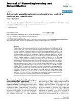
Fig. 1. Relative risks (RR) and 95% confidence limits (CL) for 1-yr mortality in 250 men
divided according to whether their heart rate was < 80 bpm or 80 bpm on the seventh day
after admission to the hospital for acute myocardial infarction. Unadj = unadjusted relative
risk; age-adj = relative risk adjusted for age; risk-adj = relative risks adjusted for age, CKMB peak, echocardiographic left ventricular ejection fraction, diabetes, history of hypertension, current smoking, history of angina, Killip class, thrombolysis and `-blocker therapy;
p-values relate to the results of Cox regression analyses.
(18) in which the total mortality was 14% in the subjects with an admission heart
rate < 60 bpm, 41% in the subjects with a heart rate > 90 bpm, and 48% in those
with a heart rate >110 bpm. In a subsequent study, Disegni et al. found a doubled
mortality risk in postmyocardial infarction patients with a heart rate > 90 bpm
compared to subjects with a heart rate < 70 bpm (19). Two analyses performed
in larger datasets confirmed the results of the above studies. In the GUSTO study
(20), a high heart rate emerged as a potent precursor of mortality, and in the
GISSI-2 trial (21), the predischarge heart rate was a stronger predictor of death
than standard indices of risk, such as left ventricular dysfunction or ventricular
arrhythmias. It is noteworthy to observe that tachycardia in postmyocardial infarction patients cannot be considered simply as a marker of heart failure, as its predictive power appeared more evident in the subjects with no or mild signs of
congestive heart failure (18,19). In a recent study, we found that the predictive
power of heart rate for mortality in subjects with acute myocardial infarction
remained significant also after adjusting for numerous confounders, including
clinical and echocardiographic signs of left ventricular dysfunction (Palatini et
al., unpublished observations) (Fig. 1).
PATHOGENETIC CONSIDERATIONS
The pathogenetic connection between fast heart rate and cardiovascular risk
can be explained according to several different mechanisms (Fig. 2). The heart
162
Part II / Circadian Variation in Cardiovascular Disease
Fig. 2. Mechanisms of the connection between heart rate and cardiovascular morbidity and
mortality. The heart rate can be a marker of risk or a consequence of an underlying disease,
but can exert a direct action in the induction of the risk as well. LV = left ventricular; BP
= blood pressure, w = increased, ¦ = decreased.
rate can be considered as a marker of an underlying clinical condition related
to the risk or a consequence of a latent chronic disease. However, experimental
evidence suggests that a high heart rate should be regarded as a pathogenetic
factor in the induction of the risk as well. In fact, tachycardia favors the occurrence of atherosclerotic lesions by increasing the arterial wall stress (22) and
impairs arterial compliance and distensibility (23). Moreover, the mean blood
Chapter 7 / Heart Rate and Cardiovascular Risk
163
Table 1
Correlation Coefficients Between Resting Heart Rate
and Other Clinical Variables in Three General and One Hypertensive Populations
Population
General
Tecumseh
Mirano
Belgian
Hypertensive
Harvest
SBP
DBP
BMI
CT
TG
GL
INS
.27
.22
.20
.26
.24
.32
.11
NS
.13
.16
.05
NS
.13
.08
NS
NS
.20*
.19*
.19
—
.20
.26
.10
NS
NS
NS
NS
—
SBP=systolic blood pressure; DBP = diastolic blood pressure; BMI = body mass index; CT =
total cholesterol; TG = triglycerides; GL = glucose; INS = fasting insulin; NS = coefficient non
significant; * = postload glucose. Data are for men only.
Data from ref. 7.
pressure has been found to be higher in subjects with faster heart rate (24). This
can be explained by the increase in the total time spent on systole because of the
shortening of diastolic time.
The experimental evidence for a direct role of tachycardia in the induction of
arterial atherosclerotic lesions was provided by studies performed in cynomolgus monkeys. Beere et al. were the first to demonstrate that reduction of heart rate
by ablation of the sinoatrial node could retard the development of coronary lesions
in these animals (25).
Bassiouny et al. studied the effect of the product of mean heart rate and mean
blood pressure (so-called hemodynamic stress) on the aorta of the monkeys (26)
and found a striking positive relationship between the hemodynamic stress index
and maximum atherosclerotic lesion thickness. Similar results were obtained by
Kaplan et al., who found a significant relationship between naturally occurring
differences in heart rate and atherosclerotic coronary lesions in monkeys (27).
As mentioned earlier, heart rate can be considered as a marker of an abnormal
clinical condition. This is suggested by the relationship found in several studies
between heart rate and many risk factors for atherosclerosis (28–30). In four
different populations studied in the Ann Arbor laboratory, we found that the
heart rate was correlated with blood pressure, degree of obesity, cholesterol, triglycerides, postload glucose, and fasting insulin (Table 1) (31,32). In other words,
subjects with a fast heart rate exhibited the features of the insulin-resistance
syndrome. If one assumes that a fast heart rate is the marker of an abnormal autonomic control of the circulation, as demonstrated by Julius et al. (33,34), it is easy
to understand why subjects with tachycardia develop atherosclerosis and cardiovascular events. In fact, several studies performed in the Ann Arbor and other
laboratories indicate that sympathetic overactivity can cause insulin resistance
(Fig. 3). This can be obtained through acute (35) as well as chronic (36) stimulation of `-adrenergic receptors. It has been shown that chronic stimulation of
164
Part II / Circadian Variation in Cardiovascular Disease
Fig. 3. Pathogenesis of the connection between tachycardia and insulin resistance. Tachycardia is a marker of the underlying sympathetic overactivity. SNS = sympathetic nervous
system, w = increased, ¦ = decreased.
`-receptors causes the conversion from a small to a larger proportion of insulinresistant fast-twitch muscles (36). An insulin-resistance state can be obtained
also through a vasoconstriction mediated by _-adrenergic receptors, as shown
by Jamerson et al. in the human forearm (37). Conversely, the _-adrenergic
blockade can improve insulin sensitivity in patients with hypertension (38).
The connection between high heart rate and mortality can be explained also
by an unrecognized underlying disease, and tachycardia can reflect poor physical fitness or loss of cardiac reserve (4,6,13). In fact, an impaired left ventricular contractility may be an early clinical finding in asymptomatic hypertensive
individuals, as demonstrated in the Padova (39) and Ann Arbor (40) laboratories.
To rule out this possibility, in some studies the subjects who died within the first
years after the baseline evaluation were eliminated (6,13,16). However, in all of
those studies, the heart rate–mortality association remained significant, indicating that tachycardia was not only a marker of latent left ventricular failure or of
loss of vigor.
Chapter 7 / Heart Rate and Cardiovascular Risk
165
Besides causing the development of atherosclerotic lesions, a fast heart rate
can also favor the occurrence of cardiovascular events, as shown by the Framingham study (6,12,16). The relationship appeared weak for nonfatal cardiovascular
events but was strong for fatal cardiovascular events. Moreover, as mentioned
earlier, tachycardia can facilitate sudden death (4,6,13). The reasons for this
connection can be of a different nature. Sympathetic overactivity underlying a
fast heart rate can facilitate the occurrence of coronary thrombosis through platelet activation and increased blood viscosity (31). Subjects with tachycardia are
more prone to ventricular arrhythmias. It is known that a heightened sympathetic
tone can promote the development of left ventricular hypertrophy (41), which
facilitates the occurrence of arrhythmias (42). Moreover, tachycardia increases
oxygen consumption and ventricular vulnerability (7,43). The latter mechanisms are important chiefly in subjects with acute myocardial infarction.
LOOKING FOR A THRESHOLD VALUE
The current definition of tachycardia is a heart rate > 100 bpm. Recent results
obtained in our laboratory with mixture analysis suggest that this value is probably too high. In fact, in three general and one hypertensive populations, we
found that the distribution of heart rate was explained by the mixture of two
homogeneous subpopulations, a larger one with a “normal” heart rate and a
smaller one with a “high” heart rate. The partition value between the two subpopulations was around 80–85 bpm. Furthermore, in almost all of the epidemiologic studies that showed an association between heart rate and death from
cardiovascular or noncardiovascular causes, the heart-rate value above which
a significant increase in risk was seen was below the 100-bpm threshold (44)
(Table 2). On the basis of the above data we suggested that the upper normal value
of heart rate should be set at 85 bpm (44).
THERAPEUTIC CONSIDERATIONS
Although there is no doubt that a fast heart rate is independently related to
cardiovascular and total mortality, it is not known whether the reduction of heart
rate can be beneficial in prolonging life. No clinical trial has been implemented
as yet in human beings with the specific purpose of studying the effect of cardiac
slowing on morbidity and mortality. This issue was dealt with by Coburn et al.
in mice by studying the effect of digoxin administration (45). Survival increased
by 29% in the digoxin-treated males and by 14% in the treated females, in comparison with two groups of untreated mice (control groups), indicating that a
heart-rate reduction may confer an advantage in terms of longevity.
A beneficial effect of heart-rate reduction in retarding the development of
atherosclerotic lesions was demonstrated by Kaplan et al. with `-blocker administration in cynomolgus monkeys (46). After 26 mo of propranolol treatment, the
166
Part II / Circadian Variation in Cardiovascular Disease
Table 2
Heart Rate Threshold Values Above Which a Significant
Increase in Mortality Was Found in Eight Epidemiologic Studies
HR threshold value
Reference
Men Women
Levy et al., 1945 (8)
99
—
Dyer et al., 1980 (4)
79
—
Dyer et al., 1980 (4)
86
—
Dyer et al., 1980 (4)
89
—
Kannel et al., 1985 (6)
87
87
Gillum et al., 1991 (5)
84
84
Gillman et al., 1993 (15)
84
84
Palatini et al., 1999 (13)
80
84
Results of the study
Increased 5-yr cardiovascular mortality
in men.
Increased 15-yr all-cause mortality in the
men of the People Gas Co. study.
Increased 5-yr all-cause mortality in the
men of the Heart Association study.
Increased 17-yr all-cause mortality in the
men of the Western Electric study.
Increased 26-yr sudden death mortality
rate in men.
Increased 10-yr all-cause mortality in
black and white men and in black women.
Increased 36-yr all-cause mortality in
hypertensive men and women.
Increased 12-yr cardiovascular mortality
in elderly men.
HR = heart rate in bpm.
socially dominant animals showed a reduced development of coronary artery
lesions in comparison to a group of untreated monkeys of the control group. This
suggests that heart-rate reduction with `-blockers is beneficial in preventing
atherosclerotic lesions, but only in animals exposed to a high environmental stress.
Most of the information on the effect of `-blockers on heart rate and morbidity
and mortality in human beings comes from results obtained in post-myocardialinfarction patients. The reduction in heart rate obtained varied greatly among the
trials, from 10.5% to 22.8%. `-Blocking treatment appeared beneficial in those
patients in whom the heart rate was reduced by 14 bpm or more, whereas for a
heart-rate reduction <8 bpm, no benefit was apparent (47). Moreover, the advantage of treatment was virtually confined to patients with a heart rate of >55 bpm.
In 26 large, placebo-controlled trials with a long-term follow-up, `-blockers
proved effective primarily in reducing sudden death and death resulting from
pump failure (47–51). An almost linear relationship was found between reduction in resting heart rate and decreased mortality (48,52). `-Blockers with intrinsic sympathomimetic activity, such as pindolol or practolol, showed only little
effect on mortality.
Similar beneficial effects were obtained in patients with congestive heart failure (53). Carvedilol caused a marked reduction in mortality in subjects with congestive heart failure (54), but only in patients with a high heart rate (>82 bpm).
Chapter 7 / Heart Rate and Cardiovascular Risk
167
The results obtained in hypertensive subjects (55) were less impressive, probably the result of the untoward effects of `-blockers on high-density lipoprotein
(HDL) cholesterol and triglycerides (56). However, the effect of `-blockers in
hypertensive patients was never examined in relation to the subjects’ heart rates
at baseline.
If the unsatisfactory effects of `-blockers in hypertension are the result of
their unfavorable effects on plasma lipids, the use of drugs which reduce blood
pressure and heart rate without altering the lipid profile appears warranted. Nondihydropyridine-calcium antagonists (57,58) have been shown to be neutral on
the metabolic profile and could, thus, be more effective in preventing cardiovascular mortality in hypertensive subjects with tachycardia. In addition to having
a peripheral action, some of them can cross the blood-brain barrier and decrease
sympathetic outflow (58).
Diltiazem and verapamil have been shown to be effective in reducing the risk
of cardiac events (59–61), but their depressive action on cardiac inotropism
makes them unsuitable for patients with acute myocardial infarction and severe
left ventricular dysfunction. The new long-acting calcium antagonists that selectively block voltage-dependent T-type calcium channels (62,63) reduce heart rate
without manifesting a depressant effect on myocardial contractility and could,
thus, be indicated also for subjects with congestive heart failure (64).
Centrally active antihypertensive drugs that decrease heart rate through reduction of the sympathetic discharge from the central nervous system should have
a good potential for the treatment of the hypertensive patient with fast heart rate.
Unfortunately, the use of clonidine, _-methyldopa, guanfacine, and guanabenz
is limited by the frequent occurrence of side effects, like dry mouth, sedation, and
impotence (65). Moxonidine and rilmenidine are new antihypertensive agents
acting on the I1-imidazoline receptors of the rostro-ventrolateral medulla of the
brainstem and do not have most of the side effects encountered with the centrally
acting agents (65,66). Moreover, these drugs proved effective in improving the
metabolic profile in the experimental animal (67) and also in human studies (68).
The goal of antihypertensive treatment should be not only to lower the blood
pressure but also to reverse those functional abnormalities that often accompany
the hypertensive condition. Therefore, a therapy that not only reduces blood
pressure effectively but also decreases the heart rate and improves metabolic
abnormalities should be sought.
REFERENCES
1. Selby JV, Friedman GD, Quesenberry CP Jr. Precursors of essential hypertension: pulmonary
function, heart rate, uric acid, serum cholesterol, and other serum chemistries. Am J Epidemiol
1990;131:1017–1027.
2. Reed D, McGee D, Yano K. Biological and social correlates of blood pressure among Japanese
men in Hawaii. Hypertension 1982;4:406–414.
168
Part II / Circadian Variation in Cardiovascular Disease
3. Garrison RJ, Kannel WB, Stokes J III. Incidence and precursors of hypertension in young
adults. Prev Med 1987;16:235–251.
4. Dyer AR, Persky V, Stamler J, et al. Heart rate as a prognostic factor for coronary heart disease and mortality: findings in three Chicago epidemiologic studies. Am J Epidemiol 1980;112:
736–749.
5. Gillum RF, Makuc DM, Feldman JJ. Pulse rate, coronary heart disease, and death: the NHANES
I epidemiologic follow-up study. Am Heart J 1991;121:172–177.
6. Kannel WB, Wilson P, Blair SN. Epidemiologic assessment of the role of physical activity
and fitness in development of cardiovascular disease. Am Heart J 1985;109:876–885.
7. Palatini P, Julius S. Heart rate and the cardiovascular risk. J Hypertens 1997;15:3–17.
8. Levy RL, White PD, Stroud WD, et al. Transient tachycardia: Prognostic significance alone
and in association with transient hypertension. JAMA 1945;129:585–588.
9. Paffenbarger RS Jr, Thorne MC, Wing AL. Chronic disease in former college students—VIII.
Characteristics in youth predisposing to hypertension in later years. Am J Epidemiol 1968;88:
25–32.
10. Schroll M, Hagerup LM. Risk factors of myocardial infarction and death in men aged 50 at
entry. A ten-year prospective study from the Glostrup population studies. Dan Med Bull 1977;
24:252–255.
11. Medalie JH, Kahn HA, Neufeld HN, et al. Five-year myocardial infarction incidence—II.
Association of single variables to age and birthplace. J Chronic Dis 1973;26:329–349.
12. Goldberg RJ, Larson M, Levy D. Factors associated with survival to 75 years of age in middleaged men and women. The Framingham study. Arch Intern Med 1996;156:505–509.
13. Palatini P, Casiglia E, Julius S, et al. Heart rate: a risk factor for cardiovascular mortality in
elderly men. Arch Int Med 1999;159:585–592.
14. Aronow WS, Ahn C, Mercando AD, et al. Association of average heart rate on 24-hour
ambulatory electrocardiograms with incidence of new coronary events at 48-month followup in 1,311 patients (mean age 81 years) with heart disease and sinus rhythm. Am J Cardiol
1996;78:1175–1176.
15. Gillman MW, Kannel WB, Belanger A, et al. Influence of heart rate on mortality among
persons with hypertension: The Framingham Study. Am Heart J 1993;125:1148–1154.
16. Kannel WB, Kannel C, Paffenbarger RS Jr, et al. Heart rate and cardiovascular mortality: The
Framingham Study. Am Heart J 1987;113:1489–1494.
17. The Norwegian Multicenter Study Group. Timolol-induced reduction in mortality and reinfarction in patients surviving acute myocardial infarction. N Engl J Med 1981;304:801–807.
18. Hjalmarson A, Gilpin EA, Kjekshus J, et al. Influence of heart rate on mortality after acute
myocardial infarction. Am J Cardiol 1990;65:547–553.
19. Disegni E, Goldbourt U, Reicher-Reiss H, et al. The predictive value of admission heart
rate on mortality in patients with acute myocardial infarction. J Clin Epidemiol 1995;48:
1197–1205.
20. Lee KL, Woodlief LH, Topol EJ, et al. Predictors of 30-day mortality in the era of reperfusion
for acute myocardial infarction. Results from an international trial of 41,021 patients. Circulation 1995;91:1659–1668.
21. Maggioni AP, Zuanetti G, Mantini L, et al. The predictive value of pre-discharge heart rate
on 8-month mortality in 7,831 patients with acute myocardial infarction in the fibrinolytic era.
Eur Heart J 1997;18(Abstract suppl):352A.
22. Gordon D, Guyton J, Karnovsky N. Intimal alterations in rat aorta induced by stressful stimuli.
Lab Invest 1983;45:14–19.
23. Mangoni AA, Mircoli L, Giannattasio C, et al. Heart rate-dependence of arterial distensibility
in vivo. J Hypertens 1996;14:897–901.
24. Palatini P. Exercise haemodynamics in the normotensive and the hypertensive subject. Clin
Sci 1994;87:275–287.
Chapter 7 / Heart Rate and Cardiovascular Risk
169
25. Beere PA, Glagov S, Zarins CK. Retarding effect of lowered heart rate on coronary atherosclerosis. Science 1984;226:180–182.
26. Bassiouny HS, Zarins CK, Kadowaki MH, et al. Hemodynamic stress and experimental aortoiliac atherosclerosis. J Vasc Surg 1994;19:426–434.
27. Kaplan JR, Manuck SB, Clarkson TB. The influence of heart rate on coronary artery atherosclerosis. J Cardiovasc Pharmacol 1987;10(Suppl 2):S100–S102.
28. Stamler J, Berkson DM, Dyer A, et al. Relationship of multiple variables to blood pressure—
findings from four Chicago epidemiologic studies. In: Paul O, ed. Epidemiology and Control
of Hypertension. Symposia Specialists, Miami, 1975, pp. 307–352.
29. Cirillo M, Laurenzi M, Trevisan M, et al. Hematocrit, blood pressure, and hypertension. The
Gubbio Population Study. Hypertension 1992;20:319–326.
30. Stern MP, Morales PA, Haffner SM, et al. Hyperdynamic circulation and the insulin resistance
syndrome (“Syndrome X”). Hypertension 1992;20(6):802–808.
31. Palatini P, Julius S. Association of tachycardia with morbidity and mortality: pathophysiological considerations. J Hum Hypertens 1997;11(Suppl 1):19–27.
32. Palatini P, Casiglia E, Pauletto P, et al. Relationship of tachycardia with high blood pressure
and metabolic abnormalities. A study with mixture analysis in three populations. Hypertension 1997;30:1267–1273.
33. Julius S, Gudbrandsson T, Jamerson K, et al. Hypothesis. The hemodynamic link between
insulin resistance and hypertension. J Hypertens 1991;9:983–986.
34. Julius S, Pascual AV, London R. Role of parasympathetic inhibition in the hyperkinetic type
of borderline hypertension. Circulation 1971;44:413–418.
35. Deibert DC, DeFronzo RA. Epinephrine-induced insulin resistance in man. J Clin Invest 1980;
65:717–721.
36. Zeman RJ, Ludemann R, Easton TG, et al. Slow to fast alterations in skeletal muscle fibers
caused by clenbuterol, a beta-2-receptor agonist. Am J Physiol 1988;254:E726–E732.
37. Jamerson KA, Julius S, Gudbrandsson T, et al. Reflex sympathetic activation induces acute
insulin resistance in the human forearm. Hypertension 1993;21(5):618–623.
38. Pollare T, Lithell H, Selinus I, et al. Application of prazosin is associated with an increase of
insulin sensitivity in obese patients with hypertension. Diabetologia 1988;31:415–420.
39. Palatini P, Visentin PA, Mormino P, et al. Left ventricular performance in the early stages of
systemic hypertension. Am J Cardiol 1998;81:418–423.
40. Julius S. Altered cardiac responsiveness and regulation in the normal cardiac output type of
borderline hypertension. Circ Res 1975;36–37(Suppl I):I-199–I-207.
41. Julius S, Li Y, Brant D, et al. Neurogenic pressor episodes fail to cause hypertension, but do
induce cardiac hypertrophy. Hypertension 1989;13:422–429.
42. Palatini P, Maraglino G, Accurso V, et al. Impaired left ventricular filling in hypertensive left
ventricular hypertrophy as a marker of the presence of an arrhytmogenic substrate. Br Heart
J 1995;73:258–262.
43. Palatini P. Heart rate as a cardiovascular risk factor. Eur Heart J 1999;20(Suppl B):B3–B9.
44. Palatini P. Need for a revision of the normal limits of resting heart rate. Hypertension 1999;
33:622–625.
45. Coburn AF, Grey RM, Rivera SM. Observations on the relation of heart rate, life span, weight
and mineralization in the digoxin-treated A/J mouse. Johns Hopkins Med J 1971;128:169–193.
46. Kaplan JR, Manuck SB, Adams MR, et al. Inhibition of coronary atherosclerosis by propranolol
in behaviorally predisposed monkeys fed an atherogenic diet. Circulation 1987;76:1364–1372.
47. Kjekshus JK. Importance of heart rate in determining beta-blocker efficacy in acute and longterm acute myocardial infarction intervention trials. Am J Cardiol 1986;57:43F–49F.
48. Teo KK, Yusuf S, Furberg CD. Effects of prophylactic antiarrhythmic drug therapy in acute
myocardial infarction: an overview of results from randomized controlled trials. JAMA 1993;
270:1589–1595.
170
Part II / Circadian Variation in Cardiovascular Disease
49. The Goteborg Metoprolol Trial in Acute Myocardial Infarction. Am J Cardiol 1984;53:
10D–50D.
50. The International Collaborative Study Group. Reduction of infarct size with the early use of
timolol in acute myocardial infarction. N Engl J Med 1984;310:9–15.
51. Taylor SH, Silke B, Ebbutt A, et al. A long-term prevention study with oxprenolol in coronary
heart disease. N Engl J Med 1982;307:1293–1301.
52. Kjekshus JK. Comments on beta-blockers: heart rate reduction, a mechanism of action. Eur
Heart J 1985;6(Suppl A):29–30.
53. MRC Working Party. Medical Research Council trial of treatment of hypertension in older
adults: principal results. Br Med J 1992;304:405–412.
54. Lehtonen A. Effect of beta blockers on blood lipid profile. Am Heart J 1985;109:1192–1198.
55. Eichorn EJ, Bristow MR. Medical therapy can improve the biological properties of the chronically failing heart. Circulation 1996;94:2285–2296.
56. Packer M, Bristow MR, Cohn JN, for the U.S. Carvedilol Heart Failure Study Group. The
effect of Carvedilol on morbidity and mortality in patients with chronic heart failure. N Engl
J Med 1996;334:1349–1355.
57. Stadler P, Leonardi L, Riesen W, et al. Cardiovascular effects of verapamil in essential hypertension. Clin Pharmacol Ther 1987;42:85–92.
58. Kailasam MT, Parmer RJ, Cervenka JH, et al. Divergent effects of dihydropyridine and phenylalkylamine calcium channel antagonist classes on autonomic function in human hypertension. Hypertension 1995;26:143–149.
59. The Danish Study Group on Verapamil in Myocardial Infarction. Effect of verapamil on mortality and major events after acute myocardial infarction. The Danish Verapamil Infarction
Trial II (DAVIT-II). Am J Cardiol 1990;66:779–785.
60. Alderman MH, Cohen H, Rogué R, et al. Effect of long-acting and short-acting calcium
antagonists on cardiovascular outcomes in hypertensive patients. Lancet 1997;349:594–598.
61. The Multicenter Diltiazem Postinfarction Trial Research Group. The effect of diltiazem on
mortality and reinfarction after myocardial infarction. N Engl J Med 1988;319:385–392.
62. Luscher TF, Clozel JP, Noll G. Pharmacology of the calcium antagonist mibefradil. J Hypertens 1997;15(Suppl 3):S11–S18.
63. Kung CF, Tschudi MR, Noll G, et al. Differential effects of the calcium antagonist mibefradil
in epicardial and intramyocardial coronary arteries. J Cardiovasc Pharmacol 1995;26:312–318.
64. Clozel J, Ertel EA, Ertel SI. Discovery and main pharmacological properties of mibefradil (Ro
40-5967), the first selective T-type calcium channel blocker. J Hypertens 1997;15(Suppl 5):
S17–S25.
65. Van Zwieten PA. Centrally acting antihypertensives: a renaissance of interest. Mechanisms
and haemodynamics. J Hypertens 1997;15(Suppl 1):S3–S8.
66. Ernsberger P, Koletsky RJ, Collins LA, et al. Sympathetic nervous system in salt-sensitive and
obese hypertension: amelioration of multiple abnormalities by a central sympatholitic agent.
Cardiovasc Drugs Ther 1996;10:275–282.
67. Rosen P, Ohly P, Gleichmann H. Experimental benefit of moxonidine on glucose metabolism
and insulin secretion in the fructose-fed rat. J Hypertens 1997;15(Suppl 1):S31–S38.
68. Ernsberger P, Friedman JE, Koletsky RJ. The I1-Imidazoline receptor: from binding site to
therapeutic target in cardiovascular disease. J Hypertens 1997;15(Suppl 1):S9–S23.
Chapter 8 / Na+, K+, the SNS, and the Renin–Angiotensin System
8
171
Sodium, Potassium,
the Sympathetic Nervous System,
and the Renin–Angiotensin System
Impact on the Circadian Variability
in Blood Pressure
Domenic A. Sica, MD
and Dawn K. Wilson, PHD
CONTENTS
INTRODUCTION
AMBULATORY BLOOD PRESSURE MONITORING AS A TOOL
ELECTROLYTES AND CIRCADIAN RHYTHMS
NEURO-HUMORAL PATTERNS AND CIRCADIAN BLOOD PRESSURE
RHYTHMS
SUMMARY
REFERENCES
INTRODUCTION
Under the usual circumstances of everyday life, the phasing of human circadian clocks and rhythms is set, or synchronized, by the sleep-in-darkness–
activity-in-light, 24-h routine. These time cues greatly influence the intrinsic
diurnal rhythm for blood pressure (BP). As an example of this, when shift workers are assigned to night duty, they ordinarily adhere to a different sleep–activity
routine than do day workers. Because of this, the timing of their peak and trough
BP differs, with reference to external clocktime, if compared to daytime active
From: Contemporary Cardiology:
Blood Pressure Monitoring in Cardiovascular Medicine and Therapeutics
Edited by: W. B. White © Humana Press Inc., Totowa, NJ
171
172
Part II / Circadian Variation in Cardiovascular Disease
individuals. The biologic time structure of man is an inherited characteristic for
a number of parameters, such as BP. Its normal expression, however, may be
influenced by either environmental/nutritional factors or an individual’s normal or pathophysiologically acquired neuro-humoral status. When normal phase
relationships change between circadian bioperiodicities, BP patterns may alter
radically and unpredictably. The purpose of this review is to characterize the neurohumoral and nutritional determinants of the ambulatory blood pressure (ABP)
profile in normotensive and hypertensive patients. In particular, this review focuses
on the sympathetic nervous system (SNS), the renin–angiotensin–aldosterone
(RAA) axis, and the role of dietary sodium (Na+) and potassium (K+) in shaping
circadian BP patterns.
AMBULATORY BLOOD
PRESSURE MONITORING AS A TOOL
Ambulatory BP monitoring is a recently developed methodology, capable of
identifying and systematically evaluating factors responsible for individual differences in BP responses in the natural environment. This approach provides a
means for studying an individual in a standardized fashion as he or she responds
to the physical and psychological demands of a typical 24-h day. Prior research
employing ABP monitoring indicates that most people display low-amplitude
diurnal variations in BP, with higher pressures during waking hours and lower
pressures during sleep (1,2). In most normotensive subjects, average BP values
decline by approx 15% during sleep (3–5). In hypertensive subjects, the circadian rhythm is generally preserved, although the 24-h BP profile shifts to higher
around-the-clock values (6).
Ambulatory BP patterns are rarely static with considerable day-to-day variability in how nocturnal BP patterns express themselves (7). It has proven tempting to assign causality to a particular dietary or neuro-humoral change in how
nocturnal BP changes occur. Unfortunately, it is the rare circumstance where a
specific neuro-humoral or dietary pattern is exclusively responsible for a particular nocturnal BP pattern, such as nondipping (minimal drop in nocturnal BP).
Rather, factors typically coalesce with different weightings assigned to individual factors in order to arrive at a final explanation for a specific BP pattern.
Comments found in this chapter should be viewed accordingly.
ELECTROLYTES AND CIRCADIAN RHYTHMS
The established associations between BP and electrolytes are for the most part
most reliable when based on data from urinary excretion and/or a validated selfreport of nutrient intake (7–9). Urinary excretion and/or dietary recall parameters
are the preferred correlates to BP, as it is widely held that they more realistically
Chapter 8 / Na+, K+, the SNS, and the Renin–Angiotensin System
173
Fig. 1. Relation between plasma K+ and 24-h systolic blood pressure (A: r = 0.336, p < 0.01)
or office systolic blood pressure (B: r = <0.018, p = NS) in 82 patients with essential hypertension. Adapted with permission of Elsevier Science from ref. (13). Copyright 1997 by
American Journal of Hypertension Ltd.
depict the true state of electrolyte balance. Interpreting the relationship between
a plasma electrolyte, such as K+, and BP is inherently difficult because nutritional intake is one of only many factors known to influence plasma K+ values.
Such factors include a circadian rhythm for plasma K+ (average peak z trough
difference F 0.60 mEq/L with lowest values at night) (10) and a tendency for K+
to migrate intracellularly, when `2-adrenergic receptors are stimulated (11).
Accordingly, very few reports have even attempted to characterize the relationship between plasma K+ and ABP patterns in hypertensive patients (12,13).
Goto et al. found significant negative correlations between daytime plasma K+
concentration and 24-h systolic and diastolic BP levels in patients with essential
hypertension (13). Plasma K+ also inversely correlated with both daytime and
nighttime systolic and diastolic BP levels. In these studies, there was no correlation between office BP readings and plasma K+ concentration. No doubt, any
such relationship was obscured by the inherent variability of office BP measurements (Fig. 1).
If the plasma K+ value in any way equates with intake, these results are consistent with prior epidemiologic studies, which have found a negative correlation
between K+ intake and BP levels (12). Goto et al. have further suggested that
decreased extracellular K+ promotes vasoconstriction in hypertensive patients
by either enhancing SNS activity or by increasing the Na+ content of vascular
smooth muscle cells (13). Additional research is needed to better understand the
relative contribution of plasma electrolytes to circadian variability in BP.
174
Part II / Circadian Variation in Cardiovascular Disease
NEURO-HUMORAL PATTERNS
AND CIRCADIAN BLOOD PRESSURE RHYTHMS
Atrial Natriuretic Peptide
As a prominent regulatory arm of volume homeostasis in man, the natriuretic
peptides are intimately involved in the regulation of BP. Atrial natriuretic peptide (ANP) release is primarily regulated by atrial pressure though a number of
other factors, such as age and level of renal and/or cardiac function, that can
arbitrate the final plasma concentration for ANP. ANP can be viewed as the
“mirror image” of the RAA axis in that it inhibits the release of renin and aldosterone while opposing the actions of angiotensin II and aldosterone through effects
on vascular tone, cells growth, and renal sodium reabsorption. When ANP is
administered to animals or humans, the BP acutely drops, a process, which is
particularly prominent when the RAA is activated. For these reasons, a relationship between the time structure of ANP, other neurohormones, and 24-h BP
patterns has been sought.
It has been observed that single-point-in-time morning ANP levels may have
either no relationship to 24-h BP (14) or may separate isolated clinic hypertension (wherein ANP levels are typically normal) from sustained hypertension
(wherein plasma ANP levels are increased) in elderly hypertensives (15). Methodologic considerations are important to the interpretation of circadian ANP
patterns. For example, Chiang et al. observed the absence of any circadian rhythm
for ANP (and thus no relationship to diurnal BP change) in a group of 14 healthy
volunteers in whom ANP was sampled every 3 h for 24 h (16).
In other studies in which subjects were synchronized to the light–dark cycle and
were given a controlled diet, a variable acrophase for ANP was found. Portaluppi
et al. originally noted an acrophase for ANP to occur at around 4:00 AM. In these
studies, BP and heart rate (HR) rhythms appeared to be in antiphase with the ANP
rhythm, with the peak of BP and HR more or less coinciding with the trough
for ANP rhythm. This pattern of response suggested a relationship between ANP
levels and BP and HR (17). Alternatively, Cugini et al. noted an acrophase timing
for ANP at about 5:00 PM in young clinically healthy subjects and no circadian
pattern for ANP in elderly subjects, although mean blood levels of ANP were
noticeably higher in the elderly cohort (18). There is no obvious explanation for
these obviously different findings. Additional studies will be required to clarify
the time pattern of ANP levels.
Plasma Renin Activity
Gordon et al. originally described a diurnal rhythm for plasma renin activity
(PRA) that was independent of posture and dietary influences (19), a finding
subsequently corroborated by a number of other investigators (17,20–22). From
these observations emerged the concept of a circadian rhythm in PRA, with a
Chapter 8 / Na+, K+, the SNS, and the Renin–Angiotensin System
175
nadir in the afternoon and a nocturnal increase culminating in the early morning
hours, despite the occasional study having failed to demonstrate any significant
variation in PRA with the time of day (23–25). What remained to be determined
was to define the relative role of endogenous circadian rhythmicity and the sleep–
wake cycle on 24-h PRA variations because sleep can make substantial contributions to the overall variations in PRA and thereby mask the characteristics of an
endogenous rhythm (26).
A strong relationship exists between nocturnal oscillations in PRA and internal sleep structure (24,27). Non-rapid-eye-movement (NREM) is invariably
linked to increasing PRA levels, with PRA decreasing during rapid-eye-movement (REM) sleep. In normal man, modifying the renal renin content modulates
only the amplitude of the nocturnal oscillations without altering their relationship to the stage of sleep (28), and in the case of sleep disorders, such as sleep
apnea, the PRA profiles reflect all facets of the sleep structure disturbance (29).
Brandenberger et al. have recently shown, using an acute shift in the normal sleep
time, that increased renin release was associated with sleep whatever time it
occurs, an observation atypical of an intrinsic circadian rhythm (Fig. 2) (26).
This group further observed that internal sleep architecture had an important
modulatory role on the characteristics of the PRA oscillations and, consequently,
on the 24-h pattern. When NREM–REM sleep cycles are disturbed, as is the case
with the fragmented sleep of obstructive sleep apnea, there is insufficient time
for PRA to increase significantly; consequently, in poor sleepers, PRA values
may not vary to any significant degree throughout a 24-h time span. This may provide an explanation for the occasional study wherein PRA values fail to increase
during sleep (23–25,30).
Although several studies have examined the 24-h cycle of PRA, few have seen
fit to examine the relationship between PRA and ABP patterns, and those that
have, fail to provide a consistent picture. For example, Watson found significant
positive correlations between PRA and variability in daytime BP readings after
adjusting for age (31). Chau et al. (32), however, reported negative correlations
between upright PRA and 24-h mean BP readings. Harshfield and colleagues (33)
examined the relationship between renin–sodium profiles and ABP patterns in
healthy children. The subjects were classified as low, intermediate, or high renin,
inferred from the relationship between PRA and Na+ excretion. The subjects
classified as high renin had elevated systolic and diastolic BP readings while
asleep more so than did subjects in the low-renin category. These studies suggest
that the relationship between the level of RAA system activity and ambulatory
BP patterns is complex, with Na+ sensitivity and/or Na+ intake emerging as important co-variables in this relationship.
In addition, the 24-h pattern for PRA oppose that for BP, which tends to fall
in the first few hours of sleep and to rise thereafter. Superimposed on these tendencies, periodic changes in BP occur that coincide with the presence of NREM
176
Part II / Circadian Variation in Cardiovascular Disease
Fig. 2. Effects of an 8-h delay of the sleep–wake cycle on the 24-h plasma renin activity
profiles in 10 subjects: (A) normal nocturnal sleep from 2300 to 0700 h and (B) daytime
sleep from 0700 to 1500 h after a night of sleep deprivation. Values are expressed as means
± SEM. Adapted from (26) by permission of Lippincott Williams & Wilkins.
sleep cycles (34). Such changes are characterized by slight decreases in the mean
BP levels during slow-wave sleep and small increases in mean BP levels during
REM sleep, during which there is a marked increase in PRA pulse activity. It is
unclear as to the relationship of PRA pulse activity to these observed oscillations
in nocturnal BP.
Angiotensin-Converting Enzyme Inhibitors
A failure to establish clear relationships between nocturnal RAA axis activity
and BP patterns may be a function of various sensitivities to external influences
Chapter 8 / Na+, K+, the SNS, and the Renin–Angiotensin System
177
Table 1
Chronopharmacology of ACE inhibitors
Authors
(ref.)
Drug/dose
(mg)
Subject No.
Study design
Weisser
et al. (35)
Palatini
et al. (36)
Enalapril/10
8
Normals
18
Hypertensives,
double-blind/
crossover
10
Hypertensives,
single blind/
crossover
8
Hypertensives,
double blind/
crossover
20
Hypertensives,
crossover
Quinapril/20
Palatini
et al. (37)
Benazepril/10
Witte
et al. (38)
Enalapril/10
Morgan
et al. (39)
Perindopril/4
Timing
(h)
Single/
multiple
dose
Observations
0800
2000
0800
2200
Yes/no
Tmax w with 2000 h dose
No/yes
0900
2100
Yes/no
Evening dose maintained
efficacy for 24 h; morning
dose lost efficacy during
nighttime and early morning
Morning had more sustained
24-h effect than did
evening dose
0700
1900
Yes/yes
0900
2100
No/yes
Evening dose maintained
efficacy for 24 h;
morning dose lost efficacy
between 0200 and 0700 h
Evening dose maintained
efficacy for 24 h; morning
dose lost efficacy after 18 h
and/or the interactions of other rhythms, which could obscure PRA cycles during
the waking periods. One way to evaluate the importance of nocturnal PRA is to
determine the nature of the vasodepressor response subsequent to administration
of ACE inhibitors. Several studies have attempted such an evaluation (Table 1).
Possible circadian changes in the pharmacokinetics and effect on serum
angiotensin-converting enzyme (ACE) activity of the ACE inhibitor enalapril
were first evaluated in the studies of Weisser et al. (35), with several subsequent
studies having been reported since that time (Table 1). Weisser et al. (35) noted
that the mean serum concentration–time profiles of enalapril and its active
metabolite enalaprilat were comparable whether enalapril was ingested at 0800
or 2000 h. Administration of enalapril at 2000 h did not markedly influence the
bioavailability of enalapril as estimated by time to maximum concentration
(Tmax), maximum drug concentration (Cmax), or area under the curve (AUC0-24)
for the active enalapril metabolite, enalaprilat. The only observed difference
was an increase in Tmax for enalapril after its evening administration (1.3 ± 0.5
[0800 h]) vs 2.4 ± 1.4 (2000 h [p < 0.05]), a phenomenon that has been observed
with a number of other drugs.
Palatini et al. subsequently examined the relationship between daytime (0800 h)
and nighttime (2200 h) administration of quinapril following 4 wk of dosing
(36). The 24-h BP profiles obtained by ABP monitoring showed a more sustained
178
Part II / Circadian Variation in Cardiovascular Disease
antihypertensive action with the evening administration (2200 h) of quinapril
compared with its morning administration (0800 h). There was a partial loss of
effectiveness for quinapril during the nighttime and early morning hours when
it was administered in the morning. In addition, measurement of ACE activity
showed that evening administration of quinapril caused a less pronounced but
more sustained decline of plasma ACE. In this regard, 24 h after the last dose
of quinapril, the residual ACE inhibition was greater (62%) with evening dosing than was the case with morning dosing (40%). These authors concluded that
evening administration of quinapril was preferable because it provided a more
homogeneous pattern of 24-h BP control, which, in part, may have related to an
extended inhibition of the ACE enzyme (36).
Palatini et al. also evaluated the influence of timing of benazepril administration on 24-h intra-arterial BP measurements. In contradistinction to their previous studies with quinapril, they noted that a single 10-mg dose of benazepril
administered at 0900 h more effectively covered the 24-h dosing interval than did
an identical dose administered at 2100 h (37). Although the single-dose nature
of these studies make their interpretation difficult, they do suggest that ACE
inhibitor pharmacokinetics are relevant to the circadian variability in response
to an ACE inhibitor.
Witte et al. (38) evaluated the cardiovascular effects and pharmacokinetics of
once-daily enalapril (10 mg) after either a single dose or following its chronic
administration. Chronic therapy (with dosing at 0700 h) significantly reduced
BP during the day but lost effectiveness between 2 and 7 AM of the succeeding
day. Chronic dosing at 1900 h significantly exaggerated the nocturnal dip in BP.
BP values slowly increased throughout the next day with the evening dosing
regimen, with no effect on elevated afternoon values. Peak concentrations of
enalaprilat were found at 3.5 h (morning) and 5.6 h (evening) after drug administration. The time-to-peak drug effect was shorter after morning dosing (7.4 ±
4.3 h [diastolic]) than evening dosing (12.1 ± 3.7 h [diastolic]). Differences in
the response to enalapril could not be attributed to timewise changes in pharmacokinetics or to a different time-course of ACE inhibition. It is more likely that
circadian changes in the sensitivity of the RAA system play an important role in
defining timewise differences in response to an ACE inhibitor.
In a final study, Morgan et al. examined the BP response following the administration of perindopril either in the morning (0900 h) or in the evening (2100 h).
It was noted in these studies that the early morning rise in BP was reduced more
with the evening administration of perindopril. However, the 2100 h dose regimen did not reduce BP over 24 h, whereas the 0900 h dose achieved better BP
control. These studies concluded that the time-related response profile obtained
with an ACE inhibitor is unique and that chronobiology has important effects on
the action of these drugs (39).
Chapter 8 / Na+, K+, the SNS, and the Renin–Angiotensin System
179
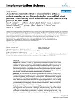
Fig. 3. Effect of an 8-h shift in sleep period cycle on 24-h profiles for plasma aldosterone
in seven subjects. Blood was sampled at 10-min intervals. In the daytime sleep condition,
the amplitude of the aldosterone pulses was significantly enhanced during the sleep period.
Values are expressed as mean ± SEM. Adapted with permission (41).
Plasma Aldosterone
Plasma aldosterone secretion follows a pattern such that mean hormone concentrations are highest during the night and early morning (20–23,40). Plasma
aldosterone values during a 24-h time period appear to be coupled to PRA, with
renin secretion being either simultaneous with or preceding aldosterone secretion by 10–20 min, with this temporal coupling enhanced in a low-sodium state
(40). Under basal conditions, the relative contribution of sleep processes and
circadian rhythm to plasma aldosterone levels remains poorly defined, particularly as relates to those systems that cojointly control aldosterone release (reninangiotensin, adrenocorticotropic, and dopaminergic systems).
Heretofore, any timewise change in the 24-h profile of aldosterone was viewed
simplistically as a circadian event. More recently, it has been recognized that the
pattern of aldosterone release is influenced by sleep architecture (41). Recent studies, employing an experimental design of abruptly shifting sleep by 8 h, show sleep
processes to have a stimulatory effect on aldosterone release, as demonstrated
by high mean levels together with high pulse amplitude and pulse frequency
observed during the sleep period and reduced levels during sleep deprivation
(Fig. 3). This pattern of secretion is similar to that observed with PRA (26). The
large increase in plasma aldosterone levels and pulse amplitude following awakening from nocturnal sleep is attributable to an increase in activity of the adrenocorticotropic axis, reflected by the surge in cortisol in the early morning. The
issue of nocturnal aldosterone change is complex, with aldosterone pulses mainly
180
Part II / Circadian Variation in Cardiovascular Disease
related to PRA oscillations during the sleep periods, whereas aldosterone pulses
are associated with cortisol pulses during the waking periods.
The influence of aldosterone circadian patterns on BP and, in particular,
nocturnal BP is poorly defined. Little meaningful information exists that might
permit an assessment of the role of aldosterone antagonism in modifying circadian BP patterns.
Sympathetic Nervous System
In both normotensive and hypertensive individuals, the BP fluctuates according to the level of both mental and physical activities. BP, HR, and SNS activity are typically highest when a hypertensive patient is awake and/or active.
Conversely, these values reach a nadir between midnight and 3:00 AM (42–44).
Although the exact interplay of all physiologic and pathophysiologic mediators
of the diurnal rhythm remains unclear, nocturnal BP and HR seems to track SNS
activity best—but not entirely so. Experiments with autonomic blocking agents
provide some insight into the importance of the SNS in diurnal BP rhythms. For
example, the BP rhythm in high spinal cord transected patients (with complete
tetraplegia) is nonexistent, despite HR variability being preserved (presumably
because cardiac vagal innervation remains intact) (45). Paraplegics and incomplete tetraplegics typically have a normal diurnal BP pattern. These findings are
consistent with the thesis that central SNS outflow is an important determinant
of the normal diurnal rhythm of BP.
Attempts to define the role of the SNS in determining nocturnal BP changes
are complicated by methodologic constraints. This being said, SNS activity
typically diminishes while asleep, with changes in the sympathoadrenal branch
(epinephrine) being governed in a dual fashion by both posture and sleep and the
noradrenergic branch (norepinephrine) being regulated more so by posture (44).
Diurnal changes in plasma catecholamine values, as markers of SNS activity, are
subject to considerable sampling error and require careful interpretation as to the
study conditions under which they were obtained. Plasma epinephrine concentrations and/or SNS activity decline during sleep (particularly during NREM
sleep) and begin to increase in conjunction with morning awakening (44,46,47)
and/or episodically during episodes of REM sleep (Fig. 4) (48). Plasma norepinephrine concentrations trend downward when asleep and do not significantly
increase until a postural stimulus to norepinephrine release, such as the upright
position, is added to changes accompanying the arousal process (44,46). Morning plasma norepinephrine concentrations, although typically higher than sleep
values, are not necessarily the highest values attained during a 24-h time interval
(46,47). Finally, microneurography, a specific marker of muscle SNS activity,
fails to show any increased neural activity in normal volunteers when performed
between the hours of 6:30 AM and 8:30 AM, a time parenthetically when the rate
of myocardial infarction is highest. This suggests that the early morning peak in
Chapter 8 / Na+, K+, the SNS, and the Renin–Angiotensin System
181
Fig. 4. Recordings of sympathetic nerve activity (SNA) and mean blood pressure (BP) in
a single subject while awake and while in stages 2, 3, and 4 and REM sleep. As non-REM
sleep deepens (stages 2–4), SNA gradually decreases and BP (mmHg) and variability in
BP are gradually reduced. Arousal stimuli elicited K complexes on the electrocardiogram
(not shown) were accompanied by increases in SNA and BP (indicated by the arrows, stage
2 sleep). In contrast to the changes during non-REM sleep, heart rate, BP, and BP variability increased during REM sleep, together with a profound increase in both the frequency
and amplitude of SNA. There was a frequent association between REM twitches (momentary periods of restoration of muscle tone, denoted by T on the tracing) and abrupt inhibition of SNA and increases in BP. Adapted with permission (48). Copyright 1993 Massachusetts Medical Society.
myocardial infarction and/or sudden cardiac death could, in part, reflect exaggerated end-organ responsiveness to norepinephrine following the relative sympathetic withdrawal that occurs during sleep (49).
Nocturnal BP can assume a number of different and now well-characterized
patterns: extreme dipping (an approximate 30% ¦ in BP while asleep), normal
dipping (a 10–20% ¦ in nighttime BP), and nondipping (minimal drop in nocturnal BP or a rise in BP at night) (7,50). Of these BP patterns, attention has recently
centered on the significance of a nighttime nondipping BP pattern, because it is
182
Part II / Circadian Variation in Cardiovascular Disease
believed to be associated with more rapid progression of renal failure (51) and/
or a greater degree of left ventricular hypertrophy (52). Aging, salt sensitivity,
and African-American ethnicity are viewed as relevant demographic markers
for this phenomenon (7).
Little is known about the pathophysiology of nocturnal nondipping in either
normotensive or essential hypertensives, although important clues to the origin
of this phenomenon can be extracted from an analysis of sleep patterns. Sleep
architecture and SNS activity are important determinants of nocturnal BP and
HR. During NREM sleep, there is a tendency for HR to slow and BP to fall, a
process characterized by a relative increase in parasympathetic or vagal activity (53–55). It is now fairly well accepted that alterations in SNS activity may
lead to relevant effects on the pathophysiology of sleep, as well as influence the
diurnal BP profile. Derangements in autonomic nervous system activity, sleepdisordered breathing, and alterations in sleep architecture and duration are wellrecognized causes of change in the circadian BP profile (54). In addition, sleep
disturbances are reported to influence the circadian BP profile. Schillaci et al.
showed that the reported duration of sleep was significantly shorter for hypertensive “nondippers” than it was for “dippers” both in males and females (56).
Kario et al. found nondippers to have increased nocturnal physical activity, as
determined by actigraphy (57). Thus, the duration and quality of sleep should be
considered in the interpretation of the diurnal BP profile.
Nutrition
Na+
K+
The intake of
and/or
is an important modulator of BP. The impact of
such nutritional modification has most typically been assessed by evaluating
change in casual BP determinations (58,59), although more recently, ABP technology has been employed to delineate the 24-h pattern of change with such interventions (60–64). Accordingly, it is only in the last decade that nocturnal BP
patterns could serve as targets for dietary intervention (60,64).
Prior research has identified demographic groups in whom the equilibrium
point for Na+ balance is set at a higher level of BP. For example, Weinberger et
al. demonstrated that blacks and older individuals (>40 yr) poorly excrete a Na+
load, and in order to achieve Na+ balance, higher BP values are required for a
longer period of time (65). Falkner et al. have also reported that salt-sensitive
adolescents with a positive family history of hypertension had greater increases
in BP with salt loading than did adolescents who were either salt resistant or had
a negative family history of hypertension (66). Harshfield et al. have also demonstrated that Na+ intake is an important determinant of ABP profiles in black
children and adolescents (67). Black subjects displayed a positive correlation
between Na+ excretion and asleep systolic BP, whereas Na+ excretion was independent of asleep BP in white subjects.
Chapter 8 / Na+, K+, the SNS, and the Renin–Angiotensin System
183
Fig. 5. Percentage of salt-sensitive versus salt-resistant normotensive adolescent blacks
who were classified as dippers (>10% decline in nocturnal blood pressure) or nondippers
(<10% decline in nocturnal blood pressure). Adapted with permission of Elsevier Science
from ref. (62). Copyright 1999 by American Journal of Hypertension Ltd.
Several investigators have probed the relationship between salt sensitivity and
the nocturnal decline in ABP. Wilson et al. examined the relationship between
salt sensitivity and ABP in healthy black adolescents (62). They classified 30%
of those studied as salt sensitive according to predetermined criteria for salt
sensitivity, with the remaining subjects designated as salt resistant. Salt-sensitive subjects showed higher daytime diastolic and mean BP than did salt-resistant subjects. A significantly greater percentage of salt-sensitive subjects were
classified as nondippers according to diastolic BP (<10% decrease in BP from
awake to asleep) as compared to salt-resistant individuals (Fig. 5). These results
were some of the first to indicate that salt sensitivity is associated with a nondipper
nocturnal BP pattern in healthy black adolescents. These findings are consistent
with prior observations by de la Sierra et al. (63), which showed higher awake
BP values in normotensive salt-sensitive adults as compared to salt-resistant adults,
and a recent meta-analysis that found American blacks to experience a smaller
dip in BP (higher levels of both systolic and diastolic BP) at night (68).
The mechanism(s) by which Na+ sensitivity (or sodium loading) alters nocturnal BP (although incompletely elucidated) likely involves increased SNS activity (65,69,70). Increased SNS activity, in turn, is known to modify Na+ handling,
albeit in a mixed fashion. For example, Harshfield et al. have found that normotensive individuals differ in Na+ handling during SNS arousal (71). In one group