Ebook Roxburgh’s common skin diseases: Part 2
Bạn đang xem bản rút gọn của tài liệu. Xem và tải ngay bản đầy đủ của tài liệu tại đây (10.62 MB, 180 trang )
C H A P T E R
10
Acne, rosacea and
similar disorders
Acne
149
Rosacea
162
Perioral dermatitis
168
Summary
169
The disorders described in this chapter are common, inflammatory, characterized
clinically by papules and occur on the face pre-eminently. These features do not
imply a common aetiopathogenesis.
Acne
Acne is one of the commonest skin disorders – if not the commonest. It has been
estimated that 70 per cent of the population have some clinically evident acne at
some stage during adolescence!
DEFINITION
Acne (acne vulgaris) is a disorder in which hair follicles develop obstructing
horny plugs (comedones), as a result of which inflammation later develops
around the obstructed follicles, causing tissue destruction and scar formation.
CLINICAL FEATURES
The lesions
The earliest feature of the disorder is an increased rate of sebum secretion, making the skin look greasy (seborrhoea). Blackheads or comedones usually accompany the greasiness. They often occur over the sides of the nose and the forehead,
but can occur anywhere (Fig. 10.1). Comedones are follicular plugs composed
149
Acne, rosacea and similar disorders
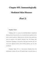
Figure 10.1 Multiple comedones and
seborrhoea in acne.
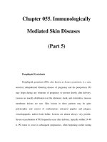
Figure 10.2 Multiple comedones in acne.
Note the blackened tips from melanin.
of follicular debris and compacted sebum. They have pigmented tips from the
melanin pigment deposited by the follicular epithelium at this level (Fig. 10.2).
Accompanying the visible comedones are numerous invisible comedones, many
of which do not have pigmented tips.
Inflamed, reddened papules develop from blocked follicles. These are often
quite tender to the touch and may be set quite deep within the skin (Fig. 10.3).
Sometimes they develop pus at their tips (pustules), but these may also arise independently. In a few patients, some of the papules become quite large and persist
for long periods – they are then referred to as nodules.
In severely affected patients, the nodules liquefy centrally so that fluctuant cysts
are formed. In reality, the lesions are pseudocysts, as they have no epithelial lining. This type of severe acne is known as cystic or nodulocystic acne and can be
very disabling and disfiguring.
When the large nodules and cysts eventually subside, they leave in their wake
firm, fibrotic, nodular scars, which sometimes become hypertrophic or even
keloidal (Fig. 10.4a). The scars are often quite irregular and tend to form ‘bridges’
(Fig. 10.4b). Even the smaller inflamed papules can cause scars and these tend to
be pock-like or are triangular indentations (‘ice-pick scars’: Fig. 10.5).
There is a very rare and severe type of cystic acne known as acne fulminans
in which the acne lesions quite suddenly become very inflamed. At the same time
the affected individual is unwell and develops fever and arthralgia. Laboratory
150
Acne
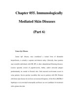
Figure 10.3 Acne papules.
(a)
(b)
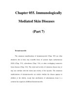
Figure 10.4 (a) Nodular scars in acne. These lesions developed following the
resolution of inflamed acne papules. (b) Hypertrophic scarring in a bridging pattern.
investigation reveals a polymorphonuclear leucocytosis and odd osteolytic lesions
in the bony skeleton. The cause of this disorder is not clear, although it has been
suggested that it is due to the presence of a vasculitis that is somehow precipitated
as a result of the underlying acne.
151
Acne, rosacea and similar disorders
Figure 10.5 Pock scarring of acne.
SITES AFFECTED
Any hair-bearing skin can develop acne, but certain areas are much more
prone than others (Fig. 10.6). These acne-prone areas tend to have hair follicles
with small terminal hairs and larger sebaceous glands (sebaceous follicles). The
face and particularly the skin of the cheeks, lower jaw, chin, nose and forehead are
usually affected. The scalp is not involved, but the back of the neck, front of the
chest, the back and shoulders are all ‘favoured areas’ for the development of
lesions.
In patients with severe acne, it is quite common for other areas to be affected,
including the outer aspects of the upper arms, the buttocks and thighs.
CLINICAL COURSE
For most of those affected, the disorder is annoying and may be troublesome, but
is not of enormous significance because it is limited in extent and only lasts a few
months or at the most a year. For the unfortunate few, the condition is a disaster,
as it is disfiguring, disabling and persistent, with wave after wave of new lesions.
Although the natural tendency is for resolution, it is difficult to know in any individual patient when the condition will improve. The majority have lost their acne
by the age of 25 years, but some tend to have the occasional lesion for very much
longer. In some women there is a pronounced premenstrual flare of their acne
some 7–10 days before the menses begin.
152
Acne
(a)
(b)
Figure 10.6 Diagram to show common sites of involvement due to acne on
(a) the front of the trunk and face, and (b) the back of the trunk.
Acne improves in the summertime and sun exposure seems to improve the
condition of many patients. However, the heat does not produce improvement
and, indeed, can make it much worse. Soldiers with acne in hot, humid climates
often become disabled by it suddenly worsening, with large areas of skin covered
by inflamed and exuding acne lesions, and have to be evacuated home or to a
cooler climate.
EPIDEMIOLOGY
Some 70 per cent of the population develop some clinically evident acne at some
point during adolescence and early adult life, but perhaps only 10–20 per cent
request medical attention for the problem. This proportion varies in different
parts of the world, depending on the racial mixture, the affluence and the sophistication of medical services.
153
Acne, rosacea and similar disorders
Figure 10.7 Infantile acne.
Figure 10.8 Steroid acne. The lesions
tend to be more uniform in appearance
than in ‘ordinary’ acne.
The variations in incidence in different ethnic groups have not been well characterized, although it does appear that Eskimos and Japanese suffer less from acne
than do Western Caucasians.
Onset is usually at puberty or a little later, although many patients do not
appear troubled until the age of 16 or 17 years. Men appear to be affected earlier
and more severely than women. Older age groups are not immune and it certainly
is not rare to develop acne in the sixth, seventh or even eighth decade.
Acne lesions sometimes appear on the cheeks and chin of infants a few weeks
or months of age and even a little later than that (Fig. 10.7). This infantile acne is
usually trivial and short lived, but can occasionally be troublesome.
SPECIAL TYPES OF ACNE
Acne from drugs and chemical agents
Androgens provide the normal ‘drive’ to the sebaceous glands. It is the increased
secretion of these hormones that is responsible for the increased sebum secretion
at puberty. When given therapeutically for any reason, they can also cause an
eruption of acne spots.
Glucocorticoids, such as prednisolone, when given to suppress the signs
of rheumatoid arthritis or some other chronic inflammation, can also induce
troublesome acne (Fig. 10.8). Why this should be so has never been adequately
154
Acne
Figure 10.9 Comedones and inflamed
follicular papules from tar application.
Figure 10.10 Acne due to cosmetics.
explained. Glucocorticoids do not seem to increase the rate of sebum secretion, and the acne that results is curiously monomorphic in that sheets of acne
lesions appear (unlike ordinary acne) all at the same stage of development.
Interestingly, corticosteroid creams can, uncommonly, also cause acne spots at the
site of application.
Oil acne
Workers who come into contact with lubricating and cutting oils develop an acnelike eruption at the sites of contact, consisting of small papules, pustules and
comedones. This is often observed on the fronts of the thighs and forearms, where
oil-soaked overalls come in contact with the skin. A similar ‘acneiform folliculitis’
sometimes arises at sites of application of tar-containing ointments during the
treatment of skin diseases (Fig. 10.9).
Some cosmetics seem to aggravate or even cause acne. This is because they
sometimes contain comedo-inducing (comedogenic) agents, such as cocoa butter
and derivatives and some mineral oils, that can induce acne. This cosmetic acne is
less of a problem now that cosmetic manufacturers are aware of it (Fig. 10.10).
Chloracne
Chloracne is an extremely severe form of industrial acne due to exposure to complex
chlorinated naphthalenic compounds and dioxin. Epidemics have occurred after
155
Acne, rosacea and similar disorders
industrial accidents such as occurred in Serveso in Italy, in which the population
around the factory was affected. The compounds responsible are extremely potent,
and lesions continue to develop for months after exposure. Typically, numerous
large, cystic-type lesions occur in this form of industrial acne.
Excoriated acne
This disorder is most often seen in young women. Small acne spots around the
chin, forehead and on the jaw line are picked, squeezed and otherwise altered by
manual interference. The resulting papules are crusted and often more inflamed
than routine acne spots. Often, the patients have little true acne and the main cosmetic problem is the results of the labour of their fingers!
PATHOLOGY, AETIOLOGY AND PATHOGENESIS
Histologically, the essential features are those of a folliculitis with considerable
inflammation. The exact histological picture depends on the stage reached at the
time of biopsy. Usually, it is possible to make out the remnants of a ruptured follicle. In the earliest stages, a follicular plug of horn (comedone) can be identified.
Later, fragments of horn appear to have provoked a violent mixed inflammatory
reaction with many polymorphs and, in places, a granulomatous reaction with
many giant cells and histiocytes (Fig. 10.11). In older lesions, fibrous tissue is
deposited, indicating scar formation.
Figure 10.11 Pathology of inflamed
acne papules showing a ruptured follicle
and a dense inflammatory cell infiltrate
composed predominantly of polymorphs.
156
Acne
What do we believe is the sequence of events? In the first place, patients with
acne have a higher rate of sebum secretion rate (SER) compared to matched
control subjects and, furthermore, there is some correlation between the extent of
the increase in the SER and the severity of the acne.
Acne first appears at puberty, at which time there is a sudden increase in the
level of circulating androgens. Eunuchs do not get acne, and the administration of
testosterone provokes the appearance of acne lesions. Sebaceous glands are predominantly ‘androgen driven’ and few other influences are as important.
Follicular obstruction also plays an important role. Comedones are early lesions
and microscopically it is commonplace to find horny plugs in the follicular canals.
Changes have been described in the follicular epithelium suggesting that there is
abnormal keratinization at the mouth of the hair follicle.
Pathogenic bacteria are not found in acne lesions and are not involved in
the pathogenesis. It is possible, nonetheless, that the normal flora has a role
to play. The flora consists of Gram-positive cocci – the micrococci (also known
as Staphylococcus epidermidis) – and Gram-positive bacteria – Propionibacterium
acnes. In addition, there are also yeast-like micro-organisms known as Pityrosporum
ovale. The Propionibacteria are microaerophilic and lipophilic, so that they are
ideally suited to living in the depths of the hair follicle in an oily milieu, and it is
not surprising that they increase in numbers during puberty when their food
supply, in the form of sebum, increases. The normal follicular flora may be
responsible for hydrolysing the lipid esters of sebum, liberating potentially irritating fatty acids. The constituents of sebum and of skin surface lipid (after bacterial hydrolysis) are given in Table 10.1.
How can these observations be linked? An acceptable hypothesis is set out in
Figure 10.12, in which it is suggested that the important inflammatory lesions of
acne are the result of follicular rupture.
Table 10.1 Main
constituents of sebum and
skin surface lipid
Sebum
Triglycerides
Cholesterol ester
Squalene
Wax esters
Skin surface lipid
Sebum lipids
Fatty acids
Monoglycerides
Diglycerides
TREATMENT
Typically, unasked for advice from the family is given in which the sufferer
is blamed in one way or another for having the disorder and accused of doing
too much of one thing or not enough of the other. Consequently, many forms
of familial or folk treatments seem to be more in the nature of punishments
than anything else. Dietetic and social restrictions are typical, as is more frequent
washing, which is another tactic adopted by well-meaning but misguided family
and friends.
Fortunately, most acne patients improve spontaneously after a few months.
Those who do not, find their way to the pharmacist and purchase preparations
containing benzoyl peroxide or other antimicrobial compounds, or sulphur or
salicylic acid. Many with milder degrees of acne will be helped by these medications. It is only those with resistant, recalcitrant and more severe types of acne
who reach the physician. Perhaps only 10 per cent of those with clinical acne in
the UK see their practitioner.
157
Acne, rosacea and similar disorders
Comedone
Irritation of follicular wall
Increased lipolysis
Ruptured wall
Follicle lumen
Increased microflora
Inflammation
Increased sebum secretion
Sebaceous gland
Figure 10.12 Diagram to show suggested events in the pathogenesis of acne.
Basic principles
Treatment may be aimed at:
●
●
●
●
reducing the bacterial population of the hair follicles to cut down the hydrolysis of lipids (antimicrobial agents)
encouraging the shedding of the follicular horny plugs to free the obstruction
(comedolytic agents)
reducing the rate of sebum production, either directly by acting on the sebaceous glands or indirectly by inhibiting the effects of androgens on the sebaceous
glands (anti-androgens)
reducing the damaging effects of acne inflammation on the skin with antiinflammatory agents (Table 10.2).
General measures
Patients with acne are often depressed and may need sympathetic counselling and
support. There is no evidence that particular foodstuffs have any deleterious effect
or that washing vigorously will help remove lesions. These and other myths
should be dispelled and replaced with a straightforward explanation of the nature
of the disorder, its natural history and treatment.
Topical treatment
Currently, the most popular form of topical preparation is a gel, cream or alcoholbased lotion.
158
Acne
Table 10.2 Treatments for acne
Topical
Antimicrobial
Oral
Comedolytic
Antimicrobial
Sebum suppressive
Benzoyl peroxide
Tretinoin
Tetracycline
Isotretinoin
Tetracycline
Isotretinoin
Minocycline
Erythromycin
Adapalene
Doxycycline
Cyproterone and
ethinylestranol
Erythromycin
Spironolactone
Clindamycin
Azelaic acid
Topical retinoids
These are comedolytic. Tretinoin-containing preparations are not bactericidal,
but are nonetheless effective. The cis-isomer of tretinoin – isotretinoin – is also
used successfully for the treatment of acne. Adapalene is a recently introduced,
effective topical retinoid that is also useful.
The side effects from the use of retinoid preparations include some pinkness
and slight scaling of the skin surface, especially in fair, sensitive-skinned individuals. For the most part, this ‘dryness’ of the treated area is tolerable and decreases
after continual usage. It is less marked with adapalene.
Sulphur (as elemental sulphur 2–10 per cent) has been used traditionally as a
treatment for acne. It seems to be helpful for some patients, but has dropped out
of fashion. Its efficacy probably depends on both its antimicrobial action and its
comedolytic activity.
Other agents employed to remove blackheads include abrasive preparations.
These contain particles of substances such as aluminium oxide or polyethylene
beads, which literally abrade the skin surface and ‘liberate’ the comedones.
Topical antibiotics
Erythromycin (1–2 per cent) and clindamycin (2 per cent) preparations are quite
effective for mild and moderate types of acne. Tetracycline preparations (2 per
cent) are slightly less effective. Fortunately, these antibiotics have a low tendency
to sensitize and are not often responsible for allergic contact dermatitis, although
they may cause a minor degree of direct primary irritation.
Other antimicrobial compounds
Bacterial resistance to erythromycin frequently develops and may prove a problem in the future.
Systemic treatment
Antibiotics
Tetracyclines
Systemic tetracyclines have been the sheet anchor of treatment for moderate and
severe acne for many years. Patients with many papular lesions involving several
159
Acne, rosacea and similar disorders
sites are suitable for systemic tetracyclines. It is usual to start treatment with a
dose of 250 mg t.i.d. or 6-hourly, and then, when there is a response, to reduce the
dose to that required to keep the patient free of new lesions. The improvement
usually begins 4–8 weeks after starting treatment and continues over the next 2–3
months. Some 70 per cent of patients can be expected to improve on this regimen.
Treatment may have to be maintained for several months or, exceptionally, even
longer. With tetracycline and oxytetracycline, the drug should be given 30 minutes
before a meal to prevent interference with absorption. The newer minocycline and
doxycycline are given in smaller doses (50 mg or 100 mg) once or twice per day
and their absorption does not seem to be affected by food.
Side effects with the tetracyclines are few and not usually serious. Gastrointestinal
discomfort and diarrhoea occasionally occur. Photosensitivity was mainly a problem with older, now no longer used, analogues. Fixed drug eruption and, rarely,
other acute drug rashes develop. Minocycline can cause a dark-brown pigmentation
of the skin or acne scars or acral areas on the exposed part of the skin after longcontinued use in a small number of patients.
Tetracyclines must not be given to pregnant women, as they are teratogenic,
and must not be given to infants, as they cause a bone and tooth dystrophy in
which these structures become deformed and discoloured.
Erythromycin
The efficacy of erythromycin in acne is similar to that of the tetracyclines. The
starting dosage is 250 mg 6-hourly for the first few weeks, with reduction after a
response has begun. Subsequently, management is as for the tetracyclines. Side
effects are usually minor and restricted to nausea.
Other antibiotics and antimicrobials
Clindamycin, the quinolines and the sulphonamides are other drugs that have
been used systemically for acne. None is more effective than the tetracyclines, but
they may be suitable for patients who are either intolerant or who no longer
respond to the tetracyclines or erythromycin. Side effects are more common and
sometimes of a serious nature (e.g. blood dyscrasias).
Isotretinoin (13-cis-retinoic acid)
The large majority of patients with acne will respond to topical or some combination of topical and systemic drugs. However, some severely affected patients
may not, and for them there is another drug that can offer relief. This agent is the
retinoid isotretinoin (the same cis-isomer of tretinoin used topically). It reduces
sebum secretion by shrinking the sebaceous glands and may also alter keratinization of the mouth of the hair follicle and have an anti-inflammatory action.
It is given in a dose of 0.5–1.0 mg/kg body weight per day, usually for a
4-month period. The response after a few weeks is to inhibit new lesions in more
than 80 per cent of patients. Patients with many large cystic lesions affecting the
trunk as well as the head and neck region take longer to respond and may need
more than one 4-month course.
160
Acne
Unfortunately, toxic side effects are frequent. They range from the trivial, of
which the most common is drying and cracking of the lips, to the very serious,
which include teratogenicity, hepatotoxicity, bone toxicity and a blood lipidelevating effect. The teratogenic effects are very worrisome, as the acne age group
is almost identical to the reproductive age group. The effects on the fetus include
facial, cardiac, renal and neural defects and are most likely to arise if the drug is
taken during the first trimester. Some 30–50 per cent of pregnancies during which
the drug was taken have been affected. Because of this, it is strongly recommended
that if it is planned to prescribe isotretinoin for women who can conceive, effective contraceptive measures must also be planned and used during and for 2 months
after stopping the drug.
Hepatotoxicity is rare, although a small rise in liver enzymes is common. A rise
in triglycerides and cholesterol, such that the ratio of very low-density lipoproteins to high-density lipoproteins is increased, regularly occurs, and overall there
is a 30 per cent rise in lipid levels. This is not likely to be a problem for most
patients with acne, but may be for older patients. The same is true for the bone
toxicity. A variety of bone anomalies have been described, including disseminated
interstitial skeletal hyperostosis and osteoporosis, but these are not likely to be a
problem for acne subjects. The drug has also been accused of causing severe
depression, leading to suicide in some cases. The evidence for this is not strong, as
severe acne patients are often depressed before starting treatment. Because of the
toxicities of this important drug it can only be prescribed from hospitals in the UK.
Case 9
Julia was 15 when she started to develop embarrassing acne. She had noticed
that her skin had been very greasy skin for the last few months. New spots
appeared every day and she spent hours in front of the mirror trying to squeeze
out blackheads and get rid of pustules. It made her quite depressed and matters
were made worse by her parents telling her that she didn’t wash her face enough
and that going to discos didn’t help her skin. Fortunately, her GP was more
sympathetic and prescribed a benzoyl peroxide preparation and oral doxycycline,
which made a big improvement after about 6 weeks.
Anti-androgens
Anti-androgens inhibit androgenic activity and reduce sebum secretion. Currently,
only one anti-androgen preparation is available – Dianette. This is a mixture of an
anti-androgen, cyproterone acetate (2 mg), and an oestrogen, ethinyl oestradiol
(35 g). It is a central anti-androgen, blocking the pituitary drive to androgen secretion. It also suppresses ovulation and acts as an oral contraceptive. It is not suitable
for men because of its feminizing properties. It improves acne after some 6–8 weeks
of use, but is not as effective as isotretinoin. It is associated with a number of minor
side effects, essentially those associated with taking oral contraceptives.
Spironolactone, the potassium-sparing diuretic, has also been found to have
anti-androgenic effects and has occasionally been used as a treatment for acne.
161
Acne, rosacea and similar disorders
Rosacea
DEFINITION
Rosacea is a chronic inflammatory disorder of the skin of the facial convexities,
characterized by persistent erythema and telangiectasia punctuated by acute
episodes of swelling, papules and pustules.
CLINICAL FEATURES
Sites affected
The cheeks, forehead, nose and chin are the most frequently affected areas, making a typical cruciate pattern of skin involvement (Fig. 10.13). The flexures and
periocular areas are conspicuously spared. Uncommonly, the neck and the bald
area of the scalp in men are also affected. Sometimes only one or two areas are
affected, and this makes diagnosis quite difficult.
The lesions
The most characteristic physical sign is that of persistent erythema, often accompanied by marked telangiectasia (Fig. 10.14). The disorder may not progress
Figure 10.13 Typical rosacea with
involvement of the cheeks, forehead
and chin.
162
Figure 10.14 Erythema and
telangiectasia in rosacea.
Rosacea
(a)
(b)
Figure 10.15 (a) and (b)
Papules of facial skin in
rosacea.
beyond this ‘erythemato-telangiectatic’ state but, even if it does not, the bright red
face causes considerable social discomfort and often marked depression. Such
patients also complain of frequent flushing at the most trivial stimuli.
Superimposed on this persistent background of erythema are episodes of
swelling and papules, which develop for no very obvious reason (Fig. 10.15). The
papules are a dull red, dome shaped and non-tender, in contrast to acne, in which
they tend to be irregular and tender. Pustules also occur, but are less frequent than
in acne; blackheads, cysts and scars do not.
DIFFERENTIAL DIAGNOSIS
Any red rash of the face may be confused with rosacea (Table 10.3).
Papular rashes of the face seem to cause most problems. Acne occurs in a
younger age group and is usually distinguished by the greasy skin, comedones and
scars as well as lesions on sites other than the face. However, in some patients, the
presence of persistent erythema can make differentiation quite difficult. Perioral
dermatitis (see page 168) should not be difficult to differentiate, as this disease is
mainly distributed around the mouth and there is no background of erythema.
Systemic lupus erythematosus may superficially resemble rosacea, become of the
symmertrical butterfly erythema but there are no symptoms of systemic disease
in rosacea. Dermatitis of the face (including seborrhoeic dermatitis) is marked by
scaling, which is not characteristic of rosacea.
Polycythaemia rubra vera gives the face a plethoric appearance. The carcinoid syndrome is characterized by reddened areas on the face in the same
163
Acne, rosacea and similar disorders
Table 10.3 Differential diagnosis of rosacea
Disorder
Positive discriminants
Skin disorders
Acne
Scars, seborrhoea, cysts, back and chest involvement
Seborrhoeic dermatitis
Scaling involvement of flexures
Perioral dermatitis
Micropapules, perioral and paranasal involvement
Systemic disorders
Systemic lupus
erythematosus
Rash on light-exposed areas, arthropathy, positive
antinuclear factor, haematological findings
Dermatomyositis
Mauve-lilac rash around the eyes, with swelling, rash
on backs of fingers, muscle tenderness, pain and
weakness, positive laboratory findings
Carcinoid syndrome
Marked telangiectasia, flushing attacks, hepatomegaly
Polycythaemia rubra vera
General facial redness and suffusion, possibly
hepatosplenomegaly
distribution as in rosacea, but the condition is accompanied by severe systemic
symptoms.
Dermatomyositis is characterized by mauvish erythema around the eyes, but
the pain, tenderness and weakness of limb girdle muscles should quickly distinguish this disease.
COMPLICATIONS
Rhinophyma
This occurs mainly in elderly men, although it occasionally occurs in women too.
The nose becomes irregularly enlarged and ‘craggy’, with accentuation of the pilosebaceous orifices (Fig. 10.16). At the same time, the nose develops a mauve or
dull-red discoloration with prominent telangiectatic vessels coursing over it (Fig.
10.17). Popular names for this include ‘whisky-drinkers nose’ and ‘grog blossom’,
but it is not due to alcoholism.
Lymphoedema
Persistent lymphoedema is another unpleasant, though uncommon, complication
of rosacea seen predominantly in men. The swollen areas are usually a shade of
red and may persist when the other manifestations of rosacea have remitted.
Ocular complications
Some 30–50 per cent of patients with acute papular rosacea have a blepharoconjunctivitis. This is usually mild, but some patients complain bitterly of soreness and
164
Rosacea
Figure 10.16 Severe, irregular, craggy
enlargement of the nose due to
rhinophyma.
Figure 10.17 Rhinophyma with
prominent telangiectasia.
grittiness of the eyes. Some of this may be the result of keratoconjunctivitis sicca,
which appears to be quite common in rosacea. Styes and chalazion are also more
common in rosacea. Keratitis is a rare, painful complication occurring in men, in
which a vascular pannus moves across the cornea, producing severe visual defects.
NATURAL HISTORY
Rosacea tends to be a persistent disease and the tendency for patients to develop
episodes of acute rosacea remains for many years after appropriate treatment has
calmed down an attack.
EPIDEMIOLOGY
Rosacea is quite a common disorder, but its exact prevalence is not known and
varies in different communities. The disorder is essentially one of fair-skinned
Caucasians. It seems particularly common in Celtic peoples and in individuals
from northwest Europe. It is only occasionally seen in darker-skinned and Asian
skin types and is rare in black-skinned individuals. It has been claimed that it is
more common in women, but this may be merely a reflection of the disorder
being of more concern to women.
165
Acne, rosacea and similar disorders
Figure 10.18 Pathology of rosacea showing marked
telangiectasia, dermal oedema and marked solar
degenerative change.
Figure 10.19 Pathology of rosacea showing
inflammatory cell infiltrate with many lymphocytes
and giant cells around blood vessels.
PATHOLOGY
There is no single pathognomonic feature, but there is a characteristic constellation of features in histological sections that makes skin biopsy a useful test when
the clinical diagnosis is uncertain. A feature common to all rosacea skin samples
is the presence of disorganization, solar damage, oedema and telangiectasia in the
upper dermis (Fig. 10.18). When there are inflammatory papules, the blood vessels are encircled by lymphocytes and histiocytes, amongst which giant cell systems are sometimes found (Fig. 10.19). In rhinophyma, apart from abnormalities
in the fibrous dermis and inflammation, there is also marked sebaceous gland
hyperplasia.
AETIOLOGY AND PATHOGENESIS
The cause of rosacea remains uncertain. Historically, dietary excess, alcoholism,
gastrointestinal inflammatory disease, malabsorption and psychiatric disturbance
have all been though to be responsible, but controlled studies fail to implicate
these agencies. The role of the mite Demodex folliculorum, a normal commensal
of the hair follicle, is also unclear. Although it is found in increased numbers in
rosacea, this increase may result from the underlying disorder in which there is
follicular distortion and dilatation.
Environmental trauma appears to play an important role in the development
of rosacea. The disorganization of upper dermal collagen, the excess of solar elastotic degenerative change and the predominance in fair-skinned types all point to
the importance of damage to the upper dermis. Inadequate dermal support to the
vasculature, which then dilates, allows pooling of the blood in this site. This pooling may then itself compromise endothelial function and ultimately result in
episodes of inflammation (Fig. 10.20).
166
Rosacea
Cold wind
Solar UV
Inherently
individual
susceptible
(Erythema and
telangiectasia)
DERMAL
DYSTROPHY
TELANGIECTASIA
Heat
Damage
to dermis
Damage to skin
sustained in acne
Pooling of blood
Endothelial damage
Inflammation
(Papules and
pustules)
Leakage of potentially
inflammatory substances
(swelling)
Figure 10.20 Possible sequence of events in rosacea.
Depressed delayed hypersensitivity and deposits of immunoprotein in facial
skin have also been reported, suggesting that the immune system is involved in the
pathogenesis.
TREATMENT
Systemic treatment
The acute episodes can be calmed with systemic tetracycline, erythromycin or
metronidazole, using the full antibacterial dosage until the condition improves
and then a dose sufficient to maintain improvement. Initial improvement usually
occurs within the first 3–4 weeks of treatment. It would be typical for a patient to
start tetracycline 250 mg 6-hourly for 3 weeks and then receive the drug three
times daily for a further 3 or 4 weeks. At that time, reduction to twice-daily dosage
would be made and maintained until stopping (perhaps at 10 or 12 weeks) did not
result in the appearance of further papules. Minocycline or doxycycline 50 mg
once or twice per day is more convenient. Erythromycin is also effective and the
same dose regimen applies as for tetracycline. Metronidazole is not often given
because of its side-effect profile. It has a disulfiram-like effect, causing alcohol
intolerance. Other side effects include nausea and blood dyscrasias.
Isotretinoin may help some patients, particularly those who have rhinophyma,
as it has been shown that it reduces the size of the enlarged nose as well as reducing the numbers of papules present.
Topical treatments
Topical corticosteroids are definitely contraindicated. Although they may suppress
the inflammatory papules, they tend to make the face redder and more telangiectatic,
167
Acne, rosacea and similar disorders
Figure 10.21 Intense erythema and
telangiectasia in rosacea due to
mistreatment with potent topical
corticosteroids.
presumably because they cause even more upper dermal wasting and exposure of the
subpapillary venous plexus (Fig. 10.21).
Facial skin may be sore and uncomfortable in rosacea and the use of emollients
can give some symptomatic relief as well as discouraging the use of topical corticosteroids! Sunscreens are of help in preventing further solar damage. Preparations
of 0.75–1.5 per cent metronidazole in either a cream or gel base seem capable of
reducing the inflammatory papules as efficiently and as quickly as systemic tetracycline. Topical azelaic acid (20 per cent) has also been shown to be effective.
How systemic antibiotics, or metronidazole, systemic or topical, achieve their
effects in rosacea is not clear. Treatment with the pulsed dye laser can greatly
improve the erythema in rosacea.
Perioral dermatitis
DEFINITION
Perioral dermatitis is a not uncommon, inflammatory disorder of the skin around
the mouth, characterized by the occurrence of micropapules and pustules.
CLINICAL FEATURES
Many minute, pink papules and pustules develop around the mouth, sparing the
area immediately next to the vermillion of the lips (Fig. 10.22). Lesions sometimes
168
Summary
Figure 10.22 Perioral dermatitis. There
are many tiny papules around the mouth.
involve the nasolabial grooves and, in severely affected patients, also affect the skin
at the sides of the nose. There is no background of erythema, distinguishing the
condition from rosacea.
The condition develops insidiously and seems to persist until treated. Recurrence is uncommon.
Perioral dermatitis is most common in young women aged 15–25 years, being
quite rare in men and in older women. Its exact incidence is unknown, but it is of
interest to know that it was first recognized in the late 1960s, seemed quite common in affluent Western communities in the 1970s and then appeared to become
less frequently observed in the 1980s, reappearing once again in the 1990s. Many
have suspected that the use of topical corticosteroids is to blame. Patients usually
respond to a course of systemic tetracycline as for rosacea for a period of 4–8
weeks. No topical treatments are indicated.
Summary
● Acne occurs in most individuals during adolescence.
It is characterized by increased sebum secretion
and the formation of comedones.
● Comedones are dilated hair follicles containing
horny plugs, the tips of which are black due
to melanin (blackheads). These blocked follicles
often leak and may rupture, causing inflammatory
papules and pustules, and when several are
involved, give rise to acne cysts (pseudocysts in
reality) form. The inflammation causes tissue
destruction and hypertrophic, keloidal, pock-like
or ice-pick scars.
● The face (cheeks, chin, forehead, lower jaw and
nose), back of the neck, back, shoulders and chest
are the commonest sites involved.
● The disorder is not troublesome for most, but
discomforting and embarrassing for many, and a
●
●
complete disaster in a few. It may only last a few
months, but can persist for years. Older subjects
are not immune and mild acne occasionally occurs
in infants. Oils and greases can aggravate or even
cause acne.
The rate of sebum secretion is increased by the
surge in testosterone levels at puberty.
Propionibacterium acnes – a major component of
the normal follicular flora – is microaerophilic and
lipophilic. These bacteria greatly increase in
numbers in the dilated and plugged follicle. The
inflammation of acne may well be caused by the
leakage of follicular content and bacterial
degradation products, including irritating fatty acids,
into the dermis.
Only a small proportion of acne sufferers (perhaps
10 per cent) are seen by their general practitioners.
169
Acne, rosacea and similar disorders
●
●
●
170
The basic principles of treatment are to reduce the
bacterial population, encourage shedding of
follicular plugs (comedolysis), reduce the rate of
sebum production and reduce the degree of
inflammation.
Topical retinoids (tretinoin, isotretinoin and
adapalene) are comedolytic agents. They are quite
effective but irritating. Topical antibiotics
(erythromycin, clindamycin and tetracycline) are
quite useful, as are preparations of benzoyl
peroxide, which are both antimicrobial and
comedolytic.
When the acne is severe, systemic treatments
are needed. Systemic tetracyclines (oxytetracycline,
doxycycline or minocycline) and erythromycin
are most often used. They may need to be
given over some months. Systemic isotretinoin
is the most effective agent for severe acne,
but is capable of causing many adverse side
effects, including fetal deformities if given to
pregnant women. An anti-androgen preparation
containing cyproterone acetate and ethinyl
oestradiol is also used in female patients and
may be helpful.
Rosacea may be defined as a chronic
inflammatory disorder of the convexities of facial
skin, characterized by persistent erythema and
telangiectasia, punctuated by acute episodes of
swelling, papules and pustules. It is quite
●
●
●
●
●
common, affecting mainly fair-complexioned
adults aged 30–60.
The cheeks, chin, nose and forehead are mainly
affected, but the neck and the bald scalp of
men my occasionally be involved. The papules
are unlike those of acne, being non-tender
and dome shaped. Blackheads, cysts and
scars are not seen. Rhinophyma (irregular
nasal swelling), keratitis and persistent
lymphoedema of facial skin are complications
seen mainly in men.
Rosacea needs to be distinguished from acne,
seborrhoeic dermatitis and other disorders with
reddened facial skin, such as lupus erythematosus
and dermatomyositis.
The cause is unknown, but the occurrence in
fair-skinned individuals on light-exposed sites and
the presence of a marked degree of solar damage
histologically suggest that photodamage plays a
major role.
The condition tends to persist, but acute episodes
usually respond to oral tetracycline or erythromycin
or topical metronidazole. Topical corticosteroids
tend to aggravate the disorder.
Perioral dermatitis is a disorder in which
micropapules and papulopustules occur
periorally and paranasally in young women.
It responds to oral tetracycline but not to topical
preparations.
C H A P T E R
11
Wound healing
and ulcers
Principles of wound healing
171
Venous hypertension, the gravitational syndrome and venous ulceration
173
Ischaemic ulceration
177
Decubitus ulceration
178
Neuropathic ulcers
179
Less common causes of ulceration
180
Diagnosis and assessment of ulcers
182
Summary
182
Principles of wound healing
Wound healing is a complex and fundamental activity of all damaged body structures. The same principles underlie the healing of cuts, abrasions, ulcers and areas
damaged by chemical attack, invasion by micro-organisms or immune reactions.
Healing of the skin damaged by a physical insult may be divided into:
●
●
●
an immediate haemostatic phase,
an early phase of re-epithelialization,
a later phase of dermal repair and remodelling (Fig. 11.1).
It is hoped that better understanding of the complex interactions and their
controls will result in new techniques and substances for the treatment of nonhealing wounds. Persistent non-healing ulcers of the skin are very common and
cause much unhappiness, disablement and economic loss.
FACTORS IMPORTANT IN THE HEALING OF WOUNDS
●
Adequate supplies of nutrients and oxygen are required for efficient healing;
when the blood supply is compromised, healing is delayed. Vitamin C and zinc
deficiencies are amongst the deficiency states also associated with delayed
wound healing.
171
Wound healing and ulcers
E
E
FC
0–12 hours
DCT
BV
(a)
ME
E
E
F, MF, M
GT
12 hours–4 days
BV
SC
(b)
BV
DC
F, M
4–10 days
(c)
●
●
172
Figure 11.1 The sequence of events after incisional
wounding of the skin. (a) 0 to 12 hours. Initially, the
small blood vessels constrict and then platelets plug the
endothelial gaps. The extravasated blood clots form a
temporary plug for the wound. White cells accumulate at
the interface between the damaged and the normal
tissue. (b) 12 hours to 4 days. After some 18–24 hours,
epidermal cells actively move on to the surface of the
defect. Epidermal cells at the sides of the wound divide
some hours later to make good the loss. Epidermis also
sprouts from the cut ends of the sweat coils and hair
follicles. After 2–4 days, new capillaries start to sprout
and vascularize the granulation tissue in the wound
cavity. Damaged connective tissue is destroyed and
removed by macrophages, and new collagen is secreted
by fibroblasts. Myofibroblasts are fibroblastic cells that
develop the power to contract and are responsible for
wound contraction. (c) 4 to 10 days. Between 4 and 10
days after wounding, the wound cavity has become
covered with new epidermis, whose stratum corneum
does not possess normal barrier efficiency until the end
of this period. The granulation tissue has been replaced
by a new dermis whose collagenous fibres are not yet
orientated. In the later stages, remodelling takes place
so that orientation of the dermal collagenous bundles to
the original lines of stress occurs. Scar formation occurs
when there has been significant damage to the dermis.
The epidermis ultimately develops a normal profile and
the vasculature is also restored to normal contractility.
E ϭ epidermis; DCT ϭ dermal connective tissue;
BV ϭ blood vessel; FC ϭ fibrin clot; ME ϭ migrating
epidermis; F ϭ fibroblasts; MF ϭ myofibroblasts;
M ϭ macrophages; DC ϭ dermal collagen;
GT ϭ granulation tissue; SC ϭ sweat coil.
Persistent infection with tuberculosis, Mycobacterium ulcerans or syphilis
causes ulcerative conditions directly due to an infection. Any ulcerated area
becomes contaminated by microbes in the environment and often this ‘secondary infection’ causes further tissue destruction.
In some uncommon congenital disorders, there is delayed wound healing
because the orderly sequence is disrupted. These disorders include factor XIII
deficiency, in which there are abnormalities of cross-linking of fibronectin and
Venous hypertension, the gravitational syndrome, venous ulceration
Table 11.1 Common causes of persistent ulcers
Condition
Aetiopathogenesis
Site(s)
Features
Venous ulcer
Venous incompetence, venous
hypertension, tissue oedema
and inflammation
Medial malleolus commonly
and ankle nearby
Sloughy; signs of venous
hypertension
Ischaemic ulcer
Atherosclerosis in most
instances
Mostly feet and lower legs
Painful; occurring in
atrophic skin
Ulcer due to vasculitis
Polyarteritis nodosa and
Henoch–Schönlein purpura
are examples of vasculitic
disorders causing ulcers
Commonly on the legs,
but anywhere can be
affected
Lesions often start as
purpuric patches or
nodules
Neuropathic ulcer
Inadvertent repetitive injury
because of sensory
loss – common in diabetes
and leprosy
Soles of feet particularly
Deeply perforating ulcers
are characteristic
Decubitus ulcer
Pressure on skin of dependent
parts in unconscious or
paralysed patients
Sacrum ischial region,
heels, scapular region,
back of head, elbows
Deep sloughy ulcers
collagen, protein C deficiency and Marfan’s syndrome, in which there are
abnormalities of dermal connective tissue repair.
The common causes of persistent leg ulcers are given in Table 11.1.
Venous hypertension, the gravitational syndrome
and venous ulceration
VENOUS HYPERTENSION
Epidemiology
It has been estimated that between 0.5 and 1.0 per cent of the population of
the UK suffers from venous ulcers at any one time. The disorder is most often seen
after the age of 60 and women are more often affected – particularly the multiparous. It is mostly a problem of the poor and underprivileged. Interestingly, it
does not occur with equal prevalence in all racial groups, for example it is rare in
Arabic peoples.
Pathology and pathogenesis
When venous return is impeded, hypertension develops in the venous circulation
behind the blockage. This results in the development of dilatation of the small
173