Ebook Reoperations in cardiac surgery: Part 2
Bạn đang xem bản rút gọn của tài liệu. Xem và tải ngay bản đầy đủ của tài liệu tại đây (21.46 MB, 197 trang )
Chapter 14
Reoperations After Mustard and Senning
Operations
J. Stark
Introduction
Senning introduced the physiological repair of
transposition of the great arteries in 1958
(Senning 1959) and Mustard published his
experience with the atrial switch in 1964
(Mustard 1964). The Mustard operation soon
became the operation of choice, and survival
rates of over 90% for patients with simple
transposition were reported (Waldhausen et al.
1971; Lindesmith et al. 1973; Ebert et al. 1974;
Stark et al. 1974a). The original concept of the
Mustard operation was a two-stage correction.
A Blalock-Hanlon atrial septectomy enabled
a sick infant to survive. Because of the fear
that the small size of the atria would preclude
successful repair, the Mustard operation was
often delayed into the second or third year
of life. The balloon atrial septostomy was
introduced by Rashkind and Miller in 1966
(Rashkind and Miller 1966). This considerably
improved the survival of infants with transposition of the great arteries. However, the
improvement achieved by a balloon sept ostomy
did not usually last as long as the improvement
following a surgical septectomy. Attempts,
therefore, were made to lower the age for an
elective Mustard operation, and soon results
which were comparable with or better than the
results achieved in older children were reported
(Aberdeen 1971; Stark et al. 1974a; Bailey et
al. 1976; Oelert et al. 1977; Turley and
Ebert 1978). The advantage of early balloon
septostomy followed by a Mustard operation
during the first year of life rapidly became
apparent.
Reports of complications of the Mustard
operation such as SVC obstruction (Mazzei
and Mulder 1971; Stark et al. 1974b) and
pulmonary venous obstruction (Stark et al.
1972; Driscoll et al. 1977; Oelert et al. 1977)
led to several technical modifications of the
original Mustard operation. Although some
of these modifications reduced postoperative
complications, others actually increased them.
Brom reintroduced the Senning operation in
1975 (Quagebeur et al. 1977); the incidence of
complications was reduced significantly but
not completely eliminated. Today, both the
Senning and Mustard operations offer excellent
early and good medium-term results. Recent
reports of the arterial switch operation for
neonates with simple transposition (Jatene et
al. 1975; Castaneda et al. 1974; Quaegebeur
et al. 1986) suggest that the J atene operation
may become the operation of choice for this
group of patients.
188
As is often the case in surgery of infants and
young children, some complications may be
growth-related, manifesting themselves only
years after the original operation. For this
reason, it is important to follow these patients
for many years. It is equally iinportant to be
familiar with the diagnosis of the complications
and with the surgical techniques of their repair,
even though the original operation may not be
in use any more.
Problems
Mustard Operation
The following problems/complications have
been reported after the Mustard operation:
1. Systemic venous obstruction (SVe, IVC)
2.
3.
4.
5.
6.
Pulmonary venous obstruction
Tricuspid valve incompetence
Baffle leaks
Residual/recurrent ventricular septal defect
Residual/recurrent left ventricular outflow
tract obstruction
7. Right and left ventricular dysfunction
8. Arrhythmias.
Some of these complications may not manifest themselves clinically, and are discovered
only on routine restudy (isolated obstruction
of the sve, small baffle leaks). Other complications may require medical treatment (arrhythmias, ventricular dysfunction). In this chapter
we shall concentrate only on those complications which require surgical treatment: systemic and pulmonary venous pathway obstruction, leaks in the baffle, and tricuspid valve
incompetence. Residual ventricular septal
defects and left ventricular outflow tract
obstruction are described in detail elsewhere
(see pp. 165-167 and 285).
Systemic Venous Obstruction
Incidence and Causes. sve obstruction is much
more common than obstruction of the IVe.
Reoperations in Cardiac Surgery
Frequently it is the intracardiac part of the
sve channel, rather than the sve itself, which
becomes narrow. In the Ive channel, the
obstruction usually occurs between the coronary sinus and the right inferior pulmonary vein.
Pathway obstruction may have several causes:
construction of too narrow a pathway, inadequate resection of the upper margin of the
interatrial septum, and thrombosis of the
pathway possibly originating on the raw area
remaining after resection of the septum. Narrowing of the sve at the cannulation site can
also occur. Late obstruction may be caused by
thickening and/or contraction of the baffle.
The incidence of systemic venous pathway
obstruction varies in different series. Venables
et al. (1974) reported 14 cases of sve obstruction among 20 restudied patients; 8 had symptoms. Park et al. (1983) reviewed 78 patients;
33 had gradients over 5 mmHg. Eighteen
obstructions were seen; of these, six required
reoperation. Silverman et al. (1981) observed
5 partial and 4 complete obstructions in a series
of 18 restudied patients. A high incidence of
systemic venous pathway obstruction was also
reported by Marx et al. (1983). Of their 59
survivors of the Mustard operation, 32 had
gradients, with 11 requiring reoperation.
In our experience a higher incidence of sve
obstruction was observed in patients in whom
a Dacron patch was used for the Mustard
repair. From the experience of other authors
it seems likely that the shape of the baffle is
more important than the material. Egloff et al.
(1978) observed systemic venous obstruction in
7 out of 10 patients in whom a "butterflyshaped" patch was used. A dumbbell-shaped
patch resulted in 25 obstructions among 84
operated patients, while only 2 obstructions
were observed among 58 patients with a trousershaped pericardial patch (Stark et al. 1974a).
Trusler et al. (1980) has reported a higher
incidence of systemic venous obstruction in the
latter part of their series (10/100 compared
with 5/105). This was probably due to the less
aggressive resection of the superior part of the
interatrial septum. Avoidance of excessive
resection decreased the incidence of arrhythmias in their series, but it increased the
incidence of sve obstruction.
Inferior vena cava obstruction is much less
common (Trusler et al. 1980 - 3/192 sur-
Reoperations After Mustard and Senning Operations
vivors). Partial obstruction of both the SVC
and IVC is a more serious complication than
isolated SVC obstruction. The patient may
present with a low cardiac output. Thrombosis
of the IVC has been described after preoperative cardiac catheterisation (Venables et al.
1974). IVC obstruction is particularly rare if
the coronary sinus is opened widely into the
left atrium during the Mustard operation.
Barratt-Boyes (Kirklin and Barratt-Boyes 1986)
has seen only one IVC obstruction among 166
patients (0.6%).
Diagnosis. Some patients with SVC obstruction are asymptomatic. Others develop puffiness of the eyelids or facial oedema, pleural
effusion or even chylothorax. Tortuous venous
collaterals on the chest wall usually develop in
the presence of severe obstruction only. An
increasing head circumference with widening
of the cranial sutures and delayed closure of
the fontanelles has been described (Silverman
et al. 1981).
Significant IVC obstruction causes hepatomegaly, ascites and leg oedema. Protein-losing
enteropathy has been described (Moodie et al.
1976). SVC obstruction can be diagnosed noninvasively by Doppler ultrasound (Wyse et al.
1979) or two-dimensional contrast echocardiography (Silverman et al. 1981). Cardiac catheterisation and cineangiography should be
performed prior to the operative revision. This
investigation demonstrates not only the· exact
location, length and severity of the obstruction,
it also visualises the width of the non-obstructed
channel. It is important to detect any other
residual/recurrent lesions so that these can be
repaired at the time of revision of the systemic
venous pathways. Asymptomatic isolated SVC
obstruction does not require treatment. All
IVC obstructions and symptomatic SVC
obstructions are indications for operative
revisions. Recently, successful balloon dilatation of partially obstructed pathways has been
described by the Boston group (Lock et
al. 1984). We have used balloon dilatation
successfully. It would seem reasonable, therefore, to attempt to dilate such pathways during
the diagnostic cardiac catheterisation. Even if
a perfect result is not achieved, dilatation can
be repeated or surgery considered at a later
189
date. In the meantime the symptoms will
usually be relieved.
Pulmonary Venous Obstruction
Causes and Incidence. Pulmonary venous
obstruction is a less frequent but much more
serious complication of the Mustard operation.
It is probably caused by a redundant baffle
which becomes adherent to the lateral right
atrial wall. It occurs more frequently when
Dacron is used for the baffle (Driscoll et al.
1977; Oelert et al. 1977). Occasionally, isolated
left pulmonary vein stenosis occurs, but more
frequently the stenosis is anterior to the entry
of the right pulmonary veins. The ostium may
be very small and divides the pulmonary
venous atrium into a posterior and anterior
compartment. Driscoll et al. (1977) observed
pulmonary venous obstruction in 9 of their 25
survivors, Oelert et al. (1977) in 7 of 43
survivors. Eight among the 48 restudied
patients at the Mayo Clinic (Hagler et al. 1978)
and 10 of 376 survivors in Toronto (Trusler
1984) developed pulmonary venous obstruction. We have reported 4 pulmonary venous
obstructions among 113 survivors of the Mustard operation (Stark et al. 1972). Pulmonary
venous obstruction was not seen by BarrattBoyes in patients in whom he used a V-Y
atrioplasty (Kirklin and Barratt-Boyes 1986).
Likewise, obstruction is rare in the series where
the pulmonary venous atrium is enlarged, even
when the operation is performed in infancy
(Turley and Ebert 1978; Stark et al. 1980).
Diagnosis. Tachypnoea, dyspnoea, cough,
fatigue and decreasing exercise tolerance are
the common symptoms. Mild cyanosis may
be present. The condition may be wrongly
diagnosed as asthma or pneumonia (Driscoll
et al. 1977). Pulmonary venous congestion or
interstitial pulmonary oedema is seen on the
chest radiograph. The diagnosis of pulmonary
venous obstruction may be made by twodimensional echocardiography. It is confirmed
by cardiac catheterisation; the catheter has
to be passed retrogradely through the right
ventricle and the tricuspid valve across the
stenotic area into the pulmonary veins. On
angiocardiography ,(direct or pulmonary artery
190
injection) narrowing is best seen in the lateral
projection. Urgent reoperation is indicated for
all patients with pulmonary venous pathway
obstruction. Recently we have successfully used
balloon dilatation of this obstruction. The
balloon was passed retrogradely through the
aortic valve, right ventricle and tricuspid valve
into the pulmonary venous atrium and through
the stenotic area.
Leaks in the Baffle
A significant baffle leak is rare. Trusler et
al. (1980) reported 12 in his series of 60
recatheterised patients (20% ), but only 3
required reoperation. Park et al. (1983)
described small baffle leaks in 22 (25%)
patients. The leak was detected by oximetry in
four, while in 18 it was visualised only by
cineangiography. None of their patients
required surgery.
Indications for surgery would be similar to
those for atrial septal defect with a left-to-right
shunt. If the SVC or IVC pathways are also
obstructed, a baffle leak above the site of
obstruction may cause considerable right-toleft shunting. This is repaired at the time of
the relief of the pathway obstruction.
Tricuspid Valve Incompetence
Causes and Incidence. Mild to moderate tricuspid valve incompetence can be seen in some
patients before the Mustard operation (Tynan
et al. 1972). The abnormalities of the tricuspid
valve are more common in patients with TGA
and VSD. Huhta et al. (1982) has reviewed
121 autopsy specimens with TGA and VSD
and found structural abnormalities of the
tricuspid valve in 38 (31%). Valve dysplasia,
straddling, double orifice, accessory tricuspid
valve tissue and abnormal chordal attachment
were observed.
The reported incidence of tricuspid valve
incompetence after the Mustard operation
varies. It is speculated that the tricuspid valve
can be injured when the baffle is sutured close
to the tricuspid valve annulus or when the VSD
is repaired through the tricuspid valve. We
have seen the tricuspid valve and/or the chordae
become adherent to the VSD patch. Incom-
Reoperations in Cardiac Surgery
petence can also develop secondary to right
ventricular dysfunction/failure or to arrhythmias. Takahashi et al. (1977) and Marx et al.
(1983) did not see serious tricuspid valve
incompetence in respective series of 110 and
59 survivors of the Mustard operation. Trusler
et al. (1980) has reported 10 cases of mild
tricuspid incompetence among 192 survivors
in Toronto. In our series of 563 Mustard
operations, tricuspid valve replacement was
performed in 6 patients.
The incidence of serious tricuspid incompetence is higher in patients operated on for TGA
and VSD. Hagler et al. (1979) reported a high
incidence of this complication from the Mayo
Clinic. They found 7 mild and 7 moderate
tricuspid valve incompetences among 33 restudied asymptomatic patients and 4 mild to
moderate and 2 severe among 16 symptomatic
patients. Four out of 17 patients operated for
TGA and VSD by Barratt-Boyes had moderate
to severe incompetence (Kirklin and BarrattBoyes 1986). In Park's series, 6/24 patients
with TGA and VSD developed incompetence;
2 required surgery (Park et al. 1983).
Diagnosis. The patients present with increasing
breathlessness, a cough and fatigue. A systolic
murmur is heard along the right sternal border.
The chest radiograph will show cardiomegaly,
pulmonary venous congestion and later pulmonary oedema.
The tricuspid valve is best assessed by crosssectional echocardiography. Anomalies, such
as straddling, overriding and prolapse, are
usually well demonstrated. Doppler echocardiography detects a regurgitant jet; the degree
of regurgitation can be estimated. Cardiac
catheterisation and angiography should exclude
additional residual/recurrent lesions and assess
the right ventricular function.
Mild to moderate tricuspid valve incompetence is often tolerated; if severe, an operation
is indicated. Tricuspid valve incompetence
secondary to right ventricular dysfunction may
be considered for an alternative treatment.
Mee (1986) has suggested that the pulmonary
artery be banded unless the left ventricular
pressure is considerably elevated. After this
preliminary banding, the Mustard operation is
changed into an arterial repair (arterial switch)
(see Chap. 16, p. 217).
191
Reoperations After Mustard and Senning Operations
Residual!Recurrent VSD
ResiduaVrecurrent VSD can occur, as it does
after VSD repair in other anomalies. The
diagnosis and treatment is described in Chapter
12 (see p. 161).
Residual!Recurrent Left Ventricular Outflow
Tract Obstruction
It is often difficult to relieve left ventricular
outflow tract obstruction (LVOTO) at the time
of .the Mustard operation. This is particularly
so If the Mustard operation is performed during
the first 3-6 months of life. L VOTO is often
~ell tolerated in patients whose LV pressure
IS less than systemic and in whom the pulmonary
artery pressure is close to normal. Under such
circumstances we do not attempt to relieve
LVOTO; the obstruction rarely progresses
(Park et al. 1983). We have serially recatheterised several patients over a period of 15 years·
the gradient remained more or less the same:
In only a few patients, the obstruction develops
late after the operation, or, if present originally,
progresses.
The diagnosis of L VOTO is made by crosssectional echocardiography, cardiac catheterisation and angiography. Detailed assessment of
~h~ anatomy of the obstruction and its severity
is Important before reoperation is indicated.
An attempt should be made to distinguish
obstruction which could be relieved at a late
operation (valvar stenosis, subvalvar membrane
or aneurysm of the membranous part of
the interventricular septum) from obstruction
which must be bypassed (long fibromuscular
tunnel or abnormal attachment of the mitral
valve).
~eoperation is indicated in symptomatic
patients whose LV pressure is at systemic level
and in asymptomatic patients with suprasystemic LV pressure.
Ventricular Dysfunction
Right ventricular (RV) function may be
reduced after the Mustard operation; impairment of the LV function is less common. The
cause of dysfunction is not clear; it has been
suggested that the morphological right ventricle
is not capable of functioning normally as a
systemic ventricle. Ventricular dysfunction is
more common in patients in whom the VSD
was ~losed in.addition to the Mustard operation,
particularly 10 those in whom the VSD was
closed through a right ventriculotomy (Park et
al. 1983).
Ventricular function after the Mustard operation was studied by measurement of RV and
LV ejection fraction and RV and LV enddiastolic volume using standard or radionuclide
angiocardiography (Graham et al. 1975· Hurwitz et al. 1985). Some have performed these
studies on patients both at rest and during
exercise (Murphy et al. 1983; Ramsay et al.
1984). Hagler et al. (1979) has studied 37
as~mptomatic patients after the Mustard operation. The RV ejection fraction calculated
for this group of patients was found to be
significantly below normal, and RV end-diastolic volume was significantly increased. LV
~u~ction was relatively well preserved. A high
IOcid~nce of RV dysfunction at rest and during
exercise was also demonstrated in a group of
26 asymptomatic patients studied by Murphy
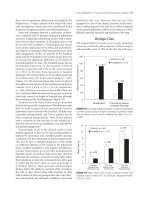
et al. (1983). In our group, Weller has shown
a statistically significant (p=O.OI) reduction in
maximal working capacity in a group of 45
asymptomatic patients studied 5-12 years after
the ~ustard operation (Stark et al. 1980).
Despite the decreased exercise tolerance all
.
'
our patients were asymptomatic and were
leading a normal life. The same observation
has been made by other authors (Mathews et
al. 1983; Ramsay et al. 1984).
The reports of RV dysfunction have prompted the exploration of alternative techniques
of ~orrec~i.on for TGA, especially techniques
which utIhse the left ventricle as a systemic
ventricle. Mee (1986) has demonstrated
recently that some patients with impaired RV
function following a Mustard operation can be
treated by pulmonary artery banding followed
by an arterial switch operation (see Chap. 16,
p. 220).
Rhythm Disturbances
Serious arrhythmias (atrial fibrillation or flutter, atrioventricular dissociation) were seen
more frequently after operations performed in
192
earlier years (Breckenridge et al. 1972; EI Said
et al. 1972; Ebert et al. 1974; Beerman et al.
1983; Hayes et al. 1986). It is not clear why
the incidence of reported rhythm disturbances
has decreased considerably in recent years
(Turley et al. 1978; Ullal et al. 1979; Trusler
et al. 1980; Deanfield et al. 1989). Possibly a
better knowledge of the exact position of the
sinus node, sinus node artery and atrioventricular node has enabled surgeons to protect these
structures better during the Mustard operation.
Ebert et al. (1974) have reported a higher
incidence of arrhythmias in patients who had
the coronary sinus cut open; however, this was
not confirmed by Clarkson et al. (1976). Fewer
arrhythmias were seen in patients in whom a
less extensive resection of the superior part of
the interatrial septum was carried out (Trusler
et al. 1980).
The incidence of post Mustard arrhythmias
varies in reported series. Southall et al. (1980)
have pointed out that some arrhythmias may
be detected on Holter monitoring even before
the Mustard operation. Our prospective study
(Deanfield et al. 1989) did not confirm this
finding. Changes of P wave amplitude and
contour are seen in almost all patients after
the Mustard operation (EI Said et al. 1972).
Atrial fibrillation, atrial flutter and atrioventricular dissociation occurred frequently in earlier
series (Breckenridge et al. 1972; Ebert et al.
1974); fortunately, the current incidence of
these is very low. Sick sinus syndrome (sinus
bradycardia with sinus arrest and junctional
escape) was the predominant arrhythmia in the
experience of Hayes et al. (1986). Junctional
rhythm does not usually cause any problems.
Episodes of supraventricular tachycardia are
more serious; they occur in a small percentage
of patients (Hayes et al. 1986). Some arrhythmias may only become "unmasked" during
maximal exercise testing (Mathews et al. 1983).
Late deaths have been described in several
series after the Mustard operation. Some of
these may possibly have been caused by
arrhythmias (Aberdeen 1971; Lewis et al. 1977;
Hayes et al. 1986).
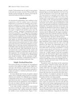
Patients discharged from hospital in sinus
rhythm may lose this rhythm later. At Green
Lane Hospital, Auckland, 72% of .patients
were in sinus rhythm 1 year after the Mustard
operation. This number decreased to 56% at
Reoperations in Cardiac Surgery
5 years and 50% at 10 years (Kirklin and
Barratt-Boyes 1986). In our postoperative study
(Deanfield et al. 1989), we have followed
patients with 24-h Holter monitoring before
and after the operation. All were in sinus
rhythm before the operation and 91 % at
discharge from the hospital. The incidence of
stable sinus rhythm decreased to 83% at 1-3
years and 66% at 6-8 years.
Benign arrhythmias do not require treatment.
Supraventricular tachycardia is treated medically. However, it may be resistant to several
drugs. Some patients with bradyarrhythmias
require insertion of a pacemaker.
Senning Operation
The Senning operation was first performed in
1958, but it was the Mustard operation which
became the operation of choice soon after its
introduction in 1964. There is a possible
explanation why the Mustard operation was
favoured. The mortality of the Senning operation was high in early reports (Kirklin et al.
1961). Today we know that the high mortality
was due to the selection of the patients rather
than to the operative technique. There were
several infants and young children with TGA
and VSD in Kirklin's series; and some of these
had pulmonary vascular obstructive disease. In
contrast, in Toronto, many patients with simple
transposition were well palliated by a
Blalock-Hanlon septectomy. These children
were stable and in a good condition in their
second, third or fourth year of life - excellent
candidates for intra-atrial repair.
Brom reintroduced the Senning operation in
1975 (Quaegebeur et al. 1977), and since then
it has become the intra-atrial repair of choice
for most cardiac centres. The idea of reviving
the Senning operation was an attempt to reduce
obstructive complications and arrhythmias. We
shall briefly review here the complications seen
after the Senning operation. In principle the
complications are similar to those seen after
the Mustard operation but are much less
frequent.
Reoperations After Mustard and Senning Operations
193
Systemic Venous Obstruction
Baffle Leaks
In several series, sve obstruction was not
detected after the Senning operation (Parenzan
et al. 1978; Quaegebeur et al. 1977; Bender et
al. 1980). Two sve obstructions required
reoperation in the early experience at Birmingham (Kirklin and Barratt-Boyes 1986). The
Boston group (Marx et al. 1983) have reported
7 obstructions in a group of 54 survivors of the
Senning operation. To our knowledge, IVe
obstruction was not reported after the Senning
operation. In our series of 196 Senning operations 1 patient required reoperation for sve
obstruction. The obstruction was due to technical error, and reoperation was performed
within 24 h after surgery. The sve and IVe
orifices were closer together than normal. Both
sve and IVe pressures were elevated after
surgery. At reoperation we found that the area
under the septum was narrow, thus presenting
obstruction to flow from both the sve and
IVe. The diagnosis and indications for reoperation are identical to those after the Mustard
operation.
Baffle leaks are uncommon after the Senning
operation. This has been our experience as
well as that of other authors.
Pulmonary Venous Obstruction
Pulmonary venous obstruction is rare after
the Senning operation. We believe that this
complication could be avoided if the technique
of Brom (Quaegebeur et al. 1977) is used for
the Senning operation. To our knowledge,
pulmonary venous obstruction only occurred
in patients in whom the pulmonary venous
atrium was enlarged with a patch: three patients
in the initial Birmingham experience and six
patients in the Boston series (Pacifico 1979;
Marx et al. 1983). We have seen this complication in 1 patient among 196 consecutive
Senning operations; reoperation to correct the
technical error was performed within 24 h
of the original operation. Pulmonary venous
obstruction did not occur in the series reported
by Parenzan et al. (1978), Quaegebeur et al.
(1977) and Bender et al. (1980). No case of
pulmonary venous obstruction was seen in the
Green Lane Hospital series and in the recent
(1977-1984) Birmingham series (Kirklin and
Barratt-Boyes 1986). The diagnosis and the
indications for reoperation are identical to
those after the Mustard operation.
Tricuspid Valve Incompetence
Tricuspid valve incompetence can occur after
the Senning operation. Penkoske et al. (1983)
has reported three mild and three severe
tricuspid valve incompetences in 39 survivors
of the Senning operation and VSD closure.
Severe incompetence required tricuspid valve
replacement in three patients. The reason why
tricuspid valve incompetence has been reported
less frequently after the Senning operation than
after the Mustard operation is probably due to
the fact that the Senning operations have been
performed more recently, when perhaps the
techniques of bypass and myocardial protection
have been improved. The lower incidence of
arrhythmias after recent atrial repairs may also
be attributed to this fact.
Residual/Recurrent VSD and LVOTO
There are no special differences between the
diagnosis and treatment of residual/recurrent
VSD or LVOTO after the Senning operation
and after the Mustard operation.
Ventricular Function
Ventricular function has not,as yet, been
extensively studied after the Senning operation.
However, Bender et al. (1980) did not show
any difference in their group of Mustards and
Sennings.
Arrhythmias
Parenzan et al. (1978) have reported a high
incidence of sinus rhythm soon after the
Senning operation. We have carried out a
prospective study (Deanfield et al. 1989),
assessing patients who underwent a Mustard
or Senning operation. The standard electrocar-
194
Reoperations in Cardiac Surgery
diogram and 24-h Holter monitoring was performed before the operation, after the operation prior to discharge from the hospital, at
1 year and at 5 years. The study showed a low
incidence of active arrhythmias . However,
there was a continuing decrease in the number
of patients remaining in a stable sinus rhythm
during the follow-up period . No statistically
significant difference was found between the
Mustard and Senning groups .
Operative Technique
Mustard
Fig. 14.2
The technique of the original operation may
influence the development and the incidence
of some late complications. Therefore, we shall
first describe some of the steps of the Mustard
operation which we consider important to avoid
complications.
Primary Operation
1. SVC Cannulation. The sve is usually cannulated directly through a purse-string suture.
This suture is placed at least 10 mm above the
CORRECT
INCORRECT
Fig. 14.1
sinus node to avoid its injury. The purse-string
is oblong (Fig. 14.1) rather than circular to
avoid narrowing of the sve when the pursestring is tied after the sve cannula is removed .
Alternatively, the purse-string is not tied after
decannulation but the partial occlusion clamp
is applied on the cava and an incision in the
sve is formally closed with a fine polypropylene stitch (Fig. 14.2a, b) . We favour the
technique of an oblong purse-string which is
tied after decannulation.
2. Shape and Material used for Baffle. The
shape and the material of the baffle and the
technique of its insertion may contribute to the
development of obstruction. It is important to
construct the sve pathway in such a manner
that the baffle forms less than 50% of its
circumference. If at least 50% of the pathway
is constructed of the atrial wall, obstruction is
unlikely to develop if the patch does not grow,
or even if it shrinks (Fig. 14.3).
In our early experience both the material
used for the baffle and the shape of the baffle
played an important role (Stark et al. 1980) .
We found that a redundant , thin Dacron patch
had a tendency to fold upon itself (Fig.
14.4a). Apposition of platelets and fibrin and
subsequent fibrosis of this tissue led to severe
thickening of the patch (Fig. 14.4b, c) .
Brom has suggested cutting the patch into a
"trouser shape" (Quagebeur and Brom 1978) .
He constructed the patch on the basis of
measurements of sve and IVe circumference
195
Reoperations After Mustard and Senning Ope rations
is to suture the patch away from the SVC and
IVC orifices and to pull the atrium onto the
patch (Kirklin and Barratt-Boyes 1986) .
. . .'---Baffle
3. Thrombosis. Thrombosis may cause SVC
pathway obstruction, especially if the lumen
was already compromised by a faulty operative
technique. Insertion of several central venous
cannulae into the internal jugular vein may be
another cause. This may be of particular
importance in young infants. Infusion of platelets and/or hypertonic solutions through these
lines may be another contributing factor.
It is useful to evaluate the adequacy of the
SVC pathway soon after the operation by
Doppler echocardiography or by an injection
of
contrast media through an internal jugular
Fig. 14.3
line and performing a chest radiograph at the
same time. Both these techniques are useful
(distances E-D and D-F in Fig. 14.5a) and the and can be easily performed in the intensive
distance between the edge of the intra-atrial care unit. The diagnosis of even mild SVC
septum and the pulmonary veins (C-D in Fig. pathway narrowing would alert us to avoid
14.5a). Subsequently, good results have been infusions of hypertonic solutions. Under such
achieved with this patch irrespective of whether circumstances, it may be safer to remove the
it was tailored from Dacron or pericardium. jugular vein cannula and place it elsewhere.
The Toronto group has always used pericardium. The original large quadrangular patch 4. Inadequate Resection of Intra-atrial Sepof the Mustard operation has been only slightly tum. Inadequate resection of the intra-atrial
modified (Trusler et al. 1980) (Fig. 14.5b). septum may leave a ridge of tissue which
Barratt-Boyes uses a small patch. His concept then causes turbulence and contributes to the
A
a
b
a
c
B
Fig. 14.4
Fig. 14.5
Reoperations in Cardiac Surgery
196
Fig. 14.6
development of obstruction. On the other
hand, too extensive resection may damage the
sinus node artery and lead to arrhythmias
(Trusler et al. 1980). If one avoids an extensive
resection, it is possible to use the superior part
of the atrial septum as a flap, which is then
sutured to the baffle (Turley and Ebert 1978).
This step is illustrated in Fig. 14.6.
manoeuvre may cause arrhythmias, it has not
been confirmed by others (Clarkson et al.
1976) . We have not cut the coronary sinus
routinely; however, we find the technique very
useful in children in whom the distance between
the SYC and IYC orifices is short. The suture
line from the left to the right pulmonary veins
should diverge to avoid pulmonary venous
obstruction. This may, on the other hand,
compromise the IYC pathway; therefore , under
such circumstances we prefer to open the
coronary sinus deep into the left atrium (Fig.
14.7). It is important to open the coronary
sinus with one cut. Repeated, short cuts may
catch the fold in the atrial wall and cut outside
the heart. This does not cause problems if
recognised in time. It is very difficult to control
the bleeding from the posterior part of the left
atrium without the aid of cardiopulmonary
bypass. Therefore, we routinely lift the heart
up before discontinuing perfusion to check for
any damage to this area.
7. Width of the Baffle. Too redundant a baffle
may form adhesions with the lateral atrial wall
and cause pulmonary venous obstruction. We
assess the width of the patch during insertion.
When the suture line around the left pulmonary
5. Coronary Sinus Cut-back. Opening the cor- veins and towards the right upper and lower
onary sinus deep into the left atrium ensures
pulmonary veins has been completed we hold
a wide IYC pathway. Although it has been
the opposite edge of the patch with forceps
suggested (Ebert et al. 1974) that this
and keep it close to the cut edge of the atrial
septum. A curved instrument then pushes the
patch from behind towards the lateral atrial
wall (Fig. 14.8). If the patch reaches the atrial
wall it is too redundant and should be trimmed.
Coronary sinus
8. Placement of the Baffle. The correct placement of the baffle is important to avoid either
systemic or pulmonary venous obstruction.
Concern about one of these complications may
cause the other one. The suture line from the
left pulmonary veins should diverge upwards
between the SYC and right pulmonary vein .
Inferiorly, the suture line runs from the left
pulmonary veins to between the right lower
pulmonary vein and the orifice of the lYe.
Figure 14.9 illustrates the correct and incorrect
placement of a baffle.
Fig. 14.7
9. Incision in the Right Atrium and Enlargement
of the Pulmonary Venous Atrium. Various
197
Reoperations After Mustard and Scnning Operations
incisions in the right atrium have been suggested
for the Mustard operation . We believe that it
is not important which type of incision is used
if the pulmonary venous atrium is subsequently
enlarged. Insertion of a generous baffle would
ensure large systemic venous pathways but
it may compromise the pulmonary venous
pathway. It is probably safer to enlarge the
pulmonary venous atrium, especially if the
operation is performed in small infants.
Enlargement can be performed with a patch.
Alternatively, a v-Y incision in the atrium
advances the flap of the atrial wall between
the right upper and lower pulmonary veins,
thus preventing narrowing at a crucial point of
the pulmonary venous pathway. This technique
Fig. 14.8 is used by Barratt-Boyes (Kirklin and BarrattBoyes 1986) (Fig. 14.10) .
Reoperations
CORRECT
1. Approach. We prefer to approach the heart
through a right anterolateral thoracotomy for
most reoperations after the Mustard operation
(Szarnicki et al. 1978). A right thoracotomy
offers several advantages over the sternotomy
approach. As the pericardium is usually used
for the baffle the anterior part of the right
ventricle may be adherent to the back of the
sternum. With a right thoracotomy, dissection
of the right ventricle is not required, thereby
avoiding potential InJunes to structures
obscured by adhesions from the first operation.
INCORRECT
Tn the presence of sve obstruction, highFig. 14.9 pressure venous collaterals may cause considerable bleeding during sternal re-entry. These
likewise are avoided with a thoracotomy. In
addition, a right chest approach places the sve
and Ive closer to the surgeon than from the
front, making dissection and cannulation easier.
Reoperations performed for sve, IVe, or
pulmonary venous obstruction, baffle leak,
tricuspid valve incompetence or residual/recurrent VSD are best performed through a right
atriotomy. The aorta is usually located anteriorly and to the right in patients with TGA;
therefore its dissection and cannulation is easy
from the right chest as well.
Reconstruction of the left pulmonary artery
or insertion of a left ventricular to pulmonary
Fig. 14.10 artery conduit cannot be performed through a
Reoperations in Cardiac Surgery
198
Fig. 14.11
right thoracotomy. For these reoperations, we
use either a median sternotomy or a left
thoracotomy (see Chapter 20, pp. 283-284). Other
authors prefer a standard approach using
sternal re-entry for all operations after Mustard
or Senning procedures (Kron et al. 1985;
Kirklin and Barratt-Boyes 1986).
For a right thoracotomy approach, the patient is placed on the operating table at about
50° (Fig. 14.11). An external defibrillator
electrode is placed between the patient's scapulae. One groin is prepped for cannulation of
the femoral/iliac vessels should it be difficult
to reach the aorta. The right thoracotomy is
usually performed through the fifth intercostal
space. This gives adequate access both to the
aorta and to the IVe. The sternum is often
transected. Extension of the thoracotomy posteriorly on the right side is usually minimal, so,
in effect, it remains an anterior thoracotomy.
2. Cannulation. The edge of the pericardium
is identified. Care is taken during the dissection
not to injure the phrenic nerve. It is easy
to avoid a phrenic nerve injury from the
thoracotomy approach because of the proximity
of the phrenic nerve to the surgeon. A pursestring suture is then placed on the ascending
aorta and on the pulmonary venous atrium
close to the atrioventricular junction (Fig.
14.12). If any bleeding occurs during the
subsequent dissection, bypass can be initiated
after cannulating the aorta and the pulmonary
venous atrium with a single venous cannula.
SVC and IVC purse-strings are placed. The
dissection around the SVC is easy; dissection
'Fig. 14.12
around the IVC is usually delayed until cardiopulmonary bypass is started. Perfusion is started
with a cold perfusate (20-25 0c). When the
heart fibrillates, the pulmonary venous atrium
is opened and caval snares are tightened.
Myocardial protection is achieved either by
cold perfusion with the heart fibrillating or
aortic cross-clamping with cardioplegia. We
favour cross-clamping of the aorta with cardioplegia for the initial stages of the operation.
Care is taken to keep the time of the crossclamping to a minimum; the operation may
therefore be completed on a cold fibrillating
heart. If the pulmonary venous atrium was not
cannulated for perfusion, a sump sucker is
introduced through the purse-string into the
pulmonary venous atrium or through the tricuspi<;l valve into the right ventricle.
3. Systemic Venous Obstruction. If the SVC
pathway is obstructed, a longitudinal cut is
made into the SVC pathway close to the atrial
septum (Fig. 14.13). The pathway should be
free at this point. The obstructed area is
inspected from below and the incision in the
roof of the pathway is extended. Care is taken
not to injure the area of the sinus node artery.
Any thrombus present is removed. If the
pathway is completely obstructed it may be
helpful to pass a probe or a curved instrument
through a stab wound in the SVc. This
identifies the point of entry of the SVC into
the intracardiac SVC channel, and an opening
from below can be made easily. If the baffle
is narrow but thin and pliable it is possible to
suture a patch into the incision in the pathway
Reoperations After Mustard and Senning Operations
199
(Fig. 14.14), using either pericardium taken
from the diaphragmatic surface or a Gore-Tex
or Dacron patch. If the baffle is thickened and
folded and the pathway severely obstructed,
we replace it. If the baffle is thickened over
the lye pathway as welI, the entire baffle
should be removed, even if the lye pathway
is not severely compromised at the time of
reoperation. We believe it is safer under such
circumstances to replace the whole baffle
because the future growth of the child and the
lack of growth of the baffle may gradually
restrict the lye channel.
If the whole baffle has to be replaced (Fig.
Fig. 14.13 14.15), we use either Gore-Tex or a patch
tailored from a tube of woven Dacron or
double-velour knitted Dacron (insert to Fig.
14.15). We have successfully used woven
Dacron for the new baffle but, in view of the
propensity to form thicker and loosely attached
neo-intima, we would currently prefer a GoreTex patch or a double-velour knitted patch.
It may be difficult to suture the new patch
if the entire baffle is removed. Deep stitches
in the area of the atrial septum close to the
tricuspid valve may damage the conduction
mechanism . For this reason we leave a narrow
rim of the old baffle in place; the new baffle
is then sutured to this rim.
When the new patch has been sewn in place,
the decision must be made as to whether the
pulmonary venous atrium is to be enlarged. In
general we prefer to enlarge it at the time of
Fig. 14.14 any reoperation after a Mustard operation,
except in patients with tricuspid valve incompetence . In such patients, the pulmonary venous
atrium is already considerably enlarged and a
patch is not required. In all other patients, we
think it is advantageous to close the atrium
with a patch. Both the previous surgery and
,the reoperation will leave scars on the atria,
thus possibly impairing future growth . The
atrial incision is extended down between the
right upper and lower pulmonary veins. A large
Gore-Tex patch is sutured in place with a 4-0
or 5-0 polypropylene suture (Fig. 14.16).
After the first stitches on the patch have
been placed , it is possible to remove the aortic
clamp or, if the heart fibrillates , to defibrillate
it and to start rewarming. Great care must be
taken to avoid air embolisation as the heart
Fig. 14.15 was not freed from adhesions . We place the
200
Reope rations in Cardiac Surgery
Fig. 14.16
patient in a mild Trendelenburg position,
and the atrial sump is advanced through the
tricuspid valve to the right ventricle to keep
the valve incompetent and to decompress the
right ventricle. The perfusion pressure is kept
high so as not to allow the aortic valve to open.
When the atrium is closed caval snares are
released. An aortic needle vent is put on
suction, the aorta cross-clamped between the
cannula and the vent, and a sump sucker pulled
from the right ventricle to the pulmonary
venous atrium and then removed . The pressure
in the pulmonary venous atrium is raised to
5-7 mmHg, and the anaesthetist starts inflating
the lungs. The de-airing procedure is repeated
a few times. When the patient is completely
rewarmed, the perfusion is discontinued in the
usual manner. The cannulae are removed from
the heart and protamine is given. A fine
polyethylene catheter is left in the pulmonary
venous atrium for pressure monitoring; atrial
and ventricular pacemaker wires are placed
and two chest drains inserted. The thoracotomy
is closed in the usual manner.
4. Pulmonary Venous Obstruction. The approach is the same as that described for systemic
venous obstruction . On cardiopulmonary
bypass the aorta is cross-clamped and cardioplegia is given. The pulmonary venous atrium is
widely opened from the atrioventricular groove .
A small opening from the posterior part of the
pulmonary venous atrium is visualised (arrow
in Fig. 14.17). The incision in the atrium is
then extended through this opening to a point
between the right upper and right lower
Fig. 14.17
pulmonary veins. If necessary, it is further
extended into the posterior wall of the left
atrium (Fig. 14.18a). A large Gore-Tex patch
is sutured into this incision in a fashion similar
to that described for closure of the pulmonary
venous atrium after the repair of systemic
venous obstruction (Fig. 14.18b). Rewarming,
discontinuation of perfusion and closure of the
thoracotomy are then performed in the usual
manner.
5. Tricuspid Valve Incompetence. The approach, cannulation, perfusion and myocardial
preservations are the same as described for
reoperations for systemic venous obstruction.
The tricuspid valve is carefully inspected to
a
Fig. 14.18
201
Reoperations After Mustard and Senning Operations
assess the pathology. The valve can rarely be
repaired (Park et al. 1983). If the valve is
severely incompetent, it is usually replaced.
We have replaced the tricuspid valve in 6
patients in our series of 563 Mustard operations;
Penkoske et al. (1983) has reported 3 patients
with severe tricuspid incompetence, all of
whom required valve replacement.
The choice of valve prosthesis is limited.
Heterografts calcify early in children. We do
not have any experience with the stent-mounted
aortic or pulmonary homografts which have
been used in adult patients in the tricuspid
position by Barratt-Boyes (Kirklin and BarrattBoyes 1986). In our patients, we have used
Bjork-Shiley valves, as do the Boston group
(Penkoske et al. 1983). Excellent results using
the St Jude medical valve have been reported
in the tricuspid position by Singh et al. (1984).
We have used either interrupted mattress
sutures with pledgets or a running stitch for
the valve insertion. The technical details are
discussed in Chapter 24 (see p. 345).
6. Baffle Leaks. There are no special technical
"tricks" for repair of baffle leaks. When the
atrium is opened the whole baffle is carefully
inspected. Small leaks are closed directly, the
large ones with a patch of pericardium, GoreTex or Dacron. If systemic or pulmonary
venous obstruction is also present, we use
the technique as described for these two
complications. Reoperations for an isolated
large residual shunt (not associated with other
defects) is easy. The perfusate is cooled to
30 DC, the aorta is cross-clamped, and the
defect in the baffle is closed with a patch.
7. Residual/Recurrent VSD. Most residual!
recurrent VSDs can be closed through the
tricuspid valve; therefore a right thoracotomy
is, again, our approach of choice. VSDs located
near the apex and multiple VSDs are best
approached from a midline sternotomy because
a left ventriculotomy or an apical fish-mouth
incision may be required for their closure. The
technical aspects of the repair of residual/
recurrent VSDs are discussed in Chapter 12
(see pp. 165-168).
8. Residual/Recurrent LVOTO. Detailed preoperative assessment, as discussed earlier in
this chapter (see p. 191) is very important for the
choice of the best approach. Repeated attempts
to relieve LVOTO are successful only in
patients with favourable anatomy. More often,
symptomatic LVOTO has to be bypassed with
a v.alved conduit placed between the apex of
the left ventricle and the main and/or left
pulmonary artery. This operation can conveniently be performed through a left thoracotomy. Details of the operative technique are
described in Chapter 20 (see pp. 283-284).
9. Right Ventricular Dysfunction. Until recently only medical treatment was offered
to patients with systemic (right) ventricular
dysfunction. Mee (1986) has suggested and
successfully performed pulmonary artery banding followed by an arterial switch operation in
five patients. The details of this approach are
discussed in Chapter 16 (see p. 217).
10. Arrhythmias. Some arrhythmias do not
require treatment; tachyarrhythmias are treated
medically. Patients with sick sinus syndrome
are considered for insertion of a pacemaker if
they have Stokes-Adam syndrome or severe
bradycardia (less than 30--40 beats/min). Patients with diminished R V function associated
with bradycardia may also benefit from pacing
(see Chap. 6, p. 68).
Senning
Obstructive complications after the Senning
operation are uncommon. As with the Mustard
operation, the technique of the original operation may influence the incidence of obstruction. Some of the important technical details
of the Senning operation will therefore be
described first.
Primary Repair
All the details of SVC cannulation in the
Mustard operation also apply for the Senning
operation (see Figs. 14.1, 14.2). The intracardiac part of the SVC channel may be constructed too narrow if the right atrial incision
is too close to the crista terminalis (Fig. 14.19).
202
Reoperations in Cardiac Surge ry
CORRECT
INCORRECT
Fig. 14.19
This may result in not enough lateral atrial flap
being available for the roof of the SVC channel.
The SVC pressure may be elevated for a few
hours after the operation but it usually regresses
quickly. Severe obstruction is rare. As the SVC
pathway is made entirely from the patient's
own atrial tissue it grows with the patient.
The area under the septum may be narrow
in some patients and does not allow free SVC
and IVC flow. This area may be enlarged by
opening the coronary sinus into the left atrium.
We now use the coronary sinus cut-back more
frequently; others use it routinely (Kirklin and
Barratt-Boyes 1986).
Enlargement of the septal flap with a piece
of pericardium or Dacron is used in patients
with a large ASD to make up the deficient
atrial septal tissue . The patch should only
enlarge the width but not the length of the flap
(Fig. 14.20). If the length is increased, the flap
may subsequently bulge into the pulmonary
venous atrium and possibly cause pulmonary
venous obstruction. With more experience it is
usually possible to use the tissue in the fossa
ovalis (Fig. 14.21a) or to augment the septal
flap with the flap created by the cut-back of
the coronary sinus without using any additional
patch (Fig. 14.21b,c). Alternatively, the narrow
atrial flap may be split and opened to increase
the width (Fig. 14.21d).
Pulmonary venous obstruction has been
described in patients in whom the pulmonary
venous atrium was enlarged with a patch (Otero
Co to et al. 1979; Pacifico 1979) (Fig. 14.22).
Using the atriotomy with cut-backs in the
superior and inferior corners of the incision
provides enough tissue to suture the medial
atrial flap across the SVC and IVC and around
the pulmonary veins. We have not used a patch
c
Fig. 14.21
Reoperations Afte r Mustard and Se nning Ope rations
INCORRECT
Fig. 14.22
to enlarge the pulmonary venous atrium in any
of our 196 Senning operations. When the
medial a~rial flap does not easily reach towards
the pulmonary veins, the pulmonary venous
atrium can be enlarged by suturing it to in situ
pericardium, as suggested by Senning (1975)
(Figs. 14.23, 14.24) .
Reoperations
1. Approach. Early reoperation is performed
through a median sternotomy. We have not
203
Fig. 14.24
had to reoperate late after the Senning operation;,we would use a right thoracotomy as for
reoperations after the Mustard operation.
The sve and Ive are cannulated. If sve
obstruction is the only lesion, enlargement of
the medial part of the pathway can be performed on a beating heart using moderate hypothermia. However, aortic cross-clamping with
cardioplegia is required for obstructions
repaired from within the pulmonary venous
atrium, obstruction of the pulmonary venous
pathway, operations on the tricuspid valve,
LVOTO or a VSD. Reoperations in infants
may be performed using deep hypothermia and
circulatory arrest with a single venous cannula
inserted into the base of the pulmonary venous
atrium .
2. Systemic Venous Obstruction. Our experience with systemic venous obstruction is limited
to one early reoperation performed within 24 h
of the original surgery . We used a midline
sternotomy, aortic and bicaval cannulation and
aortic cross-clamping with cardioplegia. The
heart was opened through an incision in
the pulmonary venous atrium close to the
atrioventricular groove (Fig. 14.25a). The
advantage of this incision is that the medial
atrial flap does not have to be detached from
the sve and the right pulmonary veins. It also
obviates the need for repeat suturing across
the area of the sinus mode. The sve channel
204
Reoperations in Cardiac Surgery
Fig. 14.25
is easily visualised from within the pulmonary
venous atrium (Fig. 14.25b) . Part of the suture
line attaching the flap to the atrial septum is
removed. This detaches the sve pathway roof
(Fig. 14.26a) ; the size of the channel is assessed .
Then an oval piece of pericardium is sutured
into this opening, either over the sve only
a
b
Fig. 14.26
Fig. 14.27
or towards the Ive if both channels were
compromised (Fig. 14.26b).
Another alternative to enlarge the narrow
pathway is to incise the medial aspect of the
pathway and place an oval patch (Fig. 14.27).
A longitudinal incision is made and the extent
of the obstruction assessed. The incision may
be facilitated by the placement of a probe or
a curved instrument through the stab wound
in the sve into the intracardiac portion of the
pathway. An oval patch of pericardium or
Gore-Tex is then sutured into this incision . We
have not used this technique; however, it
should be easy and could be performed without
aortic cross-clamping. The disadvantage of this
technique is possible injury to the sinus node
artery which may be located in the area of the
incision. It may be difficult to see the artery
in adhesions at reoperation.
3. Pulmonary Venous Obstruction. We have
seen pulmonary venous obstruction in one
patient immediately after the Senning operation . It was due to a technical error, and
reoperation was performed a few hours after
the original surgery . The pulmonary venous
atrium was opened close and parallel to the
atrioventricular groove and an oval patch of
pericardium was sutured into this incision.
Should late pulmonary venous obstruction
develop, it could be treated with the same
technique as described for pulmonary venous
obstruction after the Mustard operation (see
205
Reoperations After Mustard and Senning Operations
Figs. 14.17, 14.18). The pulmonary venous
atrium can be enlarged by suturing it to in
situ pericardium (M. Turina 1986, personal
communication) (see Figs. 14.23, 14.24).
4. Other Complications. The operative technique for tricuspid valve incompetence, baffle
leaks, residual/recurrent VSD or LVOTO does
not differ from the technique used for these
complications after the Mustard operation.
Results of Reoperations After
Mustard or Senning Procedures
The mortality rate after reoperations for
obstructive complications is not insignificant,
especially if the operation is delayed until the
patient is very ill (Kirklin and Barratt-Boyes
1986).
Reoperation for systemic venous obstruction
was required in 45 children in a combined
experience of Stark et al. (1974), Park et al.
(1983), Marx et al. (1983) and Kron et al.
(1985). Eleven patients (24%) died.
Reoperation for pulmonary venous obstruction, either complete or incomplete (left pulmonary veins only), was reported (Driscoll et
al. 1977; Oelert et al. 1977; Trusler et al. 1980;
Park et al. 1983; Marx et al. 1983; J. Stark
1987, unpublished work) in 46 children; 10
died (22%). It is not easy to estimate the exact
incidence and risk of reoperation for pulmonary
and/or systemic venous obstruction. In some
reported series, patients died before reoperation or refused surgery. Some symptomatic
patients were lost to follow-up.
The recurrence of obstruction is uncommon;
it was reported in ten patients from six
institutions (Venables et al. 1974; Hagler et al.
1978; Marx et al. 1983; Park et al. 1983; Kirklin
and Barratt-Boyes 1986). We have performed
second and third reoperations in one patient
each. Both survived.
The risk of tricuspid valve replacement after
the Mustard or Senning operation is difficult
to estimate because of the small number of
reported reoperations. We have replaced the
tricuspid valve in six children after Mustard or
Senning operations. Four survived and are
well.
References
Aberdeen E (1971) Correction of uncomplicated cases of
transposition of the great arteries. Br Heart J 33: 66--68
Bailey LL, Takeuchi Y, Williams WG, Trusler GA, Mustard
WT (1976) Surgical management of congenital cardiovascular anomalies with the use of profound hypothermia and
circulatory arrest. Analysis of 180 consecutive cases. J
Thorac Cardiovasc Surg 71: 485-492
Beerman LB, Neches WH, Fricker FJ et al. (1983) Arrhythmias in transposition of the great arteries after the Mustard
operation. Am J Cardiol 51: 1530-1534
Bender HW Jr, Graham TP Jr, Boucek RJ Jr, Walker WE,
Boerth RG (1980) Comparative operative results of the
Senning and Mustard procedures for transposition of the
great arteries. Circulation 62 (Suppl I): 1-197-203
Breckenridge 1M, Stark J, Bonham-Carter RE, Oelert H,
Graham GR, Waterston DJ (1972) Mustard's operation for
transposition of the great arteries. Review of 200 cases.
Lancet I: 1140-1142
Castaneda AR, Norwood WI, Jonas RA, Colon SD, Sanders
SP, Lang P (1984) Transposition of the great arteries and
intact ventricular septum: anatomical repair in the neonate.
Ann Thorac Surg 38: 438-443
Clarkson PM, Barratt-Boyes BG, Neutze JM (1976) Late
dysrhythmias and disturbances of conduction following
Mustard operation for complete transposition of the great
arteries. Circulation 53: 519-524
Deanfield J, Camm J, Macartney F et al. (1989) Arrhythmia
and late mortality of Mustard and Senning operation for
transposition of the great arteries: an 8 year prospective
study. J Thorac Cardiovasc Surg 96: 569-576
Driscoll DJ, Nihill MR, Vargo TA, Mullins CE, McNamara
DG (1977) Late development of pulmonary venous obstruction following Mustard's operation using a Dacron baffle.
Circulation 55: 484-488
Ebert PA, Gay WA Jr, Engle MA (1974) Correction of
transposition of the great arteries: relationship of the
coronary sinus and postoperative arrhythmias. Ann Surg
180: 433-438
Egloff LP, Freed MD, Dick Mac, Norwood WI, Castaneda
AR (1978) Early and late results with the Mustard operation
in infancy. Ann Thorac Surg 26: 474-484
EI-Said G, Rosenberg HS, Mullins CE, Hallman GL, Cooley
DA, McNamara DG (1972) Dysrhythmias after Mustard's
operation for transposition of the great arteries. Am J
Cardiol 30: 526-531
Graham TP Jr, Atwood GF, Boucek RJ Jr, Boerth RC,
Bender HW Jr (1975) Abnormalities of right ventricular
function following Mustard's operation for transposition of
the great arteries. Circulation 52: 678-684
Hagler DJ, Ritter DG, Mair DO, Davis GD, McGoon DC
(1978) Clinical, angiographic and hemodynamic assessment
of late results after Mustard operation. Circulation 57:
1214-1220
Hagler DJ, Ritter OG, Mair OD et al. (1979) Right and
left ventricular function after the Mustard procedure in
transposition of the great arteries. Am J Cardiol 44:
276-283
Hayes CJ, Gersony WM (1986) Arrhythmias after the Mustard
206
operation for transposition of the great arteries: a longterm study. 1 Am Coli Cardiol 7: 133-137
Huhta lC, Edwards WD, Danielson GK, Feldt RH (1982)
Abnormalities of the tricuspid valve in complete transposition of the great arteries with ventricular septal defect. 1
Thorac Cardiovasc Surg 83: 569-576
Hurwitz RA, Caldwell RL, Girod DA, Mahony L, Brown
1, King H (1985) Ventricular function in transposition of
the great arteries: evaluation by radionuclide angiocardiography. Am Heart 1 110: 60~5
latene AD, Fontes VF, Paulista PP et al. (1975) Successful
anatomic correction of transposition of the great vessels:
a preliminary report. Separata Arq Cardiol 28: 461-463
Kirklin lW, Barratt-Boyes BG (1986) Cardiac surgery.
Morphology, diagnostic criteria, natural history, techniques,
results and indications. Wiley, New York.
Kirklin, lW, Devloo RA, Weidman WH (1961) Open intracardiac repair for transposition of the great vessels: 11
cases. Surgery 50: 58-62
Kron IL, Rheuban KS, loob A W et al. (1985) Baffle
obstruction following the Mustard operation: cause and
treatment. Ann Thorac Surg 39: 112-115
Lewis AB, Lindesmith GG, Takahashi et al. (1977) Cardiac
rhythm following the Mustard procedure for transposition
of the great vessels. 1 Thorac Cardiovasc Surg 73: 919-926
Lindesmith GC, Stanton RE, Stiles QR et al. (1973)
Correction of transposition of the great vessels. Assessment
of clinical experience. 1 Cardiovasc Surg 6: 241-246
Lock lE, Bass lL, Castaneda-Zuniga W, Fuhrman BP,
Rashkind Wl, Lucas RV lr (1984) Dilation angioplasty of
congenital or operative narrowings of venous channels.
Circulation 70: 457-464
Marx GR, Hougen TJ, Norwood WI, Fyler DC, Castaneda
AR, Nadas AS (1983) Transposition of the great arteries
with intact ventricular septum: results of Mustard and
Senning operations in 123 consecutive patients. J Am Coli
Cardiol 1: 47fr483
Mathews RA, Fricker Fl, Beerman LB et al. (1983) Exercise
studies after the Mustard operation in transposition of the
great arteries. Am 1 Cardiol 51: 1526-1529
Mazzei EA, Mulder DG (1971) Superior vena cava syndrome
following complete correction (Mustard repair) of transposition of the great vessels. Ann Thorac Surg 11: 243-245
Mee RBB (1986) Severe right ventricular failure after Mustard
or Senning operation. Two-stage repair: pulmonary artery
banding and switch. 1 Thorac Cardiovasc Surg 92: 385-390
Moodie DS, Feldt RH, Wallace RB (1976) Transient proteinlosing enteropathy secondary to elevated caval pressures
and caval obstruction after the Mustard procedure. 1
Thorac Cardiovasc Surg 72: 379-381
Murphy lH, Barlai-Kovach MM, Mathews RA et al. (1983)
Rest and exercise right and left ventricular function late
. after the Mustard operation: assessment by radio nuclide
ventriculography. Am 1 Cardiol 51: 1520-1526
Mustard WT (1964) Successful two-stage correction of trans position of the great vessels. Surgery 55: 469-472
Oelert H, Laprell H, Piepenbrock S, Luhmer I, Kallfelz HC,
Borst HG (1977) Emergency and non-emergency intraatrial correction for transposition of the great arteries in
43 infants. Indications, details of the operative technique
and results. Thoraxchirurgie 25: 305-313
Otero Coto E, Norwood WI, Lang P, Castaneda AR (1979)
Modified Senning operation for treatment of transposition
of the great arteries. 1 Thorac Cardiovasc Surg 78: 721-729
Pacifico AD (1979) Discussion. In: Otero Coto E, Norwood
WI, Lang P, Castaneda AR (1979) Modified Senning
operation for treatment of transposition of the great
arteries. 1 Thorac Cardiovasc Surg 78: 721-729
Parenzan L, Locatelli G, Alfieri 0, Villani M, Invernizzi G,
Reoperations in Cardiac Surgery
Pacifico AD (1978) The Senning operation for transposition
of the great arteries. 1 Thorac Cardiovasc Surg 76: 305-311
Park SC, Neches WH, Mathews RA et al. (1983) Hemodynamic function after the Mustard operation for transposition
of the great arteries. Am 1 Cardiol 51: 1514-1519
Penkoske PA, Westerman GR, Marx GR et al. (1983)
Transposition of the great arteries and ventricular septal
defect: results with the Senning operation and closure of
the ventricular septal defect in infants. Ann Thorac Surg
36: 281-288
Quaegebeur 1M, Brom AG (1978) The trousers-shaped baffle
for use in the Mustard operation. Ann Thorac Surg 25:
240-242
Quaegebeur 1M, Rohmer 1, Brom AG, Tinkelenberg 1 (1977)
Revival of the Senning operation in the treatment of
transposition of the great arteries. Thorax 32: 517-524
Quaegebeur 1M, Rohmer 1, Ottenkamp 1 et al. (1986) The
arterial switch operation. An eight-year experience. 1
Thorac Cardiovasc Surg 92: 361-384
Ramsay 1M, Venables AW, Kelly Ml, Kalff V (1984) Right
and left ventricular function at rest and with exercise after
the Mustard operation for transposition of the great
arteries. Br Heart 1 51: 364-370
Rashkind Wl, Miller WW (1966) Creation of an atrial septal
defect without thoracotomy: a palliative approach to
complete transposition of the great arteries. lAMA 196:
991-992
Senning A (1959) Surgical correction of transposition of the
great vessels. Surgery 45: 966-980
Senning A (1975) Correction of the transposition of the great
arteries. Ann Surg 182: 287-292
Silverman NH, Snider R, Colo 1, Ebert PA, Turley K (1981)
Superior vena caval obstruction after Mustard's operation:
detection by two-dimensional contrast echocardiography.
Circulation 64: 392-396
Singh AK, Christian FD, Williams DO et al. (1984) Followup assessment of St lude medical prosthetic valve in the
tricuspid position: clinical and hemodynamic results. Ann
Thorac Surg 37: 324-327
Southall DP, Keeton BR, Leanage Ret al. (1980) Cardiac
rhythm and conduction before and after Mustard's operation for complete transposition of the great arteries. Br
Heart 1 43: 21-30
Stark J, Tynan MJ, Ashcraft KW, Aberdeen E, Waterston
Dl (1972) Obstruction of pulmonary veins and superior
vena cava after the Mustard operation for transposition of
the great arteries. Circulation 45/46 (Suppl I): 1-116-120
Stark 1, de Leval MR, Waterston Dl, Graham GR, BonhamCarter RE (1974a) Corrective surgery of transposition of
the great arteries in the first year of life. Results in 63
infants. 1 Thorac Cardiovasc Surg 67: 673-681
Stark 1, Silove ED, Taylor lFN, Graham GR, Kirklin lW
(1974b) Obstruction to systemic venous return following
the Mustard operation for transposition of the great
arteries. 1 Thorac Cardiovasc Surg 68: 742-749
Stark 1, Weller P, Leanage R et al. (1980) Late results of
surgical treatment of transposition of the great arteries.
Adv Cardiol 27: 254-265
Szarnicki Rl, Stark 1, de Leval M (1978) Reoperation for
complications after inflow correction of transposition of
the great arteries: technical considerations. Ann Thorac
Surg 25: 150-154
Takahashi M, Lindesmith GG, Lewis AB et al. (1977) Longterm results of the Mustard procedure. Circulation 56
(Suppl II): 11-85-90
Trusler GA (1984) The Mustard procedure: still a valid
aproach. In: Moulton A (ed.) Congenital heart surgery current techniques and controversies. Appleton Davies,
Pasadena, California, pp. 3-11
Reoperations After Mustard and Senning Operations
Trusler GA, Williams WG, Izukawa T, Olley PM (1980)
Current results with the Mustard operation in isolated
transposition of the great arteries. J Thorac Cardiovasc
Surg 80: 381-389
Turley K, Ebert PA (1978) Total correction of transposition
of the great arteries. J Thorac Cardiovasc Surg 76: 312-320
Tynan M, Aberdeen E, Stark J (1972) Tricuspid incompetence
after the Mustard operation for transposition of the great
arteries. Circulation 45/46 (Suppl I): 1-111-115
Ullal RR, Anderson RH, Lincoln C (1979) Mustard's
operation modified to avoid dysrhythmias and pulmonary
and systemic venous obstruction. J Thorac Cardiovasc Surg
78: 431-439
207
Venables AW, Edis B, Clarke CP (1974) Vena caval
obstruction complicating the Mustard operation for complete transposition of the great arteries. Eur J Cardiol 1:
401-410
Waldhausen JA, Pierce WS, Park CD, Rashkind WJ,
Friedman S (1971) Physiologic correction of transposition
of the great arteries: indications for and results of operation
in 32 patients. Circulation 43: 738-747
Wyse RKH, Haworth SG, Taylor JFN, Macartney FJ (1979)
Obstruction of superior vena caval pathway after Mustard's
repair. Reliable diagnosis by transcutaneous Doppler ultrasound. Br Heart J 42: 162-167
Chapter 15
Reoperations After Arterial Switch Operation
A.R. Castaneda
Introduction
The initial high operative mortality reported
with the arterial switch operation for repair of
complex forms of transposition of the great
arteries (TGA) has now been reduced significantly. The rather steep learning curve associated with the early development of this operation was caused, in part, by technical
difficulties related to the transfer of the coronary arteries and also by patient selection. Jatene
realised that in patients with TGA and an
intact ventricular septum the left ventricle
would soon adapt to the low resistance of the
pulmonary circuit and thus be unable to
function when faced with systemic resistance
(Jatene et al. 1976). Therefore, in his initial
experience, Jatene limited the arterial switch
operation to patients with TGA and either a
large ventricular septal defect (VSD) or with
haemodynamically significant left ventricular
outflow tract obstruction (LVOTO). However,
some of these patients had already developed
pulmonary vascular obstructive lesions or
others had significant organic LVOTO. By
now, the operative mortality for patients with
both simple and complex forms of TGA
(induding neonates) has been reduced to as
low as 0%-12% (Castaneda et al. 1984; RadleySmith and Yacoub .1984; Idriss et al. 1985;
Quaegebeur et al. 1986). This improvement
has occurred mostly because of stricter selection
criteria and a better understanding of both the
time-related functional and anatomical changes
of the left ventricle and of the variable anatomy
ofthe coronary arteries in TGA. Consequently,
in many institutions the arterial switch operation has become the operation of choice for
children with both simple and complex forms
of TGA. Encouraging late (1 year) clinical and
haemodynamic postoperative results have also
been reported (Gibbs et al. 1986; Hausdorf
1985; Helgason et al. 1985).
Complications
Potential long-term complications related to
the arterial switch operation include:
1. Stenosis of either the coronary anastomosis
and/or both aortic and pulmonary artery
anastomoses due to scarring or lack of
growth
2. Systemic semilunar valve (anatomical pulmonary valve) insufficiency
Reoperations in Cardiac Surgery
210
3. Failure of the left ventricle to maintain
long-term function after correction.
Because of the relative newness of this operation many of these questions still remain
unanswered. However, experience so far indicates that most of these possible complications
have not occurred. Only very few patients who
had postoperative cardiac catheterisation after
arterial switch operation have developed stenosis or obstruction of the transferred coronary
arteries. Of 43 patients who underwent cardiac
catheterisation and cineangiography one year
after a successful arterial switch operation at
the Children's Hospital, Boston, 2 had proximal
occlusion of the left anterior descending coronary artery with excellent retrograde perfusion;
both patients had a dominant right coronary
artery system, with a large circumflex coronary
artery originating from the right coronary artery
and a very small anterior descending coronary
artery. Both patients are asymptomatic. Systemic semilunar valve regurgitation has been
recognised after arterial switch operation, perhaps more so in patients who had a previous
pulmonary artery band placed (Yacoub et al.
1982; Gibbs et al. 1986). Martin et al. (1988a)
reported neo-aortic valve incompetence in 45%
of 55 children who had an aortogram 4--56
months after arterial switch operation. However, it was trivial in all but 1 patient.
All patients have normal left ventricular
function and virtually none have had atrial
arrhythmias (Arensman et al. 1983; Helgason
et al. 1985). Haemodynamic studies of the
above-mentioned 43 patients 1 year after
arterial switch operation, revealed the following
normal measurements:
Cardiac index (2.8-7.8, mean 4.6 litres/
min/m 2
Left ventricular end-diastolic pressure
(5-12 mmHg, mean 7 mmHg)
Right ventricular end-diastolic pressure
(1-11 mmHg, mean 4 mmHg) and
Left ventricular angiographic ejection fraction (61-81%, mean 68%).
Five patients had trivial aortic regurgitation.
In 28 patients with adequate two-dimensional
echocardiograms, regional wall motion analysis
using frame-by-frame computer digitalisation
of long and short axis views of the left ventricle
showed no differences from age-matched controls. Systolic wall stress, fractional shortening
and rate-corrected velocity of shortening and a
load independent index of contractility (ESWS/
VCFc) were also normal in 27 of the 28 patients
studied. In patients with serial contractility
studies, two had depressed contractility 7 days
after surgery, with return to normal by 6
months. The only complication of consequence
has been stenosis at the pulmonary artery
anastomosis (Yacoub et al. 1982; Helgason
et al. 1985; Sidi et al. 1987). Supravalvar
pulmonary stenosis with a gradient of greater
than 35 mmHg occurred in nine of our patients;
five required reoperation after unsucccessful
balloon angioplasty. In all of these patients
excessive tension on the anastomosis led to
elongation and flattening of the anteriorly
translocated main pulmonary artery. Late pulmonary artery stenosis has also been reported
in patients who had conduits interposed
between the proximal aorta (neopulmonary
artery) and the distal main pulmonary artery
(Yacoub et al. 1982). Kanter et al. (1985)
reported that 9 of 13 patients (69% ) at
late postoperative catheterisation had pressure
gradients ranging from 27 to 70 mmHg (mean
45 mmHg) across the right ventricular outflow
tract which had been clinically undetected. In
six of their nine patients the gradient was
supravalvar and of the remaining three patients
with infundibular gradients all had a side-byside relationship ofthe great arteries. Subvalvar
right ventricular outflow tract obstruction was
also observed intraoperatively by these authors
in five patients, three of whom died from these
complications. So far, none of our patients
with an anteroposterior, oblique or side-byside relationship of the great arteries has
demonstrated such gradients.
Recently, Muster et al. (1987) reported
haemodynamically significant kinking of the
proximal aortic arch after a Lecompte
manoeuvre in patients with either a previously
repaired coarctation and/or with a mild unrepaired coarctation. The degree of "neocoarctation" seemed to be mostly related to the
degree of pre-existing aortic arch hypoplasia.
Martin et al. (1988b) reported significant pulmonary stenosis (RV/LV ratio> 0.5) in 23 of
211
Reoperations After Arterial Switch Operation
Surgical Technique
Original Operation
This section will first describe some technical
features designed to avoid complications related
to the transfer of the coronary arteries from
the proximal aorta into the proximal pulmonary
artery (neo-aorta) and construction of both
pulmonary and aortic anastomoses . The Harvard experience with reoperations after arterial
switch operation to relieve late supravalvar
pulmonary artery obstruction will also be
reviewed.
Transfer of Coronary Arteries from the
Ascending Aorta and Reimplantation into
Proximal Neo-aorta
Fig. 15.1
their 66 restudied patients (35%). The incidence was higher in patients operated upon as
neonates (41%). Norwood (1988) has reported
reoperations in 16 of 110 survivors of arterial
switch operation (6%-13% for pulmonary
stenosis). As the incidence varies widely, it is
most probably caused by different operative
techniques and, therefore, could be reduced .
Severe subvalvar obstruction of the left ventricular outflow tract was seen by one of the
editors (J .S) 4 years after the operation (Fig.
15.1) .
The ascending aorta is transected approximately 10 mm distal to the origin of the
coronary arteries (Fig. 15.2a). The left and
right coronary arteries are then removed with
a large cuff of the aortic wall (Fig. 15.2b). The
incision extends from the free edge of the
transected proximal aorta to the bottom of
the corresponding aortic sinus. The coronary
arteries are not dissected or otherwise manipulatedbeforehand; if additional length is needed
for the aortocoronary flap to reach the neoaorta, minimal dissection of the most proximal
part of the coronary arteries is done after
d
a
b
Fig. 15.2
212
explantation. The pulmonary artery is then
transected proximal to its bifurcation. The
distal pulmonary artery is brought anterior to
the ascending aorta (Lecompte manoeuvre)
(Lecompte et al. 1981). Wedge-shaped segments corresponding to the implantation site
of the coronary arteries are cut out with
scissors, starting at the free border of the
transected proximal pulmonary artery (neoaorta). However, these excisions must not
extend deeply into the sinus because this
increases the distance across which the mobilised coronary artery must reach; downward
displacement of either the transferred left or
right coronary arteries can cause a kink and
interfere with coronary perfusion. The coronary
artery flaps are then sewn into the neo-aorta
with a continuous 7-0 monofilament suture
(Fig. 15.2c,d). Because of the relatively common variations in the origin and course of the
coronary arteries, the techniques for coronary
artery transfer mus~ be adjusted to suit each
individual case. For example, if the circumflex
coronary artery arises from the right cor()nary
artery (second most common coronary artery
pattern), the site of implantation into the neoaorta is kept slightly higher to avoid kinking
of the circumflex coronary artery. Occasionally,
coronary artery transfer can prove more difficult in a single left-sided origin or when the
aortopulmonary artery relationship is side-byside rather than anteroposterior. If, during
transfer and/or anastomosis the coronary artery
suffers undue rotation or kinking, the anastomosis must be undone and the abnormality
corrected. Occasionally, a segment of pericardium pretreated with 0.6% gluteraldehyde can
be useful in eliminating excess tension or in
facilitating alignment of a rotated coronary
artery.
Excess aortic wall tissue which extends
beyond the rim of the neo-aorta is helpful
in tailoring the aortic anastomosis and is
particularly useful in compensating for size
discrepancies between the very large proximal
neo-aorta and the much smaller distal aorta,
as is typically the case in older children with
TGA and VSD. The aortic anastomosis is
carried out with a continuous 6-0 monofilament
suture.
To reduce tension on the pulmonary artery
anastomosis it is important to first separate the
Reoperations in Cardiac Surgery
ascending aorta completely from the main
pulmonary artery. The ductus arteriosus is
divided between sutures, and the branches of
the right and left pulmonary arteries are freed
of surrounding tissue and must be mobilised
well into the hilus to the point where the
intraparenchymal branches become clearly visible. This extensive dissection and mobilisation
of the branch pulmonary arteries greatly facilitates the Lecompte manoeuvre (Lecompte et
al. 1981). Although we have successfully used
the Lecompte manoeuvre in a few patients
with side-by-side relationships of the great
arteries and very mobile pulmonary arteries,
generally it is advisable to leave the pulmonary
artery in situ in these conditions to avoid
compression of the right coronary artery anastomosis.
The coronary donor sites in the proximal
neopulmonary artery are filled with autologous
pericardial patches (fixed for 10 min in 0.6%
gluteraldehyde) (Fig. 15.3a). These pericardial
patches fulfil two purposes:
1. To decrease the distance which the distal
pulmonary artery has to reach for anastomosis with the neopulmonary artery
c
Fig. 15.3