Ebook A practical guide to fetal echocardiography normal and abnormal hearts (3E): Part 2
Bạn đang xem bản rút gọn của tài liệu. Xem và tải ngay bản đầy đủ của tài liệu tại đây (5.1 MB, 568 trang )
●UNIVENTRICULARATRIOVENTRICULAR
CONNECTION
Univentricular atrioventricular connection describes a group of cardiac
malformations where the atrioventricular connection is completely or
predominantly to a single ventricular chamber. Embryologically, this
malformation is thought to result from failure of the development of the
bulboventricular loop stage. Much debate still exists today on the various
subclassifications of cardiac anomalies within this group and what should be
included or excluded (1–4). From a clinical point of view, a congenital heart
defect with a univentricular atrioventricular connection, single ventricular
physiology, describes a heart with one functioning ventricle with inflow from
one or both atria. Numerous terms were used to describe this malformation,
including univentricular heart, primitive ventricle, common ventricle, single
ventricle, cor triloculare biatriatum, cor biloculare, dominant ventricle, and
doubleinletventricle(DIV)(3).TheclassicVanPraaghclassification(5),which
was later modified by Hallermann et al. (6), described one or two
atrioventricularvalvesthatemptyintoasingleventricleandexcludedmitralor
tricuspid atresia (TA). Anderson’s simpler classification described a single
ventricular mass with or without a rudimentary chamber and allowed for the
inclusion of mitral or TA (7, 8). In Anderson’s classification, the rudimentary
chamber, if present, should not have an inlet but may have an outlet (7, 8).
Within univentricular atrioventricular connection, three subgroups can be
identified:doubleinlet,wheretwoatriaconnecttoasingleventriclethroughtwo
patentatrioventricularvalves;singleinlet,whereoneatriumconnectstoasingle
ventriclethroughasingleatrioventricularvalve;andcommoninlet,whereboth
atriaconnecttoasingleventriclethroughasingleatrioventricularvalve(1).The
morphology of the ventricle is generally a left ventricular morphology with a
rudimentary right chamber. On rare occasions, a right ventricular morphology
with a rudimentary left chamber, or a ventricle of indeterminate morphology
without a rudimentary chamber, can be seen. A single ventricle heart, which
results from a surgical repair of a congenital heart anomaly, should not be
classified as univentricular atrioventricular connection. Table19.1 lists several
cardiac anomalies that may show a single ventricle on fetal echocardiography.
Ofthose,DIVandTAwithventricularseptaldefect(VSD)havebeencommonly
classifiedintheuniventricularatrioventricularconnectionandwillbediscussed
in this chapter. Figure 19.1 represents four-chamber views in fetuses with
differentcardiacdefectsandasingleventricleanatomy.
Figure 19.1: Spectrum of univentricular atrioventricular connection: four
differentfetalheartdefectsshowinga“singleventricle”(V)inthefour-chamber
view. The detection of one ventricle on fetal echocardiography is not
synonymouswithasingleventricle.A:Fetuswithahypoplasticleftheartwith
absentleftventricleinmitralandaorticatresia.B:Fetuswithahypoplasticright
ventricle in pulmonary atresia with intact septum. C: Common inlet single
ventricleinafetuswithrightisomerismandothercomplexanomaliesand(D)
doubleinletventricle.SeetextandTable19.1fordetails.
TABLE CardiacAnomaliesThatMayShowaSingle
19.1
VentricleonFetalEchocardiography
•Hypoplasticleftheartsyndrome
•Pulmonaryatresiawithintactseptum
•Atrioventricularseptaldefect(largeorunbalanced)
•Singleventricleinrightandleftisomerism
•Correctedtranspositionwithtricuspidatresia
•Mitralatresiawithventricularseptaldefect
•Doubleinletventricle
•Tricuspidatresiawithventricularseptaldefect
●DOUBLEINLETVENTRICLE
Definition,SpectrumofDisease,andIncidence
DIV is considered a classic and most common form of univentricular
atrioventricular connection (1). It is characterized by two normally developed
rightandleftatriathatconnectviaseparaterightandleftatrioventricularvalves
toacommonventricle(Fig.19.2).ThemostcommonformofDIVisadouble
inlettoamorphologicleftventricle,representingabout80%,andtheanomalyis
also called double inlet left ventricle (DILV) (5). In DILV, a small
underdevelopedrightventricle(notshowninFig.19.2)iscommonlypresentand
connectstothesingleventriclewithaVSD.This“remnant”ventricleisasmall
outletchamberandtheseptaldefectisusuallycalledbulboventricularforamen.
The aorta and pulmonary arteries usually arise in D- or L-malposition, and
depending on the looping, one or both vessels (double outlet) may commonly
arisefromthesmalloutletchamber.Incaseswherethebulboventricularforamen
(septaldefect)isrestrictive,thecorrespondingarisingvessel(s)fromtheremnant
chamber may be diminutive (pulmonary stenosis or aortic coarctation). Other
formsofDIVincludeadoubleinletrightventricle,aDIVofmixedmorphology,
andaDIVofundeterminedorundifferentiatedmorphology(5).DIVisrareand
isfoundin0.1per1,000livebirths(9).Theprevalenceismorecommoninfetal
seriesduetotheeasydetectionofDIVonthefour-chamberviewoftheheart.
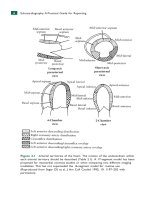
Figure19.2:Schematicdrawingofdoubleinletventricle.Notethepresenceof
right(RA)andleft(LA)atria,twopatentatrioventricularvalves,andbothatria
drain into a single ventricle. In most cases, the single ventricle is
morphologically a left ventricle. A rudimentary ventricle can occasionally be
seen(notshowninthisscheme).
UltrasoundFindings
GrayScale
Thefour-chamberviewisabnormalinDIVasitshowsasingleventriclewitha
missingventricularseptum(Fig.19.3).Identifyingthemorphologyofthesingle
ventricle on ultrasound is based on the anatomic characteristic of the
morphologic right and left ventricles as discussed in Chapter 5. The left
ventricular myocardium appears smooth with fine trabeculations, whereas the
rightventricularmyocardiumiscoarsewithanirregularsurface.Assessmentof
atrioventricular valve anatomy and/or insertion of papillary muscles cannot be
used to determine ventricular morphology in univentricular atrioventricular
connection. Occasionally, the rudimentary right ventricle is seen in the fourchamberplane(Fig. 19.4)butinmost casestheseptaldefect(bulboventricular
foramen)andtherudimentaryrightventricleinDILVareoftennotvisualizedin
thefour-chamberplanebutinamorecranialplane,whenanattempttovisualize
thegreatvesselsismade(Fig.19.5).TherudimentaryoutletchamberinDILVis
more commonly located on the left side of the main ventricle (L-looping) but
canbelocatedontherightside(D-looping)(2).Thegreatarteriesaregenerally
inL-malpositionifthesmalloutletchamberisontheleftsideoftheventricle.
When the small outlet chamber is localized on the right side, the great arteries
ariseeitherinD-malpositionorarenormallyrelatedwiththepulmonaryartery
arising from the small outlet chamber (2). Outflow tract obstructions are
recognizedduetosizediscrepancyratherthanflowdisturbances,whichmaybe
absent.Anarrowpulmonaryarterysuggeststhepresenceofpulmonarystenosis
oratresia,whereasanarrowascendingaortamaybeassociatedwithcoarctation
oftheaortaortubularaorticarchhypoplasia.
Figure19.3:Four-chamberviewsingrayscale(A)andcolorDoppler(B)ina
fetuswithadoubleinletventricle.Notethepresenceofright(RA)andleft(LA)
atria and a single ventricle (SV) in A.B shows, in color Doppler, blood flow
fromtheRAandLAthroughtworespectiveatrioventricularvalvesintotheSV.
L,left.
Figure19.4: Four-chamber view in gray scale in a fetus with a double inlet
ventricle.Notethattheright(RA)andleft(LA)atriadrainthroughtwodistinct
atrioventricularvalvesintotheleftventricle(LV).Thereisarudimentaryright
ventricle(RV)asanoutletchamberdrainedfromtheLV.L,left.
ColorDoppler
Color Doppler may be misleading since two atrioventricular valves are patent
and two color stripes are visualized, thus mimicking the virtual presence of a
separationorseptum(10)(Figs.19.3and19.6).Diagnosisistypicallymadeon
grayscaleultrasound,andcolorDopplerprovidesadditionalinformationonthe
patency of the left and right atrioventricular valves, flow across the VSD, and
great vessels (Fig. 19.5), especially to detect stenosis or atresia (Fig. 19.7).
Restrictive VSD, which may occur in this condition, is better evaluated using
colorDoppler.
Figure19.5:Long-axisviewsingrayscale(A)andcolorDoppler(B)inthe
same fetus shown in Figure 19.4 with a double inlet ventricle (SV) and a
rudimentaryoutletventricle.Therudimentaryoutletventricleisconnectedwith
the SV through a ventricular septal defect (asterisk), called bulboventricular
foramen. Aorta (Ao) and pulmonary artery (PA) arise in parallel orientation.
NotethattheAoissmallerthanthePA,duetothesmallsizeoftheventricular
septaldefect.Aorticcoarctationwasdiagnosedafterbirth.Inf.,inferior.
Figure19.6: Fetus at 15 weeks’ gestation with a double inlet ventricle, with
both right (RA) and left (LA) atria draining through two respective
atrioventricularvalvesintoasingleventricle(SV).AisingrayscaleandBisin
colorDoppler.L,left.
EarlyGestation
DIV can be detected in early gestation (Figs. 19.6 and 19.7) by detecting the
absenceofaventricularseptumonthefour-chamberviewaswellasabnormally
arisinggreatvessels.
Three-DimensionalUltrasound
The combination of three-dimensional (3D) ultrasound with tomographic
imaging permits the simultaneous visualization of the abnormality in the fourchamber plane and the demonstration of the rudimentary ventricle with the
courseofthegreatvessels.Navigatingthroughthevolumeinanofflinesetting
may facilitate the evaluation of the spatial orientation of the great arteries.
Surface rendering shows the large ventricle with inflow from two
atrioventricular valves and a rudimentary outlet chamber (Fig. 19.8) and may
helpinidentifyingthespatialrelationshipofthegreatvessels.
Figure19.7:Four-chamber(A)andlongitudinal(B)viewsincolorDopplerin
afetusat15weeks’gestationwithadoubleinletventricle(samefetusasinFig.
19.6). Note in A that the right (RA) and left (LA) atria drain through two
respective atrioventricular valves into a single ventricle (SV). The longitudinal
planeinBrevealsthepresenceofpulmonaryatresia.Thepulmonaryartery(PA)
ishypoplastic,demonstratesretrogradeflow(arrow),andislocatedposteriorto
theaorta(Ao).Inf.,inferior;L,left.
Figure19.8:Surface-renderingmodeofthefour-chamberviewinafetuswith
doubleinletventricleshowingtheright(RA)andleft(LA)atriaaswellasthe
single ventricle (SV). A small rudimentary ventricle can also be identified
(arrows).L,left;AO,descendingaorta.
AssociatedCardiacandExtracardiacFindings
Associated malformations in DIV are atresia, hypoplasia or straddling of the
atrioventricular valves, pulmonary (or subpulmonic) outflow obstruction,
(sub)aorticoutflowobstruction,andconductionabnormalities,primarilydueto
theanatomicdisruptionoftheconductionsystem(1).
The most important extracardiac abnormality to rule out is the presence of
rightorleftisomerism(seeChapter30),especiallyinthepresenceofacommon
inletventricle(11).Thesequentialapproachtotheultrasoundexaminationofthe
heart may permit detection of corresponding abnormalities. Chromosome
anomalies and other extracardiac anomalies than isomerism are possible but
ratherunusual.
DifferentialDiagnosis
Table 19.1 lists several cardiac malformations in the differential diagnosis of
DIV.DIVmaybemissedonprenatalultrasoundinalateralviewoftheheartin
diastole because the papillary muscles may mimic a ventricular septum in a
singleventricle.
PrognosisandOutcome
DIVwithpatentatrioventricularvalvesiswelltoleratedinthefetus.Follow-up
ultrasound is important prenatally as outflow tract obstruction may develop or
worsenduetoreducedflowandlackofvesselgrowth.Theneonatalcourseof
DIV is dependent on the presence of associated malformations, such as
obstructionofthegreatvesselsoratrioventricularvalveabnormalities.Surgical
treatmentcorrespondstoasingleventricularrepair.Thetypeofsurgicalrepair
(pulmonary artery banding, Fontan procedure, or other) mainly depends on
detailedevaluationofthegreatvesselarrangementandperfusion.
Anoverallmortalityrateof29%withfollow-upupto25yearsofagewas
noted in an outcome study on 105 patients with DILV and transposed arteries
(12). Multivariate analysis showed the presence of arrhythmia and pacemaker
requirementasindependentriskfactorsformortality,whereaspulmonaryatresia
or stenosis and pulmonary artery banding were associated with decreased
mortality(12). Gender, era of birth, aortic arch anomaly, and systemic outflow
obstruction were not risk factors for long-term outcome (12). Similar findings
were reported on eight fetuses with DILV with L-transposition of the great
vessels (13). Of these, four fetuses (50%) had pulmonary atresia, one fetus
(12.5%) also had TA and coarctation of the aorta (died), and one fetus had
completeheartblockandlongQTsyndrome(died)(13).Overallgoodoutcome
was noted in six (75%) infants (13). The outcome of fetuses with DIV is
generallygoodintheabsenceofassociatedrhythmabnormalities.
KEYPOINTS DoubleInletVentricle
DIVisthemostcommonformofuniventricularatrioventricular
connection.
DIVischaracterizedbytwonormallydevelopedrightandleftatriathat
connectviaseparaterightandleftatrioventricularvalvestoacommon
ventricle.
ThemostcommonformofDIVisadoubleinlettoamorphologicleft
ventricle,representingabout80%ofcases.
Thefour-chamberviewisabnormalinDIV.
InDIV,outflowtractobstructionisoftenpresentandaffectsthevessel
arisingfromtherudimentaryventricle.
AssociatedmalformationswithDIVareatresia,hypoplasiaorstraddling
oftheatrioventricularvalves,pulmonary(orsubpulmonic)outflow
obstruction,(sub)aorticoutflowobstruction,andconduction
abnormalities.
●TRICUSPIDATRESIAWITHVENTRICULAR
SEPTALDEFECT
Definition,SpectrumofDisease,andIncidence
TA is characterized by the absence of the right atrioventricular connection,
resulting in lack of communication between the right atrium and ventricle (1)
(Fig.19.9).Therightventricleisthereforediminutiveinsize.Inmostcases,the
tricuspid valve apparatus does not develop, and the right atrioventricular
junction appears as echogenic thickened tissue on ultrasound examination. An
inlet-typeVSD,typicallyperimembranous,isalwayspresent,andthesizeofthe
right ventricle is related to the size of the VSD (Fig.19.9). A large interatrial
communication, in the form of a widely patent foramen ovale or atrial septal
defect, is necessary given an obstructed tricuspid valve. TA is classified into
threetypesbasedonthespatialorientationofthegreatvessels(14).TAtype1
occurs in 70% to 80% of cases and is associated with normally oriented great
arteries(aortafromleftventricleandpulmonaryarteryfromrightventricle)(Fig.
19.9). TA type 2 occurs in 12% to 25% of cases and is associated with Dtransposition of the great vessels. TA type 3, an uncommon malformation, is
seen in the remainder of TA cases and usually denotes complex great vessel
abnormalities,suchastruncusarteriosusorL-transposition.TAisrare,withan
incidence of 0.08 per 1,000 live births (9). TA is reported in about 4% of
congenital heart disease prenatally and is more common in prenatal series
primarily as it belongs to the group of cardiac anomalies associated with an
abnormalfour-chamberview(15–18).Figure19.10isananatomicspecimenof
afetalheartwithTA.
UltrasoundFindings
GrayScale
The four-chamber view in TA is diagnostic and reveals a diminutive right
ventricle,aVSD,andtheabsenceofaright-sidedatrioventricularjunction(Figs.
19.11and19.12).Therightventricleissmallanditssizeisprimarilyrelatedto
thesizeoftheVSD:thesmallertheVSD,thesmallertherightventricle(Figs.
19.11 and 19.12). Right ventricular contractility is normal with no myocardial
thickening. The atretic right atrioventricular junction appears as an echogenic
thickened tissue and the right atrium is slightly dilated (Fig. 19.11). The
interatrial communication is large, and there is often a redundant flap of the
septumsecundumthatbulgesintotheleftatrium(Fig.19.11).Theinteratrialand
interventricular septa are malaligned (Figs. 19.11 and 19.12). In the fivechamber-, short-axis, and three-vessel-trachea views, the ventriculoarterial
connections can be evaluated for discordance (see Chapter 28 for details on
ultrasounddiagnosisoftranspositionofthegreatarteries).Thesizeofthegreat
vessel arising from the right ventricle should be carefully evaluated for the
presenceofstenosis,afairlycommonassociation.Theseverityofrightoutflow
obstruction is directly related to the size of the right ventricle and the VSD.
Pulmonaryoraorticatresiacanbefoundoccasionally.Arightaorticarchcanbe
presentandnotedtocoursetotherightofthetracheaonthethree-vessel-trachea
view.
Figure 19.9: Schematic drawing of tricuspid atresia with ventricular septal
defect(VSD).Notetheabsenceoftherightatrioventricularconnection.AVSD
with a diminutive right ventricle (RV) is noted. Also see the widely patent
foramen ovale and the right ventricular outflow tract obstruction (here
pulmonary stenosis). LA, left ventricle; RA, right ventricle; LV, left ventricle;
Ao,aorta;PA,pulmonaryartery.
Figure19.10: Anatomic specimen of a fetal heart with tricuspid atresia and
ventricular septal defect (VSD) opened at the four-chamber view plane. The
rightventricle(RV)issmallandisconnectedtotheleftventricle(LV)byaVSD
with absent right atrioventricular junction. The atretic tricuspid valve (yellow
arrows)appearsasthickenedtissue.RA,rightatrium.
Figure 19.11: Four-chamber view in a fetus at 29 weeks’ gestation with
tricuspidatresiaandventricularseptaldefect.Therightventricle(RV)issmall
and is connected to the left ventricle (LV) with a ventricular septal defect
(asterisk).Openarrowpointstotheatretic,thickenedtricuspidvalve.Note the
wideforamenovale(FO)witharedundantflapoftheinteratrialseptum(small
arrows). Interatrial and interventricular septa are malaligned. LA, left atrium;
RA,rightatrium.
Figure19.12: Four-chamber views in gray scale in a fetus at 21 weeks’ (A)
and32weeks’ (B) gestation with tricuspid atresia and ventricular septal defect
(VSD). Due to the small and restrictive VSD (arrows), the size of the right
ventricle(RV)isdiminutive.LA,leftatrium;LV,leftventricle;RA,rightatrium.
ColorDoppler
ColorDopplerconfirmsthediagnosisongrayscaleultrasoundbydemonstrating
thelackofbloodflowacrossthetricuspidvalveandapatentmitralvalve(Fig.
19.13).AliasingistypicallynotedacrossthemitralvalveoncolorDopplerdue
toincreasedbloodflow(Fig.19.13).Thepresenceofmitralvalveregurgitation
on color Doppler prenatally has been associated with poor outcome. The right
ventricularcavityisfilledinlatediastolefromtheleftventricleasleft-to-right
shuntingthroughtheVSD,andflowacrosstheVSDcanbevisualizedoncolor
Doppler (Fig. 19.13). Color Doppler is also helpful in the evaluation of flow
across the great arteries (Figs. 19.14 and 19.15). Flow across the pulmonary
artery is generally antegrade and nonturbulent. The suspicion of pulmonary
stenosisisgenerallyachievedbyadiminutivesizeofthevesselratherthanthe
demonstration of turbulent flow on color Doppler, which is typically absent in
thesecases.Flowacrosstheductusarteriosusinthethree-vessel-tracheaviewis
usuallyantegrade,butthedemonstrationofretrogradeflowinthearterialductis
a sign of ductal-dependent pulmonary circulation with possible cyanosis in the
newborn(Figs.19.14and19.15).Ductal-dependentcirculationinTAisusually
seen in severe pulmonary stenosis or atresia in association with a small right
ventricle. Due to limited flow across the foramen ovale and the subsequently
increasedpreloadintherightatrium,ductusvenosusDopplerwillshowoftena
reversedA-waveinenddiastole(19),whichshouldnotbemisinterpretedassign
ofcardiacfailure.
EarlyGestation
Due to the abnormal four-chamber view, TA can be detected in early gestation
eitherongrayscaleimagingorwhencombinedwithcolorDoppler(Fig.19.16).
TAhasbeenassociatedwithanenlargednuchaltranslucencyinearlygestation
(20). Since reversed A-wave in the ductus venosus has been reported in the
secondandthirdtrimestersinassociationwithTA,thisfindingmaybepresentat
11 to 13 weeks’ gestation and may represent an early sign of right atrial
increasedpreload(19).
Three-DimensionalUltrasound
TomographicandorthogonaldisplaymaydemonstratethemainfeaturesofTA,
suchastheabnormalfour-chamberview,thesizeofthesmallrightventricle,the
VSD, and the relationship and size of the great arteries (21, 22). Volume
renderinginsurfacemode(Fig.19.17)orotherdisplaysasinversionmodeand
glass-bodymode(Fig.19.18)mayhelpintheevaluationofventricularsizeand
greatvesselspatialrelationship.
Figure19.13: Color Doppler at the four-chamber view during early (A) and
late (B) diastole in a fetus with tricuspid atresia and ventricular septal defect
(VSD) (same fetus as in Fig. 19.11). In early diastole (A), blood entering the
rightatrium(RA)passesacrossthewideforamenovaletotheleftatrium(LA)
(whitearrow)andthroughthemitralvalvetotheleftventricle(LV)(redarrow).
Coloraliasingisseenacrossthemitralvalveduetoincreasedbloodflow(Aand
B). The right ventricle (RV) receives blood from the left ventricle (LV) across
theVSD(bluearrow)primarilyinlatediastole(B)andsystole.
Figure19.14: The three-vessel-trachea view in color (A and B) and pulsed
Doppler (C) in a fetus with tricuspid atresia and restrictive ventricular septal
defect with severe pulmonary stenosis (same fetus as in Fig.19.12). A and B
showanarrowpulmonaryartery(PA)incomparisontothedilatedaorta(Ao).A
isduringsystoleanddemonstratesantegradeflowacrosstheAoandPA.InB,
reverse flow is demonstrated in the ductus arteriosus (DA) during diastole.
Pulsed Doppler interrogation of the DA in C reveals bidirectional flow with
antegrade flow in systole and retrograde flow in diastole, a sign of severe
outflowobstructionandpostnatalductal-dependentpulmonarycirculation.
Figure19.15: Tricuspid atresia with ventricular septal defect and pulmonary
atresia. A, which is obtained at the three-vessel-trachea view, shows a single
enlarged,anteriorvessel,aorta(Ao).Bisobtainedatthethree-vesselviewand
shows hypoplastic right and left pulmonary arteries (PA). Color and pulsed
DopplerinCrevealsretrogradeflow(red)intheductusarteriosus(DA),which
isalsoconfirmedbypulsedDoppler(lowerpanel)asholosystolicreverseflow.
Thesefindingsaretypicalforpulmonaryatresia.
Figure 19.16: Transvaginal ultrasound of tricuspid atresia with ventricular
septal defect (VSD) in color Doppler in a fetus at 13 weeks’ gestation. A is
obtained at the four-chamber view and shows blood inflow through the mitral
valve into the left ventricle (LV), with blood reaching the right ventricle (RV)
throughtheVSD(arrows)(comparewithFig.19.13).Bshowsthethree-vesselstrachea view with a narrow pulmonary artery (PA) (associated pulmonary
stenosis)ascomparedtotheaorta(Ao)(similartoFig.19.14).LA,leftatrium;
RA,rightatrium.
Figure19.17:Four-chamberviewobtainedinsurface-renderingmodefroma
3Dultrasoundvolumeoftwofetuseswithtricuspidatresiaandventricularseptal
defect (VSD). Note the dilated left ventricle (LV) and the hypoplastic right
ventricle (RV). Asterisk (left) and arrows (right) point to the location of the
VSD.LA,leftatrium;RA,rightatrium.
Figure19.18:Four-chamberviewobtainedinsurface-renderingmode(Left)
andglass-bodymode(Right)froma3DcolorDopplerultrasoundvolumeofa
fetus with tricuspid atresia and ventricular septal defect (VSD). Note the
differenceinsizeinthecardiaccavities(Left)andthetypicaldirectionofflow
(Right) from right atrium (RA) across the foramen ovale into the left atrium
(LA) (white arrow), across the mitral valve (red arrow) into the left ventricle
(LV)andacrosstheVSD(bluearrow)intothehypoplasticrightventricle(RV).
Asterisk(Left)pointstothelocationoftheVSD.
AssociatedCardiacandExtracardiacFindings
Associatedcardiacfindingsincludealargeinteratrialcommunication,suchasa
patentforamenovaleoranatrialseptaldefect,transpositionofthegreatvessels,
and various degrees of ventricular outflow obstruction. Ventricular outflow
obstruction varies, from a patent pulmonary artery to stenosis and atresia and
frompatentaorticarchtoaorticstenosis,coarctation,orinterruptionoftheaortic
arch. In a multicenter study on the cardiac anatomy in 60 fetuses with TA, 9
fetuseshadpatentgreatvessels,16hadpulmonarystenosis,11hadpulmonary
atresia, 6 had aortic stenosis, 4 had coarctation of the aorta, 9 had aortic
hypoplasia, 2 had interrupted aorta, and 3 had a common arterial trunk, or
undefined ventriculoarterial connection (23). Interestingly, all fetuses with
pulmonaryoutflowobstructionhadventriculoarterialconcordanceandalmostall
fetuses with aortic outflow obstruction had ventriculoarterial discordance (23).
Otherassociatedcardiaclesionsincludepersistentleftsuperiorvenacava,right
aortic arch, pulmonary venous abnormalities, and juxtaposition of the atrial
appendages (23). On some occasions, the great vessels are in a corrected
transposition orientation, which was found in 6 of 60 cases in the series
described previously (23). Due to the atrioventricular discordance, the right
ventricleisontheleftsideandtheatreticvalveisfoundontheleftside,which
may erroneously suggest mitral atresia with VSD. In a study on the prenatal
courseandoutcomeofTAin54fetuses,28hadaconcordantventriculoarterial
connection of which 14 also had pulmonary outflow obstruction, and 25 had a
discordant ventriculoarterial connection of which 14 also had aortic outflow
obstruction (24). The peak velocity index for veins in the ductus venosus was
significantly elevated in 19 fetuses assessed and this finding did not correlate
with adverse intrauterine outcome (24). There were associated extracardiac
anomalies in 12 fetuses, with five chromosomal anomalies (24). Seventeen of
the54casesunderwentterminationofpregnancy,twodiedinutero,twodiedin
infancy,and33childrensurvivedwithamedianfollow-upof26(range,12–120)
months, resulting in a short-term overall survival in continued pregnancies
exceeded89%,withthegreatestrateoflossbeinginthefirstyearofpostnatal
life(24).
ExtracardiacanomaliescanbefoundinTA,andfetalkaryotypingshouldbe
offereddespitearareassociationwithchromosomalaberration,including22q11
microdeletion(23).
DifferentialDiagnosis
Twocardiacmalformationsarecommonlyinvolvedinthedifferentialdiagnosis
of TA: pulmonary atresia with intact septum and DIV. DIV was previously
discussed in this chapter. Table 19.2 differentiates TA with VSD from
pulmonary atresia with intact septum, both presenting with hypoplastic right
ventricleinthefour-chamberview.
TABLE DifferentiatingFeaturesofTricuspidAtresia
19.2
withVentricularSeptalDefect(TA-VSD)and
PulmonaryAtresiawithIntactVentricular
Septum(PA-IVS)
TA-VSD
PA-IVS
Rightventricle
Alwayshypoplastic
Generallyhypoplastic,butmaybeof
normalsizeordilated
Right
ventricular
wall
Normal
Hypertrophic
Interventricular
septum
Ventricularseptal
defect
Intactseptumbulgingtotheleft
ventricle
Interatrial
septum
Largeinteratrial
communicationwith
Normalforamenovale
redundantforamen
ovale
Tricuspidvalve
Thickenedechogenic
tissueandnovalve
apparatus
Generallydysplastictricuspidvalve
withlimitedvalveexcursion
occasionallywithtricuspid
regurgitation
Rightatrium
Normalsizewitha
largeinteratrial
communication
Maybedilatedduetoseveretricuspid
regurgitation
Pulmonary
arteryand
valve
Patentvalve(rarely
atretic),narrow
pulmonaryartery
Atreticvalve,narrowpulmonary
artery
Ductus
arteriosus
Generallyantegrade
flow
Alwaysretrogradeflow
Greatvessels
In80%ofcases
concordant,in20%
transposed
Concordant
Otherfeatures
No
ventriculocoronary
arterial
communications
Ventriculocoronaryarterial
communicationsmaybepresent
Postnatally
Maybestable
withoutcyanosis
Alwayscyanotic
PrognosisandOutcome
Prenatalfollow-upwithserialultrasoundexaminationisimportanttoassessthe
patency of the foramen ovale and the presence of right ventricular outflow
obstruction. Ductus venosus flow will show reverse flow during diastole in
almost all cases, but this is a reflection of right ventricular dysfunction rather
thanapoorprognosticsign(19).Pregnancyterminationisreportedinabout28%
inamulticenterseriesofTAdiagnosedprenatally(23).
Postnatal outcome is dependent on associated cardiac and extracardiac
findings. An outcome study of prenatally diagnosed TA estimated an 83%
survival at 1 year of age following active management (23). By multivariate
analysis, two independent factors were associated with an increase in timerelated mortality in the actively managed group: presence of chromosomal
anomaly or syndrome and use of extracorporeal membrane oxygenation (23).
ThisstudyshowedthatcomparedwithpublishedobservationsofTAdiagnosed
postnatally,antenataldiagnosisofTAappearstohavesimilarshort-termsurvival
inpregnanciessurvivingtobirth(23).
SurgicalcorrectionofTArevolvesaroundbypassingtherightventricleand
creating a conduit between the systemic venous blood and the pulmonary
circulation. Most TA patients are treated with the Fontan procedure, which
primarily consists of a cavopulmonary shunt. If the pulmonary artery is of
normalsize,preventingpulmonaryovercirculationandpulmonaryhypertension
is achieved by banding the pulmonary artery. The overall mortality rate in
patientswhoweretreatedwiththeFontanprocedurewasbetween7%and10%
inpediatricseries(25,26).
KEYPOINTS TricuspidAtresiawithVentricularSeptal
Defect
TAischaracterizedbytheabsenceoftherightatrioventricular
connection,resultinginlackofcommunicationbetweentheright
atriumandventricle.
Aninlet-typeVSD,typicallyperimembranous,isalwayspresentinTA.
InTA,theinteratrialcommunicationislargewitharedundantseptum
secundumvalve.
Ventriculoarterialconnectionsareconcordantin70%to80%and
discordantin12%to25%ofTAcases.
AssociatedcardiacfindingsinTAincludealargeinteratrial
communication,suchasapatentforamenovaleoranatrialseptal
defect,transpositionofthegreatvessels,andvariousdegreesofright
ventricularoutflowobstruction.
Prenatalfollow-upofTAfetuseswithserialultrasoundexaminationis
importanttoassessthepatencyoftheforamenovaleandthepresence
ofrightventricularoutflowobstruction.
AnoutcomestudyofprenatallydiagnosedTAestimatedan83%
survivalat1yearofagefollowingactivemanagement.
REFERENCES
1.EaringMG,HaglerDJ,EdwardsWD.Univentricularatrioventricular
connection.In:AllenHD,DriscollDJ,ShaddyRE,etal,eds.Mossand
Adams’HeartDiseaseinInfants,Children,andAdolescents.8thed.
Baltimore,MD:Williams&Wilkins;2012:1175–1194.
2.HornbergerLK.Double-inletventricleinthefetus.In:AllanLD,
HornbergerLK,SharlandGK,eds.TextbookofFetalCardiology.London,
England:GreenwichMedicalMedia;2000:174–182.
3.MenonSC,CabalkaAK.Univentricularatrioventricularconnections.In:
EidemBW,CettaF,O’LearyPW,eds.EchocardiographyinPediatricand
AdultCongenitalHeartDisease.Philadelphia,PA:Wolters
Kluwer/LippincottWilliams&WilkinsHealth;2010:176–195.
4.PennyDJ,AndersonRH.Otherformsoffunctionallyuniventricularhearts.
In:AndersonRH,BakerEJ,RedingtonA,etal,eds.PediatricCardiology.
3rded.Philadelphia,PA:ElsevierHealthCare-Churchill-Livingstone;
2010:665–686.
5.VanPraaghR,VanPraaghS,VladP,etal.Diagnosisoftheanatomictypes
ofsingleorcommonventricle.AmJCardiol.1965;15:345–366.
6.HallermannFJ,DavisGD,RitterDG,etal.Roentgenographicfeaturesof
commonventricle.Radiology.1966;87:409–423.
7.AndersonRH,BeckerAE,TynanM,etal.Theuniventricular
atrioventricularconnection:gettingtotherootofathornyproblem.AmJ
Cardiol.1984;54:822–828.
8.AndersonRH,TynanM,FreedomRM,etal.Ventricularmorphologyinthe
univentricularheart.Herz.1979;4:184–197.
9.HoffmanJI,KaplanS.Theincidenceofcongenitalheartdisease.JAmColl
Cardiol.2002;39:1890–1900.
10.ChaouiR,McEwingR.Threecross-sectionalplanesforfetalcolorDoppler
echocardiography.UltrasoundObstetGynecol.2003;21:81–93.
11.VanPraaghR,OngleyPA,SwanHJ.Anatomictypesofsingleorcommon
ventricleinman:morphologicandgeometricaspectsofsixtynecropsied
cases.AmJCardiol.1964;13:367–386.