Ebook Cardiovascular physiology (8th edition): Part 2
Bạn đang xem bản rút gọn của tài liệu. Xem và tải ngay bản đầy đủ của tài liệu tại đây (16.68 MB, 151 trang )
Vascular Control
OBJECTIVES
The student understands the general mechanisms involved in local vascular control:
�
Identifies the major ways in which smooth muscle differs anatomically and
functionally from striated muscle.
�
Lists the steps leading to cross-bridge cycling in smooth muscle.
�
Lists the major ion channels involved in the regulation of membrane potential in
�
Describes the processes of electromechanical and pharmacomechanical coupling
in smooth muscle.
�
Defines basal tone.
smooth muscle.
�
Lists several substances potentially involved in local metabolic control.
�
States the local metabolic vasodilator hypothesis.
�
Describes how vascular tone may be influenced by endothelin, prostaglandins,
histamine, and bradykinin.
�
Describes the myogenic response of blood vessels.
�
Defines active and reactive hyperemia and indicates a possible mechanism for each.
�
Defines autoregulation of blood flow and briefly describes the metabolic, myogenic, and tissue pressure theories of autoregulation.
�
Defines neurogenic tone of vascular muscle and describes how sympathetic neu
ral influences can alter it.
� Describes how vascular tone is influenced by circulating catecholamines, vasopres
sin, and angiotensin II.
�
Lists the major influences on venous diameters.
�
Describes how control off/ow differs between organs with strong local metabolic control of arteriolar tone and organs with strong neurogenic control of arteriolar tone.
The student knows the dominant mechanisms of flow and blood volume control in the
major body organs:
�
States the relative importance of local metabolic and neural control of coronary
blood flow.
�
Defines systolic compression and indicates its relative importance to blood flow in
the endocardial and epicardial regions of the right and left ventricular walls.
�
Describes the major mechanisms of flow and blood volume control in each of the fol
lowing systemic organs: skeletal muscle, brain, splanchnic organs, kidney, and skin.
�
States why mean pulmonary arterial pressure is lower than mean systemic arterial
pressure.
�
Describes how pulmonary vascular control differs from that in systemic organs.
126
VASCULAR CONTROL
I
127
Because the body's metabolic needs are continually changing, the cardio
vascular system must continually make adjustments in the diameter of its
vessels. The purposes of these vascular changes are (1) to efficiently
distribute the cardiac output among tissues with different current needs (the job
of arterioles) and (2) to regulate the distribution of blood volume and cardiac fill
ing (the job of veins). In this chapter, we discuss our current understanding of how
all this is accomplished.
VASCULAR SMOOTH MUSCLE
Although long-term adaptations in vascular diameters may depend on
remodeling of both the active (ie, smooth muscle) and passive (ie, struc
tural, connective tissue) components of the vascular wall, short-term vas
cular diameter adjustments are made by regulating the contractile activity of
vascular smooth muscle cells. These contractile cells are present in the walls of all
vessels except capillaries. The task of the vascular smooth muscle is unique,
because to maintain a certain vessel diameter in the face of the continual distend
ing pressure of the blood within it, the vascular smooth muscle must be able to
sustain active tension for prolonged periods.
There are many functional characteristics that distinguish smooth muscle
from either skeletal or cardiac muscle. For example, when compared with these
other muscle types, smooth muscle cells
1.
contract and relax much more slowly;
2. can change their contractile activity as a result of either action potentials or
changes in resting membrane potential;
3. can change their contractile activity in the absence of any changes in mem
brane potential;
4. can maintain tension for prolonged periods at low energy cost; and
5. can be activated by stretch.
Vascular smooth muscle cells are small (approximately 5 Jlm X 50 Jlm) spindle
shaped cells, usually arranged circumferentially or at small helical angles in mus
cular blood vessel walls. In many, but not all, vessels, adjacent smooth muscle
cells are electrically connected by gap junctions similar to those found in the
myocardium.
Contractile Processes
Just as in other muscle types, smooth muscle force development and
shortening are thought to be the result of cross-bridge interaction
between thick and thin contractile filaments composed of myosin and
actin, respectively. In smooth muscle, however, these filaments are not arranged
in regular, repeating sarcomere units. As a consequence, "smooth" muscle cells
lack the microscopically visible striations, characteristic of skeletal and cardiac
muscle cells. The actin filaments in smooth muscle are much longer than those in
128
I
CHAPTER SEVEN
striated muscle. Many of these actin filaments attach to the inner surface of the
cell at structures called dense bands. In the interior of the cell, actin filaments do
not attach to Z lines but rather anchor to small transverse structures called dense
bodies that are themselves tethered to the surface membrane by cable-like inter
mediate filaments. Myosin filaments are interspersed between the smooth muscle
actin filaments but in a more haphazard fashion than the regular interweaving
pattern of striated muscle. In striated muscle, the contractile filaments are invari
ably aligned with the long axis of the cell, whereas in smooth muscle, many
contractile filaments travel obliquely or even transversely to the long axis of the
cell. Despite the absence of organized sarcomeres, changes in smooth muscle
length affect its ability to actively develop tension. That is, smooth muscle exhib
its a "length-tension relationship" analogous to that observed in striated muscle
{see, Figure 2-8). As in striated muscle, the strength of the cross-bridge interac
tion between myosin and actin filaments in smooth muscle is controlled primar
ily by changes in the intracellular free Ca2+ level, which range from approximately
10-s M in the relaxed muscle to 10-5 M during maximal contraction. However,
the sequence of steps linking an increased free Ca2+ concentration to contractile
filament interaction is different in smooth muscle than in striated muscle. In the
smooth muscle:
1.
Intracellular free Ca2+ first forms a complex with the calcium-binding protein
calmodulin.
2. The Ca2+ -calmodulin complex then activates a phosphorylating enzyme
called myosin light-chain kinase {MLC kinase).
3. This enzyme allows the phosphorylation by adenosine triphosphate {ATP) of
the light-chain protein that is a portion of the cross-bridge head of myosin
(MLC).
4. MLC phosphorylation enables cross-bridge formation and cycling during
which energy from ATP is utilized for tension development and shortening.
Smooth muscle is also unique in that once tension is developed, it can be main
tained at very low energy costs, that is, without the need to continually split ATP
in cross-bridge cycling. The mechanisms responsible are still somewhat unclear
but presumably involve very slowly cycling or even noncycling cross-bridges. This
is often referred to as the latch state and may involve light-chain dephosphoryla
tion of attached cross-bridges.
By mechanisms that are yet incompletely understood, it is apparent that vascular
smooth muscle contractile activity is regulated not only by changes in intracellular
free Ca2+ levels but also by changes in the Ca2+ sensitivity of the contractile machin
ery. Thus, the contractile state of vascular smooth muscle may sometimes change
in the absence of changes in intracellular free Ca2+ levels. In part, this apparently
variable Ca2+ sensitivity of the activation of smooth muscle contractile apparatus
may be due to the variable activity of another enzyme, myosin phosphatase, that
facilitates some reaction that involves the phosphorylated MLC as a reactant. For
example, factors that increase the intracellular concentrations of cyclic nucleotides
VASCULAR CONTROL
often lead to relaxation of the vascular smooth muscle. Thus, the
I
129
net state of
phosphorylation of the MLC (and thus presumably contractile strength) depends
on some sort of balance between the effects of the Ca2+ -dependent enzyme MLC
kinase, and the Ca2+-independent enzyme MLC phosphatase.1
Membrane Potentials
Smooth muscle cells have resting membrane potentials ranging from -40 to
-65 mV and thus are generally less negative than those in striated muscle. As in all
cells, the resting membrane potential of the smooth muscle is determined largely
by the cell permeability to potassium. Many types ofK+ channels have been iden
tified in smooth muscle. The one that seems to be predominantly responsible for
determining the resting membrane potential is termed an
inward rectifying-type
K+ channel. There are also ATP-dependent K+ channels that are closed when cel
lular ATP levels are normal but open if ATP levels fall. Such channels have been
proposed to be important in matching organ blood flow to the metabolic state of
the tissue.
Smooth muscle cells regularly have action potentials only in certain vessels.
When they do occur, smooth muscle action potentials are initiated primarily by
inward Ca2+ current and are developed slowly like the "slow-type" cardiac action
potentials (see Figures
2-2C and D). As in the heart, this inward (depolarizing)
voltage-operated channel ( VOC) for Ca2+; this type of
Ca2+ current flows through a
channel is one of several types of calcium channels present in the smooth muscle.
The repolarization phase of the action potential occurs primarily by an outward
flux of potassium ions through both
delayed K+ channels and calcium-activated
K+ channels.
Many types of ion channels in addition to those mentioned have been identi
fied in vascular smooth muscle, but in most cases, their exact role in cardiovas
cular function remains obscure. For example, there appear to be nonselective,
stretch-sensitive cation channels that may be involved in the response of smooth
muscle to stretch. The reader should note, however, that many of the impor
tant ion channels in vascular smooth muscle are also important in heart muscle
(see Table
2-1).
1 It is very important when thinking about biological processes to keep in mind that ANY "enzyme" is
simply a chemical catalyst. As such, enzymes do not cause reactions to happen; rather, they let reactions
happen faster than they would in their absence. That is, catalysts do not determine the direction in which
chemical reactions proceed. With or without catalysts, chemical reactions ALWAYS relentlessly proceed
only in the direction of chemical equilibrium. The "case in point" example here is that although the Ca2+
activation of MLC kinase may well facilitate a reaction that would result in phosphorylated MLC as a
product, it is naive to think that Ca2+ removal from the intracellular space (and therefore lowered MLC
kinase activity) would in itself reverse the process. The absence of a catalyst cannot make a reaction
proceed backward! Moreover, it is equally erroneous to conceive there could be different catalysts for a
given chemical reaction that could make it proceed in opposite directions. Ergo, MLC kinase, and MLC
phosphatase must facilitate distinctly different chemical reactions.
130
I
CHAPTER SEVEN
Electromechanical
coupling
Pharmacomechanical
coupling
DAG
PIP2
Sarcoplasmic
reticulum
Contraction
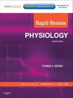
Figure 7-1. General mechanisms for activation ofthe vascular smooth muscle. VOC,
voltage-operated Ca2+ channel; ROC, receptor-operated Ca2+ channel; R, agonist-specific
receptor; G, GTP-binding protein; PIP , phosphatidylinositol biphosphate; IP3, inositol
2
triphosphate; DAG, diacylglycerol.
Electromechanical versus Pharmacomechanical Coupling
In smooth muscle, changes in intracellular free Ca2+ levels can occur both
with and without changes in membrane potential. The processes involved
are called electromechanical coupling and pharmacomechanical coupling,
respectively, and are illustrated in Figure 7-1.
Electromechanical coupling, shown in the left half of Figure 7-1, occurs
because the smooth muscle surface membrane contains VOCs for calcium (the
same VOCs that are involved in action potential generation). Membrane depo
larization increases the open-state probability of these channels and thus leads to
smooth muscle cell contraction and vessel constriction. Conversely, membrane
hyperpolarization leads to smooth muscle relaxation and vessel dilation. Because
the VOCs for Ca2+ are partially activated by the low resting membrane potential
of the vascular smooth muscle, changes in resting potential can alter the resting
calcium influx rate and therefore the basal contractile state.
With pharmacomechanical coupling, chemical agents (eg, released neurotrans
mitters) can induce smooth muscle contraction without the need for a change
in membrane potential. As illustrated on the right side of Figure 7-1, the com
bination of a vasoconstrictor agonist (such as norepinephrine) with a specific
membrane-bound receptor (such as an 0.1-adrenergic receptor) initiates events that
cause intracellular free Ca2+ levels to increase for two reasons. One, the activated
VASCULAR CONTROL
receptor may open surface membrane
I
131
receptor-operated channels for Ca2+ that
allow Ca2+ influx from the extracellular fluid. Two, the activated receptor may
induce the formation of an intracellular "second messenger," inositol trisphos
phate (IP3), which opens specific channels that release Ca2+ from the intracellular
sarcoplasmic reticulum stores. In both processes, the activated receptor first stim
ulates specific guanosine triphosphate-binding proteins (GTP-binding proteins or
G proteins). Such receptor-associated G proteins seem to represent a general first
step through which most membrane receptors operate
to
initiate their particular
cascade of events that ultimately lead to specific cellular responses.
The reader should
not conclude from Figure 7-1 that all vasoactive chemical
agents (chemical agents that cause vascular effects) produce their actions on the
smooth muscle without changing membrane potential. In fact, most vasoactive
chemical agents do cause changes in membrane potential because their receptors
can be linked, by G proteins or other means, to ion channels of many kinds.
Not shown in Figure
7-1 are the processes that remove Ca2+ from the cyto
plasm of the vascular smooth muscle, although they are important as well in
determining the free cytosolic Ca2+ levels. As in cardiac cells (see Figure
2-7),
smooth muscle cells actively pump calcium into the sarcoplasmic reticulum and
outward across the sarcolemma. Calcium is also countertransported out of the cell
in exchange for sodium.
Mechanisms for Relaxation
Hyperpolarization of the cell membrane is one mechanism for causing smooth
muscle relaxation and vessel dilation. In addition, however, there are at least
two general mechanisms by which certain chemical vasodilator agents can cause
smooth muscle relaxation by pharmacomechanical means. In Figure
7-1, the spe
cific receptor for a chemical vasoconstrictor agent is shown linked by a specific G
protein to phospholipase C. In an analogous manner, other specific receptors may
be linked by other specific G proteins to other enzymes that produce second mes
sengers other than IP3• An example is the � -adrenergic receptor that is present
2
in arterioles of the skeletal muscle and liver. � -Receptors are not innervated but
2
can sometimes be activated by abnormally elevated levels of circulating epineph
rine. The � -receptor is linked by a particular G protein (G,) to adenylate cyclase.
2
Adenylate cyclase catalyzes the conversion of ATP to cyclic adenosine monophos
phate (cAMP). Increased intracellular levels of cAMP cause the activation of pro
tein kinase A, a phosphorylating enzyme that in turn causes phosphorylation of
proteins at many sites. The overall result is stimulation of Ca2+ efflux, membrane
hyperpolarization, and decreased contractile machinery sensitivity to Ca2+ -all
of which act synergistically to cause vasodilation. In addition to epinephrine,
histamine and vasoactive intestinal peptide are other vasodilator substances that
act through the cAMP pathway.
Vascular �-receptors are designated �2-receptors and are pharmacologically distinct from the �1-receptors
found on cardiac cells.
2
132
I
CHAPTER SEVEN
In addition to cAMP, cyclic guanosine monophosphate (cGMP) is an impor
tant intracellular second messenger that causes vascular smooth muscle relaxation.
Nitric oxide is an important vasodilator substance that operates via the cGMP
pathway. Nitric oxide can be produced by endothelial cells and also by nitrates, a
clinically important class of vasodilator drugs. Nitric oxide is gaseous and easily
diffuses into smooth muscle cells, where it activates the enzyme guanylyl cyclase
that in turn causes cGMP formation.
CONTROL OF ARTERIOLAR TONE
Vascular tone is a term commonly used to characterize the general con
tractile state of a vessel or a vascular region. The "vascular tone" of a region
can be taken as an indication of the "level of activation" of the individual
smooth muscle cells in that region. As described in Chapter 6, the blood flow
through any organ is determined largely by its vascular resistance, which is depen
dent primarily on the diameter of its arterioles. Consequently, an organ's flow is
controlled by factors that influence the arteriolar smooth muscle tone.
Basal Tone
Arterioles remain in a state of partial constriction even when all external influ
ences on them are removed; hence, they are said to have a degree of basal tone
(sometimes referred to as intrimic tone). The understanding of the mechanism is
incomplete, but basal arteriolar tone may be a reflection of the fact that smooth
muscle cells inherently and actively resist being stretched as they continually are
in pressurized arterioles. Another hypothesis is that the basal tone of arterioles is
the result of a tonic production of local vasoconstrictor substances by the endo
thelial cells that line their inner surface. In any case, this basal tone establishes
a baseline of partial arteriolar constriction from which the external influences
on arterioles exert their dilating or constricting effects. These influences can be
separated into three categories: local influences, neural inf luences, and hormonal
influences.
Local Influences on Arterioles
METABOLIC INFLUENCES
The arterioles that control flow through a given organ lie within the
organ tissue itsel£ Thus, arterioles and the smooth muscle in their walls
are exposed to the chemical composition of the interstitial fluid of the
organ they serve. The interstitial concentrations of many substances reflect the
balance between the metabolic activity of the tissue and its blood supply.
Interstitial oxygen levels, for example, fall whenever the tissue cells are using oxy
gen faster than it is being supplied to the tissue by blood flow. Conversely, inter
stitial oxygen levels rise whenever excess oxygen is being delivered to a tissue from
the blood. In nearly all vascular beds, exposure to low oxygen reduces arteriolar
tone and causes vasodilation, whereas high oxygen levels cause arteriolar
VASCULAR CONTROL
Vasodilator factors
� � � �
I
133
Removal rate proportional to blood flow
--
81 od flow ----+
Arterioles
Capillaries
Veins
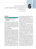
Figure 7-2. Local metabolic vasodilator hypothesis.
vasoconstriction.3 Thus, a local feedback mechanism exists that automatically
operates on arterioles to regulate a tissue's blood flow in accordance with its meta
bolic needs. Whenever blood flow and oxygen delivery fall below a tissue's oxygen
demand, the oxygen levels around arterioles fall, the arterioles dilate, and the
blood flow through the organ appropriately increases.
Many substances in addition to oxygen are present within tissues and can
affect the tone of the vascular smooth muscle. When the metabolic rate of skel
etal muscle is increased by exercise, tissue levels of oxygen decrease, but those of
carbon dioxide, H+, and K+ increase. Muscle tissue osmolarity also increases dur
ing exercise. All these chemical alterations cause arteriolar dilation. In addition,
with increased metabolic activity or oxygen deprivation, cells in many tissues may
release adenosine, which is an extremely potent vasodilator agent.
At present, it is not known which of these (and possibly other) metabolically
related chemical alterations within tissues are most important in the local meta
bolic control of blood flow. It appears likely that arteriolar tone depends on the
combined action of many factors.
For conceptual purposes, Figure 7-2 summarizes current understanding of
local metabolic control. Vasodilator factors enter the interstitial space from the
tissue cells at a rate proportional to tissue metabolism. These vasodilator fac
tors are removed from the tissue at a rate proportional to blood flow. Whenever
tissue metabolism is proceeding at a rate for which the blood flow is inade
quate, the interstitial vasodilator factor concentrations automatically build up
and cause the arterioles to dilate. This, of course, causes blood flow to increase.
The process continues until blood flow has risen sufficiently to appropriately
match the tissue metabolic rate and prevent further accumulation of vasodilator
3
An important exception to this rule occurs in the pulmonary circulation and is discussed later in this
chapter.
134
I
CHAPTER SEVEN
factors. The same system also operates to reduce blood flow when it is higher
than required by the tissue's metabolic activity, because this situation causes a
reduction in the interstitial concentrations of metabolic vasodilator factors.
Local metabolic mechanisms represent by for the most important meam of local
flow control. By these mechanisms, individual organs are able to regulate their
own flow in accordance with their specific metabolic needs.
As indicated below, several other types of local influences on blood vessels have
been identified. However, many of these represent fine-tuning mechanisms and
many are important only in certain, usually pathological, situations.
LOCAL INFLUENCES FROM ENDOTHELIAL CELLS
Endothelial cells cover the entire inner surface of the cardiovascular system. A
large number of studies have shown that blood vessels respond very differently to
certain vascular influences when their endothelial lining is missing. Acetylcholine,
for example, causes vasodilation of intact vessels but causes vasoconstriction of
vessels stripped of their endothelial lining. This and similar results led to the
realization that endothelial cells can actively participate in the control of arterio
lar diameter by producing local chemicals that affect the tone of the surrounding
smooth muscle cells. In the case of the vasodilator effect of infusing acetylcholine
through intact vessels, the vasodilator influence produced by endothelial cells has
been identified as nitric oxide. Nitric oxide is produced within endothelial cells
from the amino acid, L-arginine, by the action of an enzyme, nitric oxide syn
thase. Nitric oxide synthase is activated by a rise in the intracellular level of the
Ca2+. Nitric oxide is a small lipid-soluble molecule that, once formed, easily dif
fuses into adjacent smooth muscle cells where it causes relaxation by stimulating
cGMP production as mentioned previously.
Acetylcholine and several other agents (including bradykinin, vasoactive intes
tinal peptide, and substance P) stimulate endothelial cell nitric oxide production
because their receptors on endothelial cells are linked to receptor-operated Ca2+
channels. Probably more importantly from a physiological standpoint, flow
related shear stresses on endothelial cells stimulate their nitric oxide production
presumably because stretch-sensitive channels for Ca2+ are activated. Such flow
related endothelial cell nitric oxide production may explain why, for example,
exercise and increased blood flow through muscles of the lower leg can cause dila
tion of the blood-supplying femoral artery at points far upstream of the exercising
muscle itsel£
Agents that block nitric oxide production by inhibiting nitric oxide synthase
cause significant increases in the vascular resistances of most organs. For this
reason, it is believed that endothelial cells are normally always producing some
nitric oxide that is importantly involved, along with other factors, in reducing the
normal resting tone of arterioles throughout the body.
Endothelial cells have also been shown to produce several other locally acting
vasoactive agents including the vasodilators "endothelial-derived hyperpolarizing
factor", prostacyclin and the vasoconstrictor
endothelin.
Endothelin in particular
is the topic of intense current research. It has the greatest vasoconstrictor potency
VASCULAR CONTROL
I
135
of any known substance and appears to have many other biological effects as
well. Much recent evidence suggests that endothelin may play important roles in
such important overall process such as bodily salt handling and blood pressure
regulation.
One general unresolved issue with the concept that arteriolar tone (and there
fore local nutrient blood flow) is regulated by factors produced by arteriolar
endothelial cells is how these cells could know what the metabolic needs of the
downstream tissue are. This is because the endothelial cells lining arterioles are
exposed to arterial blood whose composition is constant regardless of flow rate or
what is happening downstream. One hypothesis is that there exists some sort of
communication system between vascular endothelial cells. That way, endothelial
cells in capillaries or venules could telegraph upstream information about whether
the blood flow is indeed adequate.
OTHER LOCAL CHEMICAL INFLUENCES
In addition to local metabolic influences on vascular tone, many specific
locally-produced and locally-reacting chemical substances have been
identified that have vascular effects and therefore could be important in
local vascular regulation in certain instances. In most cases, however, definite
information about the relative importance of these substances in cardiovascular
regulation is lacking.
Prostaglandins and thromboxane are a group of several chemically related prod
ucts of the cyclooxygenase pathway of arachidonic acid metabolism. Certain
prostaglandins are potent vasodilators, whereas others are potent vasoconstric
tors. Despite the vasoactive potency of the prostaglandins and the fact that most
tissues (including endothelial cells and vascular smooth muscle cells) are capable
of synthesizing prostaglandins, it has not been demonstrated convincingly that
prostaglandins play a crucial role in normal vascular control. It is clear, how
ever, that vasodilator prostaglandins are involved in inflammatory responses.
Consequently, inhibitors of prostaglandin synthesis, such as aspirin, are effective
anti-inflammatory drugs. Prostaglandins produced by platelets and endothelial
cells are important in the hemostatic (flow stopping, antibleeding) vasoconstric
tor and platelet-aggregating responses to vascular injury. Hence, aspirin is often
prescribed to reduce the tendency for blood dotting-especially in patients with
potential coronary flow limitations. Arachidonic acid metabolites produced via
the lipoxygenase system (eg, leukotrienes) also have vasoactive properties and may
influence blood flow and vascular permeability during inflammatory processes.
Histamine is synthesized and stored in high concentrations in secretory granules
of tissue mast cells and circulating basophils. When released, histamine produces
arteriolar vasodilation (via the cAMP pathway) and increases vascular permeabil
ity, which leads to edema formation and local tissue swelling. Histamine increases
vascular permeability by causing separations in the junctions between the endo
thelial cells that line the vascular system. Histamine release is classically associated
with antigen-antibody reactions in various allergic and immune responses. Many
drugs and physical or chemical insults that damage tissue also cause histamine
136
I
CHAPTER SEVEN
release. Histamine can stimulate sensory nerve endings to cause itching and pain
sensations. Although clearly important in many pathological situations, it seems
unlikely that histamine participates in normal cardiovascular regulation.
Bradykinin is a small polypeptide that has approximately ten times the vaso
dilator potency of histamine on a molar basis. It also acts to increase capillary
permeability by opening the junctions between endothelial cells. Bradykinin is
formed from certain plasma globulin substrates by the action of an enzyme, kal
likrein, and is subsequently rapidly degraded into inactive fragments by vari
ous tissue kinases. Like histamine, bradykinin is thought to be involved in the
vascular responses associated with tissue injury and immune reactions. It also
stimulates nociceptive nerves and may thus be involved in the pain associated
with tissue injury.
TRANSMURAL PRESSURE
The passive elastic mechanical properties of arteries and veins and how changes in
transmural pressure affect their diameters are discussed in Chapter 6. The effect
of transmural pressure on arteriolar diameter is more complex because arterioles
respond both passively and actively to changes in transmural pressure. For example,
a sudden increase in the internal pressure within an arteriole produces (I) first an
initial slight passive mechanical distention (slight because arterioles are relatively
thick-walled and muscular), and (2) then an active constriction that, within sec
onds, may completely reverse the initial distention. A sudden decrease in transmu
ral pressure elicits essentially the opposite response, that is, an immediate passive
decrease in diameter followed shortly by a decrease in active tone, which returns
the arteriolar diameter to near that which existed before the pressure change. The
active phase of such behavior is referred to as a myogenic response, because it seems
to originate within the smooth muscle itsel£ The mechanism of the myogenic
response is not known for certain, but stretch-sensitive ion channels on arteriolar
vascular smooth muscle cells are likely candidates for involvement.
All arterioles have some normal distending pressure to which they are prob
ably actively responding. Therefore, the myogenic mechanism is likely to be a
fundamentally important factor in determining the basal tone of arterioles every
where. Also, for obvious reasons and as soon discussed, the myogenic response is
potentially involved in the vascular reaction to any cardiovascular disturbance
that involves a change in arteriolar transmural pressure.
fLOW RESPONSES CAUSED BY LOCAL MECHANISMS
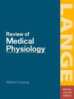
Active Hyperemia-In organs with a highly variable metabolic rate, such as skel
etal and cardiac muscles, the blood flow closely follows the tissue's metabolic rate.
For example, skeletal muscle blood flow increases within seconds of the onset of
muscle exercise and returns to control values shortly after exercise ceases. This
phenomenon, which is illustrated in Figure 7-3A, is known as exercise or active
hyperemia (hyperemia means high flow). It should be clear how active hyperemia
could result from the local metabolic vasodilator feedback on the arteriolar smooth
muscle. As alluded to previously, once initiated by local metabolic influences on
VASCULAR CONTROL
I
137
A
"'C
Active hyperemia
8
:c
c:
as
e'
0
t---·--�
I•
Period of increased
metabolic rate
�1
B
"8
0
:c
c:
as
e'
0
Reactive hyperemia
1-----..,-
I•
��
Period of arrested blood flow
Figure 7-3. Organ blood flow responses caused by local mechanisms: active and
reactive hyperemias.
small resistance vessels, endothelial flow-dependent mechanisms may assist in
propagating the vasodilation to larger vessels upstream, which helps promote the
delivery of blood to the exercising muscle.
Reactive Hyperemia-In this case, the higher-than-normal blood flow occurs
transiently after the removal of any restriction that has caused a period of lower
than-normal blood flow and is sometimes referred to as postocclusion hyperemia.
The phenomenon is illustrated in Figure 7-3B. For example, flow through an
extremity is higher than normal for a period after a tourniquet is removed from
the extremity. Both local metabolic and myogenic mechanisms may be involved in
producing reactive hyperemia. The magnitude and duration of reactive hyperemia
depend on the duration and severity of the occlusion as well as the metabolic rate
of the tissue. These findings are best explained by an interstitial accumulation of
metabolic vasodilator substances during the period of flow restriction. However,
unexpectedly large flow increases can follow arterial occlusions lasting only 1 or
2 s. These may be explained best by a myogenic dilation response to the reduced
138
I
CHAPTER SEVEN
intravascular pressure and decreased stretch of the arteriolar walls that exists dur
ing the period of occlusion.
Autoregulation-Except when displaying active and reactive hyperemia, nearly
all organs tend to keep their blood flow constant despite variations in arterial
pressure-that is, they autoregulate their blood flow. As shown in Figure 7-4A,
an abrupt increase in arterial pressure is normally accompanied by an initial
abrupt increase in organ blood flow that then gradually returns toward normal
despite the sustained elevation in arterial pressure. The initial rise in flow with
A
!!!
:::l
lll
!!!
a.
��------....1
Sustained pressure
increase
�
<(
Blood flow
autoregulation
�
q::
"C
0
0
:c
r:::
al
�
0
Steady state
1-----..1 --------------
B
�
q::
"C
0
0
:c
r:::
al
�
0
Autoregulatory
range
i
:>.
"C
al
V5
100
200
Mean arterial pressure (mm Hg)
Figure 7-4.
Autoregulation of organ blood flow.
VASCULAR CONTROL
increased pressure is expected from the basic flow equation
(Q
=
I
139
AP!R). The sub
sequent return of flow toward the normal level is caused by a gradual increase
in active arteriolar tone and resistance to blood flow. Ultimately, a new steady
state is reached with only slightly elevated blood flow because the increased driv
ing pressure is counteracted by a higher-than-normal vascular resistance. As with
the phenomenon of reactive hyperemia, blood flow autoregulation may be caused
by both local metabolic feedback mechanisms and myogenic mechanisms. The
arteriolar vasoconstriction responsible for the autoregulatory response shown in
Figure 7-4A, for example, may be partially due to
(I) a "washout" of metabolic
vasodilator factors from the interstitium by the excessive initial blood flow and
(2) a myogenic increase in arteriolar tone stimulated by the increase in stretching
tissue
pressure hypothesis of blood flow autoregulation for which it is assumed that an
forces that the increase in pressure imposes on the vessel walls. There is also a
abrupt increase in arterial pressure causes transcapillary fluid filtration and thus
leads to a gradual increase in interstitial fluid volume and pressure. Presumably
the increase in extravascular pressure would cause a decrease in vessel diameter
by simple compression. This mechanism might be especially important in organs
such as the kidney and brain whose volumes are constrained by external structures.
Although not illustrated in Figure 7-4A, autoregulatory mechanisms operate
in the opposite direction in response to a decrease in arterial pressure below the
normal value. One important general consequence of local autoregulatory mecha
nisms is that the steady-state blood flow in many organs tends to remain near the
normal value over quite a wide range of arterial pressure. This is illustrated in the
graph in Figure 7-4B. As discussed later, the inherent ability of certain organs to
maintain adequate blood flow despite lower-than-normal arterial pressure is of
considerable importance in situations such as shock from blood loss.
Neural Influences on Arterioles
SYMPATHETIC VASOCONSTRICTOR NERVES
These neural fibe�s innervate arterioles in all systemic organs and provide
by far the most Important means of reflex control of the vasculature.
Sympathetic vasoconstrictor nerves are the backbone of the system for
controlling total peripheral resistance and are thus essential participants in global
cardiovascular tasks such as regulating arterial blood pressure.
Sympathetic vasoconstrictor nerves release norepinephrine from their
terminal structures in amounts generally proportional to their action
potential frequency. Norepinephrine causes an increase in the tone of
arterioles after combining with an
Ct1-adrenergic receptor on smooth muscle cells.
Norepinephrine appears to increase vascular tone primarily by pharmacomechan
ical means. The mechanism involves G-protein linkage of a-adrenergic receptors
to phospholipase C and subsequent Ca2+ release from intracellular stores by the
action of the second messenger IP3, as illustrated on the right side of Figure 7-1.
Sympathetic vasoconstrictor nerves normally have a continual or tonic firing
activity. This tonic activity of sympathetic vasoconstrictor nerves makes the
140
I
CHAPTER SEVEN
normal contractile tone of arterioles considerably greater than their basal tone.
The additional component of vascular tone is called neurogenic tone. When the fir
ing rate of sympathetic vasoconstrictor nerves is increased above normal, arterioles
constrict and cause organ blood flow to fall below normal. Conversely, vasodila
tion and increased organ blood flow can be caused by sympathetic vasoconstrictor
nerves if their normal tonic activity level is reduced. Thus, an organ's blood flow
can either be reduced below normal or be increased above normal by changes in
the sympathetic vasoconstrictor fiber firing rate.
OTHER NEURAL INFLUENCES
Blood vessels, as a general rule, do not receive innervation from the parasympa
thetic division of the autonomic nervous system. However, parasympathetic vaso
dilator nerves, which release acetylcholine, are present in the vessels of the brain
and the heart, but their influence on arteriolar tone in these organs appears to be
inconsequential. Parasympathetic vasodilator nerves are also present in the vessels
of the salivary glands, pancreas, and gastric mucosa where they have important
influences on secretion and motility. In the external genitalia, they are respon
sible for the vasodilation of inflow vessels responsible for promoting secretion and
erection.
Hormonal Influences on Arterioles
Under normal circumstances, short-term hormonal influences on blood
vessels are generally thought to be of minor consequence in comparison
to the local metabolic and neural influences. However, it should be
emphasized that the understanding of how the cardiovascular system operates in
many situations is incomplete. Thus, the hormones discussed in the following sec
tions may play more important roles in cardiovascular regulation than is now
appreciated.
CIRCULATING CATECHOLAMINES
During activation of the sympathetic nervous system, the adrenal glands release
the catecholamines epinephrine and norepinephrine into the bloodstream. Under
normal circumstances, the blood levels of these agents are probably not high
enough to cause significant cardiovascular effects. However, circulating catechol
amines may have cardiovascular effects in situations (such as vigorous exercise or
hemorrhagic shock) that involve high activity of the sympathetic nervous system.
In general, the cardiovascular effects of high levels of circulating catecholamines
parallel the direct effects of sympathetic activation, which have already been dis
cussed; both epinephrine and norepinephrine can activate cardiac �t-adrenergic
receptors to increase the heart rate and myocardial contractility and can acti
vate vascular a-receptors to cause vasoconstriction. Recall that in addition to the
at-receptors that mediate vasoconstriction, arterioles in a few organs also possess
�2-adrenergic receptors that mediate vasodilation. Because vascular �2-receptors
are more sensitive to epinephrine than are vascular at-receptors, moderately
VASCULAR CONTROL
I
141
elevated levels of circulating epinephrine can cause vasodilation, whereas higher
levels cause a,-receptor-mediated vasoconstriction. Vascular �2-receptors are
not innervated and therefore are not activated by norepinephrine, released from
sympathetic vasoconstrictor nerves. The physiological importance of these vas
cular �2-receptors is unclear because adrenal epinephrine release occurs during
periods of increased sympathetic activity when arterioles would simultaneously
be undergoing direct neurogenic vasoconstriction. Again, under normal circum
stances, circulating catecholamines are not an important factor in cardiovascular
regulation.
VASOPRESSIN
This polypeptide hormone, also known as antidiuretic hormone (or ADH),
plays an important role in extracellular fluid homeostasis and is released
into the bloodstream from the posterior pituitary gland in response to low
blood volume and/or high extracellular fluid osmolarity. Vasopressin acts on
collecting ducts in the kidneys to decrease renal excretion of water. Its role in body
fluid balance has some very important indirect influences on cardiovascular
function, which is discussed in more detail in Chapter 9. Vasopressin, however, is
also a potent arteriolar vasoconstrictor. Although it is not thought to be signifi
cantly involved in normal vascular control, direct vascular constriction from
abnormally high levels of vasopressin may be important in the response to certain
disturbances such as severe blood loss through hemorrhage.
ANGIOTENSIN II
Angiotensin II is a circulating polypeptide that regulates aldosterone
release from the adrenal cortex as part of the system for controlling body's
sodium balance. This system, discussed in greater detail in Chapter 9, is
very important in blood volume regulation. Angiotensin II is also a very potent
vasoconstrictor agent. Although it should not be viewed as a normal regulator of
arteriolar tone, direct vasoconstriction from angiotensin II seems to be an impor
tant component of the general cardiovascular response to severe blood loss. There
is also strong evidence suggesting that direct vascular actions of angiotensin II
may be involved in intrarenal mechanisms for controlling kidney function. In
addition, angiotensin II may be partially responsible for the abnormal vasocon
striction that accompanies many forms of hypertension. Again, it should be
emphasized
that
knowledge
of
many
pathological
situations-including
hypertension-is incomplete. These situations may well involve vascular influ
ences that are not yet recognized.
CONTROLOFVENOUSTONE
Before considering the details of the control of venous tone, recall that venules
and veins play a much different role in the cardiovascular system than do arteri
oles. Arterioles are the inflow valves that control the rate of nutritive blood flow
through organs and individual regions within them. Appropriately, arterioles are
142
I
CHAPTER SEVEN
usually strongly influenced by the current local metabolic needs of the region in
which they reside, whereas veins are not. Veins do, however, collectively regu
late the distribution of available blood volume between the peripheral and central
venous compartments. Recall that central blood volume (and therefore pressure)
has a marked influence on stroke volume and cardiac output. Consequently, when
one considers what peripheral veins are doing, one should be thinking primarily
�·�
about what the effects will be on central venous pressure and cardiac output.
Veins contain the vascular smooth muscle that is influenced by
many things that influence the vascular smooth muscle of arteri
oles. Constriction of the veins (venoconstriction) is largely medi
ated through activity of the sympathetic nerves that innervate them. As in
arterioles, these sympathetic nerves release norepinephrine, which interacts with
a1-receptors and produces an increase in venous tone and a decrease in vessel
diameter. There are, however, several functionally important differences between
veins and arterioles. Compared with arterioles, veins normally have little basal
tone. Thus, veins are normally in a dilated state. One important consequence of
the lack of basal venous tone is that vasodilator metabolites that may accumulate
in the tissue have little effect on veins.
Because of their thin walls, veins are much more susceptible to physical influ
ences than are arterioles. The large effect of internal venous pressure on venous
diameter was discussed in Chapter 6 and is evident in the pooling of blood in the
veins of the lower extremities that occurs during prolonged standing (as discussed
further in Chapter 10).
Often external compressional forces are an important determinant of venous
volume. This is especially true of veins in the skeletal muscle. Very high pres
sures are developed inside skeletal muscle tissue during contraction and cause
venous vessels to collapse. Because veins and venules have one-way valves, the
blood displaced from veins during skeletal muscle contraction is forced in the for
ward direction toward the right side of the heart. In fact, rhythmic skeletal muscle
contractions may produce a considerable pumping action, often called the skeletal
muscle pump, which helps return blood to the heart during exercise.
SUMMARY OF PRIMARY VASCULAR CONTROL MECHANISMS
As is apparent from the previous discussion, vessels are subject to a wide variety
of influences, and special influences and/or situations often apply to particular
organs. Certain general factors, however, dominate the primary control of the
peripheral vasculature when it is viewed from the standpoint of overall cardio
vascular system function; these influences are summarized in Figure 7-5. Basal
tone, local metabolic vasodilator factors, and sympathetic vasoconstrictor nerves
acting through a1-receptors are the major factors controlling arteriolar tone and
therefore the blood flow rate through peripheral organs. Sympathetic vasocon
strictor nerves, internal pressure, and external compressional forces are the most
important influences on venous diameter and therefore on peripheral-central dis
tribution of blood volume.
VASCULAR CONTROL
Reflex influences
I
143
Local influences
Basal tone
Arterioles
Passive
distention
External
compression
Veins
Figure 7-5. Primary influences on arterioles and veins. NE, norepinephrine; a, alpha
adrenergic receptor; P, pressure.
� •�O
�
As evident in the remaining sections of this chapter, many
details of vascular control vary from organs to organs.
However, with regard to flow control, most organs can be
placed somewhere in a spectrum that ranges from almost total dominance by local
metabolic mechanisms to almost total dominance by sympathetic vasoconstrictor
nerves.
The flow in organs such as the brain, heart muscle, and skeletal muscle is very
strongly controlled by local metabolic control, whereas the flow in the kidneys,
skin, and splanchnic organs is very strongly controlled by sympathetic nerve activ
ity. Consequently, some organs are automatically forced to participate in overall
cardiovascular reflex responses to a greater extent than are other organs. The over
all plan seems to be that, in cardiovascular emergency, flow to the brain and heart
will be preserved at the expense of everything else if need be.
VASCULAR CONTROL IN SPECIFIC ORGANS
The general types of vascular influences outlined previously in this chapter have
different relative importance in different organs. In the following sections, we
consider how blood flow control differs between some major organs. Such differ
ences obviously influence what determines the blood flow through the particular
144
I
CHAPTER SEVEN
organ in question. But it is well to keep in perspective that all organs are part of
the overall, hydraulically interconnected cardiovascular system. What happens in
any single organ ultimately has ramifications throughout the entire system. In the
following summary of flow control in specific organs, we attempt to address both
local and global issues by listing the important and sometimes unique factors that
control flow in major organs or organ systems.
Coronary Blood Flow
1. The major right and left coronary arteries that serve the heart tissue are the
first vessels to branch off the aorta. Thus, the driving forcefor myocardial blood
flow is the systemic arterial pressure, just as it is for other systemic organs. Most of
the blood that flows through the myocardial tissue returns to the right atrium
�
by way of a large cardiac vein called the coronary sinus.
2.
Coronary blood flow is controlled primarily by local metabolic mecha
nisms. It responds rapidly and accurately to changes in myocardial
oxygen consumption. In a resting individual, the myocardium
extracts 70% to 75% of the oxygen in the blood that passes through it.
Because of this high extraction rate, coronary sinus blood normally has a
lower oxygen content than blood at any other place in the cardiovascular
system.
3. Because myocardial oxygen extraction cannot increase significantly from its
high resting value, increases in myocardial oxygen consumption must be accompa
nied by appropriate increases in coronary blood flow.
4. The issue of which metabolic vasodilator factors play the dominant role in
modulating the tone of coronary arterioles is unresolved at present. Many
suspect that adenosine, released from myocardial muscle cells in response to
increased metabolic rate, may be an important local coronary metabolic vaso
dilator influence. Regardless of the specific details, myocardial oxygen consump
tion is the most important influence on coronary blood flow.
5. Large forces and/or pressures are generated within the myocardial tissue during
cardiac muscle contraction. Such intramyocardial forces press on the outside
of coronary vessels and cause them to collapse during systole. Because of
this systolic compression and the associated collapse of coronary vessels, coro
nary vascular resistance is greatly increased during systole. The result, at least
for much of the left ventricular myocardium, is that coronary flow is lower
during systole than during diastole, even though systemic arterial pressure
(ie, coronary perfusion pressure) is highest during systole. This is illustrated
in the left coronary artery flow trace shown in Figure 7-6. Systolic com
pression has much less effect on flow through the right ventricular myocar
dium, as is evident from the right coronary artery flow trace in Figure 7-6.
This is because the peak systolic intraventricular pressure is much lower for
the right heart than for the left heart, and the systolic compressional forces
in the right ventricular wall are correspondingly less than those in the left
ventricular wall.
VASCULAR CONTROL
Left ventricular pressure
I
145
--
o --���----�--�==�--��-
Left coronary flow
Right coronary flow
0
Figure 7-6. Phasic flows in the left and right coronary arteries in relation to aortic and
left ventricular pressures.
6.
Systolic compressional forces on coronary vessels are greater in the endocardial
(inside) layers ofthe left ventricular wall than in the epicardiallayers.4 Thus, the
flow to the endocardial layers of the left ventricle is impeded more than the
flow to the epicardial layers by systolic compression. Normally, the endocar
dial region of the myocardium can make up for the lack of flow during systole
by a high flow in the diastolic interval. However, when coronary blood flow is
limited-for example, by coronary disease and stenosis-the endocardial lay
ers of the left ventricle are often the first regions of the heart to have difficulty
maintaining a flow sufficient for their metabolic needs.
Myocardial infarcts
(areas of tissue killed by lack of blood flow) occur most frequently in the endo
cardial layers of the left ventricle.
7.
Coronary arterioles are densely innervated with sympathetic vasoconstrictor
fibers, yet when the activity ofthe sympathetic nervou s system increases, the coro
nary arterioles normally vasodilate rather than vasoconstrict. This is because
an increase in sympathetic tone increases myocardial oxygen consumption
by increasing the heart rate and contractility. The increased local metabolic
4
Consider that the endocardial surface of the left ventricle is exposed to intraventricular pressure
(=120 mm Hg during systole), whereas the epicardial surface is exposed only to intrathoracic pressure
(=OmmHg).
146
I
CHAPTER SEVEN
vasodilator influence apparently outweighs the concurrent vasoconstrictor
influence of an increase in the activity of sympathetic vasoconstrictor fibers
that terminate on coronary arterioles. It has been experimentally demon
strated that a given increase in cardiac sympathetic nerve activity causes a
greater increase in coronary blood flow after the direct vasoconstrictor influ
ence of sympathetic nerves on coronary vessels has been eliminated with
a-receptor-blocking agents. However, sympathetic vasoconstrictor nerves do
not appear to influence coronary flow enough to affect the mechanical perfor
mance of normal hearts. Whether these coronary vasoconstrictor fibers might
be functionally important in certain pathological situations is still an open
question.
Skeletal Muscle Blood Flow
1.
Because ofthe large mass ofthe skeletal muscle, blood flow through it is an impor
tant foetor in overall cardiovascular hemodynamics. Collectively, the skeletal
muscles constitute 40% to 45% of body weight-more than any other single
body organ. Even at rest, approximately 15% of the cardiac output goes to
skeletal muscle, and during strenuous exercise, the skeletal muscle may receive
more than 80% of the cardiac output.
2.
Resting skeletal muscle has a high level ofintrinsic vascular tone.
Because of this
high tone of the smooth muscle in resistance vessels of resting skeletal muscles,
the blood flow per gram of tissue is quite low when compared with that of
other organs such as the kidneys. However, resting skeletal muscle blood flow
is still substantially above that required to sustain its metabolic needs. Resting
skeletal muscles normally extract only 25% to 30% of the oxygen delivered
to them in arterial blood. Thus, changes in the activity of sympathetic vaso
constrictor fibers can reduce resting muscle blood flow without compromising
�
resting tissue metabolic processes.
3.
Local metabolic control ofa�t�riolar tone is the r:zost imP_ortant influence
on blood flow through exerctstng muscle. A particularly Important char
acteristic of skeletal muscle is its very wide range of metabolic rates.
During heavy exercise, the oxygen consumption rate of and oxygen extraction
by skeletal muscle tissue can reach the high values typical of the myocardium.
In most respects, the factors that control blood flow to exercising muscle are
similar to those that control coronary blood flow. Local metabolic control of
arteriolar tone is very strong in exercising skeletal muscle, and muscle oxygen
consumption is the most important determinant of its blood flow. Blood flow
in the skeletal muscle can increase 20-fold during a bout of strenuous
•
exercise.
4.
Alterations in sympathetic neural activity can alter nonexercising skeletal
muscle blood flow. For example, maximum sympathetic discharge
rates can decrease blood flow in a resting muscle to less than one
fourth its normal value, and conversely, if all neurogenic tone is removed,
resting skeletal muscle blood flow may double. This is a modest increase in
VASCULAR CONTROL
I
147
flow compared with what can occur in an exerclSlng skeletal muscle.
Nonetheless, because of the large mass of tissue involved, changes in the vas
cular resistance of resting skeletal muscle brought about by changes in sympa
thetic activity are very important in the overall reflex regulation of arterial
pressure.
5.
Alterations in sympathetic neural activity can influence exercising skeletal muscle
blood flow. As will be discussed in Chapter 10, the cardiovascular response
to muscle exercise involves a general increase in sympathetic activity. This of
course reduces flow to susceptible organs, which include nonexercising mus
cles. In exercising muscles, the increased sympathetic vasoconstrictor nerve
activity is not evident as outright vasoconstriction but does limit the degree
of metabolic vasodilation. One important function that this seemingly coun
terproductive process may serve is that of preventing an excessive reduction in
total peripheral resistance during exercise. Indeed, if arterioles in most of the
skeletal muscles in the body were allowed to dilate to their maximum capac
ity simultaneously, total peripheral resistance would be so low that the heart
could not possibly supply enough cardiac output to maintain arterial pressure.
6.
Rhythmic contractions can increase venous return from exercising skeletal muscle.
As in the heart, muscle contraction produces large compressional forces within
the tissue, which can collapse vessels and impede blood flow. Strong, sustained
(tetanic) skeletal muscle contractions may actually stop muscle blood flow.
Approximately 10% of the total blood volume is normally contained within
the veins of the skeletal muscle, and during rhythmic exercise, the "skeletal
muscle pump" is very effective in displacing blood from skeletal muscle veins.
Valves in the veins prevent reverse flow back into the muscles. Blood displaced
from the skeletal muscle into the central venous pool is an important factor in
the hemodynamics of strenuous whole body exercise.
7.
Veins in skeletal muscle can constrict in response to increased sympathetic activity.
However, veins in the skeletal muscle are rather sparsely innervated with
sympathetic vasoconstrictor fibers, and the rather small volume of blood that
can be mobilized from the skeletal muscle by sympathetic nerve activation
is probably not of much significance to total body hemodynamics. This is in
sharp contrast to the large displacement of blood from exercising muscle by
the muscle pump mechanism. (This is discussed in more detail when postural
reflexes are considered in Chapter 10.)
Cerebral Blood Flow
1.
Interruption of cerebral blood flow for more than a few seconds leads to uncon
sciousness and to brain damage within a very short period. One rule of overall
cardiovascular system function is that, in all situations, measures are taken
that are appropriate to preserve adequate blood flow to the brain. This is nor
mally accomplished by very rapid reflex adjustments in cardiac output and
total peripheral resistance designed to keep mean arterial pressure constant
(discussed in more detail in Chapters 9 and 10).
148
2.
I
CHAPTER SEVEN
�
Cerebral blood flow is regulated almost entirely by local mechanisms. The
brain as a whole has a nearly constant rate of metabolism that, on a
per gram basis, is nearly as high as that of myocardial tissue. Flow
through the cerebrum is autoregulated very strongly and is little affected by
changes in arterial pressure unless it falls below approximately 60 mm Hg.
When arterial pressure decreases below 60 mm Hg, brain blood flow decreases
proportionately. It is presently unresolved whether metabolic mechanisms or
myogenic mechanisms or both are involved in the phenomenon of cerebral
autoregulation.
3.
Local changes in cerebral blood flow may be influenced by local metabolic
conditions. Presumably because the overall average metabolic rate of brain tis
sue shows little variation, total brain blood flow is remarkably constant over
nearly all situations. The cerebral activity in discrete locations within the
brain, however, changes from situation to situation. As a result, blood flow to
discrete regions is not constant but closely follows the local neuronal activity.
The mechanisms responsible for this strong local control of cerebral blood
flow are as yet undefined, but H+, K+, oxygen, and adenosine seem most likely
to be involved.
4.
Cerebral blood flow decreases whenever arterial blood Pco2 falls below normal.
Conversely, cerebral blood flow increases whenever the partial pressure of car
bon dioxide (Pco) is raised above normal in the arterial blood. This is the
normal state of affairs in most tissues, but it plays out importantly when it
happens in the brain. For example, the dizziness, confusion, and even faint
ing that can occur when a person hyperventilates (and "blows off'' C02) are
a direct result of cerebral vasoconstriction. It appears that cerebral arterioles
respond not to changes in Pco2 but to changes in the extracellular H+ concen
tration (ie, pH) caused by changes in Pco2• Cerebral arterioles also vasodilate
whenever the partial pressure of oxygen (Po2) in arterial blood falls signifi
cantly below normal values. However, higher-than-normal arterial blood Po2,
such as that caused by pure oxygen inhalation, produces little decrease in cere
bral blood flow.
5.
Sympathetic and parasympathetic neural influences on cerebral blood flow are
minimal. Although cerebral vessels receive both sympathetic vasoconstric
tor and parasympathetic vasodilator fiber innervation, cerebral blood flow is
influenced very little by changes in the activity of either under normal circum
stances. Sympathetic vasoconstrictor responses may, however, be important in
protecting cerebral vessels from excessive passive distention following large,
abrupt increases in arterial pressure.
6.
5
The "blood-brain barrier" refers to the tightly connected vascular endothelial
cells that severely restrict transcapillary movement of all polar and many other
substances.5 Because of this blood-brain barrier, the extracellular space of the
Brain capillaries have a special carrier system for glucose and present no barrier to oxygen and carbon
dioxide diffusion. Thus, the blood-brain barrier does not restrict nutrient supply to the brain tissue.
VASCULAR CONTROL
I
149
brain represents a special fluid compartment in which the chemical composition
is regulated separately from that in the plasma and general body extracellular
fluid compartment. The extracellular compartment of the brain encompasses
both interstitial fluid and cerebrospinal fluid (CSF), which surrounds the brain
and the spinal cord and fills the brain ventricles. The CSF is formed from
plasma by selective secretion (not simple filtration) by specialized tissues, the
choroid plexes, located within the brain ventricles. These processes regulate the
chemical composition of the CSF. The interstitial fluid of the brain takes on
the chemical composition of CSF through free diffusional exchange.
The blood-brain barrier serves to protect the cerebral cells from ionic distur
bances in the plasma. Also, by exclusion and/or endothelial cell metabolism, it
prevents many circulating hormones (and drugs) from influencing the paren
chymal cells of the brain and the vascular smooth muscle cells in brain vessels.
7. Although many organs can tolerate some level of edema (the accumulation
of excess extracellular fluid), edema in the brain represents a crisis situation.
Cerebral edema increases intracranial pressure, which must be promptly
relieved to avoid brain damage. Special mechanisms involving various specific
ion channels and transporters precisely regulate the transport of solute and
water across astrocytes and the endothelial barrier. These mechanisms contrib
ute to normal maintenance of intracellular and extracellular fluid balance.
Splanchnic Blood Flow
1.
Because ofthe high blood flow through and the high blood volume in the splanch
nic bed, its vascular control importantly influences overall cardiovascular hemo
dynamics. A number of abdominal organs, including the gastrointestinal tract,
spleen, pancreas, and liver, are collectively supplied with what is called the
splanchnic blood flow. Splanchnic blood flow is supplied to these abdominal
organs through many arteries, but it all ultimately passes through the liver and
returns to the inferior vena cava through the hepatic veins. The organs of the
splanchnic region receive approximately 25% of the resting cardiac output and
contain more than 20% of the circulating blood volume. Thus, adjustments
in either the blood flow or the blood volume of this region have extremely
•
important effects on the cardiovascular system.
2.
Sympathetic neural activity plays an important role in vascular control of
the splanchnic circulation. Collectively, the splanchnic organs have a
relatively high blood flow and extract only 15% to 20% of the oxygen
delivered to them in the arterial blood. The arteries and veins of all the organs
involved in the splanchnic circulation are richly innervated with sympathetic
vasoconstrictor nerves. Maximal activation of sympathetic vasoconstrictor
nerves can produce an 80% reduction in flow to the splanchnic region and
also cause a large shift of blood from the splanchnic organs to the central
venous pool. In humans, a large fraction of the blood mobilized from the
splanchnic circulation during periods of sympathetic activation comes from
the constriction of veins in the liver. In many other species, the spleen acts as
150
I
CHAPTER SEVEN
a major reservoir from which blood is mobilized by sympathetically mediated
contraction of the smooth muscle located in the outer capsule of the organ.
3.
�
Local metabolic activity associated with gastrointestinal motility, secretion,
and absorption is associated with local increases in splanchnic blood flow.
There is great diversity of vascular structure and function among indi
vidual organs and even regions within organs in the splanchnic region. The
mechanisms of vascular control in specific areas of the splanchnic region are not
well understood but are likely to be quite varied. Nonetheless, because most
splanchnic organs are involved in the digestion and absorption of food from the
gastrointestinal tract, overall splanchnic blood flow increases after food inges
tion. Parasympathetic neural activity is involved in many of these gastrointestinal
functions, so it is indirectly involved in increasing splanchnic blood flow. A large
meal can elicit a 30% to 100% increase in splanchnic flow, but individual organs
in the splanchnic region probably have higher percentage increases in flow at
certain times because they are involved sequentially in the digestion-absorption
process.
Renal Blood Flow
Renal blood flow plays a critical role in the kidney's main long-termjob ofregulat
ing the body's water balance and therefore circulating blood volume. However,
acute adjustments in renal blood flow also have important short-term hemody
namic consequences. The kidneys normally receive approximately 20% of the
cardiac output of a resting individual. This flow can be reduced to practically
zero during strong sympathetic activation. Thus, the control of renal blood
flow is important to overall cardiovascular function. However, because the
kidneys are such small organs, changes in renal blood volume are inconsequen
tial to overall cardiovascular hemodynamics.
2.
Renal blood flow is strongly influenced by sympathetic neural stimulation.
Alterations in sympathetic neural activity can have marked effects on
total renal blood flow by altering the neurogenic tone of renal resis
tance vessels. In fact, extreme situations involving intense and prolonged sym
pathetic vasoconstrictor activity (as may accompany severe blood loss) can lead
to dramatic reduction in renal blood flow, permanent kidney damage, and
renal failure.
3. Local metabolic mechanism may influence local vascular tone, but physiological
roles are not clear. It has long been known that experimentally isolated kid
neys (ie, kidneys deprived of their normal sympathetic input) autoregulate
their flow quite strongly. The mechanism responsible for this phenomenon
has not been definitely established, but myogenic, tissue pressure, and meta
bolic hypotheses have been advanced. The real question is what purpose such
a strong local mechanism plays in the intact organism where it seems to be
largely overridden by reflex mechanisms. In an intact individual, renal blood
flow is not constant but is highly variable, depending on the prevailing level of
sympathetic vasoconstrictor nerve activity.
1.
•