Ebook Noyes'' knee disorders surgery, rehabilitation, clinical outcomes (2nd edition): Part 2
Bạn đang xem bản rút gọn của tài liệu. Xem và tải ngay bản đầy đủ của tài liệu tại đây (28.6 MB, 743 trang )
17
Posterolateral Ligament Injuries: Diagnosis,
Operative Techniques, and Clinical Outcomes
Frank R. Noyes, Sue D. Barber-Westin
OUTLINE
Indications, 527
Contraindications, 528
Clinical Evaluation, 528
Classification and Treatment of Partial to Complete
PL Injuries, 535
Preoperative Planning: Timing of Surgery, 535
Acute Injuries, 535
Chronic Injuries, 536
Cruciate Graft Reconstruction, 538
Intraoperative Evaluation, 538
Operative Treatment of Acute PL Ruptures, 538
Operative Setup and Patient Positioning, 538
Identification of Ligament and Soft Tissue Rupture Pattern, 539
Common Peroneal Nerve Identification, 541
Surgical Repair and Reconstruction of Acute Injuries, 542
Surgical Approach and Order of Repair, 544
Operative Treatment of Chronic Posterolateral Ruptures, 544
Overview of Operative Options, 544
Anatomic Reconstruction of the Fibular Collateral Ligament and
Popliteus Muscle-Tendon-Ligament Unit, 546
Posterolateral Capsule Reconstruction for Severe Varus
Recurvatum, 550
Femoral-Fibular Reconstruction, 553
Proximal Advancement of the Posterolateral Structures, 556
Our Clinical Studies, 559
Anatomic Posterolateral Reconstruction, 560
Femoral-Fibular Allograft Reconstruction, 561
Proximal Advancement of Posterolateral Structures, 562
Causes of Failure of Posterolateral Operative Procedures, 563
Other Operative Techniques and Results, 564
Illustrative Cases, 569
VIDEO CONTENT
CRITICAL POINTS Indications
Video 2-1 The Key to the Knee: A Layer-by-Layer Demonstration of Posterior and Posterolateral Knee Anatomy
Video 3-1 Comprehensive Knee Exam: Clinical Rationale and Diagnosis
Video 11-1 Arthroscopic Treatment of Arthrofibrosis Following Major Knee
Ligament Reconstruction
Video 11-3 Cincinnati SportsMedicine Experience: Treatment of Knee
Arthrofibrosis
Video 16-5 Overview: Surgical Treatment of PCL and Posterolateral Ligament Injuries
Video 16-6 Rehabilitation Principles Following PCL and Posterolateral
Reconstruction
• 6- to 10-mm increased lateral tibiofemoral joint opening 20 degrees of
flexion.
• ≥15 degrees increased external tibial rotation 30 degrees, 90 degrees of
flexion.
• ± Varus recurvatum, standing and supine.
• ± Hyperextension gait abnormality.
• Double or triple varus knee, after osteotomy.
• Acute injuries, bony avulsions amendable to internal fixation.
INDICATIONS
The primary soft tissue–stabilizing structures of the posterolateral
(PL) aspect of the knee joint are the fibular collateral ligament
(FCL) and popliteus muscle-tendon-ligament unit (PMTL), including the popliteofibular ligament (PFL) and posterolateral capsule
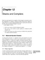
(PLC) shown in Figure 17-1. These structures function together to
resist lateral joint opening (LJO), posterior subluxation of the lateral
tibial plateau with tibial rotation, knee hyperextension, and varus
recurvatum.13,14,38,39,55,64
The mechanism of injury may be contact or noncontact and usually
involves a combined varus and hyperextension joint displacement. The
proper management of injuries involving the PL structures requires
knowledge of the complex anatomy and potential variations that may
exist, the function of the major soft tissue stabilizers, appropriate diagnostic techniques, and surgical options for reconstruction. Isolated PL
injuries are rare; however, on occasion an avulsion fracture at the
femoral attachment occurs requiring internal fixation.29 PL injuries are
frequently accompanied by anterior cruciate ligament (ACL) or posterior cruciate ligament (PCL) ligament ruptures.1,3,9,28
Although the incidence of PL injury is unknown (owing to misdiagnosis or failure to detect the injury), the consequences of untreated
PL ruptures are readily apparent. Chronic deficiency of the PL structures may be a factor in the failure of cruciate reconstructions43,44,49 and
527
528
CHAPTER 17 Posterolateral Ligament Injuries
CRITICAL POINTS Contraindications
Lateral gastrocnemius
tendon
Popliteofibular
ligament
Popliteus
tendon
• <5-mm increased lateral tibiofemoral joint opening 20 degrees of flexion.
• <10 degrees increased external tibial rotation 30 degrees, 90 degrees
flexion.
• Double varus knee, in which valgus osteotomy and subsequent adaption
(decreased laxity) of posterolateral structures eliminate abnormal lateral
joint opening and external tibial rotation.
• Triple varus knee without correction of varus malalignment.
• Prior joint infection.
• Patient noncompliant with rehabilitation, bracing, weight-bearing
restrictions.
• Hyperextension gait abnormality with no preoperative gait retraining.
• Advanced joint arthritis, <2 mm remaining joint space.
Fibular
collateral
ligament
Lateral knee ligaments
FIG 17-1 The anatomic relationships of the posterolateral structures.
may also play a role in the development of gait abnormalities and
giving-way.50,52,60 The detection and proper treatment of these problems is critical, because failure to properly treat all of the abnormalities
may result in a poor outcome. The patient will complain of a varus
type of instability with LJO during stance phase and show either a
neutral or valgus alignment. The abnormal LJO during stance phase is
always greater than that detected on the varus stress test. The patient
may demonstrate the abnormal LJO by producing a varus loading at
the knee joint while standing.
Knees that fulfill the double or triple varus diagnosis criteria (varus
osseous malalignment with increased LJO, external tibial rotation,
varus recurvatum, and knee hyperextension [see Chapter 26])47 require
high tibial osteotomy (HTO) first, followed approximately 6 months
later, with an appropriate PL reconstruction. In many instances, an
ACL or PCL deficiency also exists, which is corrected at the time of the
PL reconstruction.
There are different surgical options available for acute knee injuries,
dislocated knees with multiple ligament ruptures, chronic knees, and
revision knees. The decision-making process for determining the
appropriate PL procedure is discussed in detail under “Operative
Treatment of Acute Posterolateral Ruptures” and “Operative Treatment
of Chronic Posterolateral Ruptures” later in this chapter.
CONTRAINDICATIONS
Contraindications to PL reconstruction are findings of less than
12 mm of absolute increased lateral tibiofemoral joint opening and less
than 15 degrees of increased external tibial rotation. These findings are
frequently noted in knees with associated varus osseous malalignment
(double varus knees) that are candidates for HTO (see Chapter 26).40
Patients with varus malalignment who do not undergo HTO and
have associated chronic insufficiency of the PL structures are not candidates for a PL procedure. Untreated varus osseous malalignment is
a frequent cause of failure of PL reconstructions.46 In many cases, a
knee hyperextension gait abnormality also exists, which must be corrected before surgery with a specific gait retraining program described
in Chapter 29.50 Failure to correct a hyperextension gait abnormality
places PL reconstructions at risk for failure owing to the excessively
high tensile forces placed on the PL soft tissues with weight bearing
after surgery. Gait retraining usually decreases abnormally high knee
extension and adduction moments to normal values.50
Patients with a history of prior joint infection or who are obese
(body mass index > 30) are not candidates for PL reconstruction.
Patients with muscle atrophy of the lower extremity undergo preoperative rehabilitation before PL reconstruction.
Knees that demonstrate a loss of lateral tibiofemoral compartment
joint space, with less than 2 mm remaining on 45-degree posteroanterior (PA) weight-bearing radiographs, are usually not candidates for
PL reconstruction.
CLINICAL EVALUATION
The PL structures are injured when excessive varus, external tibial
rotation, and hyperextension forces are applied to the lower extremity.
A blow to the anteromedial tibia during sports participation appears
to be one of the most common injury mechanisms. These injuries
frequently involve rupture of other knee ligament structures, complicating the diagnosis. An isolated complete PL rupture is rare because,
usually, the injury is accompanied by an ACL or PCL rupture. In some
cases, the PL structures are only partially disrupted and do not require
surgical restoration. It is important to correctly determine the increases
in LJO, external tibial rotation, and knee hyperextension of the injured
knee (compared with the contralateral knee) preoperatively and intraoperatively. The decision of whether surgical restoration of the PL
structures is indicated is based on the abnormal knee motion limits,
joint subluxations, and the tissues disrupted.
One frequent patient presentation is a failed ACL or PCL reconstruction owing to untreated PL insufficiency. Another patient presentation is a chronic varus osseous malalignment and underlying ACL
insufficiency in which, over time, interstitial stretching and slackening
of the PL structures occurred.40,47 In these cases, HTO unloads the PL
soft tissues to the extent where physiologic remodeling and shortening
may subsequently occur in some knees and a PL reconstruction is not
required.47
A comprehensive physical examination is required, including
assessment of knee flexion and extension, patellofemoral indices, tibiofemoral crepitus, tibiofemoral joint line pain, and gait abnormalities. Pain in the medial tibiofemoral compartment occurs owing to
CHAPTER 17 Posterolateral Ligament Injuries
CRITICAL POINTS Clinical Evaluation
History
• Common injury mechanism blow to anteromedial tibia causing excessive
knee hyperextension, external tibial rotation, lateral tibiofemoral joint
opening.
• Most posterolateral injuries occur with anterior cruciate ligament or posterior cruciate ligament ruptures.
Physical Examination
• Knee flexion, extension
• Joint effusion
• Patellofemoral (medial and lateral subluxation, Q-angle, crepitus, compression pain)
• Tibiofemoral crepitus, joint line pain, compression pain
• Recurvatum (standing, supine)
• Gait (severe hyperextension stance phase)
• Muscle strength
Tibiofemoral Rotation Dial Test
• Diagnosis of posterolateral injury is based on final position of the lateral
tibial plateau.
• Subluxation of the medial and lateral tibial plateaus separately at 30
degrees, 90 degrees of knee flexion.
• Produce maximal external tibial rotation, determine change in position of
medial and lateral tibial plateaus separately.
• Qualitatively determine if anterior or posterior subluxation occurred in each
tibial plateau.
Diagnostic Clinical Tests
• External rotation recurvatum
• Lateral and medial tibiofemoral joint opening 5 degrees, 20 degrees of
flexion
• Pivot shift, Lachman
• Reverse pivot shift
• Posterior drawer, 90 degrees of flexion
• KT-2000 20 degrees of flexion, 134 N
Radiographs
• Lateral, 30 degrees of flexion
• Posteroanterior, weight-bearing, 45 degrees of flexion
• Patellofemoral axial
• Lateral stress, neutral tibial rotation select knees
• Posterior cruciate ligament ruptures: posterior stress lateral, 90 degrees of
flexion, neutral tibial rotation
• Varus malalignment: full standing radiographs, mechanical axis and
weight-bearing line
Cincinnati Knee Rating System
• Sports Activity and Function Form
• Occupational Rating Form
• Symptom Rating Form
increased compressive forces related to varus osseous malalignment.
Pain in the PL soft tissues may occur from increased soft tissue
tensile forces caused by a varus thrusting gait pattern. The abnormal
knee hyperextension involves increased extension in the sagittal
plane and is often accompanied by a varus alignment in the coronal
plane, which has been described as a varus recurvatum alignment.
Together with a varus osseous malalignment, this is referred to as a
triple varus knee (see Chapter 26). Patients with chronic PL insuffi-
529
ciency have varying amounts of altered gait mechanics and knee
hyperextension. Some individuals may present with a markedly
abnormal gait that is severely disabling and limits ambulation. Other
patients may have a less noticeable alteration because the abnormal
knee hyperextension occurs only after prolonged walking and muscle
fatigue. The abnormal gait pattern is characterized by excessive
knee hyperextension during the stance phase, which does respond
to gait retraining that initiates normal stance phase flexion (see
Chapter 29). Subjective complaints of giving-way during routine
daily activities, along with severe quadriceps atrophy, often accompany this gait abnormality.
The surgeon must determine all of the abnormal translations and
rotations in the knee joint. The ligament injuries that result in knee
hyperextension and varus recurvatum frequently involve not only the
PL structures, but also other ligament and capsular structures. The
biomechanic and kinematic studies that form the basis for the interpretation and diagnosis of the manual stress tests are described in
Chapter 15.
The increases in LJO and external tibial rotation shown in Table
17-1 are only approximations of what would be expected with clinical
injury to the PL structures. Importantly, an increase of only a few millimeters (2-5 mm) in LJO occurs with complete rupture of the FCL,
whereas an increase of 5 to 9 mm occurs with complete rupture of all
the PL structures (FCL, PMTL, and PFL). These values are based on
biomechanic studies discussed in Chapter 15. LaPrade and colleagues24
conducted a cadaveric study in which lateral stress radiography was
applied at 12 N-m (on an experimental apparatus) and the increase in
LJO over the intact state was compared with that measured during a
clinician-applied load after an isolated FCL rupture and a combined
FCL, PMTL, and PFL rupture. Compared with the intact state, LJO
induced by the clinician-applied load increased by 2.7 mm (isolated
FCL rupture) and 4.0 mm (combined PL rupture). However, the mean
values showed a wide standard deviation and variation among specimens, making extrapolation to the clinical setting difficult. In addition,
the lateral joint space measurement showed wide confidence intervals
(CI). For an isolated FCL rupture, the mean lateral gap distance was
10.99 mm (CI, 7.8-14.3 mm) and for the combined PL rupture, the
mean distance was 12.2 mm (CI, 9.3-15.2 mm). This amount of
overlap indicates that it would not be possible to accurately separate a
FCL rupture alone from a combined PL injury. The measurements are
important and useful in providing the clinician with a baseline in
interpreting lateral stress radiographs. The gap test is based on the joint
separation between articular cartilage seen at arthroscopy, and not the
tibiofemoral separation on a stress radiograph. Even so, the measurements are somewhat equivalent as to the increase in the amount of
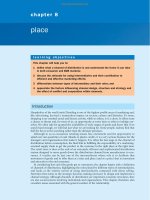
millimeters with PL injuries. For example, Figure 17-2 shows an
approximate normal lateral gap of 4 mm at the closest point of the
lateral compartment at arthroscopy. An increase of only 6 mm results
in 10 mm of absolute opening at the closest point or 12 mm at the
periphery, which is viewed as a positive gap test and indicative
of injury to the PL structures. Fortunately in most knees, these are
the lesser values and it is more common that the lateral gap exceeds
these measurements, indicating that concurrent PL reconstruction is
necessary.
An increase in external tibial rotation may occur with anterior
subluxation of the medial tibial plateau, posterior subluxation of the
lateral tibial plateau, or a combination of both subluxations. The dial
test, which the senior author (F.R.N.) published,54 allows a diagnosis
of tibial rotational subluxations of the medial and lateral tibiofemoral
compartments at 30 and 90 degrees of knee flexion (see Table 17-1).
Other variations of this test have been described.7,51,65
Text continued on p. 534
530
CHAPTER 17 Posterolateral Ligament Injuries
TABLE 17-1 Comprehensive Knee Examination
Examination
Technique
Dial test 30
degrees
Illustration
Grading
Significance
Supine position,
palpate anterior
tibial prominence,
medial and lateral
joint, maximum
external rotation,
posterior position,
lateral tibia (PL)
subluxation, anterior
position medial tibia
(anteromedial
subluxation).
Compare external
rotation between
knees.
External tibial rotation with
PL subluxation. Increase
3-5 degrees FCL tear.
Increase 6-10 degrees FCL
tear, partial PMTL.
Increase ≥15 degrees FCL,
PMTL, PLC.
Dial test 90
degrees
Maximum external
tibial rotation.
Determine PL tibial
subluxation.
Compare external
rotation between
knees.
Increase at 30 and 90
degrees means PCL and
PLC injury. With PCL tear,
degrees of external
rotation difficult to
estimate owing to
posterior tibial position.
PL external
rotation test
Knee at 90 degrees,
posterior and
external rotation
loading on tibia,
palpate posterior
subluxation lateral
tibiofemoral joint.
Qualitative PL tibial
subluxation 90 degrees
(less accurate at 30
degrees of flexion).
Similar to dial test but foot
held stationary. Dial test
allows tibia to externally
rotate fully, providing
better estimate of
increased tibial rotation.
PL subluxation 90
degrees, combined PCL,
PLC injury.
Posterior drawer
test
Knee at 90 degrees
position, posterior
load proximal tibia,
no tibial rotation.
Palpate medial
tibiofemoral
step-off.
Partial PCL: increase
0-9 mm translation;
complete PCL tear:
increase >10 mm
translation.
Stress radiography more
accurate. Increases
>10 mm indicate
secondary restraints torn
or physiologic slack
(combined injury).
CHAPTER 17 Posterolateral Ligament Injuries
531
TABLE 17-1 Comprehensive Knee Examination—cont’d
Examination
Technique
Quadriceps
active test
Lachman
(anterior
drawer, 30
degrees of
flexion)
Pivot shift
Reverse pivot
shift test
Varus stress
testing
Illustration
Grading
Significance
Knee position at
70-90 degrees.
Foot stabilized by
examiner, patient
activates quadriceps
by pushing foot
against table or
attempting to
extend knee.
Anterior load proximal
tibia.
Qualitative. Observe,
palpate tibiofemoral
position.
Confirms posterior tibial
subluxation, PCL injury at
resting position.
Quadriceps contraction
produces anterior
translation knee position
≥70 degrees.
Observe anterior
translation tibia;
compare with opposite
knee.
Estimate increase
translation in millimeters.
Soft endpoint, ACL not
resisting anterior
translation, indicates ACL
tear. Increase 3- to 5-mm
ACL tear. >5 mm ACL plus
secondary restraints.
Knee position 10-30
degrees of flexion.
Anterior load tibia,
gentle internal
tibial rotation
(subluxation)
followed by
posterior load,
gentle external
rotation (reduction).
Similar loading as
pivot shift.
Qualitative. Grade: I
slipping. II thud, clunk
with reduction. III
gross anterior
subluxation lateral
tibiofemoral joint,
anterior impingement
tibia limits reduction
event.
Grade I physiologic laxity,
no or partial ACL tear. II
ACL tear. Grade III ACL
tear plus secondary
restraints lax.
PL subluxation with
external rotation
confused for reduction
in pivot shift. No
abnormal anterior
tibial subluxation.
Observe obvious PL tibial
subluxation with posterior
and external rotation
loading. Dial test more
accurate.
Thigh supported on
examination table.
Knee position 0
degree, 30 degrees.
Varus load with no
external-internal
tibial rotation.
Palpate lateral joint
line opening.
Subtle 30 degrees
increase LJO. 2-4 mm
complete FCL tear,
further increase LJO
with PLC injury.
Stress radiography more
accurate. 30 degrees of
flexion. Increase
2- to 4-mm FCL tear.
Increase 5- to 9-mm
complete PLC tear (also
perform valgus stress
test).
Continued
532
CHAPTER 17 Posterolateral Ligament Injuries
TABLE 17-1 Comprehensive Knee Examination—cont’d
Examination
Technique
External rotation
recurvatum
test
Illustration
Grading
Significance
Grasp and hold both
feet above table,
allow gravity knee
hyperextension.
Qualitative. Tibia
externally rotates,
varus position caused
by PL joint opening,
knee.
Hyperextension, indicates
PLC injury, >10 degrees
frequently associated
ligament injury (ACL, PCL).
Standing
recurvatum
test
Patient stands, feet
together pushes
knees backward into
hyperextension,
compare knees.
Qualitative, observe
varus hyperextension
position (can be
measured with
goniometer). Increased
loading over supine
recurvatum test brings
out deformity.
10 degrees hyperextension
with varus alignment PLC
disruption, lateral, PL joint
abnormal opening, often
combined PLC, ACL tear,
confirm with other tests.
Standing frontal
alignment
Standing 0-5 degrees
of flexion. Avoid
hyperextension.
Confirm varus alignment.
Hip-knee-ankle
radiographs 0-5
degrees of flexion.
Classify primary, double,
triple varus based on all
tests (see Chapter 26).
CHAPTER 17 Posterolateral Ligament Injuries
533
TABLE 17-1 Comprehensive Knee Examination—cont’d
Examination
Technique
Knee
hyperextension
gait or varus
thrust
Illustration
Grading
Significance
Observe gait walking
to and from
examiner.
Qualitative. Knee goes
into hyperextension on
stance phase. Knee
has varus thrust
without
hyperextension.
Two hyperextension
patterns (see Chapter 29).
Forward trunk position,
loss of quadriceps control,
ankle dorsiflexion
push-off, requires gait
retraining. Varus thrust
increases medial
compartment loads and
tensile forces lateral
ligaments, osteotomy may
be required.
Range of motion
Perform passive
flexion-extension.
Normal 3-0-135 degrees.
Hyperextension to
neutral to flexion
10 degrees hyperextension
posterior capsule possible
ACL or PCL injury. ≥15
degrees multiple ligament
injury.
Effusion, soft
tissue
swelling, pain
Palpate joint for
effusion,
tenderness,
meniscus, ligament
attachments.
Qualitative. Complex
examination necessary
+ meniscus tests.
Partial to complete tears
PLC. FCL local tenderness,
pain varus, dial tests.
Patellofemoral
examination
Comprehensive
examination. All
tests. Alignment, PF
crepitus, medial/
lateral translation,
patella height.
See Chapter 35.
Increased external tibial
rotation 30 degrees, PLC
tear, produces abnormal
lateral shifting tibial
tubercle, increases
Q-angle.
Continued
534
CHAPTER 17 Posterolateral Ligament Injuries
TABLE 17-1 Comprehensive Knee Examination—cont’d
Examination
Technique
Illustration
Neurovascular
examination
Complete
examination. Both
lower extremities,
PT, DP pulses, lower
extremity muscle
function.
Grading
Significance
Peroneal nerve injuries
associated with severe
PLC disruption (10%30%). Arterial studies
indicated multiple
ligament injuries,
dislocations.
ACL, Anterior cruciate ligament; DP, dorsalis pedis; FCL, fibular collateral ligament; LJO, lateral joint opening; PCL, posterior cruciate ligament;
PF, patellofemoral; PL, posterolateral; PLC, posterolateral capsule; PMTL, popliteus muscle-tendon-ligament; PT, posterior tibial.
Abnormal lateral
joint opening (30°)
8
12 10
mm
A
Normal lateral
joint opening (30°)
2
Varus load
4
6 mm
B
C
FIG 17-2 The gap test. A, The amount of lateral tibiofemoral joint opening is measured with the knee at 25
degrees of flexion. Knees with insufficiency of the posterolateral structures will demonstrate 12 mm of joint
opening at the periphery of the lateral tibiofemoral compartment, 10 mm at the midportion of the compartment, and 8 mm at the innermost medial edge. B, Normal gap test. C, Abnormal gap test.
The position of the medial and lateral tibial plateau is assessed at
the starting position (neutral tibial rotation) with the knee flexed to
30 and 90 degrees and at the final position with the tibia in maximal
external rotation. The examiner palpates the position of the medial
and lateral tibial plateau, which is compared with the normal knee to
assess whether a subluxation (anterior or posterior) of the medial or
lateral tibial plateau is present. An increase in internal tibial rotation
occurs with both lateral ligament, medial ligament, and PCL disruption (see Chapter 15). The axis of tibial rotation is observed in the
involved knee and compared with the normal knee to detect a shift in
the medial or lateral tibiofemoral compartment during tibial rotation.
It is not recommended that the dial test be performed in the prone
position because the tibiofemoral joint cannot be accurately palpated
to distinguish an anteromedial from a PL tibial subluxation.
It is not possible to clinically determine the actual millimeters of
translation of the medial and lateral tibial plateaus in reference to the
femoral condyle. A qualitative determination is made of the anterior
or posterior subluxation of the medial or lateral tibiofemoral joint.
CHAPTER 17 Posterolateral Ligament Injuries
With PL injuries, there is an abnormal lateral deviation of the tibial
tubercle in the dial test compared with the opposite knee.
The use of the dial test in knees with PCL ruptures requires maintenance of a normal anatomic tibiofemoral position. This is accomplished by applying an anterior translation, loading the ACL in both
limbs, during the external tibial rotation. It is still necessary to use the
supine position so that the examiner can palpate the tibiofemoral position.61 The dial test is less accurate with a PCL rupture because it is
difficult to compare limbs, and other tests to be described (LJO, gap
test at arthroscopy, varus recurvatum) for the integrity of the PL structures need to be carefully assessed.
When a posterior subluxation of the lateral tibial plateau is positively identified by the tibiofemoral rotation test, additional tests must
be conducted to determine the integrity of other ligament structures.
The amount of LJO at 5 and 20 degrees of knee flexion should be
determined to further assess the integrity of the FCL and other secondary ligament restraints. The posterior tibial subluxation of the central
tibial and medial tibiofemoral joint determines the amount of increased
translation because of a PCL injury, which adds to the maximum
posterior subluxation to the lateral compartment with external tibial
rotation.
The presence of a varus recurvatum in both the supine and standing positions must be carefully assessed. Often, the varus recurvatum
reaches its maximum position when the patient is standing and asked
to maximally hyperextend both knees.
The appropriate tests to determine the integrity of the ACL and
PCL are performed, including KT-2000 (MEDmetric) arthrometer
testing at 20 degrees of flexion (134 N) to quantify total anteroposterior
(AP) displacement. The pivot shift test is recorded on a scale of 0 to
III (grade 0, no pivot shift; grade I, slip or glide; grade II, jerk or clunk;
grade III, gross subluxation with impingement of the PL aspect of the
tibial plateau against the femoral condyle). A misdiagnosis of a positive
pivot shift test may occur with PL injuries as the lateral tibial plateau
is brought to a reduced position (starting from a posterior subluxated
position) with knee extension and then posteriorly subluxates with
knee flexion (reverse pivot shift test). The medial posterior tibiofemoral
step-off on the posterior drawer test is done at 90 degrees of flexion.
Radiographs taken during the initial examination include AP, lateral
at 30 degrees of knee flexion, weight-bearing PA at 45 degrees of knee
flexion, and patellofemoral axial views. Lateral stress radiographs may
be required of both knees (20 degrees of flexion, neutral tibial rotation,
and 67-N varus force). A comparison is made of the millimeters of
lateral tibiofemoral compartment opening between knees.
A lateral radiograph is used to determine the approximate length
required for FCL anatomic grafts. The distance from the anatomic
femoral insertion site to the anatomic fibular insertion site is measured
and adjusted for magnification. A measurement of the patellar tendon
length is also made when a bone-patellar tendon-bone (B-PT-B) FCL
autograft is planned; however, in most knees, a B-PT-B allograft is
used, as will be discussed.
Posterior stress radiographs are obtained in patients with PCL ruptures, especially those in which the distinction of a partial versus complete PCL deficiency is difficult to determine on clinical examination.15
A lateral PCL stress radiograph is taken of each knee at 90 degrees
of flexion. The limb is placed in neutral rotation with the tibia
unconstrained and the quadriceps relaxed, and 89-N force applied to
the proximal tibia. Measurement is made of the millimeters of posterior tibial translation in both knees. Knees with 10 mm or more
of increased posterior tibial translation are considered candidates for
PCL reconstruction.
Full standing radiographs of both lower extremities, from the
femoral heads to the ankle joints, are done in knees with varus lower
535
extremity alignment. The mechanical axis and weight-bearing line are
measured to determine whether HTO is indicated.11
Patients complete questionnaires and are interviewed for the assessment of symptoms, functional limitations, sports and occupational
activity levels, and patient perception of the overall knee condition
according to the Cincinnati Knee Rating System (CKRS; see Chapter
41).2
CLASSIFICATION AND TREATMENT OF PARTIAL
TO COMPLETE PL INJURIES
The classification and treatment of first-, second-, and third-degree
acute PL injuries are detailed in Table 17-2. It is important to
diagnose partial tears of the PL structures, with a mild to moderate
increase in LJO and external tibial rotation, to allow protection and
maintain lateral tibiofemoral joint closure in the initial 3 weeks to
allow “stick-down” and healing of lateral soft tissues. This is a
program similar to that recommended for medial ligament ruptures
(see Chapter 19).
PREOPERATIVE PLANNING: TIMING OF SURGERY
Acute Injuries
There is a distinct advantage for repairing completely disrupted PL
structures and meniscal attachments in acute injuries (Fig. 17-3). At
the time of surgery, extensive disruption of these structures is observed.
Careful dissection is required to identify anatomic tissue planes and
maintain an intact vascular and neural supply. The so-called golden
period to perform an acute surgical repair is within 7 to 14 days of the
injury. After this time, scar tissue will obliterate tissue planes and make
the dissection and repair difficult.
A lower extremity venous ultrasound is obtained before surgery in
acute multiligament knee injuries that have swelling and soft tissue
damage to detect occult venous thrombosis that requires urgent treatment and contraindicates surgery. An initial delay in surgery for 5 to
7 days allows for observation of the neurovascular status, soft tissue
swelling, skin integrity, and some clearing of hemorrhage in soft tissues
in the injured extremity.
During this time, the lower extremity is supported in a soft-hinged
full-leg brace in extension with a well-padded compression dressing.
In knees with extensive damage to the PL structures and PCL, a
bivalved cylinder cast with a posterior plaster shell and posterior foam
calf pad may be required to provide added stability and prevent posterior tibial subluxation. Reduction of the tibiofemoral joint is verified
by a lateral radiograph. Lower limb elevation, ice, and compression are
important. The physical therapist initiates early protected knee motion,
patellar mobilization, active quadriceps function, and electrical muscle
stimulation. Dislocated knees scheduled for surgery require vascular
consultation, ankle/brachial studies (ankle/brachial index ≥90%), and
possible arteriography to exclude arterial injuries, even when intact
peripheral pulses are present.
Contraindications to acute surgical repair are excessive soft tissue
swelling, hemorrhage, and edema that are frequently present in dislocated knees with multiple ligament ruptures. The operative procedure
adds to the injury by increasing edema and soft tissue swelling, risk of
infection, vascular problems (including compartment syndromes),
and skin flap necrosis. In these cases, it is preferable to treat the acute
injury and perform ligament reconstructive procedures later after
tissue swelling is resolved and muscle function and knee motion have
been restored.
In addition, there is a significant incidence of knee arthrofibrosis
after acute surgical treatment of knee dislocations, which is lessened
536
CHAPTER 17 Posterolateral Ligament Injuries
TABLE 17-2 Diagnosis and Classification of Acute Posterolateral Injuries
First Degree
Anatomic lesion
Minor tearing
fibers
Signs
Minor tenderness
and swelling
Increase in lateral
joint opening‡
30 degrees
0 degree
Increase in external
tibial rotation (dial
test, 30 degrees)‡
Treatment
Second Degree
Third Degree*
†
Partial tears, one
third to two third
fibers
Tenderness and
swelling lateral
tissues
FCL tear
None
None
None
None
None
None
2-3 mm
None
3-5 degrees
Progress per
symptoms, no
crutches
Progress per
symptoms, soft
support brace
Bivalved cylinder cast 3 wk
ROM 0-90 degrees 2 wk
Support brace 3-6 week
Wean crutches 3-6 wk
FCL tear, partial tear
PMTL, PL capsule
FCL tear, PMTL tear, PL
capsule tear
Tenderness and
swelling lateral
tissues
2-5 mm
None
6-10 degrees
5-9 mm
3-5 mm
15 degrees
Operative repair,
reconstruction; usually
associated ACL, PCL
*Avulsion FCL, popliteus tendon: surgical indication to reattach.
†
Even though FCL shows complete tear, adjacent lateral tissues maintain ligament continuity for healing. Bivalved cylinder cast with protected
motion, maintain lateral tibiofemoral joint closure.
‡
See Chapter 15. Increases related to degrees of knee flexion, minor opening may be less under clinical conditions with lower joint loading.
ACL, Anterior cruciate ligament; FCL, fibular collateral ligament; PCL, posterior cruciate ligament; PL, posterolateral; PMTL, popliteus muscletendon-ligament; ROM, range of motion.
CRITICAL POINTS Preoperative Planning
Acute Injuries
• Golden period acute surgical repair: 7-14 days after injury.
• Lower extremity venous ultrasound, vascular consult for multiple ligament
ruptures.
• Delay surgery 5-7 days, observe neurovascular status, soft tissue swelling,
skin integrity.
• Soft hinged full-leg brace, well-padded compression dressing.
• PCL rupture: bivalved cast with posterior plaster shell, posterior calf pad.
• Verify tibiofemoral reduction with lateral radiograph in multiligament
injuries.
• MRI for location of major ligament disruptions.
• Protected knee motion, patellar mobilization, isometrics.
• Contraindications to acute surgery in dislocated knees: excessive soft
tissue swelling, hemorrhage, edema. Delay reconstruction until swelling
resolved, muscle function and knee motion restored.
Chronic Injuries
• Muscle atrophy requires preoperative rehabilitation.
• Hyperextension gait abnormality requires gait-retraining program before PL
reconstruction.
• Varus osseous malalignment requires osteotomy before PL
reconstruction.
• Absent lateral meniscus, early tibiofemoral arthritis, consider lateral
meniscus transplant.
Cruciate Graft Reconstruction
• Ensure B-PT-B, Achilles tendon allografts available.
• Determine appropriate grafts for ACL or PCL reconstruction.
ACL, Anterior cruciate ligament; B-PT-B, bone-patellar tendon-bone;
MRI, magnetic resonance imaging; PCL, posterior cruciate ligament;
PL, posterolateral.
with a staged approach. In our experience, the majority of multiligament disruptions in dislocated knees are not candidates for acute surgical procedures. A delay in surgical reconstruction results in a
decreased incidence of knee arthrofibrosis and markedly improves
surgical outcomes. Other obvious contraindications include open
wounds and skin abrasions.
Magnetic resonance imaging (MRI) provides important information regarding ligament ruptures, articular cartilage damage, and
meniscus tears. Frequently, the sites of rupture to the FCL, popliteus
muscle and tendon, PFL, and meniscal attachments may be identified
before surgery. One note of caution is the edema and swelling in the
PL tissues leads to a conclusion of greater tissue damage and disruption
than what is actually encountered at surgery.
Chronic Injuries
Patients with chronic knee injuries often present with severe lower
limb muscle atrophy requiring months of preoperative rehabilitation.
Patients with a hyperextension gait abnormality must complete a gait
retraining program50 described in detail in Chapter 29. This program
is done in addition to lower extremity muscle strengthening exercises.
In our experience, patients will convert to a more normal gait pattern
after 4 to 6 weeks of training.
Varus osseous malalignment must be corrected before chronic
PL reconstruction, as described previously. Failure to address varus
malalignment will greatly increase the risk of failure of any PL procedure (Fig. 17-4). In anatomic PL reconstructions, the ligament surgery
is staged after healing of the HTO. The indications for the various PL
procedures are described in detail under the “Operative Treatment of
Acute Posterolateral Ruptures” and “Operative Treatment of Chronic
Posterolateral Ruptures” sections.
Patients who have undergone prior lateral meniscectomy and who
demonstrate early tibiofemoral arthritis are considered for a staged
lateral meniscus transplantation after the PL reconstruction.48
CHAPTER 17 Posterolateral Ligament Injuries
Acute Injury Posterolateral Structures
Fibular collateral ligament
Avulsed
with bone:
direct repair
Midsubstance tears:
graft substitution
1. B-PT-B autograft
or allograft allows
early bone
incorporation
fibula & femoral
sites
2. Circle autograft
or allograft
through femoral
& fibular tunnels
with graft sutured
to itself. Use
semitendinosus
gracilis 2-strand
autograft or
allograft (Achilles
tendon, anterior
or posterior
tibialis tendons).
Popliteus muscle-tendon-ligament unit
Most cases
direct suture
repair is
possible.
FCL
reconstruction
protects repair
during postop
rehab.
Severe
injury direct
suture repair
not possible
PFL: direct
suture repair.
FCL graft to
popliteus
tendon restores
posterolateral
tissues, avoids
2nd tunnel
through fibula
to restore both
FCL and PFL.
Posterolateral capsule
1. Direct suture
repair, plication.
FCL, POP
tendon
grafts provide
protection of
repair postop.
2. Rare cases
of severe
hyperextension
(Ͼ15°) require
posterolateral
capsular graft
reconstruction.
1. Achilles tendonbone allograft
femoral anatomic
site, posterolateral
tibial tunnel
2. B-PT-B allograft or
autograft femoral
and tibial anatomic
site
Varus malalignment:
Tibial osteotomy at
acute repair contraindicated
due to increased
complications, arthrofibrosis.
Extra postop protection
required due to increased
risk of graft failure.
FIG 17-3 Algorithm for treatment of acute injuries to the posterolateral structures. B-PT-B, Bone-patellar
tendon-bone; FCL, fibular collateral ligament; PFL, popliteofibular ligament; POP, popliteus.
A
B
FIG 17-4 Standing anteroposterior; (A) and lateral (B) radiographs of a 28-year-old man referred to our center
14 months after failure of an acute repair of the posterolateral structures and posterior cruciate ligament
allograft reconstruction. The patient had underlying varus osseous malalignment, which likely was a factor
in the failure of the ligament reconstructions. This malalignment requires correction before revision surgery.
(From Noyes FR, Barber-Westin SD. Posterior cruciate ligament revision reconstruction. Part 1: causes of
surgical failure in 52 consecutive operations. Am J Sports Med. 2005;33:646-54.)
537
538
CHAPTER 17 Posterolateral Ligament Injuries
Chronic Injury Posterolateral Structures
Fibular collateral ligament
Graft substitution:
1. B-PT-B
for early bone
incorporation fibula
& femoral sites
2. Achilles tendon
reconstruction for
FCL, bone at fibula
or femoral site
Popliteus muscle-tendon-ligament unit
1. Prior injury, healed but
elongated: advance
tendon insertion
femoral site
2. Disrupted, scarred,
nonfunctional:
requires graft
reconstruction
PFL: direct suture
repair. FCL graft
to popliteus
tendon restores
posterolateral
tissues. Avoids
two tunnels through
fibula to restore
both FCL and PFL.
Posterolateral capsule
1. Direct suture
repair, plication
to FCL graft.
2. Rare cases
of severe
hyperextension
(Ͼ15°) require
posterolateral
capsular graft
reconstruction.
1. Achilles tendon–
bone allograft
femoral anatomic
site, posterolateral
tibial tunnel
2. B-PT-B autograft
or allograft at femoral
and tibial anatomic
site
Varus malalignment:
Valgus-producing opening
wedge osteotomy, staged
FIG 17-5 Algorithm for treatment of chronic injuries to the posterolateral structures. B-PT-B, bone-patellar
tendon-bone; FCL, fibular collateral ligament; PFL, popliteofibular ligament.
Cruciate Graft Reconstruction
The majority of patients who undergo PL reconstruction require a
concomitant ACL or PCL reconstruction (Fig. 17-5). The appropriate
grafts for the cruciate procedures should be determined; autogenous
tissues with bony fixation are preferred. However, the surgeon should
ensure that B-PT-B and Achilles tendon-bone (AT-B) allografts are
available the day of surgery. These will be required if autogenous tissue
is unavailable or not suitable for the PL or cruciate procedures.
INTRAOPERATIVE EVALUATION
All knee ligament tests are performed after the induction of anesthesia
in both the injured and contralateral limbs. The amount of increased
anterior tibial translation, posterior tibial translation, LJO, and external tibial rotation is documented. A thorough arthroscopic examination is conducted, documenting articular cartilage surface abnormalities
(see Chapter 44) and the condition of the menisci.53
The gap test is done during the arthroscopic examination.47 The
knee is flexed to 30 degrees and a varus load is applied. A calibrated
nerve hook is used to measure the amount of lateral tibiofemoral
compartment opening (see Fig. 17-2). Knees with 12 mm or more of
joint opening at the periphery of the lateral tibiofemoral compartment
require a PL reconstructive procedure.
In knees that undergo ACL reconstruction, the millimeters of joint
opening at the intercondylar area at the site of the ACL graft is the
critical distance in the gap test. Increases in LJO will occur postoperatively, allowing increases in ACL graft length. This space is normally 3
to 5 mm under varus loading.
Following the surgical exposure, the FCL and its fibular head and
femoral attachment sites, the PMTL, PL capsule, and PFL are inspected.
The distal popliteal tibia and fibula attachments of the popliteus
tendon are identified and inspected to determine the appropriate surgical treatment. All of the lateral and PL structures, including meniscus
CRITICAL POINTS Intraoperative
Evaluation
• Repeat all knee ligament tests under anesthesia, both limbs.
• Rate all articular cartilage surfaces for abnormalities, size of lesion
• Normal
• Grade 1, softening
• Grade 2A, fissuring & fragmentation <50% depth of the articular surface
• Grade 2B, fissuring & fragmentation >50% depth of the articular surface
• Grade 3, subchondral bone exposed
• Gap test at arthroscopy
• Knee 30 degrees of flexion
• Varus load
• Measure millimeters lateral tibiofemoral opening with calibrated nerve
hook
• Surgical exposure inspection
• Peroneal nerve
• FCL, fibular and femoral attachments
• PMTL, PLC, PFL
• Popliteus muscle, tendon attachments
• Meniscus attachments
FCL, Fibular collateral ligament; PFL, popliteofibular ligament; PLC,
posterolateral complex; PMTL, popliteus muscle-tendon-ligament.
attachments, are inspected in a stepwise manner, to be described. The
peroneal nerve is identified and protected at all times.
OPERATIVE TREATMENT OF ACUTE PL RUPTURES
Operative Setup and Patient Positioning
The patient is instructed to use a chlorhexidine soap scrub of the
operative limb (“toes to groin”) three days before and the morning of
CHAPTER 17 Posterolateral Ligament Injuries
surgery. Lower extremity hair is removed by clippers, not a shaver.
Antibiotic infusion is begun one hour before surgery. A nonsteroidal
antiinflammatory drug (NSAID) is given to the patient with a sip of
water upon arriving on the morning of surgery (which is continued
until the fifth postoperative day unless there are specific contraindications to the medicine). The use of an NSAID and a postoperative firm
double-cotton, double-Ace compression dressing for 72 hours (cotton,
Ace, cotton, Ace-layered dressing) has proven very effective in diminishing soft tissue swelling and is used in all knee surgery cases. In
complex multiligament surgery, the antibiotic is repeated at 4 hours
and continued for 24 hours. A urinary indwelling catheter is not used
unless there are specific indications. The patient’s urinary output and
total fluids are carefully monitored during the procedure and in the
recovery room. The knee skin area is initialed by the surgeon before
entering the operating room, with a nurse observing the procedure.
The identification process is repeated with all operative personnel with
a “time out” before surgery to verify the knee undergoing surgery,
procedure, allergies, antibiotic infusion, and special precautions that
apply. All personnel provide verbal agreement.
The patient is placed supine on the operative table and appropriately padded. The knee portion of the table is flexed 20 degrees, and
the table is tilted into a mild Trendelenburg position. A posterior thigh
pad is placed behind the proximal thigh to suspend the knee joint at
20 to 30 degrees of knee flexion. There is no pressure exerted on the
posterior popliteal space, allowing the posterior neurovascular tissues
and popliteal tissues to drop posteriorly away from the operative
approach. A common mistake is to place a posterior bolster in the
popliteal space that pushes the neurovascular structures into the operative dissection.
In cases of acute dislocation, the entire lower limb is draped free to
allow vascular checks of the anterior and posterior tibial pulses at the
foot during the operative procedure.
An initial arthroscopic examination is performed under lowpressure conditions with a free or controlled open outflow to prevent
fluid extravasation. The arthroscopic examination confirms damage to
intraarticular structures and allows photographic documentation of
the injury. If a leg holder is used, it is removed for the open surgery.
The tourniquet is placed at the proximal portion of the thigh
with appropriate padding. The tourniquet is inflated (275 to
300 mm Hg) during the initial exploration of the ligamentous injury
and identification of the common peroneal nerve (CPN). The tourniquet may often be deflated for the remainder of the procedure.
The surgeon may elect to be seated, directly facing the lateral aspect
CRITICAL POINTS Operative Treatment of
Acute Posterolateral Ruptures: Operative
Setup and Patient Positioning
• “Time out” before surgery: verify signed knee at surgery, procedure, allergies, antibiotic infusion, and special precautions.
• Complete examination of knee joint under anesthesia, compare with opposite knee.
• Acute dislocation or questionable vascular status: drape entire lower limb
free to check anterior and posterior tibial foot pulses during procedure.
• Arthroscopic examination under low pressures with free or controlled open
outflow to prevent fluid extravasation. Confirm damage to intraarticular
structures, photograph injury.
• Place tourniquet at proximal thigh with appropriate padding. Inflate
(275-300 mm Hg) during initial exploration, identify common peroneal
nerve. Deflate for surgical repair, reconstruction.
539
of the knee, with a headlight for careful dissection of the lateral soft
tissues including the CPN.
Identification of Ligament and Soft Tissue
Rupture Pattern
A 10- to 12-cm skin incision is made in a straight line centered over
the joint line and 1 cm posterior to the iliotibial band (ITB) attachment at the tibia (Fig. 17-6, A). After careful mobilization of the skin
flaps, the ITB, biceps tendon, and lateral structures are encountered.
Before dissection of the lateral aspect of the knee, the location of
the CPN must be identified. If the CPN cannot be easily palpated and
its course determined, then it is necessary at this point to expose and
identify the nerve along the entire lateral aspect of the knee. The CPN
does not have to be removed from its anatomic bed but requires protection throughout the subsequent surgery.
In the majority of knees, the ITB will be intact or demonstrate only
partial tearing. In select cases, the ITB will be completely disrupted at
the joint line or avulsed off its tibial attachment at Gerdy’s tubercle. If
the ITB is intact, an incision is made along its posterior border and the
ITB is elevated proximally to allow visualization of all of the underlying structures (see Fig. 17-6, B).
The lateral capsular tissues and meniscal attachments are the next
structures visualized. A vertical incision is made into the anterior third
of the capsule and extended to the lateral meniscus just anterior to the
anterolateral ligament attachment. The popliteus tendon and meniscus
attachments at the femoral popliteal recess are identified. Frequently,
it is necessary to repair the anterior inferior meniscal fasciculi (Fig.
17-7) and tibial meniscal attachments. Careful varus stress is placed on
the knee joint to allow inspection of the lateral meniscus attachments
and tibiofemoral articular cartilage. In some knees, an additional anterior ITB incision is required for visualization of underlying anatomy
(see Fig. 17-6, C).
The fibular head and attachments of the biceps femoris short and
long head are the next structures visualized, which have been described
in detail in Chapter 2. The two tendinous components (direct and
anterior arms) and one of the fascial components (lateral aponeurotic
expansion) make up the key portion of the long head anatomy. The
other fascial components are the reflected arm and the anterior
aponeurotic expansion.
CRITICAL POINTS Operative Treatment of
Acute Posterolateral Ruptures: Identification
of Ligament and Soft Tissue Rupture
Pattern
• Skin incision straight line 10-12 cm in length, centered over joint line, 1 cm
posterior to the ITB attachment at tibia.
• Intact ITB: incision along its posterior border to allow anterior displacement, visualize all underlying structures.
• Small bursa located superficial and anterolateral to distal portion of FCL:
open to allow better exposure of distal FCL attachment.
• Proximal third of posterior capsule attaches to proximal portion of gastrocnemius muscle and fabellum.
• Interval between posterior capsule and gastrocnemius tendon entered just
above fibula.
• Exposes PL structures, popliteus muscle tibial attachments, popliteus
muscle tendon junction, PFL, popliteus tendon attachment at the femur,
fabellofibular ligament.
FCL, Fibular collateral ligament; ITB, iliotibial band; PFL, popliteofibular
ligament; PL, posterolateral.
540
CHAPTER 17 Posterolateral Ligament Injuries
Standard
skin incision
Biceps femoris
tendon
Peroneal
nerve
A
Iliotibial
band
B
Iliotibial
band
Incision
Biceps femoris
tendon
Fibular collateral
ligament
Gastrocnemius
tendon
Posterior
joint capsule
Fibular
collateral
ligament
C
D
FIG 17-6 Posterolateral (PL) surgical technique. A, Site for the skin incision. B, Incision site in the interval
between the posterior edge of the iliotibial band (ITB) and the anterior edge of the biceps tendon. C, In
chronic cases with severe scarring, it may be necessary to add an anterior incision and displace the ITB
posteriorly during the reconstructive procedure to allow better exposure. D, With the ITB retracted anteriorly,
the interval between the lateral head of the gastrocnemius and the PL aspect of the capsule is opened
bluntly, just proximal to the fibular head, without entering the joint capsule proximally.
The most proximal component is the reflected arm. It originates
just proximal to the fibular head and ascends anteriorly to insert on
the posterior edge of the ITB. The direct arm inserts onto the PL edge
of the fibula just distal to the tip of the styloid. A portion of the anterior
arm inserts onto the lateral aspect of the fibular head, and the rest
continues distally just lateral to the FCL. Portions of the anterior arm
ascend anteriorly, forming the lateral aponeurotic expansion that
attach to the posterior and lateral aspect of the FCL. Here, a small bursa
separates the anterior arm from the distal fourth of the FCL. The
anterior arm forms the lateral wall of this bursa (see Fig. 2-15). This
is an important surgical landmark, because a small horizontal incision
can be made here, 1 cm proximal to the fibular head, to enter this bursa
and locate the insertion of the FCL into the fibular head. The anterior
arm then continues distally over the FCL, forming the anterior aponeurosis, which covers the anterior compartment of the leg. The
primary areas of injury are tendon avulsions off of the fibula, which
often have a major osseous component that can be repaired. In addition, the fascial extensions anteriorly and laterally are repaired.
The short head of the biceps courses just deep (or medial) and
anterior to the long head tendon, sending a majority of its proximal
muscular fibers to the long head tendon itself.63 It has six distal
attachments described in detail in Chapter 2. The most important
attachments are those of the direct arm, the anterior arm, and the
capsular arm.
CHAPTER 17 Posterolateral Ligament Injuries
The capsular arm originates just before the short head reaching the
fibula and continues deep to the FCL to insert onto the PL knee capsule
and fabella. Here the fibers of the capsular arm continue distally as the
fabellofibular ligament. Just distal to the capsular arm, a capsuloosseous layer forms a fascial confluence with the ITB (the bicepscapsulo-osseous iliotibial tract confluent). The direct arm of the short
head inserts onto the fibular head just posterior and proximal to the
direct arm of the long head tendon. The anterior arm then continues
medial or deep to the FCL, partially blends with the anterior tibiofibular ligament, and inserts onto the tibia 1 cm posterior to Gerdy’s
tubercle. This site is also the attachment of the mid-third lateral knee
capsule. The lateral aponeurotic expansion of the short head inserts
onto the medial aspect of the FCL. The FCL may be torn at its femoral
attachment or within its substance or avulsed along with the biceps
attachment at the fibula.
Proceeding posteriorly, the next structure encountered is the lateral
gastrocnemius muscle tendinous attachment to the femur. The proximal third of the posterior capsule attaches to the proximal portion of
the gastrocnemius muscle and fabellum (osseous or cartilaginous
analogue).
The interval between the posterior capsule and the gastrocnemius
tendon is entered just above the fibula, similar to the exposure for a
lateral meniscus repair. This exposes the popliteus muscle tibial attachments, popliteus muscle-tendon junction, PFL, popliteus tendon
attachment at the femur, and fabellofibular ligament (see Fig. 17-6, D).
In dissection studies, LaPrade and coworkers23 described a fabellum (osseous or cartilaginous) present in all specimens. This structure forms an attachment for the oblique popliteal ligament and
fabellofibular ligament, which along with the posterior capsule, are
important restraints for limiting knee hyperextension. Although
individual capsular components and structures are difficult to
discern with extensive capsular ruptures, it is important to repair
Patellar tendon
Fat pad Lateral patellar
retinaculum
Iliotibial tract
Transverse
ligament
Anterior
cruciate
ligament
Lateral meniscus
Joint capsule
Popliteus tendon
via hiatus
Posterior
cruciate
ligament
Posterior
meniscofemoral
ligament
Oblique popliteal
ligament
Politeus muscle
First layer
Second layer
Third layer
Fibular
collateral
ligament
Biceps femoris
tendon
(long head)
Fabellofibular
ligament
Common
Fibular head
Popliteofibular peroneal
Lateral inferior
nerve
ligament
geniculate artery
FIG 17-7 Illustration of the popliteus tendon and its surrounding popliteomeniscal fascicles and lateral meniscus attachments that are frequently disrupted, requiring repair.
541
disrupted posterior capsular tissues after completion of the initial
dissection.
Common Peroneal Nerve Identification
It is important at the initial stages of the dissection to palpate and
determine the location of the CPN. To expose the CPN, it is safest to
begin in the proximal aspect of the operative exposure. A large retractor is used to elevate the muscular portion of the biceps femoris,
placing the fascial tissues beneath the biceps muscle under gentle
tension. This gentle upward displacement of the biceps muscle and
vastus lateralis muscle is key to visualize and dissect the CPN, because
the normal curviform undulations are removed and the CPN assumes
a straighter appearance (Fig. 17-8). The investing crural fascia is incised
over the CPN to the fibula.
The CPN and its branches are not removed from their normal
anatomic position to avoid damaging the delicate blood supply, particularly in the region where the CPN approaches and then passes
around the fibular neck. Kadiyala and associates18 reported measurements in cadaveric specimens of the blood supply to the CPN in the
popliteal fossa and fibular neck region. These investigators hypothesized that the susceptibility of the CPN to injury or lack of a response
to operative treatment when injured may be related to deficiencies in
intraneural and extraneural vascular supply and anastomoses.
The most common source of blood supply to the proximal portion
of the CPN is a direct branch of the popliteal artery. This branch
divides into proximal and distal anastomotic vessels that run in the
connective tissue sheath of the nerve and anastomoses with the anterior recurrent tibial artery. The vessels, located in the epineurium, give
rise to many small vessels of fine caliber, which extend 20 to 30 mm
within the substance of CPN. It is important not to disturb this blood
supply. Kadiyala and associates18 noted that the blood supply of the
CPN was somewhat sparse with poor vascularization. A connection of
CRITICAL POINTS Operative Treatment of
Acute Posterolateral Ruptures: Common
Peroneal Nerve Identification
• Initial dissection: proximal aspect operative exposure, palpate, determine
location common peroneal nerve (CPN).
• Gently displace, elevate biceps muscle to visualize, dissect fascial covering
over CPN.
• CPN and its branches are not removed from normal anatomic position to
avoid damaging delicate blood supply.
• Most common source of blood supply to proximal portion of CPN from direct
branch popliteal artery. Branch divides into proximal and distal anastomotic
vessels.
• The vessels, located in the epineurium, give rise to many small vessels of
fine caliber within CPN. Do not disturb this blood supply.
• Gerdy’s safe zone: area where CPN and anterior recurrent branch define
an arc with an average radius of 45 mm.
• Region advantageous for surgical exploration, damage to peroneal nerve
and its branches avoided.
• In cases of partial to complete peroneal nerve injury, avoid added trauma
to neural tissues.
• CPN passage into the lateral and anterolateral compartment at the entrance
of the peroneal longus muscle at the fibular neck potential area for nerve
compression. Requires identification, division of variant fascial tissue
bands. Further CPN dissection avoided.
542
CHAPTER 17 Posterolateral Ligament Injuries
Biceps muscle
Biceps
muscle
CPN
A
Fibula
B
CPN
CPN discolored
Peroneus
longus
(partial cut)
CPN
C
Opening into
peroneus longus
Fibula
D
FIG 17-8 Exposure of the common peroneal nerve (CPN). A, Proximal exposure of the CPN inferior to the
long head of the biceps tendon. B, The superficial fascia over the peroneus longus is incised. C, The peroneus
longus muscle at the fibular neck is partially incised adjacent to the CPN. D, Complete exposure of the CPN
entering into the anterolateral compartment. In this knee, the CPN is shown to be distinctly abnormal and
edematous at this site. The CPN is not displaced from its normal anatomic site to protect the vascular supply.
the vasa nervorum was not found from the geniculate arteries, but
occasional contributions from muscular branches were recognized
(Fig. 17-9).
Bottomley and colleagues4 reviewed the anatomic position of the
CPN in 54 patients who had extensive traumatic disruption of the PL
structures. The CPN was noted to be displaced from its normal position in 16 of 18 patients who had biceps avulsions or associated fibular
head fractures. These authors advised that the surgeon should expect
an abnormal nerve position on surgical exploration in knees with bone
or soft tissue avulsion from the fibular head and the potential for iatrogenic damage.
Rubel and coworkers58 conducted an anatomic investigation of the
CPN in 31 cadaveric limbs by dissecting the CPN to its intramuscular
branches. The authors described Gerdy’s safe zone as the area where
the CPN and anterior recurrent branch defined an arc with an
average radius of 45 mm. The distance between the fibular head and
Gerdy’s tubercle was used to determine the radius of the safe zone.
Therefore this region in the proximal aspect of the tibia is advantageous for surgical exploration, because damage to the peroneal nerve
and its branches is avoided (Fig. 17-10). The CPN divides into three
branches as it enters the anterolateral musculature, with the anterior
recurrent branch more proximal to the superficial and deep peroneal
branches.
Dellon and associates10 reported on the anatomic variations of the
CPN at the fibular head in 29 cadavers (bilaterally) and 65 patients
treated with a CPN decompression for symptoms. Three possible anatomic variants were described that require attention and decompression in chronic neuropathies for a successful outcome. First, the
superficial fascia of the superficial head of the peroneus longus muscle
is divided by a proximal and distal transection of the fascia (found in
30% of cadavers and 78% of patients, see Fig. 17-8, B). Second, when
the peroneus longus muscle at the fibular neck is partially incised
adjacent and superior to the CPN and the peroneus muscle is lifted
anteriorly, a fibrous band may be found that requires release (soft tissue
restriction found in 43% of cadavers and 20% of patients, see Fig. 17-8,
C). Third, there may be a fibrous connection between the peroneus
longus and soleus muscle requiring division (found in 9% of cadavers
and 6% of patients). These authors advise that after CPN decompression, the surgeon’s index finger should be able to gently pass along the
CPN and into the anterolateral compartment (see Fig. 17-8, D).
In cases of partial to complete peroneal nerve injury, added trauma
to neural tissues should be avoided. The goal is to identify the nerve
pathway to avoid further damage during the ligament reconstructive
procedure. The CPN passage into the lateral and anterolateral compartment at the entrance of the peroneal longus muscle at the fibular
neck is a potential area for nerve compression. This area requires
identification and division of variant fascial tissue bands, as previously
described. Further CPN dissection is avoided.
Surgical Repair and Reconstruction of Acute Injuries
The key to restore function to the disrupted PL structures, muscle
attachments, and lateral meniscus attachments is a meticulous dissection, identification of damaged tissues, and repair of all injured structures. There is an unacceptably high risk of failure of primary repairs
of disrupted PL structures, particularly the FCL, owing to high lateral
tensile forces exerted on these tissues postoperatively.59 Therefore it is
necessary to reconstruct one or more disrupted PL structures with an
autograft or allograft. This adds tissue integrity and sufficient repair
CHAPTER 17 Posterolateral Ligament Injuries
Semimembranosus
Tibial
nerve
Common
Biceps
peroneal nerve femoris
Lateral
superior
genicular
artery
Popliteal
artery
Tibial
Common
nerve peroneal nerve
Vasa
nervorum
Popliteal
artery
Vasa
nervorum
Vasa
nervorum
Tibial nerve
Posterior
tibial artery
Popliteal
vein
(cut)
A
Lateral
sural
nerve
Communicating
sural bridge
Soleus
Anterior
Common
tibial
peroneal nerve
artery
Lateral
sural nerve
Anterior tibial
recurrent artery
B
Sural nerve
FIG 17-9 A, Gross dissection of the popliteal fossa of the right leg. The vessel branching from the popliteal
artery gives rise to vasa nervorum to the tibial nerve and common peroneal nerve (CPN) and a branch that
bifurcates into a vessel accompanying the sural nerve and the epineurial vessel running with the CPN.
B, The major vascular arrangements supplying the CPN in the popliteal fossa. (From Kadiyala, RK, Ramirez
A, Taylor AE, et al. The blood supply of the common peroneal nerve in the popliteal fossa. J Bone Joint Surg
Br. 2005;87:337-342.)
d lIl
100°
B
d ll
d ll
35°
0°
A
C
FIG 17-10 A, Cadaveric dissection of a fresh tissue specimen shows the circumferential area free of neural
structures at the level of the proximal aspect of the tibia. The center of this circumference is located at
Gerdy’s tubercle with an average radius (and standard deviation) of 45.32 ± 2.6 mm. d II, distance from the
most prominent aspect of Gerdy’s tubercle to the starting point of the superficial branch of the CPN; d III,
distance from the most prominent aspect of Gerdy’s tubercle to the anterior recurrent branch of the nerve.
B, Gerdy’s safe zone marked preoperatively. Note how the marking follows the contour of the surface in a
3-dimensional fashion on the lateral (B) and frontal (C) photographs. (From Rubel IF, Schwarzbard I, Leonard
A, Cece D. Anatomic location of the peroneal nerve at the level of the proximal aspect of the tibia: Gerdy’s
safe zone. J Bone Joint Surg Am. 2004;86:1625-1658.)
543
544
CHAPTER 17 Posterolateral Ligament Injuries
strength to resist LJO and external tibial rotation in the initial healing
period of 4 to 6 postoperative weeks.
In most acute PL injuries, the FCL is reconstructed and the other
PL soft tissues and PMTL are treated by primary repair. The FCL graft
reconstruction resists lateral tibiofemoral compartment opening and
posterolateral subluxation, protecting the overall repair process during
the initial healing stage. In more severe injuries, a graft reconstruction
of both the FCL and PMTL may be necessary. The reconstruction
procedures are discussed in detail in the “Operative Treatment of
Chronic Ruptures” section later in this chapter.
2. PMTL repair options
• Musculotendinous repair with direct suture (backup by FCL
reconstruction).
• PFL primary repair (tissues often of poor quality; FCL graft
provides suitable tissue for additional sutures).
• Popliteus tendon femoral attachment: avulsion (while rare) may
be reattached.
• Repair of disrupted biceps attachments: ITB repair of distal
insertion, posterior femoral attachments, patellar retinaculum.
Surgical Approach and Order of Repair
OPERATIVE TREATMENT OF CHRONIC
POSTEROLATERAL RUPTURES
The surgical approach the senior author prefers involves a graft reconstruction of the FCL to stabilize the lateral side of the knee joint using
a B-PT-B allograft or an AT-B allograft. A femoral-fibular reconstruction41 is a second option described later in this chapter. The FCL
reconstruction provides for secure fixation, prevents abnormal joint
displacements in the immediate postoperative period, and allows for
early protected knee motion. These procedures are not difficult because
the attachment sites on the femur and fibula are easily identifiable.
Importantly, the graft provides the cornerstone about which the
remainder of the soft tissue repair of the PL structures is performed.
A B-PT-B allograft requires an appropriate tendon graft length of
60 mm, which may not always be available. Alternative graft options
are an AT-B or anterior tibialis allograft. The bone portion of the AT-B
may be placed at either the FCL fibular attachment (preferred for bone
fixation and healing) or femoral attachment. If a soft tissue tendon
graft is selected, the graft is passed through a fibular tunnel (anteriorto-posterior), the tendon is sutured back on itself, and a soft tissue
interference screw is used at the femoral fixation site and, frequently,
at the fibular site.
After all of the anatomic structures and rupture sites are identified
and carefully exposed, the order of the operative repair starts with
deeper structures and proceeds to superficial structures. Examples of
an acute operative repair are shown in Figures 17-11 and 17-12.
1. Meniscofemoral and tibial capsular repairs
• Meniscus attachment repair.
• FCL reconstruction with femoral and fibular tunnel placement
and graft fixation.
• Posterior capsule repair with sutures tied with the knee in full
extension.
CRITICAL POINTS Operative Treatment of
Acute Posterolateral Ruptures: Surgical
Repair and Reconstruction of Acutely
Disrupted Ligament and Soft Tissues
• Unacceptably high risk for failure of primary repairs of FCL owing to high
lateral tensile forces postoperatively.
• FCL graft reconstruction, other PL soft tissues and PMTL repaired.
• Severe injuries: PMTL graft reconstruction may be necessary.
• Surgical approach, order of repair
• Prefer B-PT-B autograft or AT-B allograft replacement of FCL.
• Graft provides secure bone fixation, prevents abnormal joint displacements, and allows early protected knee motion.
• Graft provides the cornerstone for repair of other PL structures.
• Operative repair starts with deep structures and proceeds to superficial
structures.
AT-B, Achilles tendon-bone; B-PT-B, bone-patellar tendon-bone;
FCL, fibular collateral ligament; PL, posterolateral; PMTL, popliteus
muscle-tendon-ligament unit.
Overview of Operative Options
The surgical options in knees with chronic injuries to the PL structures
are based on the quality and integrity of these tissues determined at
the initial surgical dissection. The surgical approach is similar to that
described for acute dissection of the PL structures. The CPN is always
identified before any lateral dissection, as previously described. The
posterior border of the ITB is incised and elevated to allow complete
exposure. In chronic instabilities, the ITB may be lax and nonfunctional. In such knees, the attachment at Gerdy’s tubercle is osteotomized, and at the conclusion of the operative procedure, the proximal
ITB attachments (lateral intermuscular septum, femoral posterior
attachments) are sutured and the ITB osseous attachment is advanced
distally by staple fixation to the tibia. The importance of identification
of meniscus attachments, PMTL attachment, PL capsular structures,
the biceps short and long head attachments, and the peroneal nerve
has been previously discussed.
Markolf and colleagues35-37 reported a series of cadaveric studies on
the effect of a nonanatomic PL ligament reconstruction, which has the
potential to overconstrain internal tibial rotation and adduction
throughout knee flexion. The authors concluded there was no consensus on the graft tensioning routine for PL reconstructions.
The approach advocated in this chapter is an anatomic FCL
reconstruction. The FCL may be deficient from prior disruption or
replaced with scar tissue in which a well-defined structure cannot be
identified. An FCL reconstruction provides a cornerstone for the PL
reconstruction.
An anatomic PMTL reconstruction will be described later in this
chapter. In the majority of chronic unstable knees, the distal attachments of the PMTL are disrupted or replaced with scar tissue and it is
necessary to perform a graft substitution of the PMTL. In rare cases in
CRITICAL POINTS Operative Treatment of
Chronic Posterolateral Ruptures: Overview of
Operative Options
• Anatomic reconstruction for PL structures
• Unstable, disrupted, or scar tissue replacement of FCL and PMTL requires
anatomic graft substitution. If distal PMTL attachments are intact, possible
advancement and recession of popliteus tendon at femoral attachment site
performed and PFL attachment tissues repaired.
• Nonanatomic femoral-fibular graft reconstruction performed when operative time is limited (dislocated multiligament injuries).
• Proximal advancement PL structures: select knees with chronic stretching
and interstitial injury without traumatic ligament disruption.
FCL, Fibular collateral ligament; PFL, popliteofibular ligament;
PL, posterolateral; PMTL, popliteus muscle-tendon-ligament unit.
CHAPTER 17 Posterolateral Ligament Injuries
545
ITB
Popliteus
tendon
B-PT-B graft
ITB
FCL
A
Lateral meniscus,
lateral capsule repair
B
Femoral site
graft fixation
ITB
Capsular repair
PFL repair
to fibula
FCL
B-PT-B graft
C
Fibular site graft
fixation
D
FIG 17-11 Acute repair of rupture to the posterolateral (PL) structures. A, Lateral approach; anterior and
posterior incisions have been made into the iliotibial band (ITB). Sutures are placed to repair the lateral
meniscus tibial attachments. B, Fibular collateral ligament (FCL) reconstruction with a bone-patellar tendonbone (B-PT-B) allograft and suture of the popliteofibular ligament (PFL) to the fibula. The popliteus tendon
attachment at the femur was intact. C, Fixation of the FCL graft at the femoral and tibial anatomic attachments. D, Repair of PL capsule, biceps attachments, and posterior ITB.
which the distal attachments of the PMTL are intact, an advancement
and recession of the popliteus tendon at the femoral attachment site
may be performed.
A nonanatomic femoral-fibular graft reconstruction is described as
an option that is occasionally performed for acute or chronic ruptures
of the PL structures. This procedure is indicated when the FCL is
elongated or deficient and the PMTL does not require graft substitution. The operative procedure is advantageous when operative time is
limited (as in dislocated multiligament knees) or when a relatively
rapid stabilizing procedure is required. However, anatomic reconstruction and repair of both the FCL and PMTL are necessary in grossly
unstable chronic knee instabilities.
A third operative approach is described using a proximal advancement of the PL structures when chronic insufficiency of the PL
structures exists from a minor injury (without traumatic ligament
disruption). In knees with varus osseous malalignment and a varus
thrust on ambulation, there is frequently an insufficiency of the PL
structures because of chronic interstitial tearing. In these situations, a
definitive FCL of normal width and integrity (although lax) may be
identified at surgery and the PMTL attachments are intact though
elongated. A graft reconstruction of the FCL and PMTL is not indicated in these knees. Instead, the PL structures may be advanced proximally in a more simplified operative procedure that avoids the added
complexity and morbidity from major graft reconstructive procedures.
The PL structures must be carefully inspected at surgery, because this
procedure will fail if there is scar tissue replacement, lack of a normal
appearance of the structures (although lax), or if the distal attachments
of the PL structures are disrupted.
546
CHAPTER 17 Posterolateral Ligament Injuries
A
B
Biceps,
FCL avulsion
ITB
Fibular
head
Guide pin
Fibula
ITB
repair
Biceps,
FCL
C
D
E
FIG 17-12 Acute disruption of posterolateral structures with avulsion of biceps and fibular collateral ligament
(FCL) from fibula. A, Magnetic resonance imaging shows avulsion of biceps and FCL from fibular head.
B, Initial arthroscopic procedure 10 days after injury, performed under low pressure and volume conditions.
C, Surgical exploration shows avulsion of iliotibial band (ITB) from tibial insertion and biceps, FCL attachment
to fibula. The common peroneal nerve was identified to be in a normal anatomic position. D, Repair of capsular attachments and ITB (sutures). Guide pin placed in the proximal fibula for screw and washer fixation
of biceps and FCL. E, Anteroposterior postoperative radiograph shows fixation of biceps and FCL to the
proximal fibula, four-prong staple for ITB attachment to tibia, soft tissue anchor for deep capsular
attachments.
Anatomic Reconstruction of the Fibular Collateral
Ligament and Popliteus Muscle-Tendon-Ligament Unit
Patient Positioning and Surgical Approach
An operative time-out and identification of the operative limb are
performed as already described. The patient is positioned on the operative table, with a high thigh tourniquet placed as previously described
in the “Acute Injuries” section. A leg holder is only used if a meniscus
repair is anticipated to provide for limb control and opening of the
medial tibiofemoral compartment. Otherwise, the lower limb is draped
free with a bolster placed under the proximal thigh to allow the popliteal neurovascular structures to drop posteriorly away from the
dissection plane. The initial arthroscopic evaluation is performed,
including meniscus repairs and drilling of tunnels for concurrent cruciate reconstructions for placement of cruciate grafts as described in
Chapters 7 and 17. There are two options with cruciate reconstructions. The cruciate grafts may be placed with the distal fixation performed after the lateral dissection. If this approach is selected, the order
of final graft tensioning and fixation is (1) PCL, (2) ACL, (3) FCL, and
(4) PMTL. The rationale is to restore the tibiofemoral joint in the
sagittal plane and then perform the final PL graft fixation and repair
of disrupted tissues. The second option is to complete the cruciate
reconstruction and then repair and reconstruct the PL structures. If
CHAPTER 17 Posterolateral Ligament Injuries
CRITICAL POINTS Anatomic
Reconstruction of the Fibular Collateral
Ligament and Popliteus Muscle-TendonLigament: Patient Positioning and Surgical
Approach
• Initial arthroscopic evaluation performed.
• Tourniquet inflated during initial operative approach, deflated remainder of
procedure.
• Skin incision: straight line 10-12 cm in length, centered over joint line, 1 cm
posterior to ITB attachment at the tibia.
• Skin flaps created by undermining the skin in proximal, distal, anterior,
posterior directions.
• Skin dissection beneath superficial fascia, not in fatty subcutaneous plane.
• Identify, protect CPN and possible nerve branches as first priority.
• ITB incised along posterior margin at the junction of the band and biceps
muscle.
• ITB posterior attachments excised to short head of biceps femoris muscle.
• ITB lifted anteriorly to expose lateral aspect of lateral femoral condyle and
attachments of popliteus, FCL, lateral gastrocnemius muscle tendon
attachment.
• Interval anterior to lateral gastrocnemius tendon at joint line, directly at
top of fibula entered, avoid inferior geniculate artery. Visualize space
behind posterior capsule, lateral meniscus attachment, popliteus muscle
attachment, posterior gastrocnemius tendon.
• Second anterior ITB incision may be required for exposure.
• VLO lifted gently in anterior direction, S retractor placed beneath muscle
fibers.
• Vertical incision 2 cm in length into capsule just anterior to anterolateral
capsule ligament and popliteus tendon attachment. Joint entered, lateral
meniscus attachments inspected. Similar incision into PL capsule, starting
at its femoral attachment, inspect posterior meniscus attachments.
CPN, Common peroneal nerve; FCL, fibular collateral ligament;
ITB, iliotibial band; PL, posterolateral; VLO, vastus lateralis obliquus.
this sequence is followed, the surgeon must carefully control the limb
during the dissection and repair steps to make sure there is no inadvertent opening of the lateral tibiofemoral joint, which would disrupt
the cruciate graft fixation. In general, the first option is safest.
The surgeon is seated with a headlight to allow a meticulous dissection of the CPN first, followed by the PL structures. The tourniquet
is inflated during the initial dissection and then deflated during the
remainder of the surgical procedure. A skin incision 10 to 12 cm in
length is made in a straight line, centered over the joint line and 1 cm
posterior to the ITB attachment at the tibia, using the same approach
as already described (see Fig. 17-6).
Skin flaps are created by undermining the skin in proximal, distal,
anterior, and posterior directions. A cosmetic approach is used in
which the skin incision is transposed to different portions of the operative field, thereby reducing the skin incision length. The skin dissection
is accomplished beneath the superficial fascia and not in the fatty
subcutaneous plane to avoid damage to the blood and neural supply
to the skin flaps. The surgeon avoids skin necrosis by not placing
tension on the skin edges and flaps.
Peroneal Nerve Dissection and Visualization
The CPN is identified, beginning proximally as already described. A
retractor is used to elevate the proximal biceps muscle and vastus
lateralis to place tension on the lateral fascial tissues and gently
547
elongate the CPN. The fascia is incised directly anterior to the CPN,
and care is taken to avoid opening the surrounding neural sheath. At
the fibular head, the peroneal longus muscle is partially incised for a
few millimeters overlying the CPN at the fibular neck. The area is
inspected for fibrous or fascia tissues that may potentially compromise
the CPN entrance into the lateral and anterolateral muscular compartments, as described in the previous section. The yellow fatty tissue
about the CPN is protected.
The nerve is not displaced from its anatomic bed to protect its
blood supply. In cases in which scar tissue is encountered that surrounds the nerve, further dissection of the CPN is avoided because
injury may easily occur as the scar tissue prevents a safe dissection
plane. The CPN is identified proximally and distally to the scar tissue,
so its location can be protected during the ligament reconstruction.
Only in rare cases in which there is near-complete to complete loss
of CPN function and a compressive neuropathy exists, is it justified to
dissect the CPN from the encased scar tissue, because the risk of nerve
damage is high. In either situation, the surgeon must always know
where the CPN and its branches are located during the operative
procedure.
The ITB is incised at the posterior edge and anterior to the biceps
tendon. The ITB attachments are excised to the short head of the biceps
femoris muscle, and the ITB is gently lifted anteriorly to expose the
entire lateral aspect of lateral femoral condyle and attachments of the
popliteus, FCL, and lateral gastrocnemius muscle tendon attachment.
The tissues overlying the FCL and PL structures may have fascia
tissues that require dissection to identify the structures. These fascia
tissues should be grasped and gently stripped with dissection scissors,
protecting the biceps tendon attachment and underlying posterior
capsule and FCL. The bursa located anterolateral to the distal fourth
of the FCL is used as a landmark. In chronic knee injuries considerable
scar tissue may be encountered at this point that prevents clear identification of all the PL structures.
The interval anterior to the lateral gastrocnemius tendon at the
joint line, and directly at the top of the fibula, is entered, avoiding
the inferior genicular artery. The space behind the posterior capsule,
lateral meniscus attachment, popliteus muscle attachment, and posterior gastrocnemius tendon is visualized, as already described in the
“Acute Injuries” section.
A second anterior ITB incision may be required when there is
extensive scar involving all of the PL structures. The ITB is incised
along its anterior margin at the junction of the band and fascia, 10 cm
from its tibial attachment. The vastus lateralis obliquus (VLO) is carefully elevated from the lateral intermuscular septum, avoiding any
penetrating vessels. The VLO is lifted gently in an anterior direction
and an S retractor is placed beneath the muscle fibers. The surgeon
should avoid entering the suprapatellar synovial pouch by placing the
retractor adjacent to the periosteum. Occasionally, blunt dissection
with a Cobb elevator is required to gently displace the suprapatellar
synovial pouch to allow placement of the S retractor.
A vertical incision approximately 2 cm in length is made into the
capsule just anterior to the anterolateral ligament and popliteus tendon
attachment. The joint is entered and the lateral meniscus attachments
are inspected. If necessary, a vertical incision into the PL capsule may
be required to allow for further inspection and repair of the posterior
meniscus attachments. This posterior incision cannot be extended distally, because the popliteus tendon will be observed crossing into the
popliteus meniscus recess.
It may be beneficial to place a curved Kelly in the anterolateral
capsular incision, with the instrument passed beneath the popliteus
tendon and FCL, to place tension into these tissues to facilitate further
identification and inspection. The femoral anatomic attachments of all
548
CHAPTER 17 Posterolateral Ligament Injuries
the PL structures are shown in Figure 2-1, C and D, and the surgeon
should be thoroughly familiar with this anatomy because the goal of
the surgical procedure is to restore normal anatomic attachment sites.
Note the femoral FCL attachment is just posterior and superior to the
lateral epicondyle, and that the insertion of the lateral gastrocnemius
tendon is on the lateral aspect of the femoral condyle.
LaPrade and coworkers27 reported a mean distance of 18.5 mm
from the FCL insertion to the popliteus tendon insertion, indicating
that two separate grafts are required to anatomically reconstruct the
FCL and popliteal tendon femoral attachments. Although some
authors recommend a single graft placed at the femoral attachment
and split into two strands to reconstruct both the FCL and PMTL, this
procedure does not reproduce the anatomic femoral attachment sites.
For these reasons, a separate graft and femoral attachment for the FCL
and PMTL are recommended.
The PMTL, PFL attachments, and lateral meniscus attachments are
identified by careful probing to determine the integrity of these structures and the appropriate procedure required. Disruption of the popliteomeniscal attachments is usually present, requiring suture repair.
Chronic cases of rupture to the PL structures usually demonstrate
severe deficiency of the FCL and PMTL, which are encased in scar
tissue and require a two-graft anatomic reconstruction. The dissection
is limited to the respective structures to be reconstructed to avoid soft
tissue injury and devascularization.
Fibular Collateral Ligament Bone-Patellar
Tendon-Bone Reconstruction
The goal of a FCL reconstruction is to use a strong graft, attached by
bone to anatomic insertion sites on the femur and fibula. This construct provides the cornerstone for the remainder of the PL repair
and reconstruction. With an FCL graft resisting LJO and external
tibial rotation, immediate protected knee motion may be initiated
postoperatively to counteract the expected limitation of joint motion
and scar tissue that occurs after major ligament reconstructive
procedures.
Graft choices for fibular collateral ligament reconstruction. The
normal anatomic attachment sites of the FCL to the lateral femur and
anterolateral aspect of the fibular head are carefully identified.27 A
suture is placed between the two attachment sites and the length measured to determine the required graft size. The bone portion of each
end of the graft is 22 to 25 mm in length. The fibular graft attachment
is performed using a proximal fibular tunnel. The femoral graft attachment is performed by placing a femoral tunnel at the anatomic attachment site. A second option is a femoral inlay of the proximal bone
portion of the graft, which is only required if there is a 5- to 8-mm
discrepancy of graft length that will not allow full coverage of the bone
in a femoral tunnel.
The patellar tendon graft must normally be 55 to 60 mm to be
suitable for an anatomic FCL reconstruction; the senior author
keeps these lengths of allografts available. The average cross-sectional
area of the FCL (reported by LaPrade and associates22) is 11.9 ±
2.9 mm2. The FCL graft is 8 to 10 mm × 4 mm, resulting in a
32- to 40-mm2 graft. If the patient’s own tissue is of sufficient length,
an autograft harvested from the ipsilateral or contralateral patellar
tendon is an added consideration. In select cases in which a prior
FCL allograft reconstruction has failed, an autograft approach is
favored. The hypothesis is that an autograft will heal at the fibular
and femoral tunnels, with minimal remodeling and weakening of
the graft in the postoperative phase. The complications of a meticulously performed B-PT-B graft harvest from the contralateral knee
are less than 1% in terms of infection, scar formation, and graft
site pain.43
CRITICAL POINTS Fibular Collateral
Ligament Bone-Patellar Tendon-Bone
Reconstruction
• Anatomic FCL attachment sites to lateral femur, anterolateral aspect of
fibular head identified.
• Femoral graft attached via tunnel at FCL anatomic site. Inlay procedure
useful if 5- to 8-mm discrepancy of graft length that does not allow full
coverage of bone portion of graft.
• Patellar tendon graft length not counting bone: 55-60 mm.
• B-PT-B allograft favored over soft tissue FCL graft owing to prompt osseous
incorporation and healing at femoral and fibular attachment sites.
• Placement of fibular and femoral tunnels
• Anterior bare area of fibula exposed 20 mm, avoid lateral dissection that
may injure CPN.
• Fibular tunnel drilled using guide pin to depth of 25 mm.
• Avoid drilling too deep.
• Femoral tunnel placed eccentric to normal FCL attachment, allows collagen portion of graft to occupy normal FCL anatomic location.
• B-PT-B graft placement
• Bone portion of graft gently taped into fibular tunnel, bone entirely
seated into tunnel and level with proximal fibular head.
• Ideal graft fixation: one or two small fragment cortical screws.
• Proximal bone portion of graft placed in femoral tunnel.
• Graft conditioned by cycling knee 20-30 times.
• Graft fixed with interference screw, 30 degrees of flexion, neutral tibial
rotation, 22 N.
B-PT-B, Bone-patellar tendon-bone; CPN, common peroneal nerve;
FCL, fibular collateral ligament.
If an allograft is chosen, as is the usual case in multiligament reconstructions, a B-PT-B allograft is favored over a soft tissue graft owing
to more prompt osseous incorporation and healing at femoral and
fibular attachment sites. It is recognized that soft tissue allografts in
bone tunnels require added maturation time and may incompletely
remodel even under the best of circumstances.34,57
Placement of fibular and femoral tunnels. The attachment sites at
the fibula and femur are identified. The anterior bare area of the fibula
is exposed for 20 mm, avoiding lateral dissection that may injure the
CPN. The fibular tunnel is drilled first, using a guide pin to a depth of
25 mm. The drills are gradually increased in diameter to create a final
9-mm tunnel (Fig. 17-13). Care should be taken to avoid drilling too
deep, because the drill would break out the cortex distally at the fibular
neck, close to the location of the CPN. The normal cortical integrity
of the fibular head is not disrupted to maintain circumferential cortical
fixation strength at the fibular attachment site.
The femoral tunnel is placed 5 mm eccentric to the normal FCL
attachment to allow the collagenous portion of the graft to occupy the
normal FCL anatomic location. Before drilling the femoral socket, a
suture is attached to the fibular graft site and positioned at the desired
femoral location to check graft isometry. If the femoral position is too
anterior, the graft will be under high tension with knee flexion. The
opposite will occur if the graft is too posterior, because it will be under
tight tension with knee extension. The goal is to have tension in the
graft ideally in the 0 to 70 degrees knee motion limits, which replaces
the normal FCL function. A Beath guide pin is passed for the femoral
tunnel, which is angulated in an anterior and proximal direction in
line with the FCL fibers at 30 degrees of knee flexion.
If an ACL reconstruction is performed, it is necessary to diverge the
FCL tunnel in an anterior direction away from the ACL tunnel to
CHAPTER 17 Posterolateral Ligament Injuries
549
Femoral
interference
screw
fixation
FCL graft
Fibular
screw
fixation
A
Fibular
interference
screw
fixation
B
FIG 17-13 Anatomic substitution of the fibular collateral ligament (FCL) with a bone-patellar tendon-bone
autograft or allograft shows two methods for fibular graft fixation. A, Two small fragment screws are used
to fix the bone into a slot created in the proximal fibula (our choice). Interference screw fixation is used at
the femoral anatomic site of the FCL. B, A fibular tunnel is made, the graft seated, and an interference screw
is used for fixation.
maintain integrity of the two tunnels. The edges of the femoral tunnel
are smoothed with a rasp to avoid graft abrasion.
The use of a FlipCutter (Arthrex) for the ACL femoral tunnel is
ideal because it maintains the integrity of the lateral femoral cortex,
particularly if two femoral sockets are required for an FCL and PTML
graft reconstruction. Selecting grafts with a bone portion at the femoral
site is advantageous because it avoids soft tissue graft tunnels that in
the senior author’s experience always remain and potentially weaken
the lateral femoral condyle. The bone portion of the allograft will
incorporate within the femoral condyle.
Bone-patellar tendon-bone graft placement. The bone portion of
the graft is gently taped into the fibular tunnel so that the bone is
entirely seated into the tunnel and level with the proximal fibular head
to preserve graft length. The bone-tendon junction is marked with ink
to define the correct depth in the fibular tunnel. The fibular graft fixation is done with one or two small-fragment cortical screws placed at
the anterolateral bare area to engage the center bone portion of the
graft and both fibular cortices (Fig. 17-14). The angle of the screw is
posterior and medial (and never lateral) to protect the CPN. The cortical screws may be 2.7 or 3.5 mm, based on the size of the graft and
fibular head. A washer is used anteriorly. Alternative graft fixation
methods, rarely required, include an interference screw or sutures tied
over the fibular cortex.
The proximal bone of the graft is advanced into the femoral tunnel.
The graft is conditioned by cycling the knee 20 to 30 times. The graft
is fixed at the femoral site with an interference screw at 30 degrees of
knee flexion, in neutral tibial rotation, under an approximate 5-lb
tensile load (22-N) on the sutures, which have been advanced by the
Beath needle to the medial aspect of the knee joint. The graft is purposely not overtensioned to avoid overconstraining the lateral tibiofemoral joint.
Graft Replacement of Popliteus Muscle-Tendon-Ligament
Unit: Acute or Chronic Cases
The graft replacement of the PMTL is shown in Figure 17-15. An AT-B
allograft is favored, with the bone portion of the graft placed at the
anatomic femoral insertion site and the collagenous portion of the
graft passed in the tibial tunnel.
An incision is made just beneath Gerdy’s tubercle, extending from
the bare area of the anterior fibula to the tibial tubercle and then 3 cm
distally along the anterolateral tibia. Careful subperiosteal dissection
exposes the anterolateral aspect of the tibia in Gerdy’s safe zone, as
already described.
The posterolateral dissection is anterior to the lateral gastrocnemius muscle at the same interval as described for a lateral meniscus
repair. A retractor is placed anterior to the lateral gastrocnemius
muscle directly behind the posterior tibia to expose the popliteus
muscle. One mistake is to place the posterior tibial tunnel too proximally because the tibia has a normal posterior convexity. A second
mistake is to place the posterior tibial tunnel too far medially. A lateral
placement of the tibial tunnel is required to increase the moment arm
of the graft to resist external tibial rotation. Distal dissection over the
popliteus muscle and distal placement of retractors are avoided as
the anterior tibial artery courses laterally to enter the anterolateral
compartment.
The final tibial 8- to 9-mm tunnel is at the most lateral aspect of
the tibial margin and 15 mm distal to the joint line, passing through
the popliteus muscle attachment and just medial to the tibiofibular
joint (see Fig. 17-15). A guide pin is placed anterior-to-posterior, with
a retractor placed posteriorly and the correct position confirmed with
the tunnel drilled protecting the posterior structures. The total length
of the graft is measured from the femoral to tibial tunnels, plus the
length for the tibial fixation distal to the anterior tibial tunnel.
550
CHAPTER 17 Posterolateral Ligament Injuries
A
B
FIG 17-14 Postoperative anteroposterior (A) and lateral (B) radiographs of a 22-year-old man who underwent
a posterior cruciate ligament quadriceps tendon-patellar bone autograft tibial inlay two-strand reconstruction,
an anterior cruciate ligament bone-patellar tendon-bone (B-PT-B) allograft reconstruction, and a posterolateral
reconstruction. The fibular collateral ligament (FCL) B-PT-B allograft fixation is shown. The method of fixation
at the femur was a bone inlay at the anatomic FCL fixation site and at the fibula with two small fragment
screws. An advancement of the popliteus tendon at the femur and repair of the popliteofibular ligament
were also performed. (From Noyes FR, Barber-Westin SD. Posterolateral knee reconstruction with an anatomical bone-patellar tendon-bone reconstruction of the fibular collateral ligament. Am J Sports Med.
2007;35:259-273.)
CRITICAL POINTS Graft Replacement of
Popliteus Muscle-Tendon-Ligament Unit
• AT-B allograft preferred, bone portion placed at anatomic femoral insertion
site. Collagenous portion of graft passed into tibial tunnel.
• Careful subperiosteal dissection exposes anterolateral aspect of tibia in
Gerdy’s safe zone.
• Retractor placed anterior to lateral gastrocnemius muscle to expose popliteus muscle.
• Graft brought out though 8-mm tunnel placed at most lateral aspect of tibial
margin, 15 mm distal to the joint line, passes through popliteus muscle
attachment to tibia.
• Graft passed and fixed femoral tunnel, conditioned by repetitive knee
flexion-extension. Fixation interference screw in tibial tunnel, 30 degrees
of flexion, neutral tibial rotation, 22-N tension. Additional suture fixation
screw post on tibia.
• Final graft assessment determined under adequate tension, resists abnormal external tibial rotation and knee hyperextension.
• Direct suture of PMTL graft to FCL graft at the level of fibular head.
AT-B, Achilles tendon-bone; FCL, fibular collateral ligament;
PMTL, popliteus muscle-tendon-ligament unit.
The graft is passed through the femoral tunnel and then through
the tibial tunnel and fixed at the femoral site by an interference screw.
The graft is conditioned by repetitive knee flexion and extension, and
fixation is performed with a soft tissue interference screw in the tibial
tunnel with the leg at 30 degrees of knee flexion, neutral tibial rotation,
and approximately 5 lb (22 N) of tension placed on the graft. A backup
suture fixation screw post is used on the anterolateral aspect of
the tibia.
A final graft assessment is done to determine that it is under adequate tension and resists abnormal external tibial rotation and knee
hyperextension. With graft reconstructions of both the FCL and
PMTL, it is not necessary to add additional drill holes to the fibula to
perform a graft reconstruction of the PMTL. Rather, a direct suture of
the PMTL graft to the FCL graft at the level of the fibular head is
performed (see Fig. 17-15, F and G). A plication procedure is performed of the PLC at 10 degrees of flexion, avoiding overtension,
which would limit normal extension (see Fig. 17-15, H and I).
Posterolateral Capsule Reconstruction for
Severe Varus Recurvatum
In patients who demonstrate 15 degrees or more of knee varus recurvatum and hyperextension, severe deficiency exists of the entire posterior capsule and oblique popliteal ligament in addition to possible
cruciate, FCL, and PMTL damage (Fig. 17-16). In these severe knee
injuries, a PMTL reconstruction and posterior capsule plication will
551
CHAPTER 17 Posterolateral Ligament Injuries
Popliteus graft
Suture
passing
guide pin
FCL
graft
Suture
post
fixation
Placement
tibial
tunnel
Interference
screw
A
B
Interference
screw
fixation
Femoral
fixation
FCL
graft
Fibular
fixation
D
C
FIG 17-15 Anatomic popliteus muscle-tendon-ligament reconstruction and fibular collateral ligament (FCL)
reconstruction with bone-patellar tendon-bone (B-PT-B) autograft or allograft. A, Location of posterolateral
tibial tunnel and graft passage. A soft tissue interference screw and suture post are used for tibial fixation
of the popliteus graft. B, Passage of popliteus graft beneath the FCL B-PT-B graft. C to E, Final fixation of
the popliteus and FCL graft reconstructions.
not resist the severe varus recurvatum deformity. A PLC reconstruction
is required in addition to a reconstruction of the PMTL and FCL (Fig.
17-17). The operative approach and placement of the tibial tunnel for
the capsular reconstruction using an AT-B allograft (8-9 mm in diameter) is the same as described for the PMTL reconstruction. The tibial
tunnel is enlarged as required for both graft passage. The bone portion
of the graft is located adjacent to the lateral gastrocnemius tendon
origin using either a femoral tunnel or a bone inlay technique. The
inlay technique is required when a concurrent ACL reconstruction
is performed to avoid a second femoral tunnel. The fixation at the
femoral attachment requires a portion of the lateral gastrocnemius
tendon insertion (which is very broad) to be partially incised to expose