Ebook Bonney’s gynaecological surgery (12/E): Part 2
Bạn đang xem bản rút gọn của tài liệu. Xem và tải ngay bản đầy đủ của tài liệu tại đây (46.92 MB, 172 trang )
C H APT ER 1 7
Operations for urinary incontinence
Paul Hilton
Differences in study populations, the definition and
quantification of urinary incontinence and the sur
vey method used result in a wide range of preva
lence estimates. Some women may not see their
urinary incontinence as a major problem; for oth
ers, who do perceive a problem for which they
would like help, there are often barriers to presen
tation. Where the most inclusive definitions have
been used, prevalence estimates in the general pop
ulation range from 5% to 69% in women 15 years
and older, with most studies in the range 25–45%.1
The prevalence of any urinary incontinence
tends to increase up to middle age, then plateaus or
falls between 50 and 70 years, with a steady
increase with more advanced age. Slight to moder
ate incontinence is more common in younger
women, while moderate and severe incontinence
affects the elderly more often.2
Stress urinary incontinence (SUI) or exertional
incontinence is the most common type in sympto
matic terms and, overall, 50% of women in one
large epidemiological survey reported this as their
only symptom; 11% described only urgency uri
nary incontinence and 36% reported mixed incon
tinence symptoms. The trends in the prevalence of
urinary incontinence at different ages reflect a
reduction in complaints of SUI in those aged 50
years and over, with an increase in urgency and
mixed urinary incontinence in women aged 60
years and above.2,3
There are relatively few epidemiological data on
the prevalence of overactive bladder syndrome,
although surveys from the United States, Europe
and the UK all report the prevalence of urgency
urinary incontinence of the same order, at around
10% overall in women, rising from around 5% in
those aged less than 45 years to 20% in those over
65 years.4–6
It must be recognized that most urinary
incontinence can be treated without surgery, by
lifestyle adaptations, behavioural modification,
pelvic floor muscle exercises or drug treatments.
Where these methods are not effective or are
not acceptable to patients, surgery should be
considered.
Classification of procedures
There are said to have been over 200 operations,
modifications and devices used in the treatment of
SUI over the past century and a half, many with
little or no evidence base to support their use. In an
effort to rationalize procedures for the treatment, a
proposed classification system was published in
2005 (Table 17.1).7 This chapter reviews those pro
cedures (highlighted in bold text in the table) that
are currently in use and of proven value.
Alternative classifications include more compli
cated forms of urethral disruption including post‐
surgical trauma, sling erosion, other trauma,
‘drainpipe’ urethra, radiotherapy damage and con
genital abnormality, such as female epispadias.8
Many (but not all) of these pathologies can be
managed by the procedures categorized in the
table, so are not described separately here.
Bonney’s Gynaecological Surgery, Twelfth Edition. Alberto (Tito) de Barros Lopes, Nick M. Spirtos,
Paul Hilton, and John M. Monaghan.
© 2018 John Wiley & Sons Ltd. Published 2018 by John Wiley & Sons Ltd.
193
194
Chapter 17
Table 17.1 Classification of stress urinary incontinence procedures.
Procedure
Approach
Examples
Vaginal
Anterior colporrhaphy with Kelly, Kennedy
and Green modifications
Needle suspension
Stamey, Peyrera, Raz, Gittes
Suprapubic
Burch colposuspension, Marshall‐Marchetti‐
Krantz, vagino‐obturator shelf
Synthetic tapes
Tension‐free vaginal tape ‐ TVT™
Urethral/bladder neck supporting:
Vaginal wall suspension
Suburethral retropubic space slings
Biological: autologous, allograft,
xenograft – ‘traditional’ slings
Synthetic tapes
Monarc®, Obtryx®
Biological tapes
Bioarc®, Pelvilace®
Intramural urethral injection
therapy
Bulking agents
Contigen®, Macroplastique®
Extraurethral devices
Non‐circumferential retropubic adjustable
compression devices
ProACT™ balloon
Suburethral trans‐obturator
foramen
Urethral sphincter augmentation:
Fixed‐resistance perineal devices (in men)
Circumferential variable resistance devices;
i.e. artificial urinary sphincter
The role of urodynamic assessment
before surgery for stress urinary
incontinence
Urodynamic assessment has been a routine investi
gation in patients with urinary incontinence over
the past 40 years, the aim being to demonstrate
urine leakage objectively and to differentiate
between types of incontinence so that the most
effective method of treatment for the individual
patient can be determined. However, there has
been little evidence that this approach improves
clinical outcomes for patients. A Cochrane review
on the subject found some evidence that urody
namic assessment might change clinical decision
making but no evidence that this resulted in
improvements in continence rates after treat
ment.9 Despite two major trials on the subject, a
2015 feasibility study concluded that there was still
a place for a further definitive trial on the role of
urodynamic assessment prior to surgery in women
with stress or stress‐predominant mixed urinary
incontinence10 and that such further research
AMS 800™
would have added health economic value.11
Paraphrasing only slightly the current recommen
dations from the National Institute for Health and
Care Excellence (NICE) in the UK in this respect
are that, in a woman with SUI, invasive urody
namic assessment should be carried out:
• when she feels that her symptoms are bad
enough to justify treatment, and
• where conservative management (by pelvic floor
muscle exercises) has been ineffective, and
• where she wishes to consider surgery, and
• where, in addition to the above, one or more of
the following situations pertain:
in addition to SUI:
▪▪ there are symptoms of frequency, urgency or
urgency urinary incontinence (raising the
possibility of detrusor overactivity)
▪▪ there are symptoms of poor or intermittent
urinary stream or a feeling of incomplete
bladder emptying (which may indicate void
ing dysfunction)
▪▪ there is evidence of anterior vaginal wall
prolapse
Operations for urinary incontinence
there has/have been previous attempt/s to cor
rect the incontinence surgically
neurological disease that might contribute to the
urinary symptoms is known or suspected.12
Urodynamic asssessment is not required in the
absence of these additional complaints; that is,
where the only symptom is of SUI in a woman who
has not had previous surgery.12
The place of cystoscopy during
surgery for stress urinary
incontinence
Although it has not been traditional for nonspecialist
gynaecologists to undertake cystoscopy, the advent of
retropubic mid‐urethral sling procedures has meant
that cystoscopy has become an essential skill for those
carrying out any surgery for SUI. The subsequent
development of tapes introduced through the obtura
tor foramina was fostered in part by the wish to limit
the rate of bladder injury, and several authors sug
gested that cystoscopy was no longer routinely
required. Additionally, several commercial companies
encouraged this view, to make their devices accessible
to a wider range of surgeons. Nevertheless, increasing
reports of urethral injury have caused concern about
this strategy. While not evidence‐based, nor recom
mended by other guidelines, the current advice from
the American Urological Association (AUA) is that
intraoperative cystourethroscopy should be per
formed in all patients undergoing sling surgery.13
The use of bladder drainage
following surgery for stress
urinary incontinence
Historically, Foley urethral catheterization was the
standard for postoperative bladder drainage follow
ing SUI surgery. With the advent of suprapubic
procedures to stabilize the hypermobile urethra,
suprapubic catheterization gained popularity. Over
the past 20 years, as mid‐urethral slings have become
the standard, the majority of patients do not require
postoperative bladder drainage, unless regional
anaesthesia is used or concurrent prolapse surgery
undertaken. Those who do experience postoperative
difficulty in voiding are now most commonly man
aged by clean intermittent self‐catheterization.14,15
195
It has been shown that the use of intermittent
urethral catheterization following pelvic floor
surgery is associated with shorter periods of cathe
terization and hospital stay than routine suprapu
bic catheterization; the latter, however, remains in
common use following suprapubic operations for
SUI, being more comfortable, less prone to urinary
tract infection and less demanding on nursing
time.16 Although many types are available, either
the BonnanoTM (BD Worldwide), or Stamey
(Cook Medical) catheters are the author’s prefer
ences; alternatively, a Foley catheter can be inserted
at open cystostomy or by using the Robertson cys
totrocar, or pulled through into the bladder using a
forcep passed through the urethra.
Although practices vary considerably, the man
agement regimen for postoperative suprapubic
catheterization might be as follows:
• A fluid intake of 1.5–2 litres/day is encouraged
and a strict fluid chart maintained.
• The catheter tubing (not the catheter itself) is
clamped on the morning of the first postopera
tive day (or when the patient is sufficiently
mobile to be able to get to the toilet).
• If the patient is unable to void or becomes d
istressed
by the sensation of fullness, the catheter should be
released to avoid bladder overdistension.
• If the patient achieves normal voiding, the resid
ual urine volume should be checked after eight
hours (after the nearest void to this time).
The residual urine volume is checked by
emptying the catheter drainage bag, allowing
the patient to void at her next desire, then
unclamping the catheter for 5–15 minutes
(depending on the calibre of the catheter).
The habit of checking the residual urine v
olume
after each void is not recommended, as this
may mask an accumulating residual volume.
Opinions vary as to what constitutes an accept
able residual urine volume, although it is the
author’s practice to use less than 100 ml or less
than 50% of the voided volume, whichever is
achieved first.
• Generally, the catheter is left on free drainage
overnight until the residual urine volume is less
than 100 ml with voided volumes greater than
200 ml. At this stage, the catheter is clamped
overnight and the residual urine volume checked
in the morning. When the patient voids normally
196
Chapter 17
over a 24‐hour period with a residual urine vol
ume less than 100 ml the catheter is removed.
• Prophylactic antimicrobial therapy is not rou
tinely used postoperatively.17 Urine samples
should only be tested for culture and antibiotic
sensitivities in symptomatic patients.
Urethral and bladder neck
supporting procedures
Anterior colporrhaphy
The anterior colporrhaphy or anterior vaginal
repair is well established as the standard procedure
for the treatment of anterior vaginal wall prolapse
(see Chapter 16). It has also been used historically
for the treatment of SUI, where emphasis is placed
on elevating and supporting the bladder neck by
sutures inserted either into bladder muscle (Kelly
sutures), or paraurethral fascia. Although included
in earlier editions of this book, there is now good
evidence that anterior colporrhaphy is substan
tially less effective than alternative approaches for
SUI and it is not recommended by either NICE or
the AUA.12,13,18 It is not therefore discussed further
in this context.
Bladder neck needle suspension
procedures
As noted for anterior colporrhaphy above, out
comes from needle suspension procedures have
proved inadequate in the long term; these are no
longer recommended and are not discussed further
in this chapter.12
Suprapubic procedures
Similarly, outcomes from several of the suprapubic
suspension procedures listed above have proved
inadequate in the long term; these include the
Marshall‐Marchetti‐Krantz procedure, the vagino‐
obturator shelf procedure and the paravaginal
defect repair; these also are no longer recom
mended and are not considered further.12
Burch colposuspension
Burch described the procedure of urethrovaginal
fixation to Cooper’s ligament in 1961.19 Following
his report of his first nine years’ experience (albeit
median follow‐up of only around one year),20 it
became popular in the 1970s and remained the
preferred procedure of many gynaecologists and
urologists on both sides of the Atlantic until the
mid‐1990s. Although the eponymous title is well
established, it is also described simply as ‘colposus
pension’ in the UK or ‘retropubic urethropexy’ in
the United States.
Although originally described as an open retro
pubic space procedure, the laparoscopic approach
to colposuspension was first described in 199121
and, more recently, the robotic procedure has been
reported. Outcomes are similar to the open proce
dure at six months and five years.22,23 Although
earlier discharge from hospital is anticipated fol
lowing laparoscopic procedures, this was not seen
in the UK COLPO trial.23 The laparoscopic proce
dure should only be offered where skills in both
urogynaecology and laparoscopic surgery are
available;23 otherwise, the open procedure is appro
priate and only this is described below.
Indications
The aims of colposuspension are to relieve SUI and
to elevate not only the bladder neck but also the
bladder base, which makes it a suitable option
when SUI and anterior vaginal wall prolapse coex
ist. It does, however, require reasonable vaginal
capacity and mobility for satisfactory elevation of
the lateral vaginal fornices. It is therefore less likely
to be effective if elevation is restricted by scarring
from previous surgery or menopausal atrophy and
where there is intrinsic urethral sphincter defi
ciency; that is, where there is low urethral closure
force without hypermobility.
Instruments
The gynaecological general set shown in Chapter 3
is appropriate for most SUI surgery. Additionally,
the author’s practice is to use Gillies fine‐toothed
dissecting forceps (or DeBakey forceps) and fine
curved Metzenbaum scissors.
Many surgeons advocate the Denis Browne four‐
bladed ring retractor, although the author’s prefer
ence is for the three‐bladed Millin prostatectomy/
bladder retractor. This takes significantly less space
and allows the procedure to be accomplished
through a smaller incision.
The Turner‐Warwick needle holder is particularly
useful for vaginal surgery since its curved handle
means that the operator’s hand is offset from the
field of view; the curved tipped version (being
Operations for urinary incontinence
curved in two planes) is also useful for manoeuvring
a needle in inaccessible spaces or at awkward angles,
such as into the ileopectineal ligament.
An adhesive urological (transurethral resection)
drape, with a finger cot attached, allows aseptic
manipulation vaginally, while the abdomen is
open.
Anaesthesia
General or regional anaesthesia are appropriate.
Prophylactic antibiotics should be administered at
induction. Prophylaxis against thromboembolic
complications should be administered on the basis
of preoperative risk assessment.
The operation
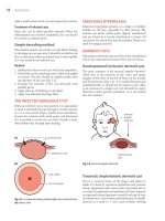
Step 1: preparation The patient should be in a
horizontal lithotomy position with legs in Lloyd‐
Davies stirrups, with the hips slightly flexed and
abducted and knees slightly flexed (Figure 17.1).
Preparation should be made as for any abdominal
procedure; in addition, the vagina should be
cleansed and an indwelling urethral catheter
inserted and the balloon inflated to facilitate
197
identification of the bladder neck. The urological
drape is secured over the perineum.
Step 2: incision This should be a low transverse
suprapubic (Pfannenstiel) incision, long enough for
access into the retropubic space; 6–8 cm is usually
adequate. After incising the skin and rectus sheath,
the Millin retractor is inserted.
Step 3: opening the retropubic space The
bladder and urethra are gently separated from
the posterior aspect of the symphysis to open up
the retropubic space (Figure 17.2). This is usually
achieved by blunt finger dissection, although if
there has been previous retropubic surgery, sharp
dissection using fine Metzenbaum scissors is
required.
Step 4: identifying the paravaginal fascia An
assistant uses a ‘swab on a holder’ or ‘sponge stick’
to retract the bladder medially. The right‐handed
surgeon (assuming they are standing on the
patient’s left side) will use their left index finger in
the vagina – covered by the urological drape – to
Figure 17.1 Patient in a horizontal lithotomy position with legs in Lloyd‐Davies stirrups.
198
Chapter 17
apply pressure upwards and laterally, at the level of
the bladder neck (not the lateral vaginal fornix as is
often described; Figure 17.3).
The upward pressure from below, with the medial
retraction above, is often sufficient to expose the
white glistening layer of paravaginal fascia. If neces
sary, a ‘peanut’ swab or Kittner dissector can be used
to achieve further separation, although the author’s
preference is to use fine Metzenbaum scissors for
this purpose, as they tend to be associated with less
tissue trauma and bleeding (Figure 17.4).
A number of venous sinuses may be encoun
tered in this area; these are best avoided; diathermy
may exacerbate bleeding so, if bleeding is trouble
some, it is better to insert the suspensory sutures as
quickly as possible, underrunning the vessels.
Step 5: inserting the suspensory sutures
When there is adequate exposure of the fascia,
two or three sutures of 0 Ethibond (coated
polyester; W975 0 Ethibond, 31 mm half‐circle
round‐bodied needle) or 0‐PDS (polydioxanone;
CT2 0 PDS, 26 mm half‐circle taper point needle)
Figure 17.4 Tips of Metzenbaum scissors used to aid
Figure 17.2 Retropubic space opened.
dissection of the paravaginal fascia.
(a)
(b)
Figure 17.3 The surgeon using their nondominant index finger in the vagina to apply pressure upwards and laterally, at
the level of the bladder neck: (a) operative view; (b) sagittal section.
Operations for urinary incontinence
are inserted into the fascia on each side; if
nonabsorbable sutures are used, care must be
taken not to penetrate into the vagina. It is wise to
check after each needle insertion that the vaginal
drape has not been caught.
The first suture should be placed at the bladder
neck and tied down on to the fascia. This should
control any venous bleeding but also provides
a ‘pulley’ to facilitate subsequent tying. The
suture is then passed through the most proximate
point on the ipsilateral ileopectineal ligament
(Figure 17.5). The two ends are then secured
with a small artery forceps until all sutures
are in place.
The second and third sutures are each placed
approximately 1 cm more cephalad and slightly
more lateral than the previous suture. Sutures
should not be placed below the level of the blad
der neck, as this may contribute to postoperative
voiding difficulties. These sutures are similarly
tied down on to the fascia and then passed
through the ileopectineal ligament, each sepa
rated by approximately 1 cm along the ligament
(Figure 17.6). Although an abnormal or acces
sory obturator artery is said to be present in
approximately 30% of people, a pubic branch of
the inferior epigastric vessels invariably crosses
the ileopectineal ligament; this should be seen as
the upper landmark for suture insertion on the
ligament.
When three sutures are in place on one side,
steps 4 and 5 are repeated on the other side.
The Step 6: the place of cystoscopy Some
surgeons advocate cystoscopy at this stage to exclude
bladder wall injury or penetration by sutures or the
possibility of other intravesical pathology; this has
not been the author’s routine practice.
Figure 17.5 Sutures inserted into the paravaginal fascia
on each side, using Turner‐Warwick needle holder.
Figure 17.6 Two or three sutures are inserted on each side, tied down onto the paravaginal fascia, and then passed
through the ileopectineal ligament.
199
200
Chapter 17
Step 7: tying the sutures When all the sutures
are in place they are tied. Tying is perhaps best
done alternately, starting from the most caudal
suture on one side, then the other side, then
moving progressively in a cephalad direction until
all are tied. In early descriptions, it was standard
practice to approximate the vaginal fascia directly
on to the ileopectineal ligament by having an
assistant apply pressure vaginally; this is not the
author’s practice. By using the ‘pulley’ suture as
described above, the application of gentle traction
to the suture limb placed through the ileopectineal
ligament brings the vaginal fascia into proximity
with the pelvic sidewall (not the ileopectineal
ligament), where it can become fixed. Some degree
of ‘bow‐stringing’ of sutures is inevitable but does
not detract from the effectiveness of the procedure,
the emphasis being on achieving support without
the necessity of extreme tension and elevation
(Figure 17.7).
Step 8: haemostasis and wound drainage
Bleeding within the retropubic space is invariably
venous and tying the suspensory sutures on to the
fascia (as in step 5) or through the ligament (as in step
7) will usually provide adequate haemostasis.
However, it is a wise precaution to leave a vacuum
drain in the retropubic space overnight postoperatively.
Step 9: wound closure The author’s preference is
to use 1 Vicryl (polyglactin; W9231 1 Vicryl, 40 mm
half‐circle round‐bodied needle) to close the rectus
sheath, and 2‐0 Prolene (polypropylene; W631 2‐0
Prolene, 65 mm straight reverse cutting needle, with
beads and collars) as a subcutaneous skin closure.
Other concurrent surgery
Hysterectomy While there is no benefit from
concurrent hysterectomy, where it is indicated for
other reasons it is best performed first, closing the
parietal peritoneum prior to opening into
retropubic space for colposuspension.
Vaginal vault or enterocoele It has long
been recognized that women who have
undergone colposuspension are at risk of
subsequent vaginal vault or posterior wall
prolapse.20 This has often been attributed to the
fact that pelvic organ prolapse reflects a systemic
connective tissue weakness and therefore
inevitably occurs in more than one site in the
same patient.24 The evidence from randomized
controlled trial (RCT) outcomes, however,
suggests that colposuspension may be a more
specific risk factor, probably related to the altered
angulation of the vagina and pressure
transmission within the pelvis.25
For this reason, several authorities have sug
gested that where an enterocoele is present, it
should be corrected, irrespective of symptoms. The
Moschowitz procedure has been advocated in this
context and was included in previous editions of
this book. This procedure was first described as a
method to close off a deep pouch of Douglas in
Figure 17.7 By using the ‘pulley’ suture the paravaginal fascia is brought into proximity with the pelvic sidewall but not
directly on to the ileopectineal ligament; ‘bow‐stringing’ of sutures does not detract from the effectiveness of the procedure.
Operations for urinary incontinence
conjunction with prolapse of the rectum.26 More
recently it has been used to close the cul‐de‐sac
during the course of several abdominal procedures.
A nonabsorbable suture material is used to place
two or three purse‐string sutures around the peri
toneum of the pouch of Douglas and close the hia
tus between the uterosacral ligaments, taking care
to avoid the pelvic ureter (Figure 17.8).
The author’s view is that a simple purse‐string, or
even a series of purse‐strings, in the peritoneum is
unlikely to achieve much in terms of long‐term
support; hence, I would never carry out the
Moschowitz procedure. My preference is to use
an abdominal sacrocolpopexy (as described in
Chapter 16) prior to the colposuspension in patients
with a symptomatic vault prolapse or enterocoele.
In those with evidence of enterocoele but no rele
vant symptoms, my preference is to carry out only
the colposuspension and then review the signs and
201
symptoms subsequently. Although one‐quarter to
one‐third of patients may experience deterioration
of findings or development of symptoms, up to
three‐quarters will not.
Rectocele As noted above, women undergoing
colposuspension are at risk of subsequent posterior
wall prolapse and several authors have suggested
that, where rectocele is present, it should be
corrected at the time of colposuspension,
irrespective of symptoms; indeed, this approach
was also advocated in the previous edition of this
book. The current author’s view, however, is that
rectocele should only be treated if causing
significant symptoms (see Chapter 16) and its
prophylactic management is therefore not justified.
Even where a rectocele is symptomatic before
embarking on colposuspension, carrying out a
posterior repair concurrently can actually be quite
difficult, in view of the extent of anterior vaginal
elevation. The author’s current preference again is
to carry out only the colposuspension and then
review the signs and symptoms subsequently,
carrying out a secondary posterior colporrhaphy
only where necessary.
Postoperative management
Postoperative bladder drainage may be by suprapu
bic catheter. This is best inserted after wound
closure (see earlier section on bladder drainage).
Patients in whom postoperative voiding difficulty is
anticipated should be taught intermittent self‐cath
eterization preoperatively; in this case, a Foley
catheter should be inserted overnight and they
can resume self‐catheterization when comfortable
enough to do so.
The wound drain can usually be removed on the
first postoperative day and the patient should be
able to mobilize and eat normally. She can be dis
charged home when voiding normally or able to
manage her catheter regimen independently.
Figure 17.8 Moschowitz procedure for enterocoele
closure; lowermost suture already tied, second in place,
and position of third (incorporating uterosacral
ligaments) shown as dotted line.
Operative complications
Bladder or urethral injury Bladder injury was
recorded in 3% of cases in one large RCT.27 Recognized
bladder or urethral injury should be repaired with 2‐0
or 3‐0 Vicryl (polyglactin; W9350 2‐0 Vicryl, 26 mm
half‐circle taper cut heavy needle or W9122 3‐0
Vicryl, 22 mm half‐circle taper cut needle); a single
layer is usually adequate, provided that the repair is
202
Chapter 17
watertight and under no tension. Catheterization
should be continued for 5 days or for 10–12 days if
hysterectomy has been carried out concurrently.
Ureteric obstruction Injury to the ureters is
uncommon, although ligation may occur if care is
not taken to identify the paravaginal fascia clearly.
On occasion, especially if there is a large cystocele
and additional sutures are inserted, distortion of
the bladder base in the region of the ureterovesical
junctions may result in kinking of the ureter(s)
with consequent obstruction unilaterally or
bilaterally (Figure 17.9).
If suspected intraoperatively, cystoscopy with
indigo carmine dye testing should be carried out. If
free efflux of dye is not seen, suspensory sutures
should be removed in sequence until free flow is
confirmed. It is probably wise to leave a double‐J
‘pigtail’ stent in place in this situation but sutures
can be replaced more laterally.
Where ureteric obstruction is suspected postop
eratively, on the basis of loin pain, persistent
nausea or, rarely, oliguria, investigation should be
Figure 17.9 Intravenous urogram in patient with bilateral
ureteric obstruction following colposuspension; point of
obstruction visible, with hydroureter and hydronephrosis
on the right; minimal function visible as faint nephro
gram only on left.
carried out as a matter of urgency, with computed
tomography urogram and isotope urography to
evaluate relative function. If ureteric stenting can
be achieved either retrogradely or via percutane
ous nephrostomy, drainage for a period of months
may allow complete resolution; otherwise laparot
omy and ureteric reimplantation would be required
(see Chapter 26).
Postoperative complications
Voiding dysfunction Delay in resumption of
spontaneous micturition may occur in up to 25% of
patients, although in most this will resolve
spontaneously if managed by the catheter regimen
described above. If voiding is delayed beyond five
days, patients are best discharged with suprapubic or
intermittent self‐catheterization.
Voiding dysfunction is also one of the most com
mon longer‐term complications of colposuspen
sion. It is seen in up to 20% of patients, although
those with early postoperative voiding difficulty
are not necessarily those who go on to have longer‐
term problems. This complication was seen more
commonly in the past, when the vaginal fascia was
elevated directly on to the ileopectineal ligament,
and is certainly less of an issue with the technique
described above. Although several operative and
pharmacological strategies have been tried in the
past, it is best managed by instituting clean inter
mittent self‐catheterization.
Bladder overactivity It has long been
recognized that women who have detrusor
overactivity preoperatively are less likely to have
a favourable outcome from surgery for SUI than
those with pure urodynamic stress incontinence
(USI). That said, the rate of resolution of SUI
symptoms in women with mixed USI and detrusor
overactivity is not significantly different from that
seen in those with pure USI. Rates of resolution of
overactive bladder symptoms between 24% and
90% have been reported.27 Although a number of
women may develop new symptoms of overactive
bladder or so‐called ‘de novo’ detrusor overactivity
following surgery for USI, particularly by
colposuspension, such surgery should not be
considered contraindicated in women with mixed
symptoms of SUI and overactive bladder or mixed
urodynamic findings of USI and detrusor
overactivity, provided that patients are fully
counselled about possible outcomes.
Operations for urinary incontinence
‘Post‐colposuspension syndrome’ In 1987,
Galloway and colleagues coined the term ‘post‐
colposuspension syndrome’ to describe the
occurrence of pain in one or other groin at the site
of suture placement. Symptoms in two‐thirds of
their small group of patients were relieved by
releasing the sutures on the affected side, without
compromising their continence. Overall, they
reported the syndrome in 12% of patients reviewed
at one to six years postoperatively.28 In the author’s
experience this situation is seen much less
commonly and may reflect a tendency to extreme
elevation in the cases reported.
203
instructions should be followed. The author’s pref
erence remains with the original Gynecare TVT™
device and it is this technique that is described
below. The tape comes packaged with introduction
needles swaged in position on either end of the
tape. The tape itself is housed within a removable
polyurethane sheath, which is split at the mid point
and overlapped in the central 4 cm to aid removal.
A reusable introduction needle handle and rigid
catheter guide are also available from the manu
facturers. Otherwise no special instruments are
required for the procedure.
Anaesthesia
Retropubic suburethral slings:
synthetic mid‐urethral slings
The description of the intravaginal slingplasty by
Ulmsten and colleagues in 1994 and the subse
quent modification that led to the introduction of
the Tension‐free Vaginal Tape (TVT™) in 1996
resulted in a paradigm shift in the practice of
surgery for SUI.29 While colposuspension repre
sented over 70% of surgery for SUI in England in
the mid‐1990s, over the next decade this had
dropped to barely 1%, with suburethral tape pro
cedures making up over 85% of procedures.30 They
have been the subject of considerable scientific
scrutiny and have been described as the most
extensively researched surgical treatment for SUI
in women,31 with RCTs extending to five‐year fol
low‐up25 and cohort studies to 17 years,32 confirm
ing that they are highly effective in the short and
medium terms, with accruing evidence demon
strating their effectiveness in the long term.
Indications
Synthetic mid‐urethral slings have been used mainly
in the situation of primary SUI due to urethral hyper
mobility. Although there are no high‐quality data to
support their use in recurrent SUI or in intrinsic
sphincter deficiency,31 many clinicians do also advo
cate their use in these more complex situations.33
Instruments
Although it is possible to carry out the procedure
using a strip of polypropylene cut from a sheet,
most surgeons would use one of the multitude of
devices designed for the purpose. Each has their
own minor design modifications and the manufacturer’s
The original reference to the TVT™ describes it as
an ambulatory procedure carried out under local
anaesthesia.29 Many surgeons continue to use this
approach, where TVT™ is carried out as an isolated
procedure; others prefer to use regional or general
anaesthesia. The benefits of the local anaesthesia
technique include more rapid recovery and earlier
discharge from hospital. In the one RCT examining
different anaesthetic techniques, the cure rate was
not found to be any better where local anaesthesia
was used, although the need for catheterization in
the postoperative period was reduced.34
Several techniques for ‘sedo‐anaesthesia’ have
been employed. The basic principle is to ensure
that the patient remains comfortable, with the
minimum level of sedation compatible with
adequate anaesthesia. The author’s preferred
technique is shown in Table 17.2.
The operation
Step 1: incisions
The patient is placed into a lithotomy position on
the operating table. Once effective local anaesthe
sia has been achieved, the points for needle exit
suprapubically are marked either by skin marker
pen or by two small stab incisions. These points are
positioned at the upper edge of the pubic symphy
sis, 2–2.5 cm from the midline, and are 0.5 cm in
length.
An incision is made in the anterior vaginal wall,
1 cm in length, centred on the mid‐urethral point.
Making the incision may be aided by grasping the
epithelium with Allis or Littlewood’s tissue forceps
either side of the midline. Limited dissection is
made using fine Metzenbaum scissors either side, to
create a space paraurethrally into which the TVT™
needle may be safely introduced (Figure 17.10).
204
Chapter 17
Table 17.2 The author’s preferred technique for for ‘sedo‐anaesthesia’.
Procedure
Technique
Monitoring
Intravenous fluid therapy with 1 litre Hartmann’s solution
Oxygen 4 litres/minute via a Hudson mask or nasal cannulae during sedation.
Continuous electrocardiography, blood pressure and pulse oximetry
Sedation
Fentanyl 50 µg (administered preferably in anaesthetic room and certainly before local anaesthesia).
Midazolam 1 mg increments slowly to maximum 3 mg.
Propofol 10–40 mg increments titrated to response slowly; continuous infusion may be used as alternative.
Local anaesthesia
80 ml 0.25% bupivacaine or 160 ml 0.25% prilocaine or lignocaine; each may be used either plain or with
adrenaline 1:200 000. The anaesthetic is injected.
A Foley catheter 18 Fr is inserted urethrally and the balloon is inflated to define the bladder neck. The local
anaesthetic is administered so as to anaesthetize the track through which the tape is to be passed. This
involves using approximately 25% of the solution suprapubically and 25% vaginally, on the right and left
sides. A 50–60‐ml syringe is used, with a 23 Fr needle to raise a skin wheal, followed by a 20 Fr spinal needle
to infiltrate the track retropubically.
Figure 17.10 Dissection using fine Metzenbaum scissors to create a space paraurethrally into which the TVT™ needle
may be safely introduced.
Step 2: tape insertion
When learning the procedure, it is probably wise to
use a rigid catheter guide to direct the bladder neck
away from the needle track, as used in the original
description; with experience this is not necessary.
The vaginal skin edge is grasped on one side with
fine dissecting forceps and the TVT™ needle tip is
placed in the paraurethral space. By ‘palming’ the
needle in the operator’s left hand, with the index
finger placed just lateral to the needle itself, the
needle is gently eased in the plane between vagina
and urethra, until it reaches the lower edge of the
Operations for urinary incontinence
205
Figure 17.11 The introduction needle is ‘palmed’ in the operator’s left hand as it is eased in the plane between vagina
and urethra.
inferior pubic ramus (Figure 17.11). The orienta
tion may be described as being towards the patient’s
ipsilateral shoulder. The endopelvic fascia is then
perforated, to bring the needle tip into the retropu
bic space, and the needle orientation then adjusted
to direct the tip more vertically. The TVT™ handle
is then lowered, (compare position of handle in
Figures 17.11 and 17.12) and, keeping the needle
tip against the back of the symphysis at all times,
the needle is moved through the retropubic space
and eventually penetrates the rectus sheath to
emerge from the suprapubic incision (Figure 17.12).
Step 3: cystoscopy
Although with experience it is possible to under
take a single cystoscopy after passage of both
needles, initially at least it is appropriate to per
form cystoscopy after each needle passage. The
whole of the bladder must be carefully inspected
using a 70‐degree telescope, with particular
attention to the area around the bladder neck;
the urethra is then inspected with a 0‐ or 12‐
degree telescope to exclude perforation by the
needle.
If perforation is identified (Figure 17.13), the
needle should be withdrawn and reinserted, being
even more careful to keep the needle against the
pubis. A more lateral approach is sometimes sug
gested as being less likely to perforate the bladder,
although the author’s view is that this probably
increases the risk of vascular injury and is best
avoided.
Once satisfactory placement is confirmed, the
needle is grasped and withdrawn from the suprapu
bic incision. The second needle passage is then
undertaken in the same manner and a second
check cystourethroscopy performed (Figure 17.14).
The tape is then pulled up by traction on both ends
of the tape (including the sheath), so as to lie a few
millimetres away from the urethra.
Step 4: ‘cough testing’
The TVT™ needles are then removed from the tape
with suture scissors and the ends of the polyure
thane sheath surrounding the tape (but not the
tape itself) are grasped with small artery forceps. If
the procedure is performed under local anaesthe
sia, a ‘cough test’ is then carried out. The bladder is
206
Chapter 17
(a)
(c)
(b)
Figure 17.12 The handle is then lowered and the needle passed through the retropubic space, to emerge from the
suprapubic incision. (a) and (c) show the needle passed on the patient’s right side (in coronal plane), and (b) on the left
(in sagittal plane).
left distended to around 300 ml after the final cys
tourethroscopy and the patient is asked to cough
several times. If no leakage of urine is observed, the
tape may be pulled down off the urethra somewhat
more, using fine scissors. The bladder may be
slightly more distended and the patient put into a
slight head‐up position or a few moments may be
allowed for the sedation to wear off, as required. If
leakage is seen, the tape is progressively adjusted
until only a slight loss of urine remains; it is
Operations for urinary incontinence
Figure 17.13 Cystoscopic photograph, showing bladder
perforation by introduction needle.
207
manoeuvres. In this case, the tape position must be
judged empirically. It should be noted that when
ever tape adjustments are made, an instrument
(e.g. fine dissecting scissors) should be placed
between the tape and the urethra, to preclude
undue tightening (Figure 17.15a,b).
Once the tension is judged to be appropriate, the
polyurethane sheath is removed from the tape by
gentle traction on the artery forceps. Although
many surgeons remove the two halves of the
sheath simultaneously, it is the author’s preference
to remove one side and then to undertake a further
check of continence before removing the second
side, as fine adjustments of the tape are still possi
ble at this stage.
In patients under regional anaesthesia, cough
testing is probably not appropriate; since pelvic
floor function cannot be adequately judged, over
correction is perhaps more likely. Similarly, in
patients under general anaesthesia, assessing tape
tension by stress testing using a Credé manoeuvre
is also likely to result in overcorrection and an
increased risk of postoperative voiding difficulty.
Step 5: wound closure
After the final tape adjustments are completed the
tape is trimmed. Gentle traction is put on the tape,
which is then trimmed below the skin level; it does
not require to be sutured in place.
The abdominal skin is closed with butterfly skin
closures (e.g. 3M Steristrips) or by a fine absorbable
suture placed subcutaneously (e.g. 3‐0 Vicryl
Rapide, polyglactin; W9927 3‐0 Vicryl Rapide,
22
mm half‐circle cutting needle); continuous
locked 2‐0 Vicryl (polyglactin) is used for the vagi
nal skin (W9350 2‐0 Vicryl, 26 mm half‐circle taper
cut heavy needle). A dilator may be passed into the
urethra and gentle traction applied away from the
pubis to ensure that elevation has not been
achieved. The bladder is drained before the patient
leaves the theatre.
Figure 17.14 Check cystoscopy may be undertaken after
each needle passage, but must as a minimum, must be
carried out after the final needle passage, before tape
adjustment (as in this image).
robably better for a drop of leakage to persist,
p
rather than complete continence to be secured per
operatively. On occasion, it may not be possible to
demonstrate urine leakage, even with these
Postoperative management
If the procedure has been carried out under regional
anaesthesia, a Foley catheter should be left in place
until sensation returns. In other patients, postop
erative catheterization is usually not required.
Patients should be encouraged to void normally
and if they are unable to do so, intermittent cathe
terization is undertaken until normal micturition
208
Chapter 17
Figure 17.15 Metzenbaum scissors placed beneath the tape when adjustments are being made: (a) leakage apparent; (b)
leakage resolved.
resumes. If there is any doubt over the complete
ness of voiding, the residual urine volume should
be checked by ultrasound or by catheterization.
Most patients will be fit for discharge on the day of
surgery or the first postoperative day. In view of the
sedation used, patients are advised not to drive for
24 hours.
There is no evidence that restriction of activity
postoperatively alters outcome. Some advise
restriction of heavy lifting and avoidance of sport
ing activities and sexual intercourse for four to six
weeks following surgery; others do not impose any
specific restrictions and encourage patients to
return to normal activities as soon as they feel able
to do so. The results from an early UK trial found
patients to be back to normal activity around the
home at two to three weeks, although return to
work was at three to four weeks on average.35 The
patient’s expectation of outcome probably has
much to do with this variation.
Operative complications
Bleeding
Despite limited vaginal dissection compared with
other continence procedures, the venous plexus in
the retropubic space is vulnerable to damage dur
ing the blind passage of an introducer. Bleeding
from the retropubic space may present as increased
intraoperative blood loss (vaginally or suprapubi
cally) or subsequently as cutaneous bruising or ret
ropubic haematoma. The symptoms of retropubic
haematoma include pain, voiding difficulty, pelvic
mass or a drop in haemoglobin. Clinically apparent
retropubic haematomas have been reported in
0.4–2.3% of women, although since many are
asymptomatic, the true incidence is not known.36
Major vascular injury during blind retropubic pro
cedures is a rare but potentially fatal occurrence;
injuries to the obturator, external iliac, femoral and
inferior epigastric arteries have been reported. The
true frequency with which this occurs is not known,
Operations for urinary incontinence
although estimates between 0.01% and 0.6% have
been reported.36 The lower estimates, coming from
larger series, are probably more reliable.
Bladder or urethral injury
Bladder perforation is a well‐recognized risk of
retropubic mid‐urethral sling procedures, reported
in up to 15% of cases. Data from the Austrian
national registry suggest that bladder perforation
rates are higher in patients with previous surgery
for incontinence or prolapse, although this has not
been found consistently in all studies. Bladder per
foration has been looked on as a surrogate
marker for training in mid‐urethral sling proce
dures.37 In one study, the perforation rate for
trainees peaked at around 10%; those who carried
out more than 20 procedures in training ultimately
achieved a perforation rate of 5% or less; no train
ees carrying out fewer cases achieved this target
rate in training.38
It is important to recognize that bladder injury
identified by cystoscopy at the time of surgery
(Figure 17.13) and managed as described above has
no long‐term sequelae. Unrecognized bladder perfo
ration can lead to immediate postoperative suprapu
bic or vaginal leakage and vulval oedema has been
described. In the long term, intravesical mesh can
cause pain, recurrent urinary tract infection, urgency
symptoms and stone formation (Figure 17.16).
209
Urethral injuries occur less frequently than blad
der injuries during retropubic mid‐urethral sling
procedures but have been recorded in one review
in around 0.1% of procedures.36 It is likely that a
proportion of women presenting late with a pparent
urethral erosion of tape will have had an undiag
nosed perforation at their initial surgery,
particularly when symptoms of pain and urinary
retention have been present since tape placement
(Figure 17.17).
Bowel injury
Bowel trauma is rare but potentially the most seri
ous complication of retropubic mid‐urethral sling
procedures. Perforation is unlikely to be recognized
at surgery and delayed diagnosis can lead to significant
morbidity and many of the deaths associated with
the procedure are attributed to bowel perforation.
Prevalence is difficult to estimate but is thought to
be of the order of 0.01%. Both ileal and colonic
perforations have occurred, as well as perforation
of the small bowel mesentery leading to small
bowel obstruction. Bowel injury usually, although
not invariably, occurs in patients who have had
previous pelvic or lower abdominal surgery. Bowel
injury can present with fever, abdominal pain or
intestinal obstruction; leakage of bowel contents
from the suprapubic incisions has been described
following perforation of the ileum.
Figure 17.16 Cystoscopic photograph, showing bladder perforation by tape, with overlying encrustation.
210
Chapter 17
Figure 17.17 Cystoscopic photograph, showing urethral perforation by tape. The open bladder neck can be seen in
distance (outlined by dotted line) in the top left of the image; fibres of the tape are seen across the right side of the
image.
Nerve injury
Injury to the ilioinguinal, obturator and femoral
nerves have been described following retropubic
mid‐urethral sling procedures. The first of these is a
recognized risk in low transverse abdominal
incisions and has been described following several
procedures, presenting with burning pain and
altered sensation in the groin, inner thigh and
labium majus. Some injuries resolve spontaneously
over time, while others may require nerve blockade
with local anaesthesia or local steroid injections.
Obturator nerve injury is a rare but serious com
plication that can occur following unduly lateral
tape insertion. Of the cases described in the literature,
some have resolved spontaneously while
others
have required tape removal or division.
Postoperative complications
Voiding dysfunction
In one early trial, 5% of women developed tran
sient voiding disorder following retropubic mid‐
urethral sling procedures, requiring intermittent
self‐catheterization for up to one month. Some
have advocated either stretching or pulling down
the tape, and tape division is widely described as a
treatment for severe voiding difficulty, with the
majority of patients (range 61–94%) remaining
continent. Given that the majority of these
roblems resolve spontaneously, however, careful
p
counselling is important before undertaking such
interventions.
Bladder overactivity
Depending on the definitions used, urgency urinary
incontinence has been reported in up to 25% of
women undergoing retropubic mid‐urethral sling
procedures; nevertheless, a reduction in urgency
and urgency urinary incontinence and the degree
of bother associated with these symptoms reduced
from 93% preoperatively to 44% five years after
surgery in one trial, with only 1% developing new
urgency urinary incontinence postoperatively.25
Pain
As noted earlier, chronic groin pain is described
following colposuspension and is often relieved by
cutting the stitch on the affected side. Similar occur
rences have been described following retropubic mid‐
urethral sling procedures in around 1.3% of cases.31
Sexual dysfunction
There are conflicting data on the effect of retropubic
mid‐urethral sling procedures on sexual function.
Although deterioration in sexual function has been
reported in up to 20% of women, longitudinal
follow‐up in trial situation suggests a significant
Operations for urinary incontinence
improvement, such as reduction in the number of
women whose sex life was spoilt due to incontinence,
in the number experiencing incontinence with inter
course, and in reporting of dyspareunia.31,35
Tape exposure or extrusion
One of the major concerns about synthetic sling
materials is their potential to extrude (the term
‘erosion’ should be eschewed) into the urinary tract
and vagina. These complications may be related to
surgical technique, host factors, wound healing,
infection or the physical properties of the implanted
material, such as pore size or multifilament con
struction. Early exposure within the vagina is most
likely to be from failure of vaginal skin healing
rather than true extrusion. The latter is generally
seen as a more gradual process and has been
reported up to five years after surgery. Whatever
the mechanism, tape exposure within the vagina
has been seen following all mid‐urethral sling pro
cedures and is reported in up to 2% of women fol
lowing retropubic mid‐urethral sling procedures.31
Early exposure or defective vaginal healing is
usually treated by simply resuturing the vaginal
skin over the tape or by trimming the exposed area
of tape and closing the skin.
Urethral extrusion following retropubic mid‐ure
thral sling procedures has been reported on a num
ber of occasions, although the incidence in national
databases appears to be less than 1%. In many
cases, extrusion was associated with severe voiding
difficulty, often from the time of surgery, with or
without urgency incontinence, haematuria and
pain. The timing of symptoms in relation to surgery
has led to the author’s view that this usually repre
sents overlooked operative urethral perforation.
Excision of the intraurethral tape with urethral
repair (with or without a Martius graft) has been
reported with variable effects on continence.
Urethrovaginal fistulae have been seen both in
association with mid‐urethral sling extrusion and
following tape excision.
Trans‐obturator foramen
suburethral slings
211
the bladder, urethra and bowel, seen with the ret
ropubic route.39 Delorme’s technique employs an
‘outside‐in’ approach; that is, with passage of the
introduction needle from the genitofemoral crease
towards the vagina. Although early devices
(Mentor‐Porges UraTape® and ObTape®) were
withdrawn because of tape‐related complications,
the method itself remains in use, and is exemplified
by the AMS Monarc® and Boston Scientific Obtryx®
systems as described below.
Although there have been numerous further
design modifications since (perhaps aimed more at
establishing a niche in the market rather than
addressing a specific clinical problem), the only
notable one was the development of the ‘inside‐out’
approach (i.e. with needle passage from vaginal to
genitofemoral aspects); this is exemplified by the
Gynecare TVT‐O® device.40 The Cochrane review of
mid‐urethral sling operations found no evidence to
support the use of one approach over the other.31
The NICE report on urinary incontinence provided a
‘future‐proofed’ recommendation by simply empha
sizing the need to use tapes of proven efficacy or cost
benefit based on (current or future) robust evidence
from RCTs.12 Although the uptake of the trans‐obtu
rator foramen approach has varied in different areas,
two‐thirds of mid‐urethral sling procedures in
England are still inserted via the retropubic route.30
In 2001, development started on further tape
modifications to produce a single‐incision sling,41
potentially reducing further the more common
complications of the retropubic and obturator
approaches. Meta‐analysis of trial data on a number
of similar devices showed that one device (since
withdrawn) was inferior to standard mid‐urethral
sling procedures; not enough data existed on other
single‐incision slings to allow reliable comparisons.42
NICE similarly found evidence on the procedure to
be inadequate and currently gives a very guarded
recommendation that the procedure should not be
used without special arrangements for clinical gov
ernance, consent and audit or research, and that
patients should understand the uncertainty about
the procedure’s safety and efficacy, including the
potential serious long‐term complications.43
Indications
The trans‐obturator foramen approach to the inser
tion of a suburethral sling was reported by Delorme
in 2001, primarily as a means of limiting injury to
Although, in general, the trans‐obturator foramen
mid‐urethral sling is considered indicated in the
same situations as the retropubic mid‐urethral sling
212
Chapter 17
described earlier, the lower rate of bladder perfora
tion perhaps makes it more suited in circumstances
where this risk is seen to be increased. It is, how
ever, the author’s view that there are few if any
situations where this actually pertains; the proce
dure certainly cannot be advocated in recurrent
SUI or in intrinsic urethral sphincter deficiency.
An incision is made in the anterior vaginal wall,
1 cm in length, centred on the mid‐urethral point;
this may be aided by grasping the epithelium with
Allis or Littlewood’s tissue forceps either side of
the midline. The subfascial plane is then dissected
out towards the ischiopubic ramus using fine
Metzenbaum scissors. Small stab incisions are made
at the previously marked points in each groin.
Anaesthesia
General, regional, local or sedo‐anaesthesia can all
be used as described for the retropubic mid‐urethral
synthetic sling.
Operation
Step 1: incisions
With the patient in a horizontal lithotomy position
on the operating table, landmarks are ascertained
and the points for needle insertion marked using a
skin marker pen; these should be 1 cm lateral to
the ischiopubic ramus, just below the adductor lon
gus muscle tendon, on a horizontal line level with
the clitoris (Figure 17.18). Local anaesthetic (as
described above for the retropubic mid‐urethral
synthetic sling) is injected into the skin and along
the proposed needle track on both sides; further
anaesthetic is used to infiltrate the anterior vaginal
wall at the mid‐urethral level.
Step 2: insertion of the tape introducer
Each of the different devices has one or more specific
introduction needles provided and the manufactur
er’s instructions should always be followed. Most
have right and left ‘halo’ introducers, shaped to the
corresponding side of the pelvis and some have a
nonlateralized curved introducer; as a general rule,
however, the following steps are common to all.
The (patient’s) left introducer is held in the
operator’s right hand when operating on the
patient’s left side; similarly, the (patient’s) right
introducer is held in the operator’s left hand when
operating on the patient’s right side. The intro
ducer is held vertically with the handle down
wards as it is introduced through the skin incision
(Figure 17.19). It is then passed through the obtu
rator membrane, when a sudden loss of resistance
is felt as a ‘pop’.
Figure 17.18 The points for needle insertion marked, 1 cm lateral to the ischiopubic ramus, just below the adductor
longus muscle tendon, on a horizontal line level with the clitoris.
Operations for urinary incontinence
213
Figure 17.19 The introducer is held vertically with the handle downwards as it is introduced through the skin incision.
Figure 17.20 The introducer is then passed around the ramus by rotation of the operator’s wrist.
The introducer handle is then rotated upwards in
the plane of the perineum, to an angle of 45 degrees
to the horizontal, bringing the tip itself into an ori
entation roughly perpendicular to the ischiopubic
ramus. The introducer is then passed around the
ramus keeping the tip in contact with the bone at all
times, by rotation of the operator’s wrist
(Figure 17.20). The index finger of the operator’s
contralateral hand is placed in the vaginal incision
to assist in maintaining the correct direction of
introducer passage. The finger should protect
the urethra from injury, while at the same time
214
Chapter 17
ensuring that the introducer passes above the lateral
vaginal sulcus and does not penetrate the vaginal
wall but rather is guided into the vaginal incision.
Step 3: insertion of the tape
Once the introducer has been passed on one side,
it is prudent to confirm that the vagina and ure
thra have not been pierced. One end of the tape is
then connected to the tip of the introducer; the
different devices have different ways of achieving
this: some require threading though an ‘eye’ in
the tip, others hook or ‘snap’ on to the end of the
device. Once securely connected, the tape is
pulled through into position by traction and more
particularly rotation of the introducer in the
reverse direction (Figure 17.21). Some tapes
constructed of thermally bonded polypropylene
are of low elasticity and can be pulled through
the tissues directly without risk of deformation;
others do stretch when traction is applied. Two
mechanisms have been developed to deal with
this issue: one is to house the tape in a polyure
thane sheath, which is removed after insertion;
the other is the use of a ‘tensioning’ suture,
passed through the tape.
Those devices intended for ‘inside‐out’ insertion
are preloaded on to the introducer, so they follow
the introducer into position and do not require to
be pulled through as a second step. The tape is then
inserted in the same way on the other side and the
introduction needles can be removed.
Step 4: positioning of the tape
The intention is that the tape is positioned beneath
the mid‐urethra under minimal tension. Ensure
that the patient is in a horizontal lithotomy or even
a slightly head‐up position. Leave a space of
2–3 mm between the tape and the urethra. This
can be achieved visually and is best achieved by
placing the blades of the Metzenbaum scissors
beneath the tape. If the tape used is one of those
housed in a polyurethane sheath, this should now
be withdrawn by traction from the groin incisions;
it is important that the tape itself does not move at
this stage. This is best achieved by counter‐traction
by the Metzenbaum scissors beneath the tape.
Step 5: wound closure
After the sheath is removed, excess tape is trimmed,
ensuring that it lies well below the skin surface. The
groin incisions are closed with butterfly skin closures
(e.g. 3M Steristrips) or by a fine absorbable suture
placed subcutaneously (e.g. 3‐0 Vicryl Rapide,
polyglactin; W9927 3‐0 Vicryl Rapide, 22 mm half‐
Figure 17.21 The tape, once connected to the tip of the introducer, is pulled through into position by traction and
rotation of the introducer in the reverse direction.
Operations for urinary incontinence
circle cutting needle); continuous locked 2‐0 Vicryl
(polyglactin; W9350 2‐0 Vicryl, 26 mm half‐circle
taper cut heavy needle) is used for the vaginal skin.
Postoperative management
Postoperative management is as described above
for the retropubic mid‐urethral synthetic sling.
Complications
Visceral injury
Although bladder perforation is significantly less
common at trans‐obturator foramen than retropubic
mid‐urethral sling, and bowel injury would be
expected to be similarly less common, urethral injury
is perhaps more common and has been reported in
up to 3% of cases.36,44 Postoperative voiding difficulty
also appears to be less common after trans‐obturator
foramen than retropubic mid‐urethral sling.31
Pain
Chronic groin pain, on the other hand, is signifi
cantly more common following tape insertion by
the trans‐obturator foramen route. The Cochrane
review of mid‐urethral slings reported an overall
incidence of 6.4% – four to five times higher than
with the retropubic route.31 This latter symptom is
certainly an increasingly common reason for patient
requests for tape removal and medicolegal claims.
Tape exposure or extrusion
Although some studies report the rate of tape
exposure/extrusion to be higher following tape
insertion by the trans‐obturator foramen than the
retropubic route,45 trial data suggest similar rates,
of around 2%.31
Retropubic suburethral slings:
traditional sling operations
The term ‘traditional’ sling is used here, in line
with the Cochrane review of these procedures,46 to
distinguish open sling procedures more usually
placed at the region of the bladder neck from the
newer minimal access mid‐urethral tape proce
dures; they are perhaps more often known as pub
ovaginal sling procedures in the United States. The
first sling operations for SUI used pyramidalis mus
cle and/or rectus sheath, and were described in
Germany in the early 1900s (Goebel‐Frangenheim‐
215
Stoeckel); they became more widely known
following the report by Aldridge in the United
States in 1942,47 although they have never been
widely used in UK, where they currently represent
only 1% of SUI surgeries.30
Numerous variations of the technique have been
described and many different materials used. Slings
may be constructed from:
• autologous materials (the patient’s own tissues;
e.g. rectus fascia, fascia lata)
• allogeneic materials (non‐patient, human/
cadaveric tissues; e.g. lyophilized dura, fascia lata)
• xenogeneic materials (non‐human organic
tissues; e.g. porcine dermis or small intestinal
submucosa)
• alloplastic materials (synthetics; e.g. Prolene,
polypropylene; Silastic, silicone‐coated Dacron;
Mersilene, polyester; Marlex, polypropylene and
high‐density polyethylene; Gore‐Tex, nylon,
polytetrafluoroethylene and polyurethane).
Although the risk of prion disease transmission has
been mitigated by the removal of cellular materials
from allogeneic and xenogeneic slings, their dura
bility remains in doubt. The morbidity associated
with alloplastic ‘traditional’ slings (as opposed to
the newer minimally invasive mid‐urethral slings
described above) makes their use largely obsolete.
Although several other materials are still available,
the author’s view is that sling procedures should
use only autologous (rectus sheath and fascia lata)
or alloplastic (polypropylene) materials.
Indications
The traditional sling procedures can be used:
• for secondary treatment of SUI after previous
failed surgery
• where there is limited vaginal access or signifi
cant reduction of vaginal capacity and mobility,
rendering a colposuspension technically difficult
or impossible
• where SUI is thought to reflect intrinsic sphincter
deficiency (i.e. low urethral closure pressure)
rather than urethral hypermobility
• for primary treatment of SUI, if the patient
chooses this over a retropubic suburethral syn
thetic sling or a colposuspension and accepts the
higher associated morbidity.
Anaesthesia
General or regional anaesthesia is required.
216
Chapter 17
Rectus sheath sling procedure (after
Aldridge)
Step 1: preparation
Antibiotic cover and antithromboembolic precau
tions, as described for the Burch colposuspension
procedure, are required.
As for colposuspension, the patient should be in
a horizontal lithotomy position with legs in Lloyd‐
Davies stirrups, with the hips slightly flexed and
abducted and knees slightly flexed (Figure 17.1).
Preparation should be made as for any abdominal
procedure. In addition, the vagina should be
cleansed and an indwelling urethral catheter
inserted and the balloon inflated to facilitate identi
fication of the bladder neck.
Two incisions are required: a low transverse
suprapubic (Pfannenstiel) incision, to harvest the sling
and an inverted U‐shaped incision over the anterior
vaginal wall to secure the sling suburethrally.
Step 2: exposure of the rectus abdominis
and preparation of the sling strip
Through the Pfannenstiel incision, the rectus
sheath is exposed. The sling may be fashioned
either as two limbs or as a T‐shape. In the first
instance, two strips are cut transversely from the
aponeurosis, each about 7–8 cm in length and
1.5 cm wide, starting from the lateral edge and end
ing 2 cm from the midline where the sling is left
attached (Figure 17.22a). The disadvantage of this
approach is the length of skin incision required,
which even with some undermining may still be
15–16 cm. The alternative is to extend the strips
downwards in the midline, towards the pubic sym
physis, forming a T‐shape, the stem of which is
then divided in the midline (Figure 17.22b). This
allows several additional centimetres of sling length
to be harvested despite a shorter abdominal incision
but carries a higher risk of midline incisional hernia
formation. Either scalpel or cutting diathermy may
be used for the dissection. A stay suture is passed
through end of each limb of the sling.
Step 3: opening of the retropubic space
The bladder and urethra are gently separated from
the posterior aspect of the symphysis to open up
the retropubic space (Figure 17.2). Although in
surgically naïve cases this is usually achieved by
blunt finger dissection, if there has been previous
retropubic surgery, sharp dissection using fine
Metzenbaum scissors is required.
Step 4: exposing the bladder neck
from the vaginal incision
With a urethral catheter in place, the bladder neck
is palpated vaginally. The anterior vaginal wall is
infiltrated with 0.5% bupivacaine and 1:200 000
adrenaline for haemostasis and definition of ana
tomical layers. A proximally based U‐shaped inci
sion is made on the anterior vaginal wall from
approximately 1 cm proximal to the external ure
thral meatus, extending to approximately 1
cm
proximal to the bladder neck. The flap is mobilized
to expose the area of pubocervical fascia overlying
the proximal urethra (Figure 17.23).
An alternative is to make a simple midline inci
sion over the proximal urethra but this obviously
provides a smaller area of fascia over which to
secure the sling.
Step 5: completing the urethral tunnel
The plane of dissection is developed laterally between
the vaginal wall and the bladder neck, using fine
Metzenbaum scissors. The endopelvic fascia is then
perforated with scissors or a finger, to gain entry into
the retropubic space from below (Figure 17.24).
Step 6: positioning the fascial sling
A curved clamp or uterine packing forceps is now
passed from the vaginal incision upwards through
the retropubic space and grasps the stay suture on
one end of the sling. It is then withdrawn, bringing
the sling down into the vaginal dissection. This is
repeated on the other side (Figure 17.25). The
author’s preference is to suture the sling ends
in a ‘waist‐coating’ or ‘double‐breasted’ fashion; if
sufficient sling length is not available, further
extension can be achieved either by further mobili
zation from the subcutaneous fat or further down
ward dissection on to the pubic symphysis.
Step 7: closure of the abdominal incision
It is probably wise to close the abdominal incision
before final positioning and suturing of the sling
at the bladder neck level. If sling strips are used,
1 Vicryl (polyglactin; W9231 1 Vicryl, 40 mm
half‐circle round‐bodied needle) may be used to
close the rectus sheath. If the T‐shaped dissection
of sheath is used, the author’s preference is to
use a nonabsorbable closure with 0 Ethibond
(coated polyester; W975 0 Ethibond, 31 mm half‐
circle round‐bodied needle). A vacuum drain
should be left in the retropubic space overnight.
Operations for urinary incontinence
217
(a)
(b)
Figure 17.22 Rectus sheath sling marked and cut from aponeurosis: (a) as two strips cut transversely; (b) the incisions
extended downwards in the midline, forming a T‐shape.
The skin is then closed with 2‐0 Prolene (poly
propylene; W631 2‐0 Prolene, 65 mm straight
reverse cutting needle, with beads and collars) as
a subcutaneous skin closure and a suprapubic
catheter is inserted.
Step 8: suturing of the sling and closure
of the vaginal incision
The two ends of the sling are positioned around the
bladder neck with minimal tension. One end is
tacked on to the underlying pubocervical fascia
with four or six interrupted sutures of 2‐0 Vicryl
(polyglactin; W9350 2‐0 Vicryl, 26 mm half‐circle
taper cut heavy needle). The other end is then
secured over this in a ‘double‐breasted’ fashion,
using a similar number of sutures, each catching
both the fascia and the other sling limb
(Figure 17.26). The vaginal skin is then closed with
a continuous locked suture of the same 2‐0 Vicryl
(polyglactin).