Ebook Back to basics in physiology - O2 and CO2 in the respiratory and cardiovascular systems: Part 1
Bạn đang xem bản rút gọn của tài liệu. Xem và tải ngay bản đầy đủ của tài liệu tại đây (1.58 MB, 91 trang )
Back to Basics in Physiology
Back to Basics in Physiology
O2 and CO2 in the Respiratory and
Cardiovascular Systems
Juan Pablo Arroyo
Internal Medicine Resident
Tinsley R. Harrison Society Scholar
Vanderbilt University À School of Medicine
Adam J. Schweickert
Attending Physician
Hospitalist Medicine À Pediatric ICU
St. Barnabas Medical Center
AMSTERDAM • BOSTON • HEIDELBERG • LONDON
NEW YORK • OXFORD • PARIS • SAN DIEGO
SAN FRANCISCO • SINGAPORE • SYDNEY • TOKYO
Academic Press is an imprint of Elsevier
Academic Press is an imprint of Elsevier
125, London Wall, EC2Y 5AS
525 B Street, Suite 1800, San Diego, CA 92101-4495, USA
225 Wyman Street, Waltham, MA 02451, USA
The Boulevard, Langford Lane, Kidlington, Oxford OX5 1GB, UK
Copyright r 2015 Elsevier Inc. All rights reserved.
No part of this publication may be reproduced or transmitted in any form or by any means,
electronic or mechanical, including photocopying, recording, or any information storage and
retrieval system, without permission in writing from the publisher. Details on how to seek
permission, further information about the Publisher’s permissions policies and our arrangements
with organizations such as the Copyright Clearance Center and the Copyright Licensing Agency,
can be found at our website: www.elsevier.com/permissions.
This book and the individual contributions contained in it are protected under copyright by the
Publisher (other than as may be noted herein).
Notices
Knowledge and best practice in this field are constantly changing. As new research and
experience broaden our understanding, changes in research methods or professional practices,
may become necessary.
Practitioners and researchers must always rely on their own experience and knowledge in
evaluating and using any information or methods described herein. In using such information or
methods they should be mindful of their own safety and the safety of others, including parties for
whom they have a professional responsibility.
To the fullest extent of the law, neither the Publisher nor the authors, contributors, or editors,
assume any liability for any injury and/or damage to persons or property as a matter of products
liability, negligence or otherwise, or from any use or operation of any methods, products,
instructions, or ideas contained in the material herein.
ISBN: 978-0-12-801768-5
Library of Congress Cataloging-in-Publication Data
A catalog record for this book is available from the Library of Congress
British Library Cataloguing-in-Publication Data
A catalogue record for this book is available from the British Library
For Information on all Academic Press publications
visit our website at />
DEDICATION
To our wives, Denise and Valentina, for their unwavering support of
our every endeavor, both aimless and not so aimless.
ACKNOWLEDGEMENTS
We wish to thank Mara Conner, Jeffrey Rossetti, and the rest of the
Elsevier staff for the time and hard work that went into helping to
make this book a reality.
We also wish to thank all those who provided their insight and suggestions throughout the writing of this book, with a special thanks to
Dr. Gary Kohn.
PREFACE
The whole idea for this series arose from the physiology classroom and
hospital teaching rounds. We realized that both in the classroom and
on the wards, students and residents had a fair amount of knowledge
regarding individual organ systems. However, there was still room for
improvement regarding how all the organ systems integrate in order to
respond to a particular situation. This book series is an attempt to
bridge the gap of knowledge that divides organ from body, and isolated action from integrated response.
Our goal is to create a series of books where the primary focus is the
integration of concepts. The books in the series are written so that hopefully they are easy to read, and can be read from beginning to end.
It is our belief that if you truly understand something, you should
be able to explain in a simple way. Therefore, we aim to tackle complicated topics with simple examples. And we hope that by the end of
any book in this series, further more complex reading (e.g., the latest
journal articles) should prove far easier to understand.
We hope you enjoy reading these books as much as we enjoyed
writing them.
Other books in the series include:
Back to Basics in Physiology: Fluids in the Renal and
Cardiovascular Systems (ISBN: 9780124071681)
Back to Basics in Physiology: Electrolytes and Nonelectrolyte
Solutes in the Body (ISBN: 9780128017692)
CHAPTER
1
Cellular Respiration and Diffusion
INTRODUCTION
Breathing in and out is key to staying alive. It’s so important that
even when we forget to breathe, our nervous system picks up the slack
and keeps going. The process of breathing provides oxygen and
removes carbon dioxide from the body. This process is essential to
sustaining each and every cellular task within our bodies. The focus of
this book is how the body achieves this seemingly simple process.
We will take you from a single cell and how it regulates oxygen and
carbon dioxide to the large-scale gas transport and delivery in the
body under normal and pathologic conditions. So, sit back, relax, and
take a deep breath!
If indeed you take a breath right now, you will breathe in air.
Air in the atmosphere is a simply a mixture of gases. Atmospheric
air, as it exists today, consists of about 21% oxygen, 78% nitrogen,
0.04% carbon dioxide, and some other miscellaneous gases such as
argon. (Carbon dioxide makes up so little of the atmospheric air that
it even gets beat out by argon, which weighs in at 1%. Seriously!)
But it wasn’t always this way. In fact, over 2.5 billion years ago,
things weren’t looking too good for our oxygen-loving brethren. There
was almost no oxygen in the atmosphere, and there was very little
food around. So, some opportunistic little buggers called cyanobacteria
took the warmth of the sun and made sustainable energy out it, much
like plants do today. In the process they gave off oxygen as “waste.”
Little by little cyanobacteria began filling up the oceans with oxygen.
The dissolved oxygen began to diffuse throughout the water (hopefully
you’ll remember the principles of diffusion from our last book “Back to
Basics in Physiology: Fluids in the Cardiovascular and Renal Systems”),
and as the oceans filled with this “waste product” it diffused into the
atmosphere. Over the next two billion years, the concentration of
oxygen in the air reached the 21% we know and enjoy today.
Back to Basics in Physiology. DOI: />© 2015 Elsevier Inc. All rights reserved.
2
Back to Basics in Physiology
As oxygen became more and more plentiful in the environment,
creatures began using this oxygen to create energy from available
food sources more efficiently, and were able to grow larger than
their non-oxygen-consuming counterparts. With size came more food
consumption and a greater need for mobility, and with mobility and
size came more energy utilization. Over time, organisms migrated
from the water to land. Cyanobacteria made room for plants in the
sea and on land, which produced even more oxygen. As organisms
developed ways to use this newfound energy (e.g., growing brains!),
they developed a larger need for oxygen, produced more carbon
dioxide, and along the way came up with some pretty ingenious
mechanisms to ensure constant oxygen delivery and carbon dioxide
removal.
In our bodies today, out of the millions of functions that need to be
carried out minute by minute in order to allow for life to proceed
“uneventfully,” oxygen (O2) and carbon dioxide (CO2) exchange are
arguably two of the most important processes our bodies require to
stay alive. If the human body is deprived of oxygen, it will die far
quicker than if deprived of food or water. If someone removed your
kidneys right now, you would live for potentially several days. If they
removed your heart or your lungs, the main organs responsible for
moving the oxygen and carbon dioxide around the body, you would
die within minutes. In fact, doctors’ primary goals in the setting of any
medical emergency always revolve around bringing back or “stabilizing” a patient’s oxygen delivery, and to a lesser extent, carbon dioxide
clearance. In fact, the classic ABCs of patient care (what doctors need
to worry about first!) stand for Airway, Breathing, and Circulation.
But why exactly are these two items so important?
O2 is consumed and CO2 is produced by all living cells in the body
every second of every day in a process called aerobic cellular respiration. This process is absolutely vital to creating the energy that keeps
the cells alive. O2 and CO2 allow for the most efficient energy extraction from the food we eat. In order to keep creating energy, these cells
need a system that will move new O2 in and take CO2 out. So, before
we go on to understand exactly how O2 and CO2 move in and out of
the body, we need to take a step “in” and first understand why O2
and CO2 are important, and how they help create energy at the
cellular level. Then we can move on to how these vital gases get in
Cellular Respiration and Diffusion
3
and out of cells and why blood is specialized to help aid this process.
In the subsequent chapters, we will apply these concepts to the lungs
and the rest of the cardiovascular system. By understanding how O2
and CO2 are used and how they move, the form and function of
the rest of the pulmonary and cardiovascular systems will make sense
intuitively.
Key
O2 is consumed and CO2 is produced in the creation of energy.
O2 AND CO2 FOR ONE CELL: MECHANICS OF SINGLE CELL
GAS EXCHANGE
A cell is the most basic unit of life (ignoring viruses, which are a bit of
a gray area). As such, it needs to be able to grow and respond to
threats in its environment long enough to reproduce before eventually
dying. Biochemically speaking, this involves a myriad of complex
tasks. However, in order to perform all of these incredibly complex
tasks, one thing is key: energy! Energy is needed for every major
process the cell undertakes: movement of ions, signaling, and
reproduction. We need energy for everything. But where does this
energy come from?
Role of Oxygen (O2) and ATP
Much like how money is used to allow us to survive in a modern economy, cells must have a form of “energy currency” that allows them to
rapidly generate and store energy that can be used at a moment’s
notice. In organisms, this energy is most commonly stored as ATP,
or adenosine triphosphate. Adenosine is a nucleoside. Nucleosides
(a nitrogenous base with a carbohydrate backbone) are some of
the most ubiquitous chemical compounds found in life. They are the
building blocks of DNA and RNA, so your body has loads of them on
hand. If multiple phosphate molecules are added to them, they become
increasingly energy rich. In short, it is energy in the form of ATP that
fuels life. As we shall soon see, oxygen makes ATP formation a heck
of a lot more efficient. And efficient is good!
Generally speaking, ATP can be made without the help of oxygen.
Many microorganisms from many walks of life live in some of the
4
Back to Basics in Physiology
most hostile and oxygen-poor environments on this earth, but they can
still thrive. They need to worry about providing fuel for only one little
cell, though. The human body, on the other hand, is made up of
trillions of cells, and within it ATP is broken down and formed and
broken down and formed over and over again, millions of times a day.
This pathway is so active that the body effectively turns over its own
body weight in ATP every day! You can imagine then that ATP production can become exceedingly expensive to produce. Thankfully,
oxygen helps us make ATP creation a lot easier.
Let’s look at ATP fabrication and recycling a little bit more closely,
shall we? As we just mentioned, oxygen allows for the efficient creation
of energy in the form of ATP. In more general terms, energy is
extracted from the food we eat. As such one of the key molecules in all
the food we eat is glucose. The process through which oxygen is used
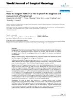
to extract energy to make ATP from glucose is called cellular
respiration (Figure 1.1):
Glucose 1 O2 -CO2 1 H2 O 1 ATP
Key
O2 is consumed and CO2 is produced during aerobic respiration.
The product is energy!
Glucose
Pyruvate
Acetyl-CoA
CO2
Kreb’s
Cycle
NADH+H+ + FADH2
CO2
O2
Oxidative Phosphorylation
ATP + H2O
Figure 1.1 Aerobic cellular respiration is the process through which cells use glucose and oxygen to produce ATP
and H2O, with CO2 as a byproduct of the biochemical reactions.
Cellular Respiration and Diffusion
5
Clinical Correlate
Ischemia
Ischemia is what happens when cells suddenly are unable to receive oxygen and get rid of carbon dioxide. Specifically, the term is used to
describe a loss of blood flow. As we’ll see in later chapters, one of the
main functions of blood is to deliver O2 to tissues and remove carbon
dioxide. When there is no blood flow, there is no O2 delivery, and there
is no CO2 removal. Therefore, cells are no longer able to produce energy,
and they begin to malfunction. One of the best examples of this is myocardial ischemia—a heart attack. When blood flow to a portion of the
heart muscle stops, the heart muscle cells can’t make energy. This causes
inflammation and abnormal functioning of these cells. Common clinical
manifestations of a myocardial infarction are pain and arrhythmias arising from the infarcted tissue.
Role of Carbon Dioxide (CO2)
The amount of CO2 that is in the air we breathe is relatively low, but
inside the body the amount of CO2 is much, much higher. As O2 is
actively being consumed during cellular respiration, CO2 is being produced as a byproduct of the same biochemical pathway (Figure 1.1).
Remember: While O2 is being consumed, CO2 is being produced.
Similar to what happens with O2, the production of CO2 by the cell is
closely linked to metabolism; the higher the metabolic rate, the more
CO2 produced. The major goal of metabolizing food is to break down
the food into its simplest chemical form (usually glucose) and then to
remove hydrogen ions and electrons from it. The removal of hydrogen
ions and electrons will ultimately power an enzyme called ATP
synthase. This enzyme creates ATP, and in doing so creates usable
energy. There are many biochemical reactions involving the removal
of hydrogen ions and electrons from food, and they differ depending
on whether the food is a sugar, a protein, or a fat. Some of these reactions, called decarboxylation reactions, result in the removal of a
carbon atom, and it is from these reactions that CO2 is generated.
CO2 is not a useless byproduct of metabolism though; it has an
extremely important role in the body as an acid base buffer, as we will
see in further chapters. For now, suffice it to say that any excess accumulation of CO2 within the cell is unwanted and could disrupt adequate cellular functioning; therefore, CO2 must be continuously
shuttled outside of the cell.
6
Back to Basics in Physiology
O2
Cell membrane
Glucose
Pyruvate
Kreb’s
Acetyl-CoA Cycle
CO2
NADH+H+ + FADH2
CO2
O2
Oxidative Phosphorylation
ATP + H2O
CO2
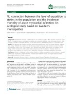
Figure 1.2 In a single cell, O2 moves from the outside of the cell to the inside (white arrow), while CO2 moves
from the inside to the outside of the cell (black arrow).
Single Cell Exchange Requirements
We’ve established that during the aerobic production of ATP, O2 is
consumed and CO2 is produced by the cell (Figure 1.2). Because O2
gets consumed by the cell, it must first be brought to the cell from the
outside, while CO2 is produced and must be shuttled from inside to
outside in order to prevent a toxic accumulation of CO2 inside the cell.
But how do these gases move across the cell membrane? Unlike ions,
which require proteins to be shuttled in and out of the cell due to a
lack of permeability, gases can freely diffuse in and out of the cell.
Because gases are freely diffusible, the only thing that regulates the
movement of O2 and CO2 across the membrane is the pressure difference between both sides of a membrane and the solubility of the gas.
Therefore in order to fully understand the movement of gases between
cells and in the body, a brief review of the basic principles regulating
the behavior of gases in the environment is warranted.
Cellular Respiration and Diffusion
7
REVIEW OF THE PHYSICAL PROPERTIES OF GASES
The physical and chemical properties that guide the diffusion of gases
are far too complex to be entirely reviewed in this book. However, we
will highlight the bare minimum that we believe is essential to understanding the movement of O2 and CO2 in the body. With that in mind,
let us push forward!
There are four fundamental states of matter: solid, liquid, gas,
and plasma. A simple way to define the differences in the states of
matter is to think of the kinetic energy of their molecules. All molecules move constantly, and this movement has the capacity to do
work. Kinetic energy is the energy that that these molecules possess
due to the movement of their molecules. The more kinetic energy,
the more they’re going to move. Solids have the least amount of
kinetic energy and plasma has the highest amount of kinetic energy.
As the kinetic energy increases, molecule movement increases. Sugarladen 4-year-olds running wild at a birthday party 5 high kinetic
energy; the same 4-year-olds asleep after the sugar crash 5 low
kinetic energy. As kinetic energy increases in the molecules that
make up a given compound, it becomes harder for the compound to
keep its shape as the intermolecular bonds weaken from all the
motion. The more kinetic energy the molecules have, the more space
they will occupy and the less likely they are to interact. Solids are
solids because of the stable interactions between molecules. Gases
have a much higher amount of kinetic energy; this means that gas
molecules moving around all over the place take up a lot more
space. (Keeping with our young child analogy, a sleeping child
equaling low kinetic energy does not occupy that much space. A
sugar-crazed toddler running around the house can feel as if no place
is big enough to contain him or her.) Thinking of the matter in this
way (and specifically, gases) leads us to the following point. There
are four basic physical properties that significantly impact the behavior of gases by impacting their molecular kinetic energy in a manner
of speaking:
•
•
•
•
Number of particles
Temperature
Volume
Pressure
8
Back to Basics in Physiology
Key
The key determinants of the behavior of a gas are the number of
particles, its temperature, its volume, and its pressure.
Of these factors let’s take a closer look at pressure, since this will
become relevant when we discuss gas movement in the body. What
exactly is pressure? Pressure is the amount of force that is applied by a
particular compound in a given area. If that compound were a gas, it
would be the force from all those collective collisions banging up
against the sides of, say, a container holding said gas:
Pressure 5
Force
Area
Pressure is therefore a function of the strength between the collisions
of the molecules in the gas and the amount of space these molecules have
to move around in. So how exactly do we quantify pressure? There are
various units that can be used: atmospheres (ATM), Pascals, pounds per
square inch (PSI), Torr, among others. We will be using two particular
units: millimeters of mercury (mmHg) and centimeters of water
(cmH2O). Both of these methods work in a similar fashion (Figure 1.3).
A graduated glass column is filled with either mercury (Hg) or water
(H2O), and it’s connected through an adaptor to wherever you want to
(A)
(B)
Pressure inside
balloon X
Pressure inside
balloon Y
X
Y
Figure 1.3 Displacement of a column of fluid (grey) allows for the measurement of pressures. (A) Low pressures
only make the column of fluid rise slightly. (B) Higher pressure makes the column of fluid rise higher. The pressure, in either mmHg or cmH2O, is the total amount of displacement measured in the column.
Cellular Respiration and Diffusion
9
measure the pressure. The pressure inside the place of interest will
displace the water or mercury a specific distance either up (high pressure)
or down (low pressure). In the case of mercury this is measured in millimeters and in the case of water it is measured in centimeters. The amount
of fluid that ends up getting displaced is measured. It’s typically much
easier to see a liquid than a gas, so this form of measurement has been
historically convenient. Given that mercury has a greater density than
water, mmHg are used for higher pressures (it is harder to displace a
dense liquid so we need higher pressures), and cmH2O are used for lower
pressures (easier to displace a less dense liquid with a lower pressure). So
whenever we mention mmHg or cmH2O, what we are referring to is how
much pressure is in a particular space. There are several pressures that we
are required to memorize: first is the atmospheric pressure at sea level.
This is the standard pressure of air at sea level, which is 760 mmHg. All
further calculations in the book will be based on sea level atmospheric
pressure!
Another concept that requires a brief mention (we’ll touch on it
again in Chapter 2) is that of partial pressures. We mentioned that the
pressure of air at sea level is 760 mmHg. But air is simply a collection
of gases! If we’re thinking of these molecules as individuals, each with
their own weight (oxygen, e.g., is heavier than nitrogen) and their own
size (nitrogen, due to different electron configuration is actually larger
than oxygen), then we can imagine that each individual gas collectively
exerts its own pressure within the air! Thus, a partial pressure is simply
the amount of pressure that an individual gas within a mixture exerts.
For example, we said earlier that O2 makes up 21% of atmospheric
air, but this tells us the relative amount. Without knowing the total
pressure, this number doesn’t help us exactly. We want to know the
amount of oxygen in absolute terms (e.g., its partial pressure in
mmHg). At sea level, where we know that the total air pressure of the
atmosphere is 760 mmHg, we can determine that 21% of this is
160 mmHg. This would be the value of the partial pressure of oxygen
within the atmosphere at sea level. If we were to hypothetically
increase the percentage of oxygen to 40%, but keep the total atmospheric air pressure the same, the partial pressure of O2 would increase
from 160 mmHg to 304 mmHg. Conversely, if we were to move much
higher up away from sea level, where there is less gravitational force
acting on molecules and a lower total air pressure (let’s say 500 mmHg
instead of 760 mmHg), the fraction of inspired O2 (FiO2) will remain
10
Back to Basics in Physiology
the same at 21%, but the ABSOLUTE pressure of O2 will decrease
from 160 mmHg to 105 mmHg. Therefore it is important to consider
both the total pressure and the fractional percentage that each gas
we’re studying represents.
REVIEW OF DIFFUSION AND GRADIENTS
In its simplest terms, diffusion is the movement of substance X from
an area where there is a lot of X to an area where there is not that
much X. When discussing gases, we can talk about a gradient from an
area of high pressure to an area of low pressure along the pressure gradient (Figure 1.4). In our previous book, Back to Basics in Physiology:
Fluids in the Cardiovascular and Renal Systems, we had defined
diffusion as the movement of substance X using the term concentration
rather than pressure. This was the case because we were talking mainly
about solutes and solvents. Since now we are referring to gases we talk
in terms of pressure. Other than pressure, the factors that modify the
diffusion across a semipermeable membrane of any one substance in
particular can be summarized with the following formula:
Diffusion α
ΔP 3 SA 3 sol
pffiffiffiffiffiffiffiffiffiffiffi
dist 3 MW
where:
ΔP 5 (P1ÀP2). The difference in pressures between compartment
1 (P1) and compartment 2 (P2). As you can see this is in the numerator; thus, the greater the pressure difference the greater the diffusion that will take place.
1
2
X
X
X
X
X
X
X
Diffusion gradient of X
X
X
X
X
X
X
Semi-permeable membrane
(permeable to X)
Figure 1.4 Diffusion of X from compartment 1 to compartment 2 follows the gradient that exists between A
(high pressure) and B (low pressure).
Cellular Respiration and Diffusion
11
SA 5 Surface area. How much membrane space is available for
difussion to occur. Again, numerator. The more membrane through
which exchange can occur, the more diffusion will take place.
sol 5 Solubility. Determined by two things: (1) the semipermeable
membrane (e.g., if something is not soluble to the membrane it will
never diffuse across no matter the pressure difference or surface
area) and (2) the states of matter on either side of the membrane
(e.g., is it diffusing from gas to gas? Liquid to gas? Gas to liquid?).
We will approach this particular concept again in the upcoming
chapters, but for now let us cover the highlights. When diffusion of
gases is occurring solely as gases and does not involve liquids, the
only impediment to diffusion will be how permeable the membrane
is to a particular gas (Figure 1.5A). In contrast, when a gas is diffusing from a gas to a liquid (Figure 1.5B), conditions change. The
gas must first dissolve in the liquid before it can diffuse throughout
the liquid. This is when solubility becomes even more important,
because how readily a gas will dissolve (e.g., in water) will have a
large impact on its rate of diffusion. (This explanation applies to
any fluid, but considering that water will be the basis of our discussions, we will continue to discuss solubility of gases in water.)
dist 5 Distance. How much distance is there from one compartment to another? As the distance increases, diffusion decreases
(in this case this variable is in the denominator). This is especially
(A)
(B)
gas
gas
membrane
membrane
gas
liquid
Figure 1.5 The solubility of a gas in a particular liquid will determine its diffusion into and through the liquid.
12
Back to Basics in Physiology
(A)
(B)
gas
gas
membrane
liquid
x
gas
membrane
liquid
gas
2x
Figure 1.6 Distance is one of the key determinants of diffusion. As distance increases (gray area), diffusion
decreases (black arrows). The converse is also true, as the distance decreases, diffusion increases.
important in certain clinical conditions, which we will approach
later in the book. But for now, consider Figure 1.5; the liquid membrane dividing both compartments has a predefined distance (x)
(Figure 1.6A), and as this distance doubles (Figure 1.6B), diffusion
decreases. If distance were to decrease, diffusion would increase
proportionally.
MW 5 Molecular weight. MW of the substance we’re analyzing.
Stated differently, how big is the molecule that is going to diffuse?
The bigger the molecule, the less easily it will diffuse.
These five factors determine diffusion across a semipermeable
membrane. We can further classify them into two groups (Figure 1.7):
1. Factors that favor diffusion; that is, as they increase, diffusion
increases as well:
• ΔP
• Surface Area
• Solubility
2. Factors that oppose diffusion; that is, as they increase, diffusion
decreases:
• Distance
A simplified version of this formula is:
Diffusion α
ΔP 3 SA
dist
Cellular Respiration and Diffusion
13
Diffusion α ΔP x SA
dist
ΔP (P1–P2)
Surface Area
Distance
P1
P2
Figure 1.7 The formula for diffusion has two nonvariable factors: solubility (sol) and MW (molecular weight).
These are intrinsic to each substance and can’t be readily altered. From the remaining three variables, two favor
diffusion (ΔP, and surface area) and one opposes diffusion (distance). From this image we can see that as ΔP or
surface area increases, diffusion (black arrows) increases. However if distance increases, diffusion decreases.
In this simplified version, we have eliminated solubility and molecular weight. This is because solubility can’t be easily altered. In fact, in
the body the solubility of different gases is relatively fixed. Therefore
it’s not going to be a variable that affects diffusion in a significant way
under steady state conditions (although we will see that it does explain
some of the differences we see later between oxygen and carbon dioxide!). Additionally for simplification we will not include MW since it
isn’t exactly something we can modify.
Key
As ΔP and surface area increase, diffusion increases. As distance
increases, diffusion decreases.
This formula explains diffusion throughout the entire body and for
every system. This means that if you understand this formula you will
be able to understand the pathophysiological mechanism of diffusion
problems everywhere in the body.
14
Back to Basics in Physiology
Clinical Correlate
Pathologic Alterations in Diffusion
In many diseases that impair adequate gas exchange, ultimately what is
altered is one of the parameters of our simplified diffusion formula. For
example, pneumonia can fill the lung with inflammatory cells and fluid
can have a significant decrease in the surface area and the ΔP, and
potentially if the pneumonia is very severe, an increase in the distance
through which gases have to diffuse! This is why patients with pneumonia can present with difficulty exchanging O2 and CO2. Likewise patients
with Chronic Obstructive Pulmonary Disease (COPD) can have
decreases in surface area and increased distance, which lead to poor O2
and CO2 exchange. Once we understand the root cause of the disease, we
can try to orient our treatment to reestablish normal function of
the lungs.
DIFFUSION AND THE CELL
After our brief review of diffusion, let’s go back to discussing our single cell example from the previous paragraphs. As we mentioned previously, cells in the body consume O2 and produce CO2. This is
happening in each and every cell. Each single cell is consuming O2 and
therefore, O2 must diffuse through the cell wall from the outside in.
Along the same lines, CO2 is being produced and it must diffuse from
the inside of the cell to the outside. Consider our simplified diffusion
formula:
Diffusion α
ΔP 3 SA
dist
If we’re talking about a single cell, surface area is going to be more
than adequate to allow for both diffusion of O2 and CO2 and the distance; that is, the width of a single cell membrane is not a huge obstacle to diffusion, therefore the most important factor determining the
diffusion of O2 and CO2 in this example is the ΔP (Figure 1.8).
For O2:
• ΔP 5 (P1ÀP2). P1 is outside the cell, and P2 is inside the cell. Given
that O2 is being consumed inside the cell, we can automatically
assume that the pressure of O2 inside the cell is going to be lower
than the pressure of O2 outside. Therefore, if P2 is less than P1, an
Cellular Respiration and Diffusion
15
High Pressure of O2
Cell membrane
O2
Low Pressure of O2
Glucose + O2 → CO2 + H2O + ATP
High Pressure of CO2
CO2
Low Pressure of CO2
Figure 1.8 The diffusion of O2 and CO2 across the cell membrane follows their individual pressure gradients
(see arrows).
O2 gradient is generated from the outside of the cell toward the
inside. This means that O2 will tend to diffuse toward the inside of
the cell.
For CO2:
• ΔP 5 (P1ÀP2). P1 is inside the cell, and P2 is outside the cell. The
situation is reversed in this case. CO2 is being produced; therefore,
we can automatically assume that the pressure of CO2 inside the cell
is higher than outside the cell. This generates a favorable CO2 diffusion gradient from the inside to the outside of the cell.
If we take all of these considerations into account, in the case
of the single cell we can say that the requirements of O2 consumption and CO2 excretion are easily met. However, more complex
organisms are made up of an increasing number of cells, and
as the number of cells increased, so did the metabolic requirements.
Unfortunately, what did not increase was the surface area!
That meant organisms had to develop a way of increasing the surface area that’s available for exchange while maintaining structural
integrity.