Study on the concentration of urine neutrophil gelatinase-associated lipocalin in acute renal failure patients
Bạn đang xem bản rút gọn của tài liệu. Xem và tải ngay bản đầy đủ của tài liệu tại đây (1.76 MB, 7 trang )
JOURNAL OF MILITARY PHARMACO-MEDICINE N09-2017
STUDY ON THE CONCENTRATION OF URINE NEUTROPHIL
GELATINASE-ASSOCIATED LIPOCALIN IN ACUTE RENAL
FAILURE PATIENTS
Pham Ngoc Huy Tuan*; Le Viet Thang**
SUMMARY
Objectives: To evaluate urine neutrophil gelatinase-associated lipocalin (uNGAL) concentration
and its relation with causes, categories, stages and biochemical indexes of acute kidney injury
(AKI) patients. Subjects and methods: A prospective, cross-sectional study in 96 patients with
AKI who admitted to General ICU, Trungvuong Hospital, Hochiminh city from 12 - 2013 to
01 - 2017 and a control group of 51 healthy people. uNGAL had been done in all 96 patients
and healthy people. Results: All of the AKI patients (100%) had uNGAL elevation. The average
concentration of uNGAL in study group (412.26 ng/mL) was significantly higher than in control
group (10.74 ng/mL) with p < 0.001. There was no relationship between AKI causes and uNGAL
concentration with p > 0.05. The concentration of uNGAL was significantly higher in oliguria
group in comparison with non-oliguria group (558.32 ng/mL vs 342.6 ng/mL) with p < 0.005.
Patients’ uNGAL concentrations at the time of ICU admission were significantly related to their
KDIGO stage (p < 0.001). Urinary NGAL had a moderate positive relationship with serum urea
concentration (r = 0.529, p < 0.001) and a strong positive linear relationship with serum creatinine
concentration (r = 0.852, p < 0.001). Conclusion: Urinary NGAL elevation was common in AKI
patients. The concentration of uNGAL depended on category and stage of AKI. It had a moderate
positive relationship with serum urea and strong positive relationship with creatinine concentration.
* Keywords: Acute kidney injury; Urine neutrophil gelatinase-associated lipocalin.
INTRODUCTION
Acute kidney injury is a common and
devastating problem with in-hospital mortality
of 40% to 80% in the intensive care setting
[10]. The traditional blood (creatinine,
blood urea nitrogen) and urine markers of
kidney injury (casts, fractional excretion of
sodium, urinary concentrating ability) that
have been used for decades in clinical
studies for diagnosis and prognosis of AKI
are insensitive and nonspecific and do not
directly reflect injury to kidney cells.
Therefore, early recognition of renal injury
is important and may help prevent further
renal damage and functional impairment.
Neutrophil gelatinase-associated lipocalin
is a small, 23 kDa protein that is an early
biomarker for ischemic, septic or nephrotoxic
kidney injury. It is normally produced at
low levels by the epithelial cells of the
kidney, but it is quickly upregulated in the
thick ascending limb (TAL) of the loop of
Henle and the collecting ducts within three
* Trungvuong Hospital
** 103 Military Hospital
Corresponding author: Pham Ngoc Huy Tuan ()
Date received: 12/09/2017
Date accepted: 22/11/2017
170
JOURNAL OF MILITARY PHARMACO-MEDICINE N09-2017
hours of tubular epithelial injury. Urinary
NGAL (uNGAL) has been evaluated as an
early biomarker of renal tubular damage
in a acute clinical settings such as the
operating room, ICU and emergency
department, and in high-risk procedures
such as cardiac surgery, radio-contrast
injection and after adult and pediatric
kidney and liver transplantation [1, 6, 7, 8, 9].
There is considerable evidence that
compared to increases in serum creatinine,
NGAL detects early or subclinical kidney
injury earlier, and predicts dialysis
requirement and mortality better[1].
In Vietnam, there are lack of studies on
the role of uNGAL in AKI diagnosis and
prognogsis in patients admitted to General
ICU. Therefore, we have conducted this
research with the aim: Evaluation of the
uNGAL concentration and its relation with
causes, categories, stages and some
biochemical indexes of AKI patients.
SUBJECTS AND METHODS
1. Subject.
The study was conducted with a study
group of 96 AKI patients who admitted
to General ICU, Trung Vuong Hospital,
Hochiminh city from 12 - 2013 to 01 - 2017
and a control group of 51 healthy people.
* Excluding criteria: Patients with chronic
kidney failure, did not fit with diagnostic
criteria, did not enough test results, anuria
patients or did not agree to participate in
the study.
2. Methods.
* Study design: A cross-sectional
descriptive study.
* uNGAL measurement: 24-hour urine
was collected. After that, the volume of urine
was measured before collecting 1 mL
sample for testing purpose. uNGAL was
measured by the sandwich ELISA method
using NGAL monoclonal antibody in the
NGAL kit. After that, the sample will be
analyzed by Achitech System of Abbott,
America to measure uNGAL concentration.
* Diagnostic criteria: KDIGO definition
and classification of AKI [5].
- Diagnostic criteria for AKI: Serum
creatinine increases ≥ 0.3 mg/dL (26.4
μmol/L compared to basic creatinine within
48 h or urine volum < 0.6 mL/kg BW/hour
at least 6 hours.
- AKI degree:
+ AKI degree 1: serum creatinine from
< 220 μmol/L.
+ AKI degree 2: serum creatinine from
220 - 353.6 μmol/L.
+ AKI degree 3: ≥ 353.6 μmol/L.
* Statistical analysis:
Statistical analyses were conducted using
SPSS 20.0.
RESULTS AND DISCUSSIONS
Table 1: uNGAL concentration in study
group.
Control
group
(n = 51)
Study
group
(n = 96)
p
X ± SD
10.74 ±
5.18
412.26 ±
324.91
< 0.001
Max
20.28
1292.38
Min
3.32
69.63
Index
uNGAL
(ng/mL)
The average concentration of uNGAL in
study group was 412.26 ng/mL which was
significantly higher than in control group
(10.74 ng/mL) with p < 0.001. The maximum
171
JOURNAL OF MILITARY PHARMACO-MEDICINE N09-2017
and minimum concentration of uNGAL was
1292.38 and 69.63 ng/mL, respectively.
With the range of urinary NGAL from 43.62
to 114.66 ng/mL, all of the AKI patients
(100%) had uNGAL elevation. Study by Au
V also showed that the mean immediate
postoperative uNGAL levels in patients who
developed sustained AKI were 204.8
ng/mL, and significantly higher than those
who had normal renal function (31.9 ±
113 ng/mL) with p < 0.001 [1]. This result
was similar to other studies by Geus H.R,
Makris K, Zappitelli M: there was a
significant higher of uNGAL concentration
in patients who diagnosed AKI compared
with non-AKI patients with p < 0.05 [6, 7, 8].
These differences in uNGAL concentration
were expected because kidney injury
associated with primary renal insults may
be more severe than that in most patients
included in our study, but our patients
were probably more severely ill.
In current clinical practice, the gold
standard for identification and classification
of AKI is dependent on serial serum
creatinine measurements, which are
especially unreliable during acute
changes in kidney function. We identified
uNGAL as one of the most upregulated
genes in the kidney soon after ischemic
injury. NGAL protein was also markedly
induced in kidney tubule cells and easily
detected in the plasma and urine in animal
models of ischemic and nephrotoxic AKI.
The expression of uNGAL protein was
also dramatically increased in kidney
tubules of humans with ischemic, septic,
and post-transplant AKI. Importantly,
NGAL in the urine was found to be an
172
early predictive biomarker of AKI in a
variety of acute clinical settings. Emerging
experimental and clinical evidence indicated
that in the early phases of AKI from
diverse etiologies, NGAL accumulates
within two distinct pools, namely, a renal
and a systemic pool. Gene expression
studies in AKI have clearly demonstrated
rapid and massive upregulation of NGAL
mRNA in the thick ascending limb of
Henle's loop and the collecting ducts, with
resultant synthesis of NGAL protein in the
distal nephron (the renal pool) and secretion
into the urine where it comprises the major
fraction of uNGAL.
This finding also confirms the need for
future research to evaluate uNGAL in
different renal disease subgroups in order
to understand fully how best to use uNGAL
to diagnose AKI.
Table 2: Relation between urine NGAL
concentration and the causes of AKI
(n = 96).
Causes
n
Urine NGAL (ng/mL)
Sepsis (1)
58
415.25 ± 312.44
Bleeding, dehydration (2)
19
388.24 ± 332.46
Cirrhosis (3)
8
614.87 ± 458.23
Shock, heart failure (4)
7
349.58 ± 260.33
Toxic (5)
4
187.48 ± 98.69
p
(1), (2), (3), (4) > 0.05
(5) and others < 0.01
In our study, sepsis was the most
common cause with the proportion of
60.4%. There was no significant difference
between these causes with p > 0.05. Our
result was similar to study by Vaidya D.S:
there was no significant difference between
uNGAL concentration and several causes
JOURNAL OF MILITARY PHARMACO-MEDICINE N09-2017
of AKI in these studies (p > 0.05) [10], but
was different with studies by Di Nardo M
and Geus H.R (there was a significant
higher concentration of uNGAL in septic
AKI patients than non-septic AKI patients
with p < 0.001 [4, 6]. Lipoproteins also
have strong affinity that trigger an innate
immune response. Therefore, it could be
postulated that these circulating ligands
that are linked to tubular epithelial TLR
activation are responsible for the increased
uNGAL concentrations, which we observed
in patients with sepsis. However, there
were no increases in their SCr levels.
However, recent studies in patients with
sepsis, septic shock, and systemic
inflammatory response syndrome has
reported contradictory findings. A possible
explanation for this difference is the
variability of the subject inclusion time
(up to 48 h after ICU admission). Intensive
resuscitation and the administration of
antibiotics may have already occurred
before study inclusion, therefore most likely
inducing rapid changes of uNGAL values.
Table 3: Relation between urine NGAL
concentration and AKI category (n = 96).
Categories
n
%
Urine NGAL
(ng/mL)
Non-anuria
65
67.7
342.60 ± 284.68
Anuria
31
32.3
558.32 ± 358.95
p
< 0.01
In our study, category of anuria occupied
32.3% all of AKI patients. The concentration
of uNGAL was significantly higher in anuria
group compared with non-anuria group
(558.32 ng/mL compared with 342.6 ng/mL)
with p < 0.01. Our findings highlight the
mechanistic insights of NGAL levels
based on the specimens being measured.
Urine NGAL is proposed to derive
predominantly from local renal synthesis
of NGAL in the thick ascending limb of the
loop of Henle and the collecting ducts
when under inflammatory and oxidative
stress. Therefore, the concentration of
uNGAL was directly related to the renal
tubule injury in AKI patients as well as
urine excretion ability.
Table 4: Relation between uNGAL
concentration and stage of AKI (n = 96).
AKI stages
(KDIGO)
n
%
Urine NGAL
(ng/mL)
1
68
70.8
230.58 ± 146.29
2
21
21.9
796.92 ± 147.77
3
7
7.3
1023.20 ± 179.70
pAnova
< 0.001
p1-2, p1-3 < 0.001,
p2-3 = 0.002
According to the KDIGO classification,
the stage 1 AKI in our study made up the
highest proportion (70.8%). Stage 2 and 3
occupied smaller proportion (21.9% and
7.3%, respectively). Our results also pointed
that patients’ uNGAL concentrations at the
time of ICU admission were significantly
related to their KDIGO stage (p < 0.001).
This result was similar to the study by
Geus H.R (p < 0.0001) and Zapittelli M
(p < 0.0002) when research on the relation
between uNGAL and RIFFLE stage [6, 8].
NGAL fulfills a central role in regulating
epithelial neogenesis, and in iron chelation
and delivery after ischemic or toxic insults
to the renal tubular epithelium. After kidney
injury, NGAL is rapidly expressed on the
apical epithelial membranes of the distal
nephron. NGAL is excreted in the urine
173
JOURNAL OF MILITARY PHARMACO-MEDICINE N09-2017
through exocytosis and has local
bacteriostatic and proapoptotic effects.
Therefore, uNGAL concentration had a
positive relation with the level of renal
damage which exhibited throughout the
high stage of KDIGO classification.
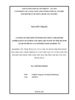
Table 5: Correlation between uNGAL and
serum urea, creatinine concentration (n = 96).
uNGAL
Indexes
Correlation equation
r
p
Urea
0.529
< 0.001
uNGAL = 17.304*urea +
169.141
Creatinine
0.852
< 0.001
uNGAL = 2.616*creatinine
- 150.730
In our study, uNGAL had a moderate
positive relationship with serum urea
concentration (r = 0.529, p < 0.001) and a
strong positive linear relationship with
serum creatinine concentration (r = 0.852,
p < 0.001). Boglignano D also pointed
that a significant correlation was also
found between serum creatinine and
uNGAL (r = 0.399, p < 0.001) [2]. NGAL
has mainly been studied in the setting
of acute renal failure. Patients who
experienced acute renal dysfunction showed
a marked increase in uNGAL levels,
which preceded the increase in serum
creatinine by a day. In a single case of acute
tubular necrosis due to heart failure induced
hypotension, NGAL tubular expression was
reported to be strongly increased [3]. Hence,
measurements of NGAL may serve as a
very early marker of worsening renal function.
Urinary (or plasma) NGAL levels could
therefore be used to adjust therapy, to
anticipate and possibly prevent expected
renal injury, even before a peak in serum
creatinine occurs. This potential of NGAL
needs to be explored further in future
studies.
uNGAL = 17.304 x ure + 169.141
1800
1600
1400
uNGAL
1200
1000
800
600
400
200
0
0
10
20
30
40
50
60
70
Ure
Chart 1: Correlation between urine NGAL and urea concentration.
174
80
JOURNAL OF MILITARY PHARMACO-MEDICINE N09-2017
uNGAL = 2.616 x creatinin - 150.730
2500
uNGAL
2000
1500
1000
500
0
0
100
200
300
400
500
Serum creatinin
600
700
800
900
Chart 2: Correlation between urine NGAL and creatinine concentration.
CONCLUSIONS
In our study, all of the AKI patients
(100%) had urine NGAL elevation. The
average concentration of uNGAL in our
study group (412.26 ng/mL) was significantly
higher than in control group (10.74 ng/mL)
with p < 0.001. There was no significant
difference between AKI causes and
uNGAL concentration with p > 0.05. The
concentration of uNGAL was significantly
higher in oliguria group compared with
non-oliguria group (558.32 ng/mL compared
with 342.6 ng/mL) with p < 0.005. Patients’
uNGAL concentrations at the time of ICU
admission were significantly related to
their KDIGO stage (p < 0.001). Urinary NGAL
had a moderate positive relationship with
serum urea concentration (r = 0.529,
p < 0.001) and a strong positive linear
relationship with serum creatinine concentration
(r = 0.852, p < 0.001).
REFFERENCES
1. Au V et al. Urinary neutrophil gelatinaseassociated lipocalin (NGAL) distinguishes
sustained from transient acute kidney injury
after general surgery. KI reports. 2016, 1 (1),
pp.3-9.
2. Bolignano D et al. Neutrophil gelatinaseassociated lipocalin (NGAL) as a marker of
kidney damage. American Journal of Kidney
Diseases. 2008, 52 (3), pp.595-605.
3. Damman K et al. Urinary neutrophil
gelatinase associated lipocalin (NGAL),
a marker of tubular damage, is increased in
patients with chronic heart failure. European
Journal of Heart Failure. 2008, 10 (10),
pp.997-1000.
175
JOURNAL OF MILITARY PHARMACO-MEDICINE N09-2017
4. Di Nardo M et al. Impact of severe
sepsis on serum and urinary biomarkers of
acute kidney injury in critically Ill children: An
observational study. Blood purification. 2013,
35 (1-3), pp.172-176.
5. Disease K. Improving global outcomes
(KDIGO) acute kidney injury work group: KDIGO
clinical practice guideline for acute kidney
injury. Kidney Int Suppl. 2012, 2, pp.1-138.
6. Geus H.R.H.D et al. Neutrophil gelatinaseassociated lipocalin at ICU admission predicts
for acute kidney injury in adult patients.
American Journal of Respiratory and Critical
Care Medicine. 2011, 183 (7), pp.907-914.
7. Makris K et al. Urinary neutrophil
gelatinase-associated lipocalin (NGAL) as an
176
early marker of acute kidney injury in critically
ill multiple trauma patients, in Clinical Chemistry
and Laboratory Medicine. 2009, p.79.
8. Zappitelli M et al. Urine neutrophil
gelatinase-associated lipocalin is an early
marker of acute kidney injury in critically ill
children: a prospective cohort study. Critical
Care. 2007, 11 (4), p.R84.
9. Chertow G.M et al. Acute kidney injury,
mortality, length of stay, and costs in hospitalized
patients. J Am Soc Nephrol. 2005, 16 (11),
pp.3365-3370.
10. Vaidya V.S et al. Urinary biomarkers
for sensitive and specific detection of acute
kidney injury in humans. Clin Transl Sci. 2008,
1 (3), pp.200-208.