Ebook Oncology in primary care: Part 2
Bạn đang xem bản rút gọn của tài liệu. Xem và tải ngay bản đầy đủ của tài liệu tại đây (9.09 MB, 189 trang )
SECTION
VI
Cancer
Survivorship
(c) 2015 Wolters Kluwer. All Rights Reserved.
CHAPTER
38 Cancer Survivors,
Oncologists, and
Primary Care Clinicians
Kevin C. Oeffinger, MD
KEY POINTS
• The population of cancer survivors is growing rapidly;
many cancer survivors have complex health care needs.
• Risk-based health care of cancer survivors is lifelong care
that integrates the cancer and survivorship experience
in the overall health care needs of the individual and
includes a systematic plan for surveillance and prevention
that incorporates risks based on the previous cancer,
cancer therapy, genetic factors, lifestyle behaviors, and
comorbid health conditions.
• The survivorship care plan is a key component of
risk-based health care.
• Many cancer survivors are lost in transition from active
therapy to posttreatment health care and have many
health care needs that are not addressed.
• The primary care clinician’s role in the care of cancer
survivors is critically important.
strategies that incorporate prevention and early detection.
The Institute of Medicine (IOM) published two seminal
reports on survivors of childhood and adult cancer.2,7 The
latter report, subtitled Lost in Transition, highlighted the
fact that the transition of patients from active cancer therapy
to posttreatment care is often suboptimal.2 Through these
reports, the concept of risk-based health care for cancer survivors was developed. Risk-based health care (Table 38-1)
is an approach to lifelong care that integrates the cancer and
survivorship experience in the overall health care needs of
the individual and includes a systematic plan for surveillance
and prevention that incorporates risks based on the previous cancer, cancer therapy, genetic factors, lifestyle behaviors, and comorbid health conditions. The document that is
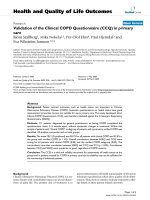
the cornerstone of this process is the survivorship care plan
(SCP), which includes an individualized cancer treatment
summary, information on potential late effects, and guidelines for follow-up care. Figure 38-1 provides an example
TABLE 38-1
Long-term cancer survivors represent a significant proportion
of the US population. Currently, there are more than 12 million cancer survivors; by 2020, it is estimated that this number will increase to 20 million.1 As the number of long-term
survivors has increased, there has been a growing realization that many will develop health conditions as a direct or
an indirect consequence of their cancer therapy.2–6 Although
some of these conditions occur during therapy and persist well
after the therapy has been completed (or become permanent),
such as ifosfamide-induced renal dysfunction or steroidinduced osteonecrosis, many outcomes are not evident until
10 to 20 years later such as second cancers and therapy-related
heart failure or ischemic coronary artery disease. Collectively,
these outcomes are referred to as “late effects.”
Fortunately, the incidence and severity of many late
effects of cancer therapy can be substantially reduced with
Basic Tenets of Risk-Based Health
Care of Cancer Survivors
• Longitudinal care that is considered a continuum from cancer diagnosis to
eventual death regardless of age
• Continuity of care consisting of a partnership between the survivor and a health
care provider who can coordinate necessary services
• Comprehensive, anticipatory, personalized, and proactive care that includes a
systematic plan of prevention and surveillance
• Multidisciplinary team approach with communication between the primary health
care provider, cancer specialists, and allied/ancillary service providers
• Health care of the whole person, not a specific disease or organ system, which
includes the individual’s family and his or her cultural and spiritual values
• Sensitivity to the issues of the cancer experience, including expressed and
unexpressed fears of the survivor and his or her family/spouse
238
(c) 2015 Wolters Kluwer. All Rights Reserved.
Chapter 38 / Cancer Sur vivors, Oncologists, and Primar y Care Clinicians
239
CANCER TREATMENT SUMMARY / SURVIVORSHIP CARE PLAN
Date of preparation: June 14, 2012
Name: Jane Doe
Date of Birth: 7/1/1972
Cancer Diagnosis: Hodgkin lymphoma, nodular sclerosing, stage IV B
Treatment center: Best Cancer Center, USA
Date of diagnosis: 12/1/1998
Age at diagnosis: 26 y
Date of completion of therapy: 7/29/1999
Surgery
Date
Procedure
12/1/1998
Left supraclavicular lymph node biopsy
Radiation Therapy
Date Start
Date Stop
Field
6/1/1999
6/24/1999
Modified mantle (cervical, supraclavicular, infraclavicular,
mediastinal, and left axillary nodes)
7/12/1999
7/29/1999
Spleen and para-aortic lymph nodes
Chemotherapy:
Drug Name
Dose (units or mg/m2)
Doxorubicin
300 mg/m2
Bleomycin
100 U/m2
Vincristine
Etoposide
Prednisone
Cyclophosphamide
4g
Potential Late Effects
Screening Recommendations**
•
•
•
•
•
•
•
Cardiovascular problems
Lung problems
Thyroid problems
Fertility problems
Second cancers
Musculoskeletal problems
Psychosocial problems including
anxiety or depression
•
•
•
•
•
•
•
•
•
Dose (cGy)
2,100
2,100
Complete physical exam every year
Echocardiogram annually
EKG baseline and as clinically indicated
Breast MRI/mammogram annually
DXA baseline and as clinically indicated
Pulmonary function test baseline and as clinically indicated
Annual blood work: CBC, BUN, creatinine, fasting lipids, TSH,
urinalysis
Counseling as needed
Yearly evaluation of skin in radiation field
**Screening recommendations adapted from the
Children’s Oncology Group Long-Term Follow-Up Guidelines
.
For any questions, please contact:
Dr. Mary Doe
Best Cancer Center, Anywhere USA
1111 Main Street, USA
Phone: 888-888-8888
FIGURE 38-1. Example of one-page cancer treatment summary/survivorship care plan. EKG, electrocardiogram; MRI, magnetic resonance imaging; DXA, dual energy X-ray
absorptiometry; CBC, complete blood count; BUN, blood urea nitrogen; TSH, thyroid-stimulating hormone.
(c) 2015 Wolters Kluwer. All Rights Reserved.
240
Oncology in Primar y Care
Pre
CA
Low risk for future cancer-related
health problems:
All of the following:
• Surgery only or chemotherapy that
did not include alkylating agent,
anthracycline, bleomycin, or
epipodophyllotoxin
• No radiation
• Low risk of recurrence
• Mild or no persistent toxicity of
therapy
PCC
CA
DX
Off
RX
2y
Off RX
a
High risk:
Any of the following:
• High-dose radiation
• High-dose alkylating agent,
anthracycline, bleomycin, or
epipodophyllotoxin
• Allogeneic stem cell transplant
• High risk of recurrence
• Multi-organ persistent toxicity of
therapy
10 y
Off RX
c
c
LTFU
Onc
Cancer center *
PCC
Moderate risk:
Any of the following:
• Low- or moderate-dose alkylating
agent, anthracycline, bleomycin,
or epipodophyllotoxin
• Low-to-moderate dose radiation
• Autologous stem cell transplant
• Moderate risk of recurrence
• Moderate persistent toxicity of
therapy
b
5y
Off RX
b
c
c
c
LTFU
Onc
Cancer center *
PCC
a
b
d
d
c
LTFU
Onc
Cancer center *
Communication points with PCC
a. CA DX and planned therapeutic approach, brief overview of chemotherapy, radiation therapy, and/or surgery
b. Survivorship care plan: CA DX, cancer therapy, surveillance recommendations, contact information
c. Periodic update with changes in surveillance recommendations and new information regarding potential late effects
d. Periodic update of survivor’s health for PCC record
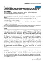
FIGURE 38-2. Risk-stratified shared care model for cancer survivors. Solid line denotes primary responsibility for cancer-related care; PCC continues care to manage noncancer comorbidities and routine preventive health maintenance. *Cancer center or oncologist/oncology group practice; if there is not an LTFU/survivor program available,
care in the gray box is provided by the primary oncologist. CA, cancer; DX, diagnosis; Off RX, completion of cancer therapy; PCC, primary care clinician; LTFU, long-term
follow-up (survivor) program; Onc, oncologist.
(c) 2015 Wolters Kluwer. All Rights Reserved.
Chapter 38 / Cancer Sur vivors, Oncologists, and Primar y Care Clinicians
of a simple one-page SCP. Despite recommendations from
the IOM and numerous other national groups, studies indicate that most survivors do not have an SCP; they are often
unsure about the details of their cancer therapies; and most
community physicians are unaware of their risks. Thus, most
survivors, including those at highest risk, are not engaged in
risk-based follow-up care that is aimed on optimizing their
health and quality of life.
241
The following chapters highlight some of the more serious late effects and key aspects of integrating the health care
needs of the cancer survivor with his or her routine health care
needs. The primary care clinician, with expertise in preventive
care and the management of chronic conditions, is critically
important in this process. Figure 38-2 presents an approach,
stratified by risk, for shared care between the primary care
clinician and the cancer specialist(s).
References
1. Parry C, Kent EE, Mariotto AB, et al. Cancer survivors: a booming population. Cancer Epidemiol Biomarkers Prev. 2011;20(10):1996–2005.
4. Oeffinger KC, Robison LL. Childhood cancer survivors, late effects, and a
new model for understanding survivorship. JAMA. 2007;297(24):2762–2764.
2. Hewitt M, Greenfield S, Stovall E, eds. From Cancer Patient to Cancer
Survivor: Lost in Transition. Washington, DC: Committee on Cancer
Survivorship: Improving Care and Quality of Life, National Cancer Policy
Board, Institute of Medicine, and National Research Council, National
Academies Press; 2006.
5. Bhatia S, Robison LL. Cancer survivorship research: opportunities
and future needs for expanding the research base. Cancer Epidemiol
Biomarkers Prev. 2008;17(7):1551–1557.
3. Ganz PA. Why and how to study the fate of cancer survivors: observations
from the clinic and the research laboratory. Eur J Cancer. 2003;39(15):
2136–2141.
7. Hewitt M, Weinger SL, Simone JV, eds. Childhood Cancer Survivorship:
Improving Care and Quality of Life. Washington, DC: National Academies
Press; 2003.
6. Oeffinger KC, McCabe MS. Models for delivering survivorship care.
J Clin Oncol. 2006;24(32):5117–5124.
(c) 2015 Wolters Kluwer. All Rights Reserved.
CHAPTER
39 Cardiac and Pulmonary
Sequelae of Cancer
and Its Treatment
Jennifer E. Liu, MD, FACC • Kevin C. Oeffinger, MD
KEY POINTS
• Cardiac and pulmonary sequelae are major contributing
factors to serious morbidity and premature mortality
among survivors of cancer.
• Chest (mediastinal) radiation frequently causes ischemic
coronary artery disease. Traditional risk factors increase
this risk and therefore should be aggressively managed.
• Anthracycline therapy frequently causes asymptomatic
left ventricular dysfunction, which occasionally can
progress to overt heart failure.
• Pulmonary disease including pulmonary fibrosis and
restrictive and obstructive lung disease can result from
radiation to the chest and/or bleomycin and other
pulmonary toxic chemotherapeutic agents.
Cardiac and pulmonary disease are the most common noncancer causes of serious morbidity and premature mortality
among long-term survivors of cancer.1–8 Thus, preventive
interventions and identification and management of earlystage disease are essential for the health and well-being
of many survivors of cancer.9–11 The primary care clinician
is integral in this process, particularly for cardiac sequelae,
because most outcomes will not be clinically evident until 10
or 20 years after the cancer therapy.
CARDIAC SEQUELAE
Depending on treatment exposures, there is an excess risk of
ischemic coronary artery disease (CAD), heart failure (HF),
valvular heart disease, arrhythmias, and pericardial disease
(Table 39-1). As illustrated in Figure 39-1, CAD or HF can
result from direct injury to the heart muscle and coronary arteries, respectively. Comorbidities, unhealthy lifestyle behaviors,
and genetic factors interact with treatment exposures and further
increase risk. Alternatively, indirect multifactorial pathways may
lead to CAD. Lastly, patients with cancer often are disconnected
from their primary care provider as they are treated for their cancer and followed for recurrence. This can result in suboptimal
management of traditional cardiovascular risk factors, such as
diabetes and hypertension, hastening the development of CAD.
Ischemic Coronary Artery Disease
Radiation fields that include the mediastinum, often used in
the therapy of Hodgkin and non-Hodgkin lymphoma, can
cause direct injury to the proximal coronary arteries and
accelerate atherosclerotic plaque formation leading to CAD
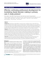
(Fig. 39-2) and myocardial infarction (MI).
Following mediastinal radiation:
• By 20 years, the cumulative incidence of symptomatic
CAD is 21%.12
• By 30 years, the cumulative incidence of MI is 13%.1
• A survivor of cancer with an MI has a threefold increased
risk of dying compared with a noncancer person with an
MI.7 This is because the proximal coronary arteries, including the left main and left anterior descending arteries, are
directly in the field of radiation.
Heart disease risk prediction models are often used in
practice to estimate the 10-year risk of a serious cardiac
event and then intervene with high-risk individuals by targeting risk factors.13,14 Unfortunately, traditional risk prediction
models for cardiovascular disease fail to account for cancer
treatment–related risk factors. Take, for example, a 52-yearold female with a history of Hodgkin lymphoma diagnosed
at the age of 22 years and treated with mediastinal radiation
and chemotherapy, including cyclophosphamide, vincristine,
procarbazine, and prednisone. She is asymptomatic, does not
smoke, has a total cholesterol of 210 mg per dL and a highdensity lipoprotein (HDL) of 44 mg per dL, and a systolic
blood pressure of 138 mm Hg. Using the cardiovascular risk
calculator on the National Heart, Lung, and Blood Institute15
website based on the Framingham Study, her risk is Ͻ1% for
having an MI or coronary death in the next 10 years. However,
242
(c) 2015 Wolters Kluwer. All Rights Reserved.
Chapter 39 / Cardiac and Pulmonar y Sequelae of Cancer and Its Treatment
TABLE 39-1
243
Cancer Therapies Associated with Cardiac Sequelae
Antitumor Class/Drug
Most Frequent Toxicity
Comments
LV dysfunction/HF
Toxicity can be acute (within 24 hr), chronic (within 1 y) or late onset (after 1 y).
Antitumor antibiotic
Anthracycline
Doxorubicin
Daunorubicin
Idarubicin
Monoclonal antibodies/small molecule inhibitors
Trastuzumab
LV dysfunction/HF
Increased incidence when combined with anthracyclines. Toxicity is not dose dependent
and usually reversible.
Bevacizumab
MI, CVA, HF
Increased toxicity in age Ͼ65 y and preexisting CVD.
Hypertension
Sunitinib
Hypertension, HF
Tyrosine kinase inhibitor that targets the vascular endothelial growth factor pathway; potential
for LV dysfunction recovery with interruption of drug and initiation of cardiac treatment
Acute ischemic events, CAD, MI
Increased risk of CAD in male germ cell tumor survivors treated with cisplatin.
Platinum agents
Cisplatin
Arrhythmia, hypertension, HF
Antimicrotubules
Paclitaxel
Bradyarrhythmia, HF, ischemia
Increased risk of LV dysfunction when combined with anthracycline.
Fluorouracil
Myocardial ischemia/MI
Possibly secondary to vasospasm; risk increased with co-existing CAD and concomitant
cisplatin therapy
Capecitabine
Same as mentioned previously
Radiotherapy
Myocardial fibrosis with restrictive heart
disease, valvular disease, accelerated
atherosclerosis, pericardial disease
Antimetabolites
Atrial or ventricular arrhythmia
Cardiac effects worsen over time with long latency between exposure and onset of symptoms
LV, left ventricular; HF, heart failure; MI, myocardial infarction; CVA, cerebrovascular accident; CVD, cardiovascular disease; CAD, coronary artery disease.
because of her mediastinal radiation, we know that her
10-year risk of MI or coronary death has been substantially
underestimated—based on available evidence, her risk is 10%
to 15%.16 Despite an apparently low-risk profile based on a
traditional risk calculator, this patient’s cancer treatment history necessitates aggressive risk-reducing measures to prevent
a serious coronary event. This vignette illustrates the lack of
appropriate tools available to clinicians when managing longterm survivors of cancer. Current studies are in progress to
develop risk prediction models for survivors of cancer.
Ischemic CAD can result from indirect pathways. For
example, therapy for childhood acute lymphoblastic leukemia
Lifestyle: tobacco, alcohol,
diet, physical activity
results in obesity, insulin resistance, decreased levels of
physical activity, and ultimately to increased rates of CAD.17
Cisplatin-based chemotherapy used in the treatment of men
with testicular cancer has been associated with an increased
risk of CAD and MI. This may be the result of direct endothelial damage caused by cisplatin and/or increasing the risk of
developing hypertension and lipid abnormalities.18–24
To date, studies of the use of stress exercise testing, echocardiography, and radionucleotide imaging to screen for
obstructive CAD in asymptomatic survivors have been inconclusive.8 Stress echocardiography appears to be more sensitive and specific than other methods.25 However, this area of
Therapy: mediastinal/neck radiation, anthracyclines,
alkylating agents, stem cell transplantation
Cardiovascular Outcomes:
coronary artery disease/
myocardial infarction,
cardiomyopathy, heart failure,
stroke
Biology/Genetics
Comorbidities: diabetes, hypertension, dyslipidemia, renal disease
FIGURE 39-1. Factors associated with cardiac sequelae in survivors of cancer.
(c) 2015 Wolters Kluwer. All Rights Reserved.
244
Oncology in Primar y Care
FIGURE 39-2. A 39-year-old man who was treated for Hodgkin lymphoma
25 years ago with 45 Gy mantle field radiation. The curved reconstruction of coronary computed tomography (CT) angiogram shows two areas of severe stenosis (straight arrows) in left anterior descending coronary artery (LAD) and multiple
plaques (arrowhead). More distal LAD has relatively wide diameter and might
represent normal vessel or region of ectasia (curved arrow). (From Rademaker J,
Schoder H, Ariaratnam NS, et al. Coronary artery disease after radiation therapy for
Hodgkin lymphoma: coronary CT angiography findings and calcium scores in nine
asymptomatic patients. AJR Am J Roentgenol. 2008;191:32–37, with permission.)
research is limited because of the relatively small number of
survivors available for study. Because of the substantially
heightened risk of CAD and elevated risk of death from an MI
among pediatric and young adult survivors of cancer treated
with high-dose mediastinal radiation (Ն40 Gy), the Children’s
Oncology Group recommends consideration of cardiology
consultation 5 to 10 years after radiation.26
Regardless, studies consistently emphasize the importance
of modifiable traditional cardiovascular risk factors.1,2,7,8
Smoking and comorbid hypertension, dyslipidemia, and diabetes mellitus substantially increase the risk of ischemic CAD
in individuals treated with mediastinal radiation. Thus, the
primary care clinician’s role in the care of survivors of cancer is critically important. As with other high-risk populations
(i.e., patients with type 2 diabetes), it is essential that the clinician screen for and manage hypertension, lipid disorders, and
diabetes and implement strategies for smoking cessation or
increasing level of physical activity as necessary.
Left Ventricular Dysfunction and Heart Failure
Anthracycline chemotherapy (e.g., doxorubicin, daunorubicin, epirubicin) is an important component in the treatment
of several types of cancer including breast, lung, endometrial,
and ovarian cancer; lymphoma; leukemia; and sarcoma. In a
seminal study, von Hoff et al.27 reported that anthracyclineinduced cardiac injury is characterized by dose-dependent and
progressive left ventricular (LV) dysfunction, which can lead
to HF, developing within 1 year of treatment in 3% of patients
treated with a cumulative dose of 400 mg per m2 of doxorubicin, 7% at 550 mg per m2, and 18% at 700 mg per m2. Subsequent studies have established that anthracycline-induced
LV dysfunction is common, risk increases with increasing
interval from therapy, and can occur even with low cumulative doses.28–35 Although the incidence of overt HF is low
with conventional regimens, subclinical echocardiographic
abnormalities of LV structure and function has been reported
in more than half of patients in the first few years after anthracycline exposure and the abnormalities worsened over time.
Importantly, HF can develop a decade or two after completion
of the anthracycline therapy. Risk factors for anthracyclineinduced HF include young age at therapy, cumulative doxorubicin dose, rate of administration, concurrent mediastinal
or chest radiation, female gender, preexisting heart disease,
and hypertension. Recent studies have identified modifying genetic factors associated with anthracycline-related
cardiomyopathy.36–38
The primary care clinician is an important member of the
team for patients who may be treated with anthracycline chemotherapy as well as those who have completed their therapy.
Before a patient starts on potentially LV cardiotoxic therapy,
risk stratification should be formulated based on treatmentrelated factors (type of drug, cumulative dose, combination
of potentially cardiotoxic treatment) and patient-specific risk
factors (age, coexisting cardiovascular conditions, and prior
history of cardiotoxic treatment). In high-risk patients, there
should be a discussion between the oncologist, the primary
care clinician, and a cardiologist assessing the oncologic
benefit of treatment and possible adverse cardiac risk, with
consideration of cardioprotective measures or alteration of
the treatment. Optimization of the cardiovascular status (e.g.,
management of hypertension) prior to initiation of chemotherapy is recommended with close cardiac monitoring during
treatment so that an intervention can be initiated as soon as
signs of cardiotoxicity are detected. The American College of
Cardiology (ACC)/American Heart Association (AHA) recommend echocardiographic monitoring in patients who are at
risk for HF (class I indication).39
For children, adolescents, and young adults who have
completed anthracycline-based chemotherapy, the Children’s
Oncology Group has developed evidence-informed recommendations for screening.26 The frequency of monitoring is
based on cumulative anthracycline dose, age at exposure, and
whether or not the patient was treated with chest radiation.
Guidelines for posttherapy cardiac screening and follow-up
in asymptomatic survivors of adult cancer have not been
established.8
The most common method for monitoring LV function
during or after cancer therapy is measurement of LV ejection fraction (LVEF) either by echocardiography or multigated acquisition (MUGA) scan. Other newer methods
include cardiac magnetic resonance imaging (MRI) and
3-D echocardiography (Table 39-2). Because a broad range
LVEF can be seen in healthy individuals, changes in LVEF
indicative of cardiac damage can be identified only when
comparison between serial studies and pretreatment study
are made. Cardiotoxicity in recent major clinical trials has
been defined as reduction of LVEF Ͼ5% to Ͻ55% with
symptoms of HF or an asymptomatic reduction of LVEF of
Ͼ10% to Ͻ55%.
The natural history of anthracycline-induced LV dysfunction and its response to modern HF therapy has not
been well established. Mortality rates up to 50% within 2
years of diagnosis have been reported in the past, which is
worse than many other forms of cardiomyopathy.40 Although
ACC/AHA has published evidence-based treatment guidelines
(c) 2015 Wolters Kluwer. All Rights Reserved.
Chapter 39 / Cardiac and Pulmonar y Sequelae of Cancer and Its Treatment
TABLE 39-2
245
Assesment of Cardiac Function
Methods
Advantages
Disadvantages
MUGA scan
Reproducible LVEF measurement with low interobserver and
intraobserver variability
Radiation exposure; limited information on cardiac structure and
diastolic function
2-D echocardiography
Low cost, easy to perform and widely available; no radiation
exposure; comprehensive evaluation of cardiac structure and
function
High intraobserver and interobserver variability of LVEF
calculation because of dependency on image quality,
geometric assumption, and operator expertise. May fail to
detect subtle changes in LVEF
3-D echocardiography
Same as 2-D echo; highly reliable LVEF calculation
Limited data on its use in monitoring cardiotoxicity; not yet
incorporated into routine clinical practice
MRI
Accurate and reliable assessment of LVEF; gold standard in the
measurement of LV volume, structure, and systolic function; can
detect myocardial fibrosis and scarring when combined with late
gadolinium contrast enhancement
High cost and not widely available
MUGA, multigated acquisition scan; LVEF, left ventricular ejection fraction; MRI, magnetic resonance imaging; LV, left ventricular.
for HF in general,41,42 the effectiveness of therapy in anthracycline-related HF has not been well established. Given the
well-established final common pathway of remodeling and
compensation in systolic HF, treatment for chemotherapyrelated LV dysfunction based on current HF management
guideline is recommended.
Valvular Heart Disease and Arrhythmias
Mediastinal (chest radiation) occasionally causes valvular
heart disease, predominantly involving the aortic and mitral
valves.43 About 6% of survivors treated with moderate- to
high-dose mediastinal radiation develop clinically significant
valvular disease and have an eightfold higher likelihood of
valve surgery.44 Evaluation for and monitoring of valvular
heart disease in survivors treated with mediastinal radiation
can be accomplished with periodic echocardiography.8,26
Importantly, survivors of cancer with valvular heart disease
following mediastinal radiation have a higher incidence of
perioperative morbidity.45
Life-threatening arrhythmias, including complete heart
block, are rare outcomes following cancer therapy and are
generally attributable to mediastinal radiation. Prolongation
of QTc infrequently occurs following anthracycline therapy.46
As in the general population, the patient should be counseled
about the use of medications that may prolong the QT interval
such as antifungal agents and metronidazole.
PULMONARY SEQUELAE
Cancer therapy–related pulmonary sequelae include restrictive and obstructive lung disease and pulmonary fibrosis.
In addition, patients with cancer treated with a hematopoietic stem cell transplant may develop an array of pulmonary
problems, as described in Chapter 46. In contrast to cardiac
outcomes, most pulmonary sequelae develop either during
therapy or soon thereafter.
Dose-related bleomycin-induced pneumonitis has long
been recognized. With contemporary therapy for germ cell
tumors in men, this outcome is very uncommon because of
limits in the total dose of bleomycin.47–49 Other chemotherapeutic agents that are associated with pulmonary disease
include busulfan, carmustine, and lomustine. Combination of
pulmonary toxic chemotherapy with chest radiation increases
the risk of pulmonary disease. Survivors of Hodgkin lymphoma treated with chest radiation in combination with bleomycin frequently have pulmonary problems; fortunately, these
are generally mild to moderate in severity.50,51
The natural history of treatment-related pulmonary disease, particularly 10 years or more after therapy, is not well
described. Thus, the optimum frequency and duration of
monitoring pulmonary function is not known.8 As previously mentioned, it is imperative that survivors of cancer
treated with potentially pulmonary toxic therapy avoid or
stop smoking.
References
1. Aleman BM, van den Belt-Dusebout AW, De Bruin ML, et al. Late cardiotoxicity after treatment for Hodgkin lymphoma. Blood. 2007;109:
1878–1886.
3. Chapman JA, Meng D, Shepherd L, et al. Competing causes of death
from a randomized trial of extended adjuvant endocrine therapy for
breast cancer. J Natl Cancer Inst. 2008;100:252–260.
2. Aleman BM, van den Belt-Dusebout AW, Klokman WJ, et al. Long-term
cause-specific mortality of patients treated for Hodgkin’s disease. J Clin
Oncol. 2003;21:3431–3439.
4. Hooning MJ, Aleman BM, van Rosmalen AJ, et al. Cause-specific mortality in long-term survivors of breast cancer: a 25-year follow-up study.
Int J Radiat Oncol Biol Phys. 2006;64:1081–1091.
(c) 2015 Wolters Kluwer. All Rights Reserved.
246
Oncology in Primar y Care
5. Mertens AC, Liu Q, Neglia JP, et al. Cause-specific late mortality among
5-year survivors of childhood cancer: the Childhood Cancer Survivor
Study. J Natl Cancer Inst. 2008;100:1368–1379.
6. Ng AK, Bernardo MP, Weller E, et al. Long-term survival and competing
causes of death in patients with early-stage Hodgkin’s disease treated at
age 50 or younger. J Clin Oncol. 2002;20:2101–2108.
26. Shankar SM, Marina N, Hudson MM, et al. Monitoring for cardiovascular disease in survivors of childhood cancer: report from the Cardiovascular Disease Task Force of the Children’s Oncology Group. Pediatrics.
2008;121:e387–e396.
27. Von Hoff DD, Layard MW, Basa P, et al. Risk factors for doxorubicininduced congestive heart failure. Ann Intern Med. 1979;91:710–717.
7. Swerdlow AJ, Higgins CD, Smith P, et al. Myocardial infarction mortality risk after treatment for Hodgkin disease: a collaborative British cohort
study. J Natl Cancer Inst. 2007;99:206–214.
28. Du XL, Xia R, Burau K, et al. Cardiac risk associated with the receipt
of anthracycline and trastuzumab in a large nationwide cohort of older
women with breast cancer. Med Oncol. 2010:1998–2005.
8. Carver JR, Shapiro CL, Ng A, et al. American Society of Clinical Oncology clinical evidence review on the ongoing care of adult cancer survivors: cardiac and pulmonary late effects. J Clin Oncol. 2007;25:
3991–4008.
29. Du XL, Xia R, Liu C, et al. Cardiac toxicity associated with anthracyclinecontaining chemotherapy in older women with breast cancer. Cancer.
2009;115:5296–5308.
9. Hewitt M, Greenfield S, Stovall E, eds. From Cancer Patient to Cancer
Survivor: Lost in Transition. Washington, DC: Committee on Cancer
Survivorship: Improving Care and Quality of Life, National Cancer
Policy Board, Institute of Medicine, and National Research Council,
National Academies Press; 2005.
10. Oeffinger KC, Hudson MM, Landier W. Survivorship: childhood cancer
survivors. Prim Care. 2009;36:743–780.
11. Oeffinger KC, McCabe MS. Models for delivering survivorship care.
J Clin Oncol. 2006;24:5117–5124.
12. Reinders JG, Heijmen BJ, Olofsen-van Acht MJ, et al. Ischemic heart
disease after mantlefield irradiation for Hodgkin’s disease in long-term
follow-up. Radiother Oncol. 1999;51:35–42.
13. Mosca L, Benjamin EJ, Berra K, et al. Effectiveness-based guidelines for the prevention of cardiovascular disease in women—2011
update: a guideline from the American Heart Association. Circulation.
2011;123:1243–1262.
14. U.S. Preventive Services Task Force. Aspirin for the prevention of cardiovascular disease: U.S. Preventive Services Task Force recommendation statement. Ann Intern Med. 2009;150:396–404.
15. National Heart Lung and Blood Institute. />Accessed April 10, 2012.
16. Aleman BMP, van den Belt-Dusebout AW, De Bruin ML, et al.
Late cardiotoxicity after treatment for Hodgkin lymphoma. Blood.
2007;109:1878–1886.
17. Oeffinger KC. Are survivors of acute lymphoblastic leukemia (ALL)
at increased risk of cardiovascular disease? Pediatr Blood Cancer.
2008;50:462–467; discussion 468.
18. Feldman DR, Bosl GJ, Sheinfeld J, et al. Medical treatment of advanced
testicular cancer. JAMA. 2008;299:672–684.
19. Haugnes HS, Aass N, Fossa SD, et al. Components of the metabolic
syndrome in long-term survivors of testicular cancer. Ann Oncol.
2007;18:241–248.
20. Haugnes HS, Aass N, Fossa SD, et al. Predicted cardiovascular mortality and reported cardiovascular morbidity in testicular cancer survivors.
J Cancer Surviv. 2008;2:128–137.
21. Haugnes HS, Wethal T, Aass N, et al. Cardiovascular risk factors and
morbidity in long-term survivors of testicular cancer: a 20-year follow-up
study. J Clin Oncol. 2010;28:4649–4657.
22. van den Belt-Dusebout AW, Nuver J, de Wit R, et al. Long-term risk
of cardiovascular disease in 5-year survivors of testicular cancer. J Clin
Oncol. 2006;24:467–475.
30. Lipshultz SE, Colan SD, Gelber RD, et al. Late cardiac effects of doxorubicin therapy for acute lymphoblastic leukemia in childhood. N Engl J
Med. 1991;324:808–815.
31. Lipshultz SE, Lipsitz SR, Sallan SE, et al. Chronic progressive cardiac
dysfunction years after doxorubicin therapy for childhood acute lymphoblastic leukemia. J Clin Oncol. 2005;23:2629–2636.
32. Pinder MC, Duan Z, Goodwin JS, et al. Congestive heart failure in older
women treated with adjuvant anthracycline chemotherapy for breast
cancer. J Clin Oncol. 2007;25:3808–3815.
33. Sawaya H, Sebag IA, Plana JC, et al. Early detection and prediction of
cardiotoxicity in chemotherapy-treated patients. Am J Cardiol. 2011;107:
1375–1380.
34. Swain SM, Whaley FS, Ewer MS. Congestive heart failure in patients
treated with doxorubicin: a retrospective analysis of three trials. Cancer.
2003;97:2869–2879.
35. van Dalen EC, van der Pal HJ, Kok WE, et al. Clinical heart failure in
a cohort of children treated with anthracyclines: a long-term follow-up
study. Eur J Cancer. 2006;42:3191–3198.
36. Blanco JG, Sun CL, Landier W, et al. Anthracycline-related cardiomyopathy after childhood cancer: role of polymorphisms in carbonyl
reductase genes—A report from the Children’s Oncology Group. J Clin
Oncol. 2011.
37. Visscher H, Ross CJ, Rassekh SR, et al. Pharmacogenomic prediction of
anthracycline-induced cardiotoxicity in children. J Clin Oncol. 2011.
38. Wojnowski L, Kulle B, Schirmer M, et al. NAD(P)H oxidase and multidrug resistance protein genetic polymorphisms are associated with
doxorubicin-induced cardiotoxicity. Circulation. 2005;112:3754–3762.
39. Cheitlin MD, Armstrong WF, Aurigemma GP, et al. Guideline update
for the clinical application of echocardiography—summary article: a
report of the American College of Cardiology/American Heart Association Task Force on Practice Guidelines (ACC/AHA/ASE Committee to
Update the 1997 Guidelines for the Clinical Application of Echocardiography). J Am Coll Cardiol. 2003;42:954–970.
40. Felker GM, Thompson RE, Hare JM, et al. Underlying causes and longterm survival in patients with initially unexplained cardiomyopathy.
N Engl J Med. 2000;342:1077–1084.
41. Hunt SA, Abraham WT, Chin MH, et al. 2009 Focused update incorporated into the ACC/AHA 2005 guidelines for the diagnosis and management of heart failure in adults. A report of the American College
of Cardiology Foundation/American Heart Association Task Force
on practice guidelines developed in collaboration with the International Society for Heart and Lung Transplantation. J Am Coll Cardiol.
2009;53:e1–e90.
24. Vaughn DJ, Palmer SC, Carver JR, et al. Cardiovascular risk in long-term
survivors of testicular cancer. Cancer. 2008;112:1949–1953.
42. Hunt SA, Baker DW, Chin MH, et al. ACC/AHA guidelines for the evaluation and management of chronic heart failure in the adult: executive
summary. A report of the American College of Cardiology/American
Heart Association Task Force on practice guidelines (Committee to
revise the 1995 guidelines for the evaluation and management of heart
failure). J Am Coll Cardiol. 2001;38:2101–2113.
25. Heidenreich PA, Schnittger I, Strauss HW, et al. Screening for coronary
artery disease after mediastinal irradiation for Hodgkin’s disease. J Clin
Oncol. 2007;25:43–49.
43. Adams MJ, Lipsitz SR, Colan SD, et al. Cardiovascular status in longterm survivors of Hodgkin’s disease treated with chest radiotherapy.
J Clin Oncol. 2004;22:3139–3148.
23. Feldman DR, Schaffer WL, Steingart RM. Late cardiovascular toxicity
following chemotherapy for germ cell tumors. J Natl Compr Canc Netw.
2012;10:537–544.
(c) 2015 Wolters Kluwer. All Rights Reserved.
Chapter 39 / Cardiac and Pulmonar y Sequelae of Cancer and Its Treatment
247
44. Hull MC, Morris CG, Pepine CJ, et al. Valvular dysfunction and
carotid, subclavian, and coronary artery disease in survivors of hodgkin
lymphoma treated with radiation therapy. JAMA. 2003;290:2831–2837.
48. Loehrer PJ Sr, Johnson D, Elson P, et al. Importance of bleomycin in
favorable-prognosis disseminated germ cell tumors: an Eastern Cooperative Oncology Group trial. J Clin Oncol. 1995;13:470–476.
45. Chang AS, Smedira NG, Chang CL, et al. Cardiac surgery after mediastinal radiation: extent of exposure influences outcome. J Thorac Cardiovasc Surg. 2007;133:404–413.
49. Nichols CR, Catalano PJ, Crawford ED, et al. Randomized comparison of cisplatin and etoposide and either bleomycin or ifosfamide in treatment of advanced disseminated germ cell tumors: an
Eastern Cooperative Oncology Group, Southwest Oncology Group,
and Cancer and Leukemia Group B Study. J Clin Oncol. 1998;16:
1287–1293.
46. Gupta M, Thaler HT, Friedman D, et al. Presence of prolonged dispersion of qt intervals in late survivors of childhood anthracycline therapy.
Pediatr Hematol Oncol. 2002;19:533–542.
47. de Wit R, Roberts JT, Wilkinson PM, et al. Equivalence of three or four
cycles of bleomycin, etoposide, and cisplatin chemotherapy and of a
3- or 5-day schedule in good-prognosis germ cell cancer: a randomized study of the European Organization for Research and Treatment of
Cancer Genitourinary Tract Cancer Cooperative Group and the Medical
Research Council. J Clin Oncol. 2001;19:1629–1640.
50. Duggan DB, Petroni GR, Johnson JL, et al. Randomized comparison of ABVD and MOPP/ABV hybrid for the treatment of advanced
Hodgkin’s disease: report of an intergroup trial. J Clin Oncol. 2003;21:
607–614.
51. Lund MB, Kongerud J, Nome O, et al. Lung function impairment in
long-term survivors of Hodgkin’s disease. Ann Oncol. 1995;6:495–501.
(c) 2015 Wolters Kluwer. All Rights Reserved.
CHAPTER
40 Bone Health
Susan Hong, MD, MPH • Marina Rozenberg, MD • Kevin C. Oeffinger, MD
KEY POINTS
• Cancer and cancer therapy can cause a failure to reach
peak bone mass and/or accelerate bone loss via several
mechanisms.
• Bone density evaluation should be considered for children
and adolescents treated with cancer therapies that prevent
peak bone mass and for all survivors of cancer treated
with therapies associated with accelerated bone loss.
• For individuals younger than the age of 50 years, z scores
are used to assess bone mineral density.
• Recommendations for initiation of antiresorptive therapy
for survivors of adult cancer are similar to persons without
a history of cancer.
• Referral to an endocrinologist should be considered for
survivors of childhood cancer with very low bone mass
( z score Յ Ϫ2.5).
Osteoporosis is a systemic disorder of the skeletal system
characterized by low bone mass and deterioration in the
bone tissue microarchitecture leading to an increased risk
of bone fractures.1 Cancer and cancer treatments often
negatively impact bone health, resulting in higher rates of
osteoporosis and subsequent fractures among survivors of
cancer.
Bone remodeling continues throughout life. Peak bone
mass is achieved by 18 to 20 years of age. After the age
of 35 years, bone resorption exceeds formation. Adequate
bone mineralization is crucial to bone health and is dependent on vitamin D, calcium, magnesium, phosphorus, and
other trace elements.2 Important factors in bone remodeling include the receptor activator for nuclear factor B
(RANK) pathway, which stimulates bone resorption via
osteoclast activation, and hormones such as estrogen and
growth hormone (GH). Estrogen inhibits osteoclast-driven
resorption and promotes bone formation by stimulating osteoblast activity.3 In males, estrogen is formed by
the aromatization of testosterone and is thus dependent
on adequate testosterone levels.4 Adequate levels of GH
are essential for bone density acquisition in children and
adolescents. Thus, bone remodeling involves a complex
network of cells and signals, which, if disrupted, can negatively impact bone health.
Childhood cancer and its treatment coincide with a vital
period of bone growth, interfering with the acquisition of
maximal bone density and leading to increased bone loss
via several mechanisms (Table 40-1). The Children’s Oncology Group (COG) provides updated evidence-based guidelines for screening for early- and late-occurring sequelae
following therapy for pediatric cancer, including bonerelated morbidity.5 Table 40-2 provides a synopsis of these
recommendations.
Survivors of adult cancer are at increased risk for accelerated bone loss through several mechanisms (Table 40-3).
The American Society of Clinical Oncology (ASCO) and
the National Comprehensive Cancer Network (NCCN) recommend monitoring of bone mineral density (BMD) in men
and women who have undergone cancer therapy that negatively impacts bone health. Dual-energy X-ray absorptiometry
(DEXA) scans are used to measure BMD; however, there are
limitations with this approach in children.
Lifestyle modification is recommended for everyone
regardless of BMD (Table 40-4). The World Health Organization (WHO) fracture risk algorithm (FRAX) calculates
the 10-year probability of hip and major osteoporotic fracture risk. The NCCN Task Force on Bone Health in Cancer
Care recommends using the WHO FRAX algorithm to
assess baseline fracture risk for all patients with cancer at
risk for bone loss.6 Pharmacotherapy is generally indicated
for patients with osteoporosis or a history of fragility fractures (Tables 40-2 and 40-5). As in persons without a history of cancer, bisphosphonates are usually first line to treat
bone loss when clinically indicated. Denosumab is a newly
U.S. Food and Drug Administration (FDA)–approved
monoclonal antibody that interferes with RANK ligand
binding and is also approved to treat bone loss.7 Teriparatide is a recombinant human parathyroid hormone, which
can be used to build bone in individuals with severe osteoporosis. It is seldom used in survivors of cancer because
of concerns about the risk of subsequent osteosarcoma.8
Treatment for survivors of childhood cancer with bisphosphonates may be considered, but evaluation with an endocrinologist is recommended.
248
(c) 2015 Wolters Kluwer. All Rights Reserved.
Chapter 40 / Bone Health
TABLE 40-1
249
Childhood Cancer Therapy Associated with Reduced Bone Mineral Density
Therapy
Used for
Mechanism of Bone Loss
Corticosteroids
Supportive therapy, chemotherapy
(ALL, NHL, HSCT)
Impair osteoblastic function and differentiation
Interfere with GH
Impair calcium absorption
Increased risk of bone loss if total prednisone equivalent dose is Ն9 g/m2.8,9
Methotrexate
Ewing sarcoma
Directly toxic to osteoblasts
Osteosarcoma
Increases osteoclast formation
NHL
Total dose Ն40 g/m2 is associated with highest risk for osteopenia.9
Leukemias (ALL)
Alkylating agents
HSCT
Confer risk of premature menopause/ovarian failure/Leydig cell dysfunction
Cyclophosphamide
Hodgkin lymphoma
Concurrent radiation potentiates gonadal toxicity.10
Ifosfamide
Ewing sarcoma
Busulfan
Osteosarcoma
Rhabdomyosarcoma
Radiation therapy
Cranial radiation
Brain tumors, ALL
Doses Ն18 Gy to the HPA associated with GH deficiency.
Doses Ն40 Gy to the HPA may cause gonadotropin deficiency.10
Radiation to abdomen & pelvis
or TBI
Hodgkin lymphoma
Prepubertal girls Ն10 Gy
Neuroblastoma
Pubertal girls Ն5 Gy
Wilms tumor
Males Ն20 Gy
HSCT
Surgical castration
Testicular cancer
Ovarian failure/estrogen deficiency and Leydig cell dysfunction/androgen deficiency10
Pelvic sarcoma
Rapid loss of androgens result in loss of estrogen
Orchiectomy
ALL, acute lymphoblastic leukemia; NHL, non-Hodgkin lymphoma; HSCT, hematopoietic stem cell transplant; GH, growth hormone; Gy, gray; HPA, hypothalamic-pituitary-adrenal axis;
TBI, total body irradiation.
TABLE 40-2
Evaluation and Management of Bone Health in Childhood Cancer Survivors
Recommendations
BMD testing
Initiate 2 y after completion of cancer therapy
For patients who received therapies that have negative impact on
bone health (see Table 40-1)
For individuals Ͻ50 y, use z scores, which compares measured BMD to BMD of age-, gender-,
and ethnicity-matched controls.
Based on z-score results
Normal (z score Ͼ Ϫ1.0)
If not at risk for ongoing bone loss, consider stopping until menopause. Consider restarting
screening if clinically indicated.
Osteopenia (Ϫ1.0 Ն; z Ͼ Ϫ2.5)
Repeat as clinically indicated—usually every 2 y
Osteoporosis (z Յ Ϫ2.5 or fragility fracture, i.e., a fracture that
results from a fall from a standing height or less)
Refer to endocrinology for consideration of possible contributing factors for severe bone loss.
Consider treatments when appropriate. Repeat BMD as clinically indicated (usually every 2 y)
BMD, bone mineral density.
(c) 2015 Wolters Kluwer. All Rights Reserved.
250
Oncology in Primar y Care
TABLE 40-3
Adult Cancer Therapy Associated with Reduced Bone Mineral Density
Therapy
Used in
Mechanism of Bone Loss
Degree and Site of Bone Loss
Aromatase inhibitors
Hormone sensitive breast
cancer
Inhibit peripheral conversion of androgen to estrogen (reduces
estrone sulfate, estradiol, estrogen)
↓ 4.1% in LS after 2 y11
Tamoxifen
Premenopausal hormone
sensitive breast cancer
Potentially interferes with estrogen action on bone when used in
premenopausal women but not in postmenopausal women
↓ 1.44%/y in LS (unclear if increased
fracture risk)12
GnRH agonists
Prostate cancer
Decrease LH and FSH receptors
Premenopausal breast cancer
Decrease testosterone
↓ 4%–10% in LS the first year, then ↓
4%–5% per year with sustained use13
Buserelin
Goserelin
Decrease estrogen (via decreased testosterone conversion to
estradiol)
Histrelin
Leuprolide
Antiandrogens
Prostate cancer
Block androgen receptors
↓ 2%–5% in BMD at LS spine after
12 mo; 40%–50% increase in RR of
vertebral and hip fractures14,15
Supportive therapy,
chemotherapy
Impair osteoblastic function and differentiation
Impair calcium absorption
Impact greater on cancellous bone than
cortical bone.
No safe dose; however, risk increased when Ն5 mg/d for 3 mo or if
total dose Ն10 g.8
Fractures typically occur at higher BMD
than with natural menopause.
Bicalutamide
Flutamide
Corticosteroids
Impair calcium absorption
Chemotherapy
Ovarian cancer
Cisplatin
Breast cancer
Carboplatin
Germ cell tumors
Chemotherapy
Premenopausal breast cancer
Cyclophosphamide
Magnesium wasting leads to increased osteoclast activity through
activation of the RANK pathway.8
No data on degree of bone loss
Premature menopause
Depletion of estrogen and androgens
Greater loss of BMD than with natural
menopause
Rapid depletion of sex hormones
Rapid loss of BMD; increased fracture risk
Doxorubicin
Methotrexate
High-dose ifosfamide
Surgical castration
Prostate cancer
Orchiectomy
Testicular cancer
Oophorectomy
Breast cancer
Ovarian cancer
↓, decrease; LS, lumbar spine; GnRH, gonadotropin-releasing hormone; LH, luteinizing hormone; FSH, follicle-stimulating hormone; BMD, bone mineral density; RR, relative risk; RANK, receptor activator of nuclear factor B
TABLE 40-4
Recommendations by NCCN for All Cancer Survivors Regardless of Menopausal Statusa
For All Cancer Survivors
Recommendations
Calcium from food is best supplement if/when needed (calcium citrate is
better absorbed than carbonate)
1,200 mg/d in divided doses
Vitamin D3
800–1,000 IU/d
T score less than Ϫ1.0, check 25-OH vitamin D levels and target to levels Ն 30 ng/mL6
Encourage
Weight-bearing exercises
Avoid
Smoking, intake of excess alcohol, caffeine, and carbonated beverages
Consider
Replacement of GH (for children) and sex steroids (for adults and children) when appropriate
a
For all cancer survivors regardless of age, calcium and vitamin D recommendations are the equivalent National Osteoporosis Foundation guidelines for individuals aged 50 years and older.
NCCN, National Comprehensive Cancer Network; GH, growth hormone.
(c) 2015 Wolters Kluwer. All Rights Reserved.
Chapter 40 / Bone Health
TABLE 40-5
251
Evaluation and Management of Bone Health in Adults
Evaluation and Management
Recommendations
For women Ͼ50 y, use t score which compares measured BMD to peak bone mass of young
healthy adults.
ASCO, NCCN
Initiate BMD testing for women and men who have undergone treatments that negatively impact
bone health (see Table 40-3).
Recommendations based on t-score results
Normal (t Ͼ Ϫ1)
If not at increased risk for ongoing bone loss, consider stopping BMD testing until menopause.8
Osteopenia (Ϫ1.0 Ն t Ͼ Ϫ2.5), and if all the following apply:
Repeat BMD as clinically indicated, usually every 2 y.4,6
1) No history of fragility fracture
Check 25-OH vitamin D level and treat to levels Ն30 ng/mL.6
2) FRAX 10-y hip fracture risk Ͻ3%
NCCN guidelines—start antiresorptive therapy for t score Ͻ Ϫ2.0.6
3) FRAX 10-y osteoporotic fracture risk Ͻ20%
Osteoporosis (t Յ Ϫ2.5) or if any of the following apply:
Antiresorptive therapy
1) History of fragility fracture
Continue BMD testing (in some individuals, may be appropriate to retest after a year).
2) FRAX 10-y hip fracture risk Ͼ3%
Check 25-OH vitamin D level and target values Ն30 ng/mL.6
3) FRAX 10-y osteoporotic fracture risk Ͼ20%
BMD, bone mineral density; ASCO, American Society of Clinical Oncology; NCCN, National Comprehensive Cancer Network.
From Children’s Oncology Group. Long-term Follow-up Guidelines for Survivors of Childhood, Adolescents, and Young Adult Cancers. Version 3.0. . Accessed December 16, 2012;
NCCN Task Force Report: bone health in cancer care. J Natl Compr Canc Netw. 2009; 7(suppl 3):S1–S32; quiz S33–S35.
References
1. Consensus development conference: diagnosis, prophylaxis, and treatment of osteoporosis. Am J Med. 1993;94(6):646–650.
2. Santen RJ. Clinical review: effect of endocrine therapies on bone in
breast cancer patients. J Clin Endocrinol Metab. 2011;96(2):308–319.
3. Lee BL, Higgins MJ, Goss PE. Denosumab and the current status of bonemodifying drugs in breast cancer. Acta Oncol. 2012;51(2):157–167.
4. Sandhu SK, Hampson G. The pathogenesis, diagnosis, investigation and
management of osteoporosis. J Clin Pathol. 2011;64(12):1042–1050.
5. Children’s Oncology Group. Long-Term Follow-up Guidelines for Survivors of Childhood, Adolescent, and Young Adult Cancers. Version 3.0.
. Accessed December 16, 2012.
6. NCCN Task Force Report: bone health in Cancer Care. J Natl Compr
Canc Netw. 2009;7(suppl 3):S1–S32; quiz S33–S55.
7. Brown JE, Coleman RE. Denosumab in patients with cancer—a surgical
strike against the osteoclast. Nat Rev Clin Oncol. 2011;9(2):110–118.
10. Landier W, eds. Establishing and Enhancing Services for Childhood
Cancer Survivors Long-term Follow-up Program Resource Guide.
Arcadia, CA: CureSearch Children’s Oncology Group; 2007.
11. Hadji P. Aromatase inhibitor-associated bone loss in breast cancer
patients is distinct from postmenopausal osteoporosis. Crit Rev Oncol
Hematol. 2009;69(1):73–82.
12. Powles TJ, Hickish T, Kanis JA, et al. Effect of tamoxifen on bone mineral density measured by dual-energy x-ray absorptiometry in healthy
premenopausal and postmenopausal women. J Clin Oncol. 1996;14(1):
78–84.
13. Body JJ, Bergmann P, Boonen S, et al. Management of cancer treatment-induced bone loss in early breast and prostate cancer—a consensus paper of the Belgian Bone Club. Osteoporos Int. 2007;18(11):
1439–1450.
8. Wickham R. Osteoporosis related to disease or therapy in patients with
cancer. Clin J Oncol Nurs. 2011;15(6):E90–E104.
14. Smith MR, Lee WC, Brandman J, et al. Gonadotropin-releasing
hormone agonists and fracture risk: a claims-based cohort study of
men with nonmetastatic prostate cancer. J Clin Oncol. 2005;23(31):
7897–7903.
9. Wasilewski-Masker K, Kaste SC, Hudson MM, et al. Bone mineral density deficits in survivors of childhood cancer: long-term follow-up guidelines and review of the literature. Pediatrics. 2008;121(3):e705–e713.
15. Shahinian VB, Kuo YF, Freeman JL, et al. Risk of fracture after
androgen deprivation for prostate cancer. N Engl J Med. 2005;352(2):
154–164.
(c) 2015 Wolters Kluwer. All Rights Reserved.
CHAPTER
41 Fertility
Joanne Frankel Kelvin, RN, MSN • Glenn L. Schattman, MD
the cessation of menses.16 Additional effects of cancer treatment on fertility are described in Table 41-2.
KEY POINTS
• Many cancer treatments affect fertility.
• Many cancer survivors want to be parents after cancer
treatment.
• Most postpubertal patients can preserve fertility before
treatment begins if they are informed of the risks and
options early during treatment planning.
About 164,000 men and women younger than 45 years of age
are diagnosed with cancer each year in the United States.1
Treatments including surgery, chemotherapy, and radiation
have resulted in improved survival; however, they can negatively affect future fertility.2 Unfortunately, many patients are
not informed of these risks before beginning treatment3–5 and
thus cannot take advantage of advances in reproductive technology that may enable them to preserve fertility potential
before treatment. Primary care clinicians (PCCs) can play a
significant role as advocates for their patients—ensuring they
get the information and referrals they need to understand their
risks and decide whether or not to pursue fertility preservation
(FP) before treatment begins and to learn of options for building a family after treatment is completed.
EFFECTS OF TREATMENT
The impact of chemotherapy or radiation on future reproductive capability depends on several factors, including the
quantity and quality of gametes in the gonads prior to treatment, the gonadotoxicity of the agents used, the dose of each
agent, and the number of potentially gonadotoxic agents
given. Risks of selected chemotherapy agents are outlined
in Table 41-12,6–12; however, there are many new drugs and
regimens for which the risks are unknown. It is impossible
to predict with certainty who will have permanent gonadal
failure. Men continually produce new gametes after puberty
and may recover spermatogenesis after treatment.13 Women
are born with a finite supply of gametes and they continually
deplete with age.14,15 This loss is hastened by gonadotoxic
therapy, potentially resulting in premature ovarian failure. The
difficulty in predicting risk is compounded by the fact that
research on fertility risks in females often uses amenorrhea as
the outcome; however, fertility declines many years prior to
BEFORE BEGINNING TREATMENT
Postpubertal males can cryopreserve sperm prior to treatment and should be encouraged to bank at least three semen
samples. Sperm banking is noninvasive, does not delay treatment, and is relatively inexpensive.2 Later use of this limited
quantity of cryopreserved sperm is most efficient if used in
conjunction with in vitro fertilization.17 Other FP options are
available for postpubertal males who are unable to masturbate
or who have impaired fertility before treatment begins and for
prepubertal males who have not yet initiated spermatogenesis.2,18 These are summarized in Table 41-3.
Women can cryopreserve oocytes or embryos, but this is
expensive and takes 2 to 3 weeks. It requires daily hormone
injections, monitoring with regular blood tests and ultrasound
examinations, and a transvaginal needle aspiration under
sedation to retrieve oocytes. Early referrals to reproductive
specialists can ensure patients have time to do this without
significantly delaying treatment. Other FP options are available for postpubertal and prepubertal females5,19–21 and are
summarized in Table 41-3.
FP decisions must be made before treatment begins,
because once the patient has received gonadal radiation or
systemic chemotherapy, collection of gametes is discouraged
because of risk of damage and poor outcomes.22 With knowledge of their patients’ desires for children, health concerns,
values and beliefs, and social situation, PCCs can effectively
counsel patients while they make decisions whether or not to
pursue FP. Ensuring patients are informed and participate in
the decision making minimizes the likelihood of regret in the
future regardless of their reproductive outcomes.23
AFTER TREATMENT IS COMPLETED
Evaluating gonadal function after treatment helps individuals
understand their fertility potential. In males, a semen analysis
will evaluate for the presence of sperm and measure density,
motility, and morphology. Some men will be infertile immediately after treatment but will recover spermatogenesis. This
occurs most often within 3 years but has been seen to occur
even many years after treatment is completed.13
Many women will cease menstruation during treatment
because of the effects of treatment on developing follicles but
252
(c) 2015 Wolters Kluwer. All Rights Reserved.
Chapter 41 / Fertility
TABLE 41-1
Risk of Infertility from Chemotherapy
Single Agents
Males
• Depletion of spermatogonial germ cells with oligospermia or azoospermia (C, RT)
Busulfan
Mechlorethamine
Chlorambucil
Melphalan
Cyclophosphamide
Procarbazine
High
• Injury to genitourinary nerves and blood vessels with erectile or ejaculatory
dysfunction (RT, S)
Antimetabolites
Cytarabine
Carboplatin
Intermediate
Nitrosoureas
Oxaliplatin
Carmustine
Anthracyclines
Lomustine
• Leydig cell dysfunction with reduced testosterone production (C, RT, S)
• Injury to genitourinary ductal system with impaired transport of sperm during
ejaculation (RT, S)
Ifosfamide
Cisplatin
Potential Fertility Effects of
Cancer Treatment
Risk of Infertility
Alkylating agents
Platinum analogues
TABLE 41-2
253
• Injury to pituitary gland with impaired hormonal regulation of spermatogenesis
(RT, S)
Females
• Depletion of primordial follicles with decrease in ovarian reserve, premature
ovarian failure, infertility, and/or early menopause (C, RT)
Dacarbazine
Doxorubicin
Multiagent Regimens
Risk of Infertility
Testicular cancer
• Fibrotic changes in uterus leading to endometrial damage, vascular insufficiency,
and loss of elasticity with inability to support embryo implantation and/or
accommodate a growing fetus (RT)
• Loss of reproductive structures with inability to conceive or carry a pregnancy (S)
Any regimen with cisplatin or carboplatin
Intermediate
Breast cancer
• Injury to pituitary gland with impaired hormonal regulation of menses (RT, S)
C, chemotherapy; RT, radiation therapy; S, surgery.
CMF (cyclophosphamide, methotrexate, fluorouracil)
Intermediate high
AC (doxorubicin, cyclophosphamide)
Low intermediate
Hodgkin lymphoma
ABVD (doxorubicin, bleomycin, vinblastine, dacarbazine)
Low
Any regimen with procarbazine
High
Non-Hodgkin lymphoma
CHOP (cyclophosphamide, doxorubicin, vincristine,
prednisone) Ϯ rituximab
Intermediate
Hyper-CVAD (cyclophosphamide, vincristine, doxorubicin,
dexamethasone)
VAPEC-B (vincristine, doxorubicin, prednisone, etoposide,
cyclophosphamide, and bleomycin)
Low
VACOP-B (vinblastine, doxorubicin, cyclophosphamide,
vincristine, prednisone, and bleomycin)
MACOP-B (methotrexate, doxorubicin, cyclophosphamide,
vincristine, prednisone, and bleomycin)
VEEP (vincristine, etoposide, epirubicin, and prednisolone)
Hematopoietic cell transplant
All conditioning regimens (↑ risk with total body irradiation)
gametes have been eliminated, and to ensure the patient has
recovered from treatment. This time is generally 1 to 3 years.
If semen parameters are normal or ovarian function is
present, patients should first try to conceive naturally. If
unsuccessful after 3 to 6 months, referral to a reproductive
specialist for evaluation and treatment is warranted. Patients
may be able to use their own gametes to conceive; others will
be interested in pursuing alternative options for building a
family. These include use of donor sperm or eggs, gestational
carriers (for women who have had a hysterectomy, received
high-dose pelvic radiation, or are at risk for recurrence if they
were to carry a pregnancy), or adoption. A history of cancer
does not preclude adoption, but patients generally have to be
cancer free for at least 5 years. These alternative options for
building a family are summarized in Table 41-4.
Young women who are not ready to start a family but are
at risk for premature ovarian failure can consider fertility
preservation with oocyte or embryo cryopreservation after
treatment once cleared by their oncologist.
High
Risks of specific agents are dose related, and in females, are age related, with increased risk at
increased age.
↑, high/increase.
depending on their age and treatment may resume menses
within the first year after treatment is completed. However, as
discussed previously, resumption of menses does not guarantee
fertility. Measures of ovarian reserve to evaluate fertility include
transvaginal ultrasound to count potential follicles in the ovaries, anti-müllerian hormone (AMH) levels, and, in menstruating females, day 3 follicle-stimulating hormone (FSH) levels.24
The oncologist should determine when it is safe for the patient
to try to start a family—to pass the time interval when he or she
is at the greatest risk of an early recurrence, to ensure damaged
RESOURCES
The treating oncologist should have a network of sperm banks
and reproductive specialists to whom they can refer patients
interested in pursuing one of these options. The process can be
complicated, time consuming, costly, and stressful. However,
with the support of a multidisciplinary team and the ongoing
advances in reproductive technology, the process can be
extremely rewarding for your patients. PCCs can encourage
their patients to speak with their oncologists about their desires
and concerns, provide resources for them to access information at their own pace, and guide them toward resources for
financial assistance. Table 41-5 lists resources you can share
with your patients.
(c) 2015 Wolters Kluwer. All Rights Reserved.
254
Oncology in Primar y Care
TABLE 41-3
Options for Fertility Preservation Before Treatment
Males
Sperm cryopreservation
Sperm banking
For postpubertal males able to obtain a semen sample by masturbation
• Home collection kits are available if no local sperm bank: Live:On (Fertile Hope), OverNite Male (Reprotech)
Electroejaculation
For males unable to masturbate for physical, emotional, religious, or cultural reasons
• Collected by a reproductive urologist in the OR under anesthesia; ejaculation stimulated by an electrical current from a rectal probe placed over the prostate gland
Testicular sperm extraction/epididymal aspiration
For males with obstruction of the vas deferens or impaired spermatogenesis and who are azoospermic on semen analysis
• Collected by a reproductive urologist in the OR, under anesthesia, through testicular biopsy, microsurgical epididymal aspiration, or percutaneous aspiration
Testicular tissue cryopreservation
For prepubertal males
• Collected by a reproductive urologist in the OR under anesthesia, through testicular biopsy
• Investigational; no live human births from reimplantation of tissue to date.
Testicular shielding
For males getting pelvic radiation
• Use of external shields to protect the testes from the effects of radiation
Females
Embryo cryopreservation
For females with a partner or willing to use donor sperm
• Freezing of embryos obtained by ovarian stimulation, egg retrieval, and in vitro fertilization
Oocyte cryopreservation
For females with no partner and unwilling to use donor sperm or patients with ethical concerns about freezing embryos
• Freezing of unfertilized eggs obtained by ovarian stimulation and egg retrieval
Ovarian tissue cryopreservation
For prepubertal females or those who cannot delay treatment for ovarian stimulation
• Collected in the OR under anesthesia
• Investigational; only 18 live human births reported from reimplantation of tissue to date.
Ovarian transposition
For females getting pelvic radiation
• Surgical placement of ovaries out of the field of radiation
Ovarian suppression
For females getting chemotherapy
• Use of GnRH agonists to suppress ovarian function
• Investigational; data on effectiveness is conflicting.
OR, operating room; GnRH, gonadotropin-releasing hormone.
(c) 2015 Wolters Kluwer. All Rights Reserved.
Chapter 41 / Fertility
TABLE 41-4
Alternative Options for Building a
Family After Treatment Is Completed
TABLE 41-5
255
Resources
Resources
Males
Cancer and fertility
Patient’s frozen sperm
• Fertile Hope/LIVESTRONG (www.fertilehope.org)
• Sperm thawed and used for in vitro fertilization
• MyOncofertility (myoncofertility.org)
Testicular sperm extraction
Fertility
• For azoospermic males, search for sperm by a reproductive urologist in the OR,
under anesthesia, through testicular biopsy; used for in vitro fertilization
Donor sperm
• Obtained from a sperm bank; used for intrauterine insemination
Females
• American Society of Reproductive Medicine, ReproductiveFacts
(www.reproductivefacts.org)
• InterNational Council on Infertility Information Dissemination (INCIID)
(www.inciid.org)
• RESOLVE: The National Infertility Association (www.resolve.org)
Ovarian stimulation
• Society for Assisted Reproductive Technology (www.sart.org)
• For females with decreased ovarian reserve, attempt to achieve pregnancy
through ovarian stimulation, egg retrieval, in vitro fertilization, and transfer of
embryos into the uterus
Financial assistance (for FP before treatment)
Patient’s frozen embryos or oocytes
• Fertile Hope ( />Adoption
• Adoption.com (www.adoption.com)
• Transfer of thawed embryos (or embryos created from thawed oocytes) into
the uterus
Donor oocytes or embryos
• Adoption.org (www.adoption.org)
• Adoptive Families (www.adoptivefamilies.com)
• Adoptive Parents Committee (adoptiveparents.org)
• Oocytes obtained from a younger woman; fertilized with partner or donor sperm
and transferred into the uterus
• Yahoo! Groups: Adoption after Cancer (groups.yahoo.com)
FP, fertility preservation.
Gestational carrier
• Arranging for another woman to carry a pregnancy; embryos transferred to
her uterus
OR, operating room.
References
1. Surveillance Epidemiology and End Results. Age-distribution of incidence cases. SEER Cancer Statistics Review 1975–2008. http://seer
.cancer.gov/csr/1975_2008/browse_csr.php?section=1&page=sect_01
_table.10.html. Accessed November 7, 2011.
2. Lee SJ, Schover LR, Partridge AH, et al. American society of clinical
oncology recommendations on fertility preservation in cancer patients.
J Clin Oncol. 2006;24(18):2917–2931.
3. Peate M, Meiser B, Hickey M, et al. The fertility-related concerns, needs
and preferences of younger women with breast cancer: a systematic
review. Breast Cancer Res Treat. 2009;(116):215–223.
4. Tschudin S, Bitzer J. Psychological aspects of fertility preservation in
men and women affected by cancer and other life-threatening diseases.
Hum Reprod Update. 2009;15(5):587–597.
10. Meirow D, Biederman H, Anderson RA, et al. Toxicity of chemotherapy and radiation on female reproduction. Clin obstet Gynecol.
2010;53(4):727–739. doi:10.1097/GRF.0b013e3181f96b54.
11. Stroud JS, Mutch D, Rader J, et al. Effects of cancer treatment on ovarian
function. Fertil Steril. 2009;92(2):417–427.
12. Wo JY, Viswanathan AN. Impact of radiotherapy on fertility, pregnancy,
and neonatal outcomes in female cancer patients. Int J Radiat Oncol Biol
Phys. 2009;73(5):1304–1312.
13. Howell SJ, Shalet SM. Spermatogenesis after cancer treatment: damage
and recovery. J Natl Cancer Inst Monographs. 2005;2005(34):12–17.
14. Oktem O, Oktay K. The ovary: anatomy and function throughout human
life. Ann N Y Acad Sci. 2008;1127:1–9.
5. Duffy C, Allen S. Medical and psychosocial aspects of fertility after
cancer. Cancer J. 2009;15(1):27–33.
15. de Bruin JP, Dorland M, Spek ER, et al. Age-related changes in the
ultrastructure of the resting follicle pool in human ovaries. Biol Reprod.
2004;70(2):419–424.
6. Magelssen H, Brydoy M, Fossa SD. The effects of cancer and cancer
treatments on male reproductive function. Nat Clin Pract Urol. 2006;
3(6):312–322.
16. Letourneau JM, Ebbel EE, Katz PP, et al. Acute ovarian failure
underestimates age-specific reproductive impairment for young women
undergoing chemotherapy for cancer. Cancer. 2011.
7. Meistrich ML. Male gonadal toxicity. Pediatr Blood Cancer. 2009;
53(2):261–266.
8. Yamaguchi K, Fujisawa M. Anticancer chemotherapeutic agents and
testicular dysfunction. Reprod Med Biol. 2011;10:81–87.
17. Hourvitz A, Goldschlag DA, Davis OK, et al. Intracytoplasmic
sperm injection (ICSI) using cryopreserved sperm from men with
malignant neoplasm yields high pregnancy rates. Fertil Steril. 2008;
90(3):557–563.
9. Maltaris T. Seufert R, Fischl F, et al. The effect of cancer treatment
on female fertility and strategies for preserving fertility. Eur J Obstet
Gynecol Reprod Biol. 2007;130(2):148–155.
18. Ginsberg JP, Carlson CA, Lin K, et al. An experimental protocol for
fertility preservation in prepubertal boys recently diagnosed with cancer:
a report of acceptability and safety. Hum Reprod. 2010;25(1):37–41.
(c) 2015 Wolters Kluwer. All Rights Reserved.
256
Oncology in Primar y Care
19. Agarwal SK, Chang RJ. Fertility management for women with cancer.
Cancer Treat Res. 2007;138:15–27.
20. Grifo JA, Noyes N. Delivery rate using cryopreserved oocytes is
comparable to conventional in vitro fertilization using fresh oocytes:
potential fertility preservation for female cancer patients. Fertil Steril.
2010;93(2):391–396.
21. Letourneau JM, Melisko ME, Cedars MI, et al. A changing perspective:
improving access to fertility preservation. Nat Rev Clin Oncol. 2011;
8(1):56–60.
22. Dolmans MM, Demylle D, Martinez-Madrid B, et al. Efficacy of
in vitro fertilization after chemotherapy. Fertil Steril. 2005;83(4):
897–901.
23. Letourneau JM, Ebbell EE, Katz PP, et al. Pretreatment fertility counseling and fertility preservation improve quality of life
in reproductive age women with cancer. Cancer. 2012;118(6):
1710–1717.
24. Broekmans FJ. A systematic review of tests predicting ovarian reserve
and IVF outcome. Hum Reprod Update. 2006;12(6):685–718.
(c) 2015 Wolters Kluwer. All Rights Reserved.
CHAPTER
42 Sexual Dysfunction
Shari Goldfarb, MD • Kevin C. Oeffinger, MD • Aarati D. Didwania, MD
KEY POINTS
• Male and female survivors at highest risk for treatmentrelated sexual dysfunction are those with pelvic tumors,
breast cancer, testicular cancer, or those whose treatments affect hormone levels and pathways mediating
sexual desire and pleasure.
• Primary care clinicians can help direct care by exploring
the extent of sexual dysfunction and basing therapeutic
options on the etiology of dysfunction.
• Testosterone effects are complex and use of standard
replacement for sexual dysfunction needs further
evaluation.
• Women with cancer often experience abrupt or premature
menopause from their treatment, which causes them to
have greater intensity and duration of symptoms such
as hot flashes, vaginal dryness, dyspareunia, decreased
libido, and changes in sexual response.
• Treatment options for sexual dysfunction in men
depend on etiology of the problem and concomitant
medical conditions. Some possible options include
phosphodiesterase-5 inhibitors, SSRIs, penile suppositories, penile injections (alprostadil or phentolamine),
vacuum pumps, or implantable prostheses.
• Treatment options for sexual dysfunction in women also
depend on etiology of the problem and concomitant
medical conditions. Some possible options include
lubricants, moisturizers, counseling/sex therapy, altering
contributing medications, physical therapy for pelvic
floor disorders, mechanical devices/vibrators, and local
intravaginal estrogens.
Quality of life issues are exceedingly important in caring
for cancer survivors, and sexual dysfunction is one of the
significant challenges faced by this population. Effectively
addressing sexual dysfunction can be difficult given the varied
etiologies, multifactorial nature of the disorder, and the comfort level of the clinician in addressing it. It is important for
primary care providers to address this topic with cancer survivors to improve their quality of life. The National Health and
Social Life Survey (NHSLS) defines sexual dysfunction as
symptoms or problems associated with (1) desire for sex, (2)
arousal difficulties, (3) inability to achieve climax or ejaculation, (4) anxiety about sexual performance, (5) climaxing or
ejaculating too rapidly, (6) physical pain during intercourse,
and (7) not finding sex pleasurable.1 Both cancer and its treatment can impact sexual function. Survivors at highest risk
for treatment-related sexual dysfunction are those with pelvic tumors and those whose treatment affects the hormonal
systems mediating sexual desire and pleasure. Emotional distress, relationship conflict, and having a partner with sexual
dysfunction can also increase the risk of sexual dysfunction
in survivors.2
Specific cancer types are associated with higher rates of
sexual dysfunction. Men treated for prostate or testicular
cancer have an increased risk of sexual dysfunction. Erectile
dysfunction (ED) rates among these survivors can be related
to extent of surgery, increased doses of external beam radiation, and need for hormonal therapy.3,4 Studies attempting
to modify surgery or radiation therapy for prostate cancer to
spare sexual function suggest that 75% to 85% of men treated
for localized disease still have long-term problems with ED.5
In addressing survivorship care, primary care clinicians can
help direct care by exploring the extent of sexual dysfunction
and basing therapeutic options on etiology of dysfunction if
this can be determined. Factors correlated with better outcome include having more counseling sessions, younger age,
absence of depression, and absence of marital conflict.6 The
role of hormonal assessment and treatment for male cancer
survivors is not clear. Low normal to low levels of testosterone are common in young men treated with high-dose alkylating agent chemotherapy (e.g., Hodgkin and non-Hodgkin
lymphoma). Male cancer survivors with androgen deficiency
report impairment in sexual functioning, but studies of
replacement have not consistently demonstrated improvement. Testosterone effects are complex, and use of standard
replacement for sexual dysfunction needs further evaluation.7,8 Determining primary tumor type, treatment dose,
and common side effects of specific treatment modalities
can help guide evaluation and management. To date, most
of the efforts in improving sexual dysfunction in male survivors focus on mechanically restoring erectile rigidity. A few
studies of outcome in impotence clinics where men were not
selected for health or etiology of ED demonstrated that only
30% to 40% of men were sexually active and considered their
problem resolved up to 5 years after evaluation despite trying
a mean of two treatments.9
257
(c) 2015 Wolters Kluwer. All Rights Reserved.
258
Oncology in Primar y Care
In women, treatment of cancers that affect the sexual
organs such as breast, endometrial, ovarian, cervical, fallopian
tube, and vulva directly impacts sexual function. However,
even cancers that do not directly involve sexual organs can
impact sexual health through side effects of the multimodality
treatment. Surgery, chemotherapy, endocrine therapy, and
radiation therapy all can cause body image concerns including
decreased feelings of attractiveness and femininity, alopecia,
scars, and weight changes.10,11 Cancer and its treatment can
also lead to fatigue, neuropathy, decreased libido, change in
physical capacity for sex, hormonal changes, anxiety, stress,
depression, infertility, transient or permanent amenorrhea,
and premature menopause.
Menopause in the patient with cancer is different than
natural menopause.12 Estrogen depletion from transient or
permanent ovarian suppression leads to instability of the
hypothalamic thermoregulatory set point and allows changes
in body temperatures and hot flushing sensations. Women
with cancer often experience abrupt or premature menopause
TABLE 42-1
Vaginal Health Products to
Address Sexual Side Effects in
Patients Treated for Cancer
Over-the-Counter Products
Water-based
lubricants
• Improve dryness
• Increase comfort with sexual activity and decrease pain
with intercourse
• Safe to use with latex condoms
• Apply to both partners during sexual activity
• Examples: K-Y Jelly, Astroglide, vitamin E, Eros for Women,
almond oil, and Liquid Silk
Silicone-based
lubricants
from their treatment, which causes them to have greater intensity and duration of symptoms such as hot flashes, vaginal
dryness, dyspareunia, decreased libido, and changes in sexual
response.10,11 These symptoms have been shown to negatively
impact quality of life. Even in women already in menopause,
treatment can have significant sexual health effects.13
Changes in sexual health often cause distress. When distress is high, libido often declines. A decreased libido may
cause confusion and embarrassment. Many women and men
are not cognizant that their sexual problems are related to
their treatment. Available treatments should be discussed
with patients, and, for women, some possibilities are lubricants, moisturizers, counseling/sex therapy, altering contributing medications, physical therapy for pelvic floor disorders,
mechanical devices/vibrators, and local intravaginal estrogens
(Tables 42-1 and 42-2).14–17 There are currently no U.S. Food
and Drug Administration (FDA)–approved medications for
decreased libido, arousal, or orgasmic difficulties in women.
However, these are areas of active drug development by pharmaceutical companies. Men experiencing sexual dysfunction
have a few options for treatment depending on etiology and
concomitant medical conditions (Table 42-3).18–19 Exploration
of sexual dysfunction and referral to appropriate specialists
for treatment can improve quality of life for survivors.
Sexuality in patients with cancer is understudied, and a
better understanding of the impact of specific treatments on
sexual function is needed to appropriately counsel patients
about the relative morbidity of cancer treatment strategies.
Additional research is warranted to improve prevention, diagnosis, and treatment of sexual concerns throughout cancer
treatment and survivorship. Safe and effective interventions
to ameliorate sexual dysfunction in survivors are needed to
improve quality of life. Because primary care clinicians are
often the first level of interaction with the medical community,
it is important that they address this topic with survivors.
• Longer lasting than water-based lubricants
• Increase comfort with sexual activity and decrease pain
with intercourse
TABLE 42-2
• Safe to use with latex condoms
Sexual Health Strategies to Address
Pain and Promote Pelvic Floor Health
in Women Treated for Cancer
• Apply to both partners during sexual activity
Vaginal
moisturizers
• Cannot be used with silicone sex toys
Therapeutic Approach
• Examples: K-Y Intrigue, Eros Body Glide, Wet Platinum Silver
Dilator therapy
• Mechanically stretches vaginal tissue
• Suppositories that hydrate vaginal tissue
• Use to decrease pain with intercourse or gynecologic exams
• Improve dryness, pruritus, elasticity, and irritation
• Use to prevent or treat vaginal stenosis/adhesions
• Not uncommon for patients with cancer to use three to
five times per week
• Dilators usually come in a set of increasing size
• Help to reduce anxiety about pain and increases confidence
• Take 2 mo to realize full benefit
• Use for 5–10 min several times per week
• May cause watery discharge
• Examples: Replens, K-Y Aquabeads, vitamin E vaginally
Pelvic floor
exercises
• Stretch and relax pelvic floor muscles
• Improve control and strength of pelvic muscles
Prescription Products
• Use to decrease pain with intercourse or gynecologic exams
Intravaginal
estrogens
• Reestrogenize vaginal epithelium
• May promote circulation and pelvic blood flow
• Effective in improving vaginal dryness and comfort
• Daily use recommended
• May cause transient estradiol elevation
• Controversial in women with breast cancer or hormone
receptor-positive cancers; safety unclear
• Examples: Vagifem, Estring, Estrace, and Premarin
Increase
blood flow
to pelvic
floor
• May promote circulation and arousal response
• May have rehabilitative effects by drawing oxygenated blood
• Methods include pelvic floor exercises, vibrators, and
self-stimulation
(c) 2015 Wolters Kluwer. All Rights Reserved.
Chapter 42 / Sexual Dysfunction
TABLE 42-3
Treatment Options for Male Sexual Dysfunction
Therapeutic Approach
Phosphodiesterase-5
inhibitors
Therapeutic Approach
• Sildenafil, vardenafil, tadalafil
• Allows accumulation of cyclic GMP within the penis
Penile injection
(phentolamine)
• Contraindicated if patient is also using nitrates
Vacuum pump
• Draws blood into penile cavernosae
• Inhibits serotonin reuptake by neurons
• Inflated before sexual activity and effective until
elastic ring at base is removed
• May help patients with premature ejaculation
• Erection not to be maintained more than 1 h
• Effective dose is dependent on specific drug.
Penile injection
(alprostadil)
• Injected 10–20 min before sexual activity
• Tourniquet at base holds blood in penis.
• Taken 1 h before sexual activity and effective for
up to 4 h
Penile suppository
• Causes relaxation of penile vascular smooth muscle
• Requires stimulation to have erection
• 100 mg dose effective in 75% of men
SSRI
259
• Alprostadil is prostaglandin E1.
Inflatable penile
implants
• Offered to patients unresponsive to medical therapy
• Surgically implanted
• Causes smooth muscle relaxation in corpus cavernosum
• Provides reliable long-term erectile function
• Delivered in gel formulation into meatus of penis
• High rate of satisfaction among patients
• Can be used twice daily
• Available in two or three piece models
• Inserted up to 10 min prior to sexual activity and
effective for 1 h
• Autoinflation can occur with abdominal straining.
• Prostaglandin E1 injected into base of penis
• Effective in 50%–85% of patients
• Risk of infection with implanted device
Noninflatable
penile implants
• Semirigid surgically implanted rod
• Permanent erection
• Priapism is an uncommon side effect.
• Rod is malleable to allow manipulation by patient.
• Injected 10–20 min prior to sexual activity and
effective for up to 1 h
• Used much less frequently than inflatable device
• May be a good option for older men with limited
mental or manual dexterity
GMP, guanosine monophosphate; SSRI, selective serotonin reuptake inhibitor.
References
1. Laumann EO, Paik A, Rosen RC. Sexual dysfunction in the United
States. Prevalence and predictors. JAMA. 1999;281:537–544.
2. Nicolosi A, Laumann EO, Glasser DB, et al. Global Study of Sexual
Attitudes and Behaviors Investigators’ Group. Sexual behavior and
sexual dysfunction after age 40: the global study of sexual attitudes and
behaviors. Urology. 2004;64:991–997.
3. Hollenbeck BK, Dunn RL, Wei JT, et al. Determinants of long-term sexual health outcome after radical prostatectomy measured by a validated
instrument. J Urol. 2003;169:1453–1457.
4. Jonker-Pool G, Van de Wile HBM, Hoekstra HJ, et al. Sexual functioning after treatment for testicular cancer: review and meta-analysis of 36
empirical studies between 1975–2000. Arch Sex Behav. 2001;30:55–74.
10. Mourits MJ, Bockermann I, de Vries EG, et al. Tamoxifen effects on subjective and psychosexual well-being, in a randomised breast cancer study
comparing high-dose and standard-dose chemotherapy. Br J Cancer.
2002;86:1546–1550.
11. Su HI, Sammel MD, SpringerE, et al. Weight gain is associated with
increased risk of hot flashes in breast cancer survivors on aromatase
inhibitors. Breast Cancer Res Treat. 2010.
12. Ganz PA, Greendale GA, Petersen L, et al. Managing menopausal symptoms in breast cancer survivors: results of a randomized controlled trial.
J Natl Cancer Inst. 2000;92:1054–1064.
13. Smith IE, Dowsett M. Aromatase inhibitors in breast cancer. N Engl J
Med. 2003;348:2431–2442.
5. Steineck G, Helgesen F, Adolfsson J, et al. Quality of life after radical
prostatectomy or watchful waiting. N Eng J Med. 2002;347:790–796.
14. Mac Bride M, Rhodes D, Shuster L. Vulvovaginal atrophy. Mayo Clin
Proc. 2010;85(1):8794.
6. Schover LS, Evans RB, von Eschenbach AC. Sexual rehabilitation in a
cancer center: diagnosis and outcome in 384 consultations. Arch Sex Beh.
1987;16:445–461.
15. Harris G, Markowski M. Successful treatment of orgasmic dysfunction using
specialized physical therapy: a case report. J Reprod Med. 2009;54:520–522.
7. Greenfield DM, Walters SJ, Coleman RE, et al. Quality of life, selfesteem, fatigue, and sexual function in young men after cancer. Cancer.
2010;116:1592–1601.
8. Howell SJ, Radford JA, Adams JE, et al. Randomized placebocontrolled trial of testosterone replacement in men with mild Leydig
cell insufficiency following cytotoxic chemotherapy. Clin Endocrinol.
2001;55:315–324.
9. Hanash KA. Comparative results of goal oriented therapy for erectile
dysfunction. J Urol. 1997;157:2135–2138.
16. Rosenbaum T, Owens A. The role of pelvic floor physical therapy in the
treatment of pelvic and genital pain-related sexual dysfunction (CME).
J Sex Med. 2008;5(3):513–523.
17. McGuire H, Hawton K. Interventions for vaginismus. Cochrane Database
Syst Rev. 2003;(1):CD001760.
18. Qaseem A, Snow V, Denberg TD, et al. Hormonal testing and pharmacologic treatment of erectile dysfunction: a clinical practice guideline from
the American College of Physicians. Ann Intern Med. 2009;151:639.
19. Montague DK, Jarow JP, Broderick GA, et al. Chapter 1: the management of erectile dysfunction: an AUA update. J Urol. 2005;174:230.
(c) 2015 Wolters Kluwer. All Rights Reserved.
CHAPTER
43 Endocrinopathies
Emily S. Tonorezos, MD, MPH • Danielle N. Friedman, MD •
Charles A. Sklar, MD
KEY POINTS
• Survivors of cancer are at risk for the development of a
wide range of endocrine health conditions as a result of
prior cancer therapies, particularly radiation therapy or
high-dose alkylating agents.
• Hypothalamic–pituitary dysfunction is a dose- and timedependent specific late effect following cranial irradiation.
• Continued lifelong surveillance is required in both children
and adults for the development of endocrine dysfunction.
• Referral to an endocrinologist is recommended for
management of hormonal issues.
INTRODUCTION
With improvements in cancer detection and treatment,
the population of survivors of cancer in the United States
is growing. Unfortunately, exposure to cancer therapies
including surgery, chemotherapy, and radiation can lead to
persistent or late-occurring health outcomes collectively
termed “late effects.” Although endocrine disorders among
survivors of childhood cancers have been well described, the
adult survivorship literature in this area is limited. Nonetheless, it is important for the adult primary care clinician to
have a basic understanding of common endocrine complications among survivors. In this chapter, we will touch briefly
on three common cancer treatment–related endocrinopathies:
disorders of the gonads, thyroid, and hypothalamic–pituitary
axis (HPA) as well as the metabolic syndrome. Table 43-1
outlines common cancer treatments and their endocrinerelated late effects. For detailed clinical guidelines pertaining
to survivors of childhood cancer, the reader is directed to the
Children’s Oncology Group (COG) recommendations regarding cancer-related exposures and potential late effects, which
are publically available at www.survivorshipguidelines.org.
GONADAL DYSFUNCTION
Gonadal dysfunction is likely to be the most common late effect
of cancer therapy.1 A functioning gonadal system requires intact
hypothalamus, pituitary, and gonads. Therefore, damage to any
part of the system may result in dysfunction. Among males, primary Leydig or germ cell dysfunction can result from alkylating
agent chemotherapy or radiotherapy to the testes. Although
Leydig cell dysfunction may require testosterone replacement,
germ cell dysfunction will result in oligoazoospermia. Among
females, the ovaries of prepubertal girls are more resistant to
chemotherapy than the ovaries of older women, but high-dose
alkylating agents or radiation to the ovaries can cause ovarian
failure even in younger subjects.2,3 Finally, it should be noted
that premature menopause is a common side effect of chemotherapy among women older than the age of 40 years.4
THYROID GLAND DISORDERS
Thyroid gland disorders following cancer treatment are
extremely common; a study of 5-year survivors of Hodgkin
lymphoma found a cumulative incidence of thyroid chronic
conditions exceeding 50% by 30 years from diagnosis
(Fig. 43-1).5 Radiation therapy to the thyroid gland itself,
including craniospinal irradiation (doses Ն15 Gy), may not
only lead to central or primary hypothyroidism but can also
cause hyperthyroidism (doses Ն35 Gy), thyroiditis, or multinodular goiter. Primary hypothyroidism can also be caused
by cytokine treatment or interleukin-based immunotherapy.
Tyrosine kinase inhibitors, such as sunitinib and sorafenib,
have also been frequently associated with primary hypothyroidism. Thyroid neoplasms, both benign and malignant, are
frequently seen following radiation to the gland. Children
treated prior to the age of 10 years and doses of 20 to 29 Gy
to the thyroid gland are the highest risk groups for these
tumors.6,7
HYPOTHALAMIC–PITUITARY DISORDERS
Exposure to high doses of radiation therapy or surgery in the
vicinity of HPA places survivors of cancer at risk for multiple
hormone deficiencies, including thyroid-stimulating hormone
(TSH), growth hormone (GH), adrenocorticotropic hormone
(ACTH), antidiuretic hormone (ADH), and gonadotropin
(luteinizing hormone [LH]/follicle-stimulating hormone
[FSH]) deficiencies. Patients treated with cranial irradiation
are at risk for other endocrinopathies such as hyperprolactinemia and central precocious puberty as well.
Growth Hormone Deficiency
GH deficiency (GHD) is the most common endocrinopathy seen in survivors of cancer following cranial irradiation,
260
(c) 2015 Wolters Kluwer. All Rights Reserved.
Chapter 43 / Endocrinopathies
TABLE 43-1
60.0
Cancer Therapies and Potential
Endocrine Late Effects
Thyroid Chronic Condition
Cardiac Chronic Condition
Pulmonary Chronic Condition
Cancer Therapy
Gonadal dysfunction
• Alkylating agents
• Nitrosoureas
• Cisplatin
• Tamoxifen (transient)
• Radiation to the gonads
Precocious puberty
• Radiation to the hypothalamic–pituitary axis (Ն10 Gy)
Growth hormone
deficiency
• Radiation to the hypothalamic–pituitary axis
(Ն18 Gy)
Cumulative Incidence (%)
50.0
Potential Late
Effect
40.0
30.0
20.0
10.0
0
0
5
• Surgery
Skeletal dysplasia
• Radiotherapy to the spine
Osteoporosis
• Methotrexate
10
15
20
Years Since Diagnosis
25
30
FIGURE 43-1. Cumulative incidence of nonneoplastic chronic conditions in 5-year
survivors of childhood Hodgkin lymphoma. (Reprinted from Castellino SM, Geiger
AM, Mertens AC, et al. Morbidity and mortality in long-term survivors of childhood
Hodgkin lymphoma: a report from the Childhood Cancer Survivor Study. Blood. 2011:
1806–1816, with permission.)
• Glucocorticoids
• Cranial radiotherapy
Diabetes mellitus
261
• Cranial radiotherapy
• Abdominal irradiation and total body irradiation
Thyroid dysfunction
• Radiotherapy to neck or scatter
• Total body irradiation
• Cytokines and immune therapy
• Tyrosine kinase inhibitors
LH/FSH deficiency
• Radiation to the hypothalamic–pituitary axis (Ն30 Gy)
TSH deficiency
• Cranial radiotherapy (Ն30 Gy)
ACTH deficiency
• Cranial radiotherapy (Ն30 Gy)
• Injury to the adrenals (surgery, tumoral expansion)
• Glucocorticoids (transient)
SIADH (transient)
• Cisplatin
• Cyclophosphamide
activation of the hypothalamic-pituitary-gonadal axis, puberty
with accelerated progression, and delayed or arrested puberty
because of complete or partial gonadotropin deficiency
resulting in hypogonadotropic hypogonadism.10 Gonadotropin
deficiency in adults is associated with infertility and sexual
dysfunction. Estrogen and testosterone deficiencies may be
treated with hormone replacement preparations.
Thyrotropin Deficiency
Radiation therapy to the HPA (typically in doses Ն30 Gy may
result in TSH deficiency). Although TSH deficiency is easily
treated with thyroid hormone replacement, clinicians must be
careful to follow free thyroxine (T4) levels, not TSH.
• Melphalan
Hyperprolactinemia
• Vinca alkaloids
Other Hormonal Derangements
• Cranial radiotherapy (Ͼ40–50 Gy)
Adrenal insufficiency resulting from loss of ACTH secretion is
a relatively rare occurrence in survivors of cancer, but it may be
seen in patients treated with surgery in the region of the HPA
or high-dose radiation (HPA doses Ն30 Gy).7,11 Clinical manifestations include fatigue, weakness, nausea, vomiting, diarrhea, hypotension, and temperature instability. Treatment is
with lifelong glucocorticoid replacement therapy. Patients who
receive very high doses of cranial radiation with HPA doses
Ͼ40 to 50 Gy may experience elevated levels of prolactin
(PRL). Radiation-induced hyperprolactinemia is often clinically silent, but it can cause pubertal delay in children, galactorrhea or amenorrhea in women, and decreased libido and
impotence in men.12 Treatment is with dopamine agonists,
which lead to inhibition of PRL secretion and synthesis.
LH, luteinizing hormone; FSH, follicle-stimulating hormone; TSH, thyroid-stimulating hormone;
ACTH, corticotropin; SIADH, syndrome of inappropriate secretion of antidiuretic hormone.
particularly with doses Ն18 Gy to the HPA. GHD following
irradiation occurs in a dose- and time-related fashion, with risk
increasing with higher doses of radiation and longer interval
from treatment.7 Other risk factors for GHD include younger
age at exposure and female sex. GHD should be suspected in
patients with decreased growth velocity over a 6-month period
or a drop in two percentiles on standardized growth curves.8
Adults with GHD often experience increased adiposity as well
as decreased lean mass, strength, bone density, and quality of
life.9 Referral to an endocrinologist should be made for both
children and adults considering GH replacement therapy.
Gonadotropin Deficiency and Precocious Puberty
Survivors who have been treated with cranial radiation are
at risk for central precocious puberty because of premature
METABOLIC SYNDROME
The metabolic syndrome is a cluster of cardiovascular risk
factors including hypertension, dyslipidemia, and central or
visceral adiposity associated with an increased risk for the
development of type 2 diabetes and atherosclerotic disease.
(c) 2015 Wolters Kluwer. All Rights Reserved.