Ebook Pediatric cardiology - The essential pocket guide (3rd edition): Part 2
Bạn đang xem bản rút gọn của tài liệu. Xem và tải ngay bản đầy đủ của tài liệu tại đây (4.51 MB, 207 trang )
Chapter 6
Congenital heart disease with a
right-to-left shunt in children
Admixture lesions
Complete transposition of the great arteries (d-TGA or d-TGV)
Total anomalous pulmonary venous connection (TAPVC or TAPVR)
Common arterial trunk (truncus arteriosus)
Cyanosis and diminished pulmonary blood flow
Tetralogy of Fallot
Tetralogy “variants”
Tricuspid atresia
Pulmonary atresia with intact ventricular septum
Ebstein’s malformation of the tricuspid valve
187
188
196
204
209
209
219
219
225
228
In most patients with cyanosis related to congenital cardiac abnormalities, an
abnormality permits a portion of the systemic venous return to bypass the lungs
and enter the systemic circulation directly. Therefore, this creates a right-to-left
shunt and results from two general types of cardiac malformations: (a) admixture
of the systemic and pulmonary venous returns or (b) a combination of an intracardiac defect and obstruction to pulmonary blood flow. The first group shows
increased pulmonary vascularity, but the second shows diminished pulmonary vascularity. Therefore, the most common conditions resulting in cyanosis are divided
between these two categories (Table 6.1).
Regardless of the type of cardiac malformation leading to cyanosis, a risk of polycythemia, clubbing, slow growth, and brain abscess exists. The first three findings
related to tissue hypoxia have been discussed previously. Brain abscess results from
the direct access of bacteria to the systemic circuit from the right-to-left shunt of
venous blood.
These cyanotic conditions usually present in the early neonatal period and need
prompt recognition and management. Most can be palliated by prostaglandin
Pediatric Cardiology: The Essential Pocket Guide, Third Edition.
Walter H. Johnson, Jr. and James H. Moller.
© 2014 John Wiley & Sons, Ltd. Published 2014 by John Wiley & Sons, Ltd.
186
6 Congenital heart disease with a right-to-left shunt in children
187
Table 6.1 Physiologic Classification of Cyanotic Malformations.
Admixture lesions (increased pulmonary vascularity):
Complete transposition of the great arteries
Total anomalous pulmonary venous connection
Persistent truncus arteriosus
Obstruction to pulmonary blood flow and an intracardiac defect (decreased pulmonary
vascularity):
Tetralogy of Fallot
Tricuspid atresia
Pulmonary atresia with intact ventricular septum
Ebstein’s malformation of the tricuspid valve
administration until the patient can be transferred to a center or stabilized in the
center in preparation for an operation.
Early recognition, careful stabilization and timely operation are the keys to an
excellent outcome.
ADMIXTURE LESIONS
The combination of cyanosis and increased pulmonary blood flow indicates an
admixture lesion. In most cardiac malformations classified in this group, a single
cardiac chamber receives the entire systemic and pulmonary venous blood flows
as they return to the heart. These two blood flows mix and then the mixture
leaves the heart into both the aorta and pulmonary artery. The admixture of blood
can occur at any cardiac level: venous (e.g. total anomalous pulmonary venous
connection), atrial (e.g. single atrium), ventricular (e.g. single ventricle), or great
vessel (e.g. persistent truncus arteriosus).
Near-uniform mixing of the two venous returns occurs. Complete transposition
of the great arteries is included in the admixture group because the patients are
cyanotic with increased pulmonary blood flow. They have, however, only partial
admixture of the two venous returns; this incomplete mixing leads to symptoms
of severe hypoxia.
The hemodynamics of the admixture lesions resemble those of the
left-to-right shunts that occur at the same level. The direction and magnitude
of blood flow in total anomalous pulmonary venous connection and single
atrium are governed, as in isolated atrial septal defect, by the relative
ventricular compliances. Relative resistances to systemic and pulmonary flow
188
Pediatric cardiology
determine the distribution of blood in patients with single ventricle and
persistent truncus arteriosus, similarly to ventricular septal defect. Thus, the
natural history and many of the clinical and laboratory findings of the
admixture lesions resemble those of similar left-to-right shunts, including the
development of pulmonary vascular disease.
In an admixture lesion, the systemic arterial oxygen saturation is a valuable
indicator of the volume of pulmonary blood flow, since the degree of cyanosis
is inversely related to the volume of pulmonary blood flow.
In patients with large pulmonary blood flow, the degree of cyanosis is slight
because large amounts of fully saturated blood return from the lungs and mix with
a relatively smaller volume of systemic venous return (Figure 6.1). If the patient
develops pulmonary vascular disease or pulmonary stenosis that limits pulmonary
blood flow, the amount of fully oxygenated blood returning from the lungs and
mixing with the systemic venous return is reduced, so the patient becomes more
cyanotic and the hemoglobin and hematocrit values rise.
Complete transposition of the great arteries (d-TGA or
d-TGV)
This is the most frequently occurring condition with cyanosis and increased pulmonary blood flow.
The term transposition indicates an anatomic reversal in anteroposterior,
not left–right relationships. Normally, the pulmonary artery lies anterior to and
slightly to the left of the aorta. In complete transposition of the great arteries
(Figure 6.2a), the aorta lies anterior to the pulmonary artery. Normally, the anterior
blood vessel arises from the infundibulum, which is the conus portion of the right
ventricle. The aorta in complete transposition arises from the infundibulum of the
right ventricle. The pulmonary trunk, on the other hand, originates posteriorly
from the left ventricle.
Because of the transposition of the great arteries and their anomalous relationship to the ventricles, two independent circulations exist. The systemic venous
blood returns to the right atrium, enters the right ventricle, and is ejected into the
aorta, while the pulmonary venous blood flows through the left side of the heart
into the pulmonary artery and returns to the lungs.
A communication must exist between the left and right sides of the heart to
allow bidirectional shunting between of these two venous returns. The communication exists in one or more of the following: patent foramen ovale, atrial septal
defect, ventricular septal defect, or patent ductus arteriosus. In about 60% of the
patients, the ventricular septum is intact and the shunt occurs at the atrial level.
6 Congenital heart disease with a right-to-left shunt in children
70%
100%
70%
Severe PS
70%
QP 0.5
=
= 0.5
QS
1
100%
No PS
Mild PS
Severe PS
Pulmonary venous blood
(100% saturation)
100%
189
Mild PS
QP 1
= =1
QS 1
No PS
QP 4
= =4
QS 1
0.5 part
1 part
4 parts
1 part
1 part
1 part
80%
85%
94%
Plus
Systemic venous blood
(70% saturation)
Equals
Systemic artery saturation
PS, pulmonary stenosis; QP/QS, ratio of pulmonary blood flow to systemic blood flow.
Figure 6.1 Estimation of the pulmonary blood flow in admixture lesions. Using a single
ventricle, three clinical examples are shown, each with different degrees of pulmonary
stenosis and pulmonary blood flow. Cyanosis is inversely related to the pulmonary blood
flow. Assuming healthy lungs and complete mixture of the systemic and pulmonary venous
return, the systemic arterial oxygen saturation represents the average of the contribution of
the pulmonary blood flow (QP ), represented by the pulmonary venous return, and the
systemic blood flow (QS ), represented by the systemic venous return. QP /QS can be
estimated from the pulse oximetry value. PS, pulmonary stenosis; QP /QS , ratio of pulmonary
blood flow to systemic blood flow.
190
Pediatric cardiology
(a)
(b)
(c)
Figure 6.2 Complete transposition of the great vessels (d-TGV). (a) Central circulation.
Surgical options: (b) venous switch; (c) arterial switch.
6 Congenital heart disease with a right-to-left shunt in children
191
In the other 40%, a ventricular septal defect is present. Pulmonary stenosis, often
valvar and subpulmonic, may coexist.
In patients with an intact ventricular septum, the communication (either a
patent foramen ovale or a patent ductus arteriosus) between the two sides of the
circulation is often small. As these communications follow the normal neonatal
course and close, neonates with transposition and an intact septum develop
profound cyanosis. Because a greater degree of mixing usually occurs in patients
with a coexistent ventricular septal defect, cyanosis is mild in such infants with
transposition and diagnosis is sometimes delayed.
History
Complete transposition of the great arteries occurs more frequently in males.
Cyanosis becomes evident shortly after birth. Without intervention, almost all
infants exhibit dyspnea and other signs of cardiac failure in the first month of
life; infants with intact ventricular septum develop cardiac symptoms in the first
2 days of life and are more intensely cyanotic than those with coexistent ventricular septal defect. In the absence of operation, death occurs, usually in neonates,
and in nearly every patient by 6 months of age. Patients with ventricular septal
defect and pulmonary stenosis are often the least symptomatic because the pulmonary stenosis prevents excessive pulmonary blood flow and enhances the flow
of fully saturated blood through the ventricular septal defect into the aorta; these
patients resemble those with tetralogy of Fallot.
Physical examination
Infants may be large for gestational age. Setting aside cyanosis and congestive
cardiac failure, physical findings vary with the coexistent defect associated with the
complete transposition. Neonates on the first day of life are often asymptomatic,
except for cyanosis, but quickly develop tachypnea.
With an intact ventricular septum and an atrial shunt, either no murmur or a
soft, nonspecific murmur is present. With an associated ventricular septal defect,
a louder murmur is present. The second heart sound is single and loud along the
upper left sternal border, representing closure of the anteriorly placed aortic valve.
Although the murmur does not diagnose complete transposition, it can indicate
the type of associated defect. If pulmonary stenosis coexists, the murmur often
radiates to the right side of the back.
Electrocardiogram
Since the aorta arises from the right ventricle, its pressure is elevated to systemic
levels and is associated with a thick-walled right ventricle. The electrocardiogram
reflects this by a pattern of right-axis deviation and right ventricular hypertrophy.
192
Pediatric cardiology
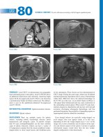
Figure 6.3 Chest X-ray in complete transposition of the great vessels: cardiomegaly, narrow
mediastinum, and increased pulmonary vasculature.
The latter is manifested by tall R waves in the right precordial leads. Right atrial
enlargement is also possible. In neonates it may be indistinguishable from normal
for the age.
Patients with a large volume of pulmonary blood flow, as with coexistent ventricular septal defect, also may have left ventricular enlargement/hypertrophy because
of the volume load on the left ventricle.
Chest X-ray
Cardiomegaly is generally present. The cardiac silhouette has a characteristic eggshaped appearance (Figure 6.3); the superior mediastinum is narrow because the
great vessels lie one in front of the other; the thymus is usually small. Left atrial
enlargement exists in the unoperated patient.
Summary of clinical findings
The diagnosis of complete transposition is usually indicated by a combination
of rather intense cyanosis in the neonatal period, roentgenographic findings
of increased pulmonary vasculature, and characteristic cardiac contour.
6 Congenital heart disease with a right-to-left shunt in children
193
Echocardiogram
The key to the echocardiographic diagnosis of complete transposition is the recognition of an anteriorly arising aorta and a posteriorly arising pulmonary artery. In
views parallel to the long axis of the left ventricle, both arteries course parallel to
each other for a short distance. This appearance is not seen in a normal heart,
where the great arteries cross each other at an acute angle. In views profiling the
short axis of the left ventricle, the aorta is seen arising anterior and rightward of the
central and posterior pulmonary artery (hence the term d-transposition, or dextrotransposition). A cross-sectional view of the aortic root allows demonstration of
the origins, branching, and proximal courses of the coronary arteries.
In neonates with transposition, the interventricular septum usually has a flat
contour when viewed in cross-section; however, as the infant ages, the septum
gradually bows away from the right (systemic) ventricle and bulges into the left
(pulmonary) ventricle.
Ventricular septal defect represents the most important associated lesion diagnosed by echocardiography; the shunt through it and any atrial septal defect or
ductus is bidirectional, consistent with the physiology of transposition described
earlier. The atrial septal defect may be small and restrictive (Doppler signals are
high velocity) before balloon septostomy; after, it is typically large and unrestrictive, with a mobile flap of the torn fossa ovalis waving to and fro across the defect.
Balloon septostomy may be performed under echocardiographic guidance.
Cardiac catheterization
Since echocardiography shows the diagnosis, the primary purpose of cardiac
catheterization is the performance of interventional creation of an atrial septal
defect (Rashkind procedure). In patients with an intact septum, oximetry data
show little increase in oxygen saturation values through the right side of the
heart, and little decrease through the left side. Among those with coexistent
ventricular septal defect, larger changes in oxygen values are found. The oxygen
saturation values in the pulmonary artery are higher than those in the aorta, a
finding virtually diagnostic of transposition of the great arteries.
In all patients, right ventricular systolic pressure is elevated. When the ventricular
septum is intact, the left ventricular pressure may be low; but in most patients
with coexistent ventricular septal defect or in those with a large patent ductus
arteriosus, the left ventricular pressure is elevated and equals that of the right
(systemic) ventricle.
Angiography confirms the diagnosis by showing the aorta arising from the
right ventricle and the pulmonary artery arising from the left ventricle, and it
identifies coexistent malformations. Aortic root injection demonstrates coronary
194
Pediatric cardiology
artery anatomy in preparation for surgery. A left ventricular injection is indicated
to demonstrate ventricular septal defect(s) and pulmonic stenosis.
Palliative procedures
Hypoxia, one of the major symptom of infants with transposition of the great
vessels, results from inadequate mixing of the two venous returns, and palliation is
directed towards improvement of mixing by two means. Unless hypoxia is treated,
it becomes severe, leading to metabolic acidosis and death.
Intravenous prostaglandin. This substance opens and/or maintains patency of
the ductus arteriosus and improves blood flow from aorta to pulmonary artery.
Rashkind balloon atrial septostomy procedure. Patients with inadequate
mixing benefit from the creation of an atrial septal defect (enlargement of the
foramen ovale). At cardiac catheterization or by echocardiographic guidance, a
balloon catheter is inserted through a systemic vein and advanced into the left
atrium through the foramen ovale. The balloon is inflated and then rapidly and
forcefully withdrawn across the septum, creating a larger defect and often improving the hypoxia.
Infants who do not experience adequate improvement of cyanosis despite a
large atrial defect and patent ductus are rare. Factors responsible in these neonates
include nearly identical ventricular compliances, which limits mixing through the
atrial defect, and elevated pulmonary vascular resistance, which limits the ductal
shunt and pulmonary blood flow. Increased intravenous fluids may benefit the
patient by increasing blood volume.
Rarely, an atrial defect is created surgically by atrial septectomy, an open-heart
procedure. A closed-heart technique, the Blalock–Hanlon procedure, was used
previously, but frequently resulted in scarring of the pulmonary veins.
Corrective operation
Atrial (venous) switch (see Figure 6.2). The first successful corrective procedure was performed by Senning in the 1950s and later modified by Mustard. These
procedures invoke the principle that two negatives make a positive. Since the circulation of transposition is reversed at the arterial level, these operations reverse it
the atrial level. This procedure involves removal of the atrial septum and creation
of an intra-atrial baffle to divert the systemic venous return into the left ventricle
and thus to the lungs, whereas the pulmonary venous return is directed to the
right ventricle and thus to the aorta.
It can be performed at low risk in patients with an intact ventricular septum and
at a higher risk in patients with ventricular septal defect. Serious complications,
6 Congenital heart disease with a right-to-left shunt in children
195
stroke, or death can occur in infants before an atrial (venous) switch procedure,
which is usually done after 3–6 months of age.
The long-term results of the atrial switch procedure have been identified.
Arrhythmias, the most frequent long-term complication, are often related to
abnormalities of the sinoatrial node and of the atrial surgical scar. Sometimes
these are life threatening, although the exact mechanism of sudden death in
the rare child who succumbs is not usually known. Scarring can also cause
systemic or pulmonary obstruction of the venous return. The most common
significant complication is not sudden death but progressive dysfunction of the
right ventricle, leading to death from chronic heart failure in adulthood. This
complication is related to the right ventricle functioning as the systemic ventricle.
Predicting which patients will develop failure and at the age postoperatively is
not possible.
Arterial switch (Jatene) (see Figure 6.2c). This operation, developed in the
1970s, avoids the complications inherent with the atrial (venous) switch and
involves switching the aorta and pulmonary artery to the correct ventricle. The
great vessels are transected and reanastomosed, so blood flows from left ventricle
to aorta and from right ventricle to pulmonary arteries. Since the coronary arteries
arise from the aortic root, they are transferred to the pulmonary (neoaortic)
root. Certain variations of coronary artery origins or branching make transfer
more risky. The arterial switch operation must occur early in life (within the first
2 weeks) before the pulmonary resistance falls and the left ventricle becomes
“deconditioned” to eject the systemic pressure load.
Arterial switch is not free from complications: coronary artery compromise may
result in left ventricular infarct or failure; pulmonary artery stenosis can result from
stretching or kinking during the surgical repositioning of the great vessels; and the
operative mortality may be higher, partly because of the risks of neonatal openheart surgery.
The short- and long-term outcomes favor those receiving the arterial switch
procedure.
Summary
Complete transposition of the great arteries is a common cardiac anomaly
that results in neonatal cyanosis and ultimately in cardiac failure. Many
neonates are initially asymptomatic, but quickly become cyanotic. The
physical findings and electrocardiogram vary with associated malformations.
The chest X-ray reveals cardiomegaly and increased pulmonary vascularity.
Palliative and corrective procedures are available.
196
Pediatric cardiology
Total anomalous pulmonary venous connection (TAPVC
or TAPVR) (see Figure 6.4)
The pulmonary veins, instead of entering the left atrium, connect with a systemic
venous channel that delivers pulmonary venous blood to the right atrium. Developmentally, this anomaly results from failure of incorporation of the pulmonary
veins into the left atrium, so that the pulmonary venous system retains earlier
embryologic communications to the systemic venous system.
In the embryo, the pulmonary veins communicate with both the left and right
anterior cardinal veins and the umbilical vitelline system, both precursors of systemic veins. If the pulmonary veins, which form with the lungs as outpouchings of
the foregut, are not incorporated into the left atrium, the result is anomalous pulmonary venous connection to one of the following structures: right superior vena
cava (right anterior cardinal vein), left superior vena cava (distal left anterior cardinal vein), coronary sinus (proximal left anterior cardinal vein), or infradiaphragmatic
site (umbilical–vitelline system), usually a tributary of the portal system.
Therefore, the right atrium receives not only the entire systemic venous return,
but also the entire pulmonary venous return. The left atrium has no direct venous
supply. An obligatory right-to-left shunt exists at the atrial level through either a
patent foramen ovale or usually an atrial septal defect.
The volume of blood shunted from the right to the left atrium and the volume of
blood that enters each ventricle depends upon their relative compliances. Ventricular compliance is influenced by ventricular pressures and vascular resistances. Right
ventricular compliance normally increases following birth as pulmonary vascular
resistance and pulmonary arterial pressure fall. Therefore, in most patients with
total anomalous pulmonary venous connection, pulmonary blood flow becomes
considerably greater than normal; systemic blood flow is usually normal. Since a
disparity exists between the volume of blood being carried by the right and left
sides of the heart, the right side becomes dilated and hypertrophied, whereas the
left side is relatively smaller but near-normal size.
In patients with total anomalous pulmonary venous connection, the degree of
cyanosis inversely relates to the volume of pulmonary blood flow. As the volume
of pulmonary blood flow becomes larger, the proportion of the pulmonary venous
blood to total venous blood returning to the right atrium becomes greater. As a
result, the saturation of blood shunted to the left side of the heart is higher, being
only slightly reduced from normal.
On the other hand, in hemodynamic situations in which the resistance to flow
through the lungs is increased (e.g. the neonatal period), the volume of blood flow
through the lungs is nearly normal (i.e. equal to systemic blood flow). Therefore,
the pulmonary and systemic venous systems contribute nearly equal volumes of
blood to the right atrium, and these neonates exhibit noticeable cyanosis.
6 Congenital heart disease with a right-to-left shunt in children
(a)
(b)
Figure 6.4 Total anomalous pulmonary venous connection. (a) Central circulation and
surgical repair of unobstructed type; (b) central circulation in obstructed type.
197
198
Pediatric cardiology
Total anomalous pulmonary venous connection is an example of bidirectional
shunting: a left-to-right shunt at the venous level and a right-to-left shunt at the
atrial level since all the pulmonary venous blood returns to the right atrium.
Total anomalous pulmonary venous connection presents two clinical pictures.
One resembles atrial septal defect and has no obstruction to the venous channel. The other shows intense cyanosis and a radiographic pattern of pulmonary
venous obstruction. In this form, the connecting venous channel is narrowed and
obstructed. These two are discussed separately in the following.
Total anomalous pulmonary venous connection without
obstruction (see Figure 6.4a)
History. The clinical manifestations vary considerably. Usually, the anomaly is
recognized in the neonatal period or with fetal echocardiography. If not operated
upon in early infancy, most patients develop congestive cardiac failure, grow
slowly, and have frequent respiratory infections, but a few may be asymptomatic
into later childhood.
Physical examination. The degree of cyanosis varies because of differences in
the volume of pulmonary blood flow. Although systemic arterial desaturation
is always present, children with greatly increased pulmonary blood flow appear
acyanotic or show only slight cyanosis.
The physical findings mimic isolated atrial septal defect. Cardiomegaly, precordial bulge, and right ventricular heave are found in older unoperated infants. A
grade 2/6–3/6 pulmonary systolic ejection murmur due to excess flow across the
pulmonary valve is present along the upper left sternal border. Wide, fixed splitting
of the second heart sound is heard and the pulmonary component may be accentuated, reflecting elevated pulmonary pressure. A mid-diastolic murmur caused
by increased blood flow across the tricuspid valve is found along the lower left
sternal border and is associated with greatly increased pulmonary blood flow. In
total anomalous pulmonary venous connection to the superior vena cava, a venous
hum may exist along the upper right sternal border because of the large venous
blood flow.
Electrocardiogram. The electrocardiogram reveals enlargement of the rightsided cardiac chambers with right-axis deviation, right atrial enlargement, and
right ventricular enlargement/hypertrophy. Usually, the pattern reflecting volume
overload, is an rSR′ pattern in lead V1 .
Chest X-ray. Chest X-ray findings also resemble isolated atrial septal defect. Cardiomegaly, primarily of right-sided chambers, and increased pulmonary blood flow
6 Congenital heart disease with a right-to-left shunt in children
199
are found. In contrast to most other admixture lesions, the left atrium is not
enlarged because blood flow through this chamber is normal.
Except for total anomalous pulmonary venous connection to a left superior vena
cava (“vertical vein”), the roentgenographic contour is not characteristic. In this
form, the cardiac silhouette can be described as a figure-of-eight or as a “snowman heart” (Figure 6.5a). The upper portion of the cardiac contour is formed by
the enlarged left and right superior venae cavae. The lower portion of the contour
is formed by the cardiac chambers.
Summary of clinical findings
The clinical, electrocardiographic, and roentgenographic findings resemble
those of atrial septal defect because the effects on the heart are similar.
Cyanosis distinguishes the conditions; although it may be minimal or not
clinically evident, it is easily detectable by pulse oximetry. Unlike
uncomplicated atrial septal defect, congestive cardiac failure and elevated
pulmonary arterial pressure may be found in total anomalous pulmonary
venous connection.
Echocardiogram. Cross-sectional echocardiography reveals an atrial septal
defect and enlarged right atrium, right ventricle, and pulmonary arteries. The left
atrium and left ventricle appear smaller than normal. In contrast to most normal
neonates, with an atrial septal defect the shunt is from right atrium to left atrium.
Doppler demonstrates a right-to-left atrial septal defect shunt because the only
blood entering the left atrium is through the atrial septal defect. The individual
pulmonary veins are visualized as they join a common pulmonary vein, which
then connects to the coronary sinus, the superior vena cava by way of a vertical
vein (the left-sided superior vena cava), or the hepatic portal venous system after
a descent into the abdomen.
Cardiac catheterization. Oxygen saturation values in each cardiac chamber and
in both great vessels are virtually identical. An increase in oxygen saturation is
found in the vena cava, coronary sinus, or other systemic venous sites into which
the pulmonary venous blood flows. The saturation of blood in the left atrium and
left ventricle is reduced because of the obligatory right-to-left atrial shunt.
Pulmonary hypertension may be found in infants, but some patients, particularly
older ones, show near-normal levels of pulmonary arterial pressures.
Pulmonary angiography is indicated. During the later phases of the angiogram
(the so-called levophase), the pulmonary veins opacify and subsequently fill the
200
Pediatric cardiology
(a)
(b)
Figure 6.5 Chest X-ray in total anomalous pulmonary venous connection. (a) Unobstructed
(supracardiac) connection to the left superior vena cava (“snowman” heart). Upper portion
of cardiac silhouette formed by dilated right and left superior venae cavae. (b) Obstructed
(infradiaphragmatic) type. Pulmonary vascular congestion, a pleural effusion, and a small
heart shadow.
6 Congenital heart disease with a right-to-left shunt in children
201
connecting venous channel, delineating the anatomic form of anomalous pulmonary venous connection.
Operative considerations. Under cardiopulmonary bypass, the confluence of
pulmonary veins, which lies directly behind the left atrium, is opened and connected to it (Figure 6.4a). The atrial communication is closed, and the connecting
vessel is divided. This operation can be performed with low risk, even in neonates
and younger infants.
Summary
Each of the anatomic types of total anomalous pulmonary venous connection
is associated with cyanosis of variable extent. The physical findings are those
of atrial septal defect; pulmonary hypertension may also be found. Both the
electrocardiogram and the chest X-ray reveal enlargement of the right-sided
cardiac chambers. Corrective operations can be performed successfully for
each of the forms of total anomalous pulmonary venous connection.
Total anomalous pulmonary venous connection with obstruction
(see Figure 6.4b)
In total anomalous pulmonary venous connection, an obstruction can be present
in the channel returning pulmonary venous blood to the right side of the heart.
Obstruction is always present in patients with an infradiaphragmatic connection
and occasionally in patients with a supradiaphragmatic connection. In the latter,
obstruction may occur intrinsically from narrowing of the channel or extrinsically
if the channel passes between the bronchus and the ipsilateral branch pulmonary
artery.
In infradiaphragmatic connection, four mechanisms contribute to obstruction
in pulmonary venous flow: (1) the venous channel is long; (2) the channel
traverses the diaphragm through the esophageal hiatus and is compressed by
either esophageal or diaphragmatic action; (3) the channel narrows at its junction
with the portal venous system; and (4) the pulmonary venous blood must traverse
the hepatic capillary system before returning to the right atrium by way of the
hepatic veins.
The obstruction elevates pulmonary venous pressure. Consequently, pulmonary
capillary pressure is raised, leading to pulmonary edema and a dilated pulmonary
lymphatic system. Pulmonary arterial pressure is elevated because of both elevated
pulmonary capillary pressure and reflex pulmonary vasoconstriction. Because of
the pulmonary hypertension, the right ventricle remains thick walled, does not
undergo its normal evolution following birth, and remains relatively noncompliant.
202
Pediatric cardiology
As a result, the volume of flow into the right ventricle is limited. Because of the
reduced pulmonary blood flow, the patients show more intense cyanosis than
those with without pulmonary venous obstruction.
The clinical features of total anomalous pulmonary venous connection with
obstruction relate to the consequences of pulmonary venous obstruction and to
the limited pulmonary blood flow.
History. Patients with obstruction present as neonates with significant cyanosis
and respiratory distress. Cyanosis is often intense because of the limited volume
of pulmonary flow. The cyanosis is accentuated by the pulmonary edema that
interferes with oxygen transport from the alveolus to the pulmonary capillary. Respiratory symptoms of tachypnea and dyspnea result from the altered pulmonary
compliance from pulmonary edema and hypertensive pulmonary arteries.
Physical examination. Cyanosis is present, and increased respiratory effort is
manifested by intercostal retractions and tachypnea. On clinical examination the
heart size is normal. Since the volume of flow through the right side of the heart
is normal, no murmurs appear. The accentuated pulmonic component of the second heart sound reflects pulmonary hypertension. The cyanosis without cardiac
findings of these neonates usually suggests a pulmonary rather than a cardiac
condition.
Beyond the immediate neonatal period, the infants appear scrawny and malnourished.
Electrocardiogram. Right ventricular hypertrophy, right-axis deviation, and right
atrial enlargement are found. In a normal neonate, however, the QRS axis is usually
directed towards the right, the P waves may approach 3 mm in amplitude, and the
R waves are tall in the right precordial leads. Therefore, the electrocardiograms of
neonates with obstructed pulmonary venous connection appear similar to those
of normal neonates. Such a pattern, however, is compatible with the diagnosis.
Chest X-ray (see Figure 6.5b). Cardiac size is normal because the volume of
systemic and pulmonary blood flows is normal. The pulmonary vasculature shows
a diffuse reticular pattern of pulmonary edema. Even in young children, Kerley B
lines, which are small horizontal lines at the margins of the pleura mostly in the
lower lung fields, are present. The radiographic pattern, although similar to that
of hyaline membrane disease, differs from it because it does not usually show air
bronchograms.
6 Congenital heart disease with a right-to-left shunt in children
203
Summary of clinical findings
This form of total anomalous pulmonary venous connection is very difficult
to distinguish from neonatal pulmonary disease because of similar clinical
and laboratory findings. In both, the patients present with respiratory distress
and cyanosis in the neonatal period. No murmurs are present. The
electrocardiogram may be normal for age and the chest X-ray shows a
normal-sized heart and a diffuse, hazy pattern. Echocardiography may be
misleading, so cardiac catheterization and angiography may be necessary to
distinguish pulmonary disease from this form of cardiac disease.
Echocardiogram. Because the intracardiac anatomy appears normal and visualization is often limited by pulmonary hyperinflation from aggressive mechanical
ventilation used in these neonates, the echocardiographic detection of this lesion
is challenging. An atrial septal defect with a right-to-left shunt exists, typical of
total anomalous pulmonary venous connection, but this finding is also found with
severe primary lung disease or persistent pulmonary hypertension. The atrial septal defect flow is much lower than in the unobstructed form because pulmonary
venous obstruction results in very low pulmonary blood flow. The ductus may
be large and have bidirectional or predominantly pulmonary artery-to-aorta shunt
because of elevated pulmonary arteriolar resistance. Doppler shows no pulmonary
venous return to the left atrium; in the most common form, the pulmonary veins
return to a common pulmonary vein that courses caudad to the abdomen, usually
slightly to the left of the spine.
Cardiac catheterization. As in the unobstructed form, the oxygen saturations
are identical in each cardiac chamber, but with this lesion oxygen saturations are
extremely low. Pulmonary hypertension is present, and also the pulmonary wedge
pressure is elevated. Angiography shows the anomalous pulmonary venous connection, which is usually connected to an infradiaphragmatic site.
Operative considerations. Infants with total anomalous pulmonary venous
connection to an infradiaphragmatic site often die in the neonatal period. As
soon as the diagnosis is made, operation is indicated, using the technique
described previously. In some infants, pulmonary hypertension persists in the
postoperative period for a few days and requires management with mechanical
ventilation, creation of an alkalotic state, and administration of nitric oxide and
other pulmonary vasodilators.
204
Pediatric cardiology
Summary
Total anomalous pulmonary venous connection, although of several
anatomic forms, presents with one of two clinical pictures. In one, the
pulmonary arterial pressures and right ventricular compliance are normal or
slightly elevated. These patients’ features resemble atrial septal defect but
show mild cyanosis. In the other, pulmonary arterial pressure and pulmonary
resistance are elevated because of pulmonary venous obstruction. Therefore,
right ventricular compliance is reduced and pulmonary blood flow is limited.
These patients show a radiographic pattern of pulmonary venous obstruction
or severe cyanosis and major respiratory symptoms. The clinical and
laboratory findings resemble neonatal respiratory distress or persistent
pulmonary hypertension syndromes.
Common arterial trunk (truncus arteriosus)
In common arterial trunk or persistent truncus arteriosus (Figure 6.6), a single
arterial vessel leaves the heart and gives rise to the three major circulations,
Figure 6.6 Truncus arteriosus. Central circulation.
6 Congenital heart disease with a right-to-left shunt in children
205
pulmonary, systemic, and coronary circulations. This malformation is associated
with a ventricular septal defect through which both ventricles eject into the
common arterial trunk. Because the defect is large and the common trunk
originates from both ventricles, the right ventricular systolic pressure is identical
with that of the left ventricle.
The hemodynamics are similar to those of ventricular septal defect and patent
ductus arteriosus. The volumes of systemic and pulmonary blood. flow depend on
the relative resistances to flow into the systemic pulmonary circulations.
The resistance to flow through the lungs is governed by two factors: (1) the
caliber of the pulmonary arterial branches arising from the common trunk and
(2) the pulmonary vascular resistance. Although differences in the size of the pulmonary arterial branches vary as they originate from the common trunk, ordinarily
their size does not offer significant resistance to pulmonary blood flow, so the
pulmonary arterial pressure equals that of the aorta. Therefore, the pulmonary
arteriolar resistance is the primary determinant of pulmonary blood flow. In the
neonatal period, when pulmonary vascular resistance is elevated, the volume of
blood flow through the lungs is similar to the systemic blood flow. As the pulmonary vasculature matures, the pulmonary blood flow increases progressively.
Many of the clinical and laboratory findings of truncus arteriosus depend on
the volume of pulmonary blood flow. Increased pulmonary blood flow leads to
three effects: (1) the degree of cyanosis and the volume of pulmonary blood flow
are inversely related, and the degree of cyanosis lessens as pulmonary blood flow
increases because of the larger quantities of fully saturated pulmonary venous
return mixing with the relatively fixed systemic venous return; (2) congestive cardiac failure develops because of left ventricular volume overload; and (3) the pulse
pressure widens because the blood leaves the common trunk during diastole to
enter the pulmonary arteries.
Although the truncal valve is usually tricuspid, it becomes regurgitant in some
patients. Therefore, the additional volume load of regurgitation is incurred by the
ventricles. Some truncal valves have four or more cusps; these are both stenotic
and regurgitant, adding pressure overload to the already volume-overloaded
ventricles.
Approximately 40% of truncus patients show deletion of a portion of chromosome 22 and other laboratory findings of DiGeorge syndrome, such as hypocalcemia and reduced T lymphocytes.
History
The symptoms vary with the volume of pulmonary blood flow. In the neonatal
period, cyanosis is the major symptom because the elevated pulmonary vascular resistance limits the pulmonary blood flow. As pulmonary vascular resistance
falls, cyanosis lessens, but congestive cardiac failure develops, usually after several
206
Pediatric cardiology
weeks of age. Patients with common trunk and congestive cardiac failure mimic
those with ventricular septal defect at this time because cyanosis is mild or absent.
Dyspnea on exertion, easy fatigability, and frequent respiratory infections are common symptoms.
Patients whose pulmonary blood flow is limited, owing either to the development of pulmonary vascular disease or to the presence of small pulmonary
arteries arising from the truncus, show predominant symptoms of cyanosis rather
than congestive cardiac failure, unless significant regurgitation through the truncal
valve coexists.
Physical examination
Cyanosis may or may not be clinically evident but is easily detected with pulse
oximetry. Manifestations of a wide pulse pressure may appear if increased pulmonary blood flow or significant truncal valve regurgitation exists. Cardiomegaly
and a precordial bulge are common. The auscultatory findings may initially
resemble ventricular septal defect. The major auscultatory finding is a loud systolic
murmur along the left sternal border. An apical mid-diastolic rumble present in
most patients indicates large blood flow across the mitral valve from increased
pulmonary blood flow.
Common arterial trunk shows three distinctive auscultatory findings: (1) the
second heart sound is single since only a single semilunar valve is present; (2) a
high-pitched early diastolic decrescendo murmur is present if truncal valve regurgitation coexists; and (3) an apical systolic ejection click that is usually heard indicates
the presence of a dilated great vessel, the common trunk. The click, especially if
heard at an early age, suggests that the truncal valve is stenotic to some extent.
Electrocardiogram
The electrocardiogram usually shows a normal QRS axis and biventricular enlargement/hypertrophy. The left ventricular enlargement is related to left ventricular
volume overload; the right ventricular hypertrophy is related to the elevated right
ventricular systolic pressure. If pulmonary vascular disease develops and reduces
pulmonary blood flow, the left ventricular enlargement may disappear. Truncal
regurgitation and truncal stenosis modify these findings by augmenting the ventricular volume and by increasing ventricular pressures, respectively.
Chest X-ray
The pulmonary vasculature is increased. The prominent “ascending aorta” that
is usually seen represents the enlarged common trunk. Because the branch
pulmonary arteries arise from the truncus arteriosus, a main pulmonary artery
6 Congenital heart disease with a right-to-left shunt in children
207
Figure 6.7 Chest X-ray in truncus arteriosus. Cardiomegaly, right aortic arch, and increased
pulmonary vascularity.
silhouette is absent. Most patients show cardiomegaly proportional to the volume
of pulmonary blood flow and the amount of truncal regurgitation. Left atrial
enlargement is present in patients with increased pulmonary blood flow.
A right aortic arch is found in one-fourth of patients; this finding, when combined with that of increased pulmonary vascular markings and cyanosis, is virtually
diagnostic of truncus arteriosus (Figure 6.7).
Summary of clinical findings
Persistent truncus arteriosus is suspected in a cyanotic patient who has a
murmur suggesting ventricular septal defect and two characteristic features:
a single second heart sound and a systolic ejection click. The volume of
pulmonary blood flow is reflected by the degree of cyanosis and the amount
of left atrial enlargement. The degree of cardiomegaly on chest X-ray or left
ventricular hypertrophy on electrocardiogram is not the sole reflection of
pulmonary blood flow, since coexistent truncal insufficiency can also cause
these particular findings. DiGeorge syndrome is common.
208
Pediatric cardiology
Natural history
The course of common arterial trunk resembles that of ventricular septal defect but
is more severe, and the development of pulmonary vascular disease, the ultimate
threat to longevity and operability, is greatly accelerated. Truncal regurgitation usually progresses.
Echocardiogram
Cross-sectional echocardiography in views parallel to the long axis of the left ventricular outflow tract shows a large great vessel (the common trunk) “overriding”
a large ventricular septal defect, similar to images seen in tetralogy of Fallot. A
separate pulmonary artery cannot be demonstrated arising from the heart; the
pulmonary arteries arise from the common trunk and their pattern of origin is
seen by echocardiography. The ductus arteriosus is usually absent unless coexisting
interruption of the aortic arch is present. The truncal valve may be trileaflet, with
apparent movement similar to that of a normal aortic valve, or it may be deformed,
usually as a quadricuspid or multicuspid valve, with both stenosis and regurgitation. Left atrial enlargement parallels the degree of pulmonary overcirculation.
Cardiac catheterization
Usually, a venous catheter is passed through the right ventricle into the common
trunk and then into the pulmonary arteries. The systolic pressures are identical in
both ventricles and in the common trunk, unless truncal valve stenosis is present.
In that case, ventricular systolic pressures exceed the systolic pressure in the trunk.
A wide pulse pressure is often present in the trunk. An increase in oxygen saturation is found in the right ventricle with further increase in the common trunk. The
blood is not fully saturated in the latter site. Truncal root injection demonstrates
the origin and course of the pulmonary arteries but requires a large volume of
contrast that must be administered rapidly to overcome excessive dilution from
high pulmonary blood flow.
Operative considerations
For infants manifesting severe cardiac failure who do not to respond to medical
management, banding of the pulmonary artery is sometimes performed. Although
the cardiac failure is improved and the infant grows, the band may complicate and
increase the risk of repair. Banding surgery may also be difficult to perform when
the pulmonary artery branches arise from separate origins from the truncus.
Corrective operation is almost always preferable. In this procedure, the ventricular septal defect is closed so that left ventricular blood passes into the common
trunk. The pulmonary arteries are detached from the truncal wall and connected
to one end of a valved conduit; its other end is inserted into the right ventricle.
6 Congenital heart disease with a right-to-left shunt in children
209
If severe, truncal regurgitation can be corrected simultaneously by valvuloplasty or
insertion of a prosthetic valve. The risk is considerably higher for patients with truncal regurgitation, stenosis, or any element of pulmonary vascular disease. Since
the conduit from the right ventricle to pulmonary arteries has a fixed diameter,
reoperation is necessary as the child grows.
Summary
Common arterial trunk (persistent truncus arteriosus) is an infrequently
occurring cardiac anomaly whose clinical and laboratory features resemble
ventricular septal defect and patent ductus arteriosus, with similarities in
hemodynamics and natural history. Early corrective operation is advised, but
considerable operative risks remain, partially due to the frequent coexistence
of DiGeorge syndrome.
C YA N O S I S A N D D I M I N I S H E D P U L M O N A R Y
BLOOD FLOW
Patients with cyanosis and roentgenographic evidence of diminished pulmonary
blood flow have a cardiac malformation in which both obstruction to pulmonary
blood flow and an intracardiac defect that permits a right-to-left shunt are found.
The degree of cyanosis varies inversely with the volume of pulmonary blood flow.
The amount by which pulmonary blood flow is reduced equals the volume of blood
shunted in a right-to-left direction.
The intracardiac right-to-left shunt can occur at either the ventricular or the atrial
level. In patients with a ventricular shunt, the cardiac size is usually normal, as in
tetralogy of Fallot, whereas those with an atrial shunt often show cardiomegaly,
as in tricuspid atresia or Ebstein’s malformation.
Tetralogy of Fallot
This is probably the most widely known cardiac condition resulting in cyanosis and
is the most common anomaly in this category (Figure 6.8).
Classically, tetralogy of Fallot has four components: ventricular septal defect;
aorta overriding the ventricular septal defect; pulmonary stenosis, generally
infundibular in location; and right ventricular hypertrophy. Because of the large
ventricular septal defect, right ventricular systolic pressure is at systemic levels.
Hemodynamically, tetralogy of Fallot can be considered a combination of two
lesions: a large ventricular septal defect, allowing equalization of ventricular systolic pressures, and severe pulmonary stenosis.
The magnitude of the shunt through the ventricular communication depends
on the relative resistances of the pulmonary stenosis and the systemic circulation.
210
Pediatric cardiology
Figure 6.8 Tetralogy of Fallot. Central circulation and surgical repair.
Because the pulmonary stenosis is frequently related to a narrowed infundibulum,
it responds to catecholamines and other stimuli. Therefore, the amount of rightto-left shunt and the degree of cyanosis vary considerably with factors such as
emotion or exercise. Many of the symptoms of tetralogy of Fallot are related to
sudden changes in either of these resistance factors.
Tetralogy of Fallot with pulmonary valve atresia (Figure 6.9) has also been called
pseudotruncus arteriosus. In this anomaly, blood cannot flow directly from the
right ventricle into the pulmonary artery, so the entire output of both ventricles
passes into the aorta. The pulmonary circulation is supplied either by multiple
major aortopulmonary collateral arteries (MAPCAs) and/or through a patent ductus arteriosus. Severe hypoxic symptoms may develop in the neonatal period if the
patent ductus arteriosus closes or if the MAPCAs are narrow.
History
The children often become cyanotic in the first year of life, often in the neonatal
period. The time of appearance and the severity of cyanosis are directly related
to the severity of pulmonary stenosis and the degree pulmonary blood flow is
reduced.