Ebook Bates’ poktet guide to physical examination and history taking (7E): Part 2
Bạn đang xem bản rút gọn của tài liệu. Xem và tải ngay bản đầy đủ của tài liệu tại đây (5.41 MB, 252 trang )
CHAPTER
The Breasts and Axillae
10
The Health History
Common or Concerning Symptoms
◗ Breast lump or mass
◗ Breast pain or discomfort
◗ Nipple discharge
Ask, “Do you examine your breasts?” . . . “How often?” Ask about
any discomfort, pain, or lumps in the breasts. Also ask about any discharge from the nipples, change in breast contour, dimpling, swelling,
or puckering of the skin over the breasts.
Health Promotion and Counseling:
Evidence and Recommendations
Important Topics for Health Promotion and Counseling
◗
◗
◗
◗
Palpable masses of the breast
Assessing risk of breast cancer
Breast cancer screening
Breast self-examination (BSE)
Palpable Masses of the Breast. Breast masses show marked
variation in etiology, from fibroadenomas and cysts seen in younger
women, to abscess or mastitis, to primary breast cancer. All breast
masses warrant careful evaluation, and definitive diagnostic measures
should be pursued.
167
168
Bates’ Pocket Guide to Physical Examination and History Taking
Palpable Masses of the Breast
Age
Common Lesion
Characteristics
15–25
Fibroadenoma
25–50
Cysts
Usually smooth, rubbery, round,
mobile, nontender
Usually soft to firm, round, mobile; often tender
Nodular, ropelike
Irregular, firm, may be mobile or
fixed to surrounding tissue
As above
Fibrocystic changes
Cancer
Over 50
Pregnancy/
lactation
Cancer until proven
otherwise
Lactating adenomas, cysts,
mastitis, and cancer
As above
Adapted from Schultz MZ, Ward BA, Reiss M. Breast diseases. In: Noble J, Greene HL,
Levinson W, et al., eds: Primary Care Medicine, 2nd ed. St. Louis: Mosby, 1996. See also
Venet L, Strax P, Venet W, et al. Adequacies and inadequacies of breast examinations by
physicians in mass screenings. Cancer 1971;28(6):1546–1551.
Assessing Risk of Breast Cancer.
Although 70% of affected
women have no known predisposing factors, selected risk factors are
well established. Use the Breast Cancer Risk Assessment Tool of the
National Cancer Institute ( or
other available clinical models, such as the Gail model, to individualize
risk factor assessment for your patients. Ask women beginning in their
20s about any family history of breast or ovarian cancer, or both, on
the maternal or paternal side, to help assess risk of BRCA1 or BRCA2
gene mutation. (See http: astor.som.jhmi.edu/Bayesmendel/brcapro.
html). See also Table 10-1, Breast Cancer in Women: Factors That
Increase Relative Risk, p. 175.
Breast Cancer Screening.
The American Cancer Society recommendations, listed below, vary slightly from those of the U.S. Preventive Services Task Force.
●
Yearly mammography for women 40 years of age and older. For
women at increased risk, many clinicians advise initiating screening
mammography between ages 30 and 40, then every 2 to 3 years
until 50 years of age.
Chapter 10
| The Breasts and Axillae
169
●
Clinical breast examination (CBE) by a health care professional every
3 years for women between 20 and 39 years of age, and annually
after 40 years of age
●
Regular breast self-examination (BSE), in conjunction with mammography and CBE, to help promote health awareness
Techniques of Examination
EXAMINATION TECHNIQUES
Subclavian vein
POSSIBLE FINDINGS
Subclavian
lymph nodes
Pectoralis major
Axillary vein
Axillary
lymph nodes
Axillary tail
of breast
Fat
Gland lobules
Upper
outer
Upper
inner
Lower
outer
Lower
inner
Areola
Serratus anterior
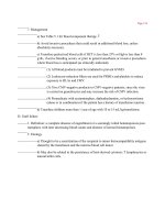
THE FEMALE BREAST
Inspect the breasts in four
positions.
Note:
●
Size and symmetry
See Table 10-2, Visible Signs of Breast
Cancer, pp. 176–177, development,
asymmetry.
●
Contour
Flattening, dimpling
170
Bates’ Pocket Guide to Physical Examination and History Taking
EXAMINATION TECHNIQUES
●
Appearance of the skin
ARMS AT SIDES
HANDS PRESSED AGAINST HIPS
POSSIBLE FINDINGS
Edema (peau d’orange) in breast
cancer
ARMS OVER HEAD
LEANING FORWARD
Inspect the nipples.
●
Compare their size, shape,
and direction of pointing.
Inversion, retraction, deviation
●
Note any rashes, ulcerations,
or discharge.
Paget’s disease of the nipple,
galactorrhea
Palpate the breasts, including augmented breasts. Breast
tissue should be flattened and
the patient supine. Palpate a
rectangular area extending from
the clavicle to the inframammary fold, and from the
midsternal line to the posterior
axillary line and well into the
axilla for the tail of Spence.
Chapter 10
| The Breasts and Axillae
EXAMINATION TECHNIQUES
171
POSSIBLE FINDINGS
Note:
●
Consistency
Physiologic nodularity
●
Tenderness
Infection, premenstrual tenderness
●
Nodules. If present, note
location, size, shape, consistency, delimitation, tenderness,
and mobility.
Cyst, fibroadenoma, cancer
Use vertical strip pattern
(currently the best validated
technique) or a circular or
wedge pattern. Palpate in small,
concentric circles.
●
For the lateral portion of the
breast, ask the patient to roll
onto the opposite hip, place
her hand on her forehead,
but keep shoulders pressed
against the bed or examining
table.
●
For the medial portion of the
breast, ask the patient to lie
with her shoulders flat against
the bed or examining table,
place her hand at her neck,
and lift up her elbow until it is
even with her shoulder.
Palpate each nipple.
Thickening in cancer
Palpate and inspect along the
incision lines of mastectomy.
Local recurrences of breast cancer
172
Bates’ Pocket Guide to Physical Examination and History Taking
EXAMINATION TECHNIQUES
POSSIBLE FINDINGS
THE MALE BREAST
/
Inspect and palpate the
nipple and areola.
Gynecomastia, mass suspicious for
cancer, fat
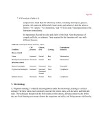
AXILLAE
Inspect for rashes, infection,
and pigmentation.
Hidradenitis suppurativa, acanthosis
nigricans
Palpate the axillary nodes,
including the central, pectoral,
lateral, and subscapular groups.
Lymphadenopathy
Supraclavicular
Lateral
Infraclavicular
Central
(deep within axilla)
Subscapular
(posterior)
Pectoral
(anterior)
ARROWS INDICATE DIRECTION OF
LYMPH FLOW
SPECIAL TECHNIQUE
BREAST DISCHARGE
Compress the areola in a
spokelike pattern around the
nipple. Watch for discharge.
Type and source of discharge may be
identified.
Chapter 10
/
| The Breasts and Axillae
173
BREAST SELF-EXAMINATION
Patient Instructions for the Breast Self-Examination (BSE)
Supine
1. Lie down with a pillow under
your right shoulder. Place your
right arm behind your head.
2. Use the finger pads of the three
middle fingers on your left hand
to feel for lumps in the right
breast. The finger pads are the
top third of each finger.
3. Press firmly enough to know
how your breast feels. A firm
ridge in the lower curve of each
breast is normal. If you’re not
sure how hard to press, talk with
your health care provider, or try
to copy the way the doctor or
nurse does it.
4. Press firmly on the breast in an
up-and-down or “strip” pattern.
You can also use a circular or
wedge pattern, but be sure to
use the same pattern every
time. Check the entire breast
area, and remember how your
breast feels from month to
month.
5. Repeat the examination on your
left breast, using the finger pads
of the right hand.
6. If you find any changes, see
your doctor right away.
(continued)
174
Bates’ Pocket Guide to Physical Examination and History Taking
Patient Instructions for the Breast
Self-Examination (BSE) (continued)
Standing
1. While standing in front of a
mirror with your hands
pressing firmly down on your
hips, look at your breasts for
any changes of size, shape,
contour, or dimpling, or redness
or scaliness of the nipple or
breast skin. (The pressing down
on the hips position contracts
the chest wall muscles and
enhances any breast changes.)
2. Examine each underarm while
sitting up or standing and with
your arm only slightly raised so
you can easily feel in this area.
Raising your arm straight up
tightens the tissue in this area
and makes it harder to examine.
Adapted from the American Cancer Society, updated September 2010. Available at http://
www.cancer.org/Cancer/BreastCancer/MoreInformation/BreastCancerEarlyDetection/
breast-cancer-early-detection-a-c-s-recs-b-s-e. Accessed December 3, 2010.
Recording Your Findings
Recording the Physical Examination—
Breasts and Axillae
“Breasts symmetric and smooth, without masses. Nipples without discharge.”
(Axillary adenopathy usually included after Neck in section on Lymph Nodes;
see p. 123.)
OR
“Breasts pendulous with diffuse fibrocystic changes. Single firm 1 × 1 cm mass,
mobile and nontender, with overlying peau d’orange appearance in right
breast, upper outer quadrant at 11 o’clock, 2 cm from the nipple.” (Suggests
possible breast cancer.)
Chapter 10
| The Breasts and Axillae
175
Aids to Interpretation
Table 10-1
Breast Cancer in Women: Factors That
Increase Relative Risk
Relative Risk
Factor
>4.0
●
●
●
●
●
●
●
2.1–4.0
●
●
●
Female
Age (65+ versus <65 years, although risk
increases across all ages until age 80)
Certain inherited genetic mutations for
breast cancer (BRCA1 and/or BRCA2)
Two or more first-degree relatives with
breast cancer diagnosed at an early age
Personal history of breast cancer
High breast tissue density
Biopsy-confirmed atypical hyperplasia
One first-degree relative with breast
cancer
High-dose radiation to chest
High bone density (postmenopausal)
1.1–2.0
Factors that affect
circulating hormones
●
●
●
●
●
●
●
●
Other factors
●
●
●
●
●
Late age at first full-term pregnancy
(>30 years)
Early menarche (<12 years)
Late menopause (>55 years)
No full-term pregnancies
Never breast-fed a child
Recent oral contraceptive use
Recent and long-term use of hormone
replacement therapy
Obesity (postmenopausal)
Personal history of endometrium, ovary,
or colon cancer
Alcohol consumption
Height (tall)
High socioeconomic status
Jewish heritage
Source: American Cancer Society. Breast Cancer Facts and Figures 2009–2010, p. 11.
Available at: www.cancer.org/acs/groups/content/cnho/documents/document/
f861009final90809pdf.pdf. Accessed July 31, 2012.
176
Table 10-2
Bates’ Pocket Guide to Physical Examination and History Taking
Visible Signs of Breast Cancer
Retraction Signs
Fibrosis from breast cancer
produces retraction signs:
dimpling, changes in contour,
and retraction or deviation of the
nipple. Other causes of retraction
include fat necrosis and mammary
duct ectasia.
Cancer
Dimpling
Retracted
nipple
Skin Dimpling
Abnormal Contours
Look for any variation in the normal
convexity of each breast, and
compare one side with the other.
Nipple Retraction and Deviation
A retracted nipple is flattened or
pulled inward. It may also be
broadened and feel thickened.
The nipple may deviate, or point
in a different direction, typically
toward the underlying cancer.
Chapter 10
| The Breasts and Axillae
Table 10-2
177
Visible Signs of Breast Cancer (continued)
Edema of the Skin
From lymphatic blockade, appearing
as thickened skin with enlarged
pores—the so-called peau
d’orange (orange peel) sign.
Paget’s Disease of the Nipple
An uncommon form of breast
cancer that usually starts as a
scaly, eczemalike lesion. The skin
may also weep, crust, or erode.
A breast mass may be present.
Suspect Paget’s disease in any
persisting dermatitis of the nipple
and areola.
Dermatitis of
areola
Erosion of
nipple
CHAPTER
The Abdomen
11
The Health History
Common or Concerning Symptoms
Gastrointestinal Disorders
Urinary and Renal Disorders
◗ Abdominal pain, acute and chronic
◗ Indigestion, nausea, vomiting including blood, loss of appetite, early
satiety
◗ Dysphagia and/or odynophagia
◗ Change in bowel function
◗ Diarrhea, constipation
◗ Jaundice
◗ Suprapubic pain
◗ Dysuria, urgency, or frequency
◗ Hesitancy, decreased stream
in males
◗ Polyuria or nocturia
◗ Urinary incontinence
◗ Hematuria
◗ Kidney or flank pain
◗ Ureteral colic
PATTERNS AND MECHANISMS OF ABDOMINAL PAIN
Be familiar with three broad
categories:
Visceral pain—occurs when hollow
abdominal organs such as the
intestine or biliary tree contract
unusually forcefully or are distended
or stretched.
●
May be difficult to localize
●
Varies in quality; may be gnawing,
burning, cramping, or aching
Visceral pain in the right upper
quadrant (RUQ) from liver distention against its capsule in alcoholic
hepatitis
179
180
●
Bates’ Pocket Guide to Physical Examination and History Taking
When severe, may be associated
with sweating, pallor, nausea,
vomiting, restlessness.
Parietal pain—from inflammation
of the parietal peritoneum.
●
Steady, aching
●
Usually more severe
●
Usually more precisely localized
over the involved structure than
visceral pain
Visceral periumbilical pain in early
acute appendicitis from distention
of inflamed appendix gradually
changes to parietal pain in the right
lower quadrant (RLQ) from inflammation of the adjacent parietal
peritoneum.
Referred pain—occurs in
more distant sites innervated at
approximately the same spinal levels
as the disordered structure.
Pain of duodenal or pancreatic
origin may be referred to the back;
pain from the biliary tree—to the
right shoulder or right posterior
chest.
Pain from the chest, spine, or pelvis
may be referred to the abdomen.
Pain from pleurisy or acute myocardial infarction may be referred to
the upper abdomen.
THE GASTROINTESTINAL TRACT
Ask patients to describe the
abdominal pain in their own words,
especially timing of the pain (acute
or chronic); then ask them to point
to the pain.
Pursue important details:
“Where does the pain start?”
“Does it radiate or travel?”
“What is the pain like?”
“How severe is it?”
“How about on a scale of 1 to 10?”
“What makes it better or worse?”
Chapter 11
| The Abdomen
181
Elicit any symptoms associated with
the pain, such as fever or chills; ask
their sequence.
Upper Abdominal Pain,
Discomfort, or Heartburn. Ask
about chronic or recurrent upper
abdominal discomfort, or dyspepsia.
Related symptoms include bloating,
nausea, upper abdominal fullness,
and heartburn.
Find out just what your patient
means. Possibilities include:
●
Bloating from excessive gas,
especially with frequent belching,
abdominal distention, or flatus,
the passage of gas by rectum
●
Nausea and vomiting
●
Unpleasant abdominal fullness
after normal meals or early satiety,
the inability to eat a full meal
Consider diabetic gastroparesis,
anticholinergic drugs, gastric outlet
obstruction, gastric cancer. Early
satiety may signify hepatitis.
●
Heartburn
Suggests gastroesophageal reflux
disease (GERD)
Lower Abdominal Pain
or Discomfort—Acute and
Chronic. If acute, is the pain sharp
and continuous or intermittent and
cramping?
Right lower quadrant (RLQ) pain,
or pain migrating from periumbilical region in appendicitis; in
women with RLQ pain, possible
pelvic inflammatory disease, ectopic
pregnancy
Left lower quadrant (LLQ) pain in
diverticulitis
182
Bates’ Pocket Guide to Physical Examination and History Taking
If chronic, is there a change in
bowel habits? Alternating
diarrhea and constipation?
Colon cancer; irritable bowel
syndrome
Other GI Symptoms
●
Anorexia
Liver disease, pregnancy, diabetic
ketoacidosis, adrenal insufficiency,
uremia, anorexia nervosa
●
Dysphagia or difficulty
swallowing
If solids and liquids, neuromuscular disorders affecting
motility. If only solids, consider
structural conditions like Zenker’s
diverticulum, Schatzki’s ring, stricture, neoplasm
●
Odynophagia, or painful
swallowing
Radiation; caustic ingestion,
infection from cytomegalovirus,
herpes simplex, HIV
●
Diarrhea, acute (<2 weeks)
and chronic
Acute infection (viral, salmonella,
shigella, etc.); chronic in Crohn’s
disease, ulcerative colitis; oily
diarrhea (steatorrhea)—in pancreatic insufficiency. See Table 11-1,
Diarrhea, pp. 194–195.
●
Constipation
Medications, especially anticholinergic agents and opioids; colon
cancer
●
Melena, or black tarry stools
GI bleed
●
Jaundice from increased levels of
bilirubin: Intrahepatic jaundice can
be hepatocellular, from damage to
the hepatocytes, or cholestatic, from
impaired excretion caused by damaged hepatocytes or intrahepatic
bile ducts
Impaired excretion of conjugated
bilirubin in viral hepatitis, cirrhosis,
primary biliary cirrhosis, druginduced cholestasis
Extrahepatic jaundice arises from
obstructed extrahepatic bile ducts,
commonly the cystic and common
bile ducts
Chapter 11
| The Abdomen
Ask about the color of the urine
and stool.
183
Dark urine from increased conjugated bilirubin excreted in urine;
acholic clay-colored stool when
excretion of bilirubin into intestine
is obstructed
Risk Factors for Liver Disease
◗ Hepatitis A: Travel or meals in areas with poor sanitation, ingestion of contaminated water or foodstuffs
◗ Hepatitis B: Parenteral or mucous membrane exposure to infectious body fluids
such as blood, serum, semen, and saliva, especially through sexual contact
with an infected partner or use of shared needles for injection drug use
◗ Hepatitis C: Illicit intravenous drug use or blood transfusion
◗ Alcoholic hepatitis or alcoholic cirrhosis: Interview the patient carefully about
alcohol use
◗ Toxic liver damage from medications, industrial solvents, environmental
toxins or some anesthetic agents
◗ Extrahepatic biliary obstruction that may result from gallbladder disease or
surgery
◗ Hereditary disorders reported in the Family History
THE URINARY TRACT
Ask about pain on urination,
usually a burning sensation, sometimes termed dysuria (also refers to
difficulty voiding).
Bladder infection
Also, consider bladder stones,
foreign bodies, tumors, and acute
prostatitis. In women, internal burning in urethritis, external burning in
vulvovaginitis
Other associated symptoms include:
●
Urgency, an unusually intense and
immediate desire to void
●
Urinary frequency, or abnormally
frequent voiding
●
Fever or chills; blood in the urine
●
Any pain in the abdomen, flank,
or back
May lead to urge incontinence
Dull, steady pain in pyelonephritis;
severe colicky pain in ureteral
obstruction from renal stone
184
Bates’ Pocket Guide to Physical Examination and History Taking
In men, hesitancy in starting the
urine stream, straining to void,
reduced caliber and force of the
urine stream, or dribbling as they
complete voiding.
Prostatitis, urethritis
Assess any:
●
Polyuria, a significant increase in
24-hour urine volume
Diabetes mellitus, diabetes insipidus
●
Nocturia, urinary frequency at
night
Bladder obstruction
●
Urinary incontinence,
involuntary loss of urine:
See Table 11-2, Urinary Incontinence, pp. 196–197.
●
From coughing, sneezing,
lifting
Stress incontinence (poor urethral
sphincter tone)
●
From urge to void
Urge incontinence (detrusor overactivity)
●
From bladder fullness with
leaking but incomplete
emptying
Overflow incontinence (anatomic
obstruction, impaired neural
innervation to bladder)
Health
H
eallth P
Promotion
romotio
on and
dC
Counseling:
ou
unsseling
g:
Evidence
E
viide
ence a
and
nd Re
Recommendations
eco
ommend
dattion
ns
Important Topics for Health Promotion
and Counseling
◗ Screening for alcohol abuse
◗ Risk factors for hepatitis A, B, and C
◗ Screening for colon cancer
Alcohol Abuse. Assessing use of alcohol is an important clinician
responsibility. Focus on detection, counseling, and, for significant
impairment, specific treatment recommendations. Use the four CAGE
questions to screen for alcohol dependence or abuse in all adolescents
and adults, including pregnant women (see Chapter 3, p. 46). Brief
Chapter 11
| The Abdomen
185
counseling interventions have been shown to reduce alcohol consumption by 13% to 34% over 6 to 12 months.
Hepatitis. Protective measures against infectious hepatitis include
counseling about transmission:
●
Hepatitis A: Transmission is fecal–oral. Illness occurs approximately
30 days after exposure. Hepatitis A vaccine is recommended for children after age 1 and groups at risk: travelers to endemic areas; food
handlers; military personnel; caretakers of children; Native Americans
and Alaska Natives; selected health care, sanitation, and laboratory
workers; homosexual men; and injection drug users.
●
Hepatitis B: Transmission occurs during contact with infected body
fluids, such as blood, semen, saliva, and vaginal secretions. Infection increases risk of fulminant hepatitis, chronic infection, and subsequent cirrhosis and hepatocellular carcinoma. Provide counseling
and serologic screening for patients at risk. Hepatitis B vaccine
is recommended for infants at birth and groups at risk: all young
adults not previously immunized, injection drug users and their
sexual partners, people at risk for sexually transmitted infections,
travelers to endemic areas, recipients of blood products as in hemodialysis, and health care workers with frequent exposure to blood
products. Many of these groups also should be screened for HIV
infection, especially pregnant women at their first prenatal visit.
●
Hepatitis C: Hepatitis C, now the most common form, is spread by
blood exposure and is associated with injection drug use. No vaccine
is available.
Colorectal Cancer.
The U.S. Preventive Services Task Force made
the recommendations below in 2008.
Screening for Colorectal Cancer
Assess Risk: Begin screening at age 20 years. If high risk, refer for more complex management. If average risk at age 50 (high-risk conditions absent), offer
the screening options listed.
◗ Common high-risk conditions (25% of colorectal cancers)
◗ Personal history of colorectal cancer or adenoma
◗ First-degree relative with colorectal cancer or adenomatous polyps
◗ Personal history of breast, ovarian, or endometrial cancer
◗ Personal history of ulcerative or Crohn’s colitis
(continued)
186
Bates’ Pocket Guide to Physical Examination and History Taking
Screening for Colorectal Cancer (continued)
◗ Hereditary high-risk conditions (6% of colorectal cancers)
◗ Familial adenomatous polyposis
◗ Hereditary nonpolyposis colorectal cancer
Screening recommendations—U.S. Preventive Services Task Force 2008
◗ Adults age 50 to 75 years—options
◗ High-sensitivity fecal occult blood testing (FOBT) annually
◗ Sigmoidoscopy every 5 years with FOBT every 3 years
◗ Screening colonoscopy every 10 years
◗ Adults age 76 to 85 years—do not screen routinely, as gain in life-years is
small compared to colonoscopy risks, and screening benefits not seen for
7 years; use individual decision making if screening for the first time
◗ Adults older than age 85—do not screen, as “competing causes of mortality
preclude a mortality benefit that outweighs harms”
Detection rates for colorectal cancer and insertion depths of colonoscopy are roughly as follows: 25% to 30% at 20 cm; 50% to 55% at
35 cm; 40% to 65% at 40 cm to 50 cm. Full colonoscopy or air contrast barium enema detects 80% to 95% of colorectal cancers.
Techniques
T
ecchn
nique
es off Examination
Exa
amin
nattion
n
EXAMINATION TECHNIQUES
POSSIBLE FINDINGS
THE ABDOMEN
Inspect the abdomen,
including:
●
Skin
Scars, striae, veins, ecchymoses (in intraor retroperitoneal hemorrhages)
●
Umbilicus
Hernia, inflammation
●
Contours for shape, symmetry,
enlarged organs or masses
Bulging flanks of ascites, suprapubic
bulge, large liver or spleen, tumors
●
Any peristaltic waves
Increase in GI obstruction
●
Any pulsations
Increased in aortic aneurysm
Chapter 11
| The Abdomen
EXAMINATION TECHNIQUES
187
POSSIBLE FINDINGS
Auscultate the abdomen for:
●
Bowel sounds
Increased or decreased motility
●
Bruits
Bruit of renal artery stenosis
●
Friction rubs
Liver tumor, splenic infarct
Bowel Sounds and Bruits
Change
Seen With
Increased bowel sounds
Diarrhea
Early intestinal obstruction
Adynamic ileus
Peritonitis
Intestinal fluid
Air under tension in a dilated bowel
Intestinal obstruction
Decreased, then absent bowel sounds
High-pitched tinkling bowel sounds
High-pitched rushing bowel sounds
with cramping
Hepatic bruit
Arterial bruits
Carcinoma of the liver
Alcoholic hepatitis
Partial obstruction of the aorta or
renal, iliac or femoral arteries
Aorta
Renal artery
Iliac artery
Femoral artery
Percuss the abdomen for patterns
of tympany and dullness.
Ascites, GI obstruction, pregnant uterus,
ovarian tumor
Palpate all quadrants of the
abdomen:
See Table 11-3, Abdominal Tenderness,
p. 197.
188
Bates’ Pocket Guide to Physical Examination and History Taking
EXAMINATION TECHNIQUES
●
●
Lightly for guarding, rebound,
and tenderness
Deeply for masses or
tenderness
POSSIBLE FINDINGS
“Acute abdomen” or peritonitis if:
•
Firm, boardlike abdominal wall—
suggests peritoneal inflammation.
•
Guarding if the patient flinches,
grimaces, or reports pain during
palpation.
•
Rebound tenderness from peritoneal
inflammation; pain is greater when
you withdraw your hand than when
you press down. Press slowly on a
tender area, then quickly “let go.”
Tumors, a distended viscus
THE LIVER
Percuss span of liver dullness in
the midclavicular line (MCL).
Hepatomegaly
4–8 cm in
midsternal line
6–12 cm
in right
midclavicular
line
Feel the liver edge, if possible,
as patient breathes in.
Normal liver spans
Firm edge of cirrhosis
Chapter 11
| The Abdomen
EXAMINATION TECHNIQUES
189
POSSIBLE FINDINGS
Measure its distance from the
costal margin in the MCL.
Increased in hepatomegaly—may be
missed (as below) by starting palpation
too high in the RUQ
Note any tenderness or masses.
Tender liver of hepatitis or heart failure;
tumor mass
THE SPLEEN
Percuss across left lower anterior
chest, noting change from tympany to dullness.
Try to feel spleen with the
patient:
●
Supine
●
Lying on the right side
with legs flexed at hips and
knees
Splenomegaly
190
Bates’ Pocket Guide to Physical Examination and History Taking
EXAMINATION TECHNIQUES
POSSIBLE FINDINGS
THE KIDNEYS
Try to palpate each kidney.
Check for costovertebral angle
(CVA) tenderness.
Enlargement from cysts, cancer,
hydronephrosis
Tender in pyelonephritis
THE AORTA
Palpate the aorta’s pulsations. In older people, estimate
its width.
Periumbilical mass with expansile pulsations ≥3 cm in diameter in abdominal
aortic aneurysm. Assess further due to
risk of rupture.
Chapter 11
| The Abdomen
191
EXAMINATION TECHNIQUES
POSSIBLE FINDINGS
ASSESSING ASCITES
/
Palpate for shifting
dullness. Map areas of tympany
and dullness with patient supine,
then lying on side (see below).
Ascitic fluid usually shifts to dependent
side, changing the margin of dullness
(see below)
Tympany
Tympany
Dullness
Shifting
dullness
Check for a fluid wave. Ask
patient or an assistant to press
edges of both hands into midline
of abdomen. Tap one side and
feel for a wave transmitted to the
other side.
A palpable wave suggests but does not
prove ascites.