Ebook Guyton and hall textbook of medical physiology (12/E): Part 2
Bạn đang xem bản rút gọn của tài liệu. Xem và tải ngay bản đầy đủ của tài liệu tại đây (23.79 MB, 567 trang )
Aviation, Space, and Deep-Sea
Diving Physiology
43. Aviation, High Altitude, and Space
Physiology
44. Physiology of Deep-Sea Diving and
Other Hyperbaric Conditions
Unit
vIII
This page intentionally left blank
chapter 43
As humans have ascended to
higher and higher altitudes
in aviation, mountain climbing, and space vehicles, it
has become progressively
more important to understand the effects of altitude
and low gas pressures on the human body. This chapter
deals with these problems, as well as acceleratory forces,
weightlessness, and other challenges to body homeostasis
that occur at high altitude and in space flight.
Effects of Low Oxygen Pressure
on the Body
Barometric Pressures at Different Altitudes.
Table 43-1 gives the approximate barometric and oxygen
pressures at different altitudes, showing that at sea level,
the barometric pressure is 760 mm Hg; at 10,000 feet, only
523 mm Hg; and at 50,000 feet, 87 mm Hg. This decrease
in barometric pressure is the basic cause of all the hypoxia
problems in high-altitude physiology because, as the
barometric pressure decreases, the atmospheric oxygen
partial pressure (Po2) decreases proportionately, remaining at all times slightly less than 21 percent of the total
barometric pressure; at sea level Po2 is about 159 mm Hg,
but at 50,000 feet Po2 is only 18 mm Hg.
Alveolar Po2 at Different Elevations
Carbon Dioxide and Water Vapor Decrease the
Alveolar Oxygen. Even at high altitudes, carbon dioxide is continually excreted from the pulmonary blood
into the alveoli. Also, water vaporizes into the inspired
air from the respiratory surfaces. These two gases dilute
the oxygen in the alveoli, thus reducing the oxygen concentration. Water vapor pressure in the alveoli remains
at 47 mm Hg as long as the body temperature is normal,
regardless of altitude.
In the case of carbon dioxide, during exposure to
very high altitudes, the alveolar Pco2 falls from the sealevel value of 40 mm Hg to lower values. In the acclimatized person, who increases his or her ventilation about
f ivefold, the Pco2 falls to about 7 mm Hg because of
increased respiration.
Now let us see how the pressures of these two gases
affect the alveolar oxygen. For instance, assume that the
barometric pressure falls from the normal sea-level value
of 760 mm Hg to 253 mm Hg, which is the usual measured value at the top of 29,028-foot Mount Everest.
Forty-seven mm Hg of this must be water vapor, leaving
only 206 mm Hg for all the other gases. In the acclimatized person, 7 mm of the 206 mm Hg must be carbon
dioxide, leaving only 199 mm Hg. If there were no use of
oxygen by the body, one fifth of this 199 mm Hg would
be oxygen and four fifths would be nitrogen; that is, the
Po2 in the alveoli would be 40 mm Hg. However, some
of this remaining alveolar oxygen is continually being
absorbed into the blood, leaving about 35 mm Hg oxygen
pressure in the alveoli. At the summit of Mount Everest,
only the best of acclimatized people can barely survive
when breathing air. But the effect is very different when
the person is breathing pure oxygen, as we see in the following discussions.
Alveolar Po2 at Different Altitudes. The fifth
c olumn of Table 43-1 shows the approximate Po2s in the
alveoli at different altitudes when one is breathing air for
both the unacclimatized and the acclimatized person. At
sea level, the alveolar Po2 is 104 mm Hg; at 20,000 feet
altitude, it falls to about 40 mm Hg in the unacclimatized
person but only to 53 mm Hg in the acclimatized person.
The difference between these two is that alveolar ventilation increases much more in the acclimatized person than
in the unacclimatized person, as we discuss later.
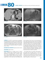
Saturation of Hemoglobin with Oxygen at Different
Altitudes. Figure 43-1 shows arterial blood oxygen satu-
ration at different altitudes while a person is breathing air
and while breathing oxygen. Up to an altitude of about
10,000 feet, even when air is breathed, the arterial oxygen
saturation remains at least as high as 90 percent. Above
10,000 feet, the arterial oxygen saturation falls rapidly, as
shown by the blue curve of the figure, until it is slightly
less than 70 percent at 20,000 feet and much less at still
higher altitudes.
527
U n i t v III
Aviation, High Altitude, and Space Physiology
Unit VIII Aviation, Space, and Deep-Sea Diving Physiology
Table 43-1 Effects of Acute Exposure to Low Atmospheric Pressures on Alveolar Gas Concentrations and Arterial Oxygen Saturation*
Breathing Air
Altitude
(ft/meters)
Breathing Pure Oxygen
Barometric
Pressure
(mm Hg)
Po2 in
Air
(mm Hg)
Pco2 in
Alveoli
(mm Hg)
Po2 in
Alveoli
(mm Hg)
Arterial
Oxygen
Saturation
(%)
Pco2 in
Alveoli
(mm Hg)
Po2 in
Alveoli
(mm Hg)
Arterial
Oxygen
Saturation
(%)
0
760
159
40 (40)
104 (104)
97 (97)
40
673
100
10,000/3048
523
110
36 (23)
67 (77)
90 (92)
40
436
100
20,000/6096
349
73
24 (10)
40 (53)
73 (85)
40
262
100
30,000/9144
226
47
24 (7)
18 (30)
24 (38)
40
139
99
40,000/12,192
141
29
36
58
84
50,000/15,240
87
18
24
16
15
Arterial oxygen saturation (percent)
*Numbers in parentheses are acclimatized values.
the arterial saturation at 47,000 feet when one is breathing
oxygen is about 50 percent and is equivalent to the arterial oxygen saturation at 23,000 feet when one is breathing
air. In addition, because an unacclimatized person usually
can remain conscious until the arterial oxygen saturation
falls to 50 percent, for short exposure times the ceiling for
an aviator in an unpressurized airplane when breathing
air is about 23,000 feet and when breathing pure oxygen is
about 47,000 feet, provided the oxygen-supplying equipment operates perfectly.
Breathing pure oxygen
100
90
80
Breathing air
70
60
Acute Effects of Hypoxia
50
0
10
20
30
40
50
Altitude (thousands of feet)
Figure 43-1 Effect of high altitude on arterial oxygen saturation
when breathing air and when breathing pure oxygen.
Effect of Breathing Pure Oxygen on Alveolar
Po2 at Different Altitudes
When a person breathes pure oxygen instead of air, most
of the space in the alveoli formerly occupied by nitrogen
becomes occupied by oxygen. At 30,000 feet, an aviator
could have an alveolar Po2 as high as 139 mm Hg instead
of the 18 mm Hg when breathing air (see Table 43-1).
The red curve of Figure 43-1 shows arterial blood
hemoglobin oxygen saturation at different altitudes when
one is breathing pure oxygen. Note that the saturation
remains above 90 percent until the aviator ascends to
about 39,000 feet; then it falls rapidly to about 50 percent
at about 47,000 feet.
The “Ceiling” When Breathing Air and When
Breathing Oxygen in an Unpressurized Airplane
Comparing the two arterial blood oxygen saturation
curves in Figure 43-1, one notes that an aviator breathing
pure oxygen in an unpressurized airplane can ascend to
far higher altitudes than one breathing air. For instance,
528
Some of the important acute effects of hypoxia in the
unacclimatized person breathing air, beginning at an
altitude of about 12,000 feet, are drowsiness, lassitude,
mental and muscle fatigue, sometimes headache, occasionally nausea, and sometimes euphoria. These effects
progress to a stage of twitchings or seizures above 18,000
feet and end, above 23,000 feet in the unacclimatized person, in coma, followed shortly thereafter by death.
One of the most important effects of hypoxia is
decreased mental proficiency, which decreases judgment,
memory, and performance of discrete motor movements.
For instance, if an unacclimatized aviator stays at 15,000
feet for 1 hour, mental proficiency ordinarily falls to about
50 percent of normal, and after 18 hours at this level it
falls to about 20 percent of normal.
Acclimatization to Low Po2
A person remaining at high altitudes for days, weeks, or
years becomes more and more acclimatized to the low
Po2, so it causes fewer deleterious effects on the body. And
it becomes possible for the person to work harder without
hypoxic effects or to ascend to still higher altitudes.
The principal means by which acclimatization comes
about are (1) a great increase in pulmonary ventilation, (2)
increased numbers of red blood cells, (3) increased diffusing capacity of the lungs, (4) increased vascularity of the
Chapter 43 Aviation, High Altitude, and Space Physiology
peripheral tissues, and (5) increased ability of the tissue
cells to use oxygen despite low Po2.
lates the arterial chemoreceptors, and this increases alveolar ventilation to a maximum of about 1.65 times normal.
Therefore, compensation occurs within seconds for the
high altitude, and it alone allows the person to rise several
thousand feet higher than would be possible without the
increased ventilation. Then, if the person remains at very
high altitude for several days, the chemoreceptors increase
ventilation still more, up to about five times normal.
The immediate increase in pulmonary ventilation on
rising to a high altitude blows off large quantities of carbon dioxide, reducing the Pco2 and increasing the pH
of the body fluids. These changes inhibit the brain stem
respiratory center and thereby oppose the effect of low Po2
to stimulate respiration by way of the peripheral arterial
chemoreceptors in the carotid and aortic bodies. But during the ensuing 2 to 5 days, this inhibition fades away,
allowing the respiratory center to respond with full force
to the peripheral chemoreceptor stimulus from hypoxia,
and ventilation increases to about five times normal.
The cause of this fading inhibition is believed to be
mainly a reduction of bicarbonate ion concentration in
the cerebrospinal fluid, as well as in the brain tissues.
This in turn decreases the pH in the fluids surrounding
the chemosensitive neurons of the respiratory center,
thus increasing the respiratory stimulatory activity of the
center.
An important mechanism for the gradual decrease
in bicarbonate concentration is compensation by the
kidneys for the respiratory alkalosis, as discussed in
Chapter 30. The kidneys respond to decreased Pco2 by
reducing hydrogen ion secretion and increasing bicarbonate excretion. This metabolic compensation for
the respiratory alkalosis gradually reduces plasma and
cerebrospinal fluid bicarbonate concentration and pH
toward normal and removes part of the inhibitory effect
on respiration of low hydrogen ion concentration. Thus,
the respiratory centers are much more responsive to
the peripheral chemoreceptor stimulus caused by the
hypoxia after the kidneys compensate for the alkalosis.
Increase in Red Blood Cells and Hemoglobin
Concentration During Acclimatization. As discussed
in Chapter 32, hypoxia is the principal stimulus for causing an increase in red blood cell production. Ordinarily,
when a person remains exposed to low oxygen for weeks
at a time, the hematocrit rises slowly from a normal value
of 40 to 45 to an average of about 60, with an average
increase in whole blood hemoglobin concentration from
normal of 15 g/dl to about 20 g/dl.
In addition, the blood volume also increases, often by
20 to 30 percent, and this increase times the increased
blood hemoglobin concentration gives an increase in total
body hemoglobin of 50 or more percent.
Peripheral Circulatory System Changes During
Acclimatization—Increased Tissue Capillarity. The
cardiac output often increases as much as 30 percent
immediately after a person ascends to high altitude but
then decreases back toward normal over a period of weeks
as the blood hematocrit increases, so the amount of oxygen
transported to the peripheral body tissues remains about
normal.
Another circulatory adaptation is growth of increased
numbers of systemic circulatory capillaries in the nonpulmonary tissues, which is called increased tissue capillarity
(or angiogenesis). This occurs especially in animals born
and bred at high altitudes but less so in animals that later
in life become exposed to high altitude.
In active tissues exposed to chronic hypoxia, the
increase in capillarity is especially marked. For instance,
capillary density in right ventricular muscle increases
markedly because of the combined effects of hypoxia and
excess workload on the right ventricle caused by pulmonary hypertension at high altitude.
Cellular Acclimatization. In animals native to altitudes of 13,000 to 17,000 feet, cell mitochondria and
cellular oxidative enzyme systems are slightly more plentiful than in sea-level inhabitants. Therefore, it is presumed
that the tissue cells of high altitude–acclimatized human
beings also can use oxygen more effectively than can their
sea-level counterparts.
Natural Acclimatization of Native Human Beings
Living at High Altitudes
Many native human beings in the Andes and in the
Himalayas live at altitudes above 13,000 feet—one group
in the Peruvian Andes lives at an altitude of 17,500 feet
and works a mine at an altitude of 19,000 feet. Many of
these natives are born at these altitudes and live there all
their lives. In all aspects of acclimatization, the natives are
superior to even the best-acclimatized lowlanders, even
though the lowlanders might also have lived at high altitudes for 10 or more years. Acclimatization of the natives
529
U n i t v III
Increased Pulmonary Ventilation—Role of Arterial
Chemoreceptors. Immediate exposure to low Po2 stimu-
Increased Diffusing Capacity After Acclimatization.
The normal diffusing capacity for oxygen through the
pulmonary membrane is about 21 ml/mm Hg/min, and
this diffusing capacity can increase as much as threefold
during exercise. A similar increase in diffusing capacity
occurs at high altitude.
Part of the increase results from increased pulmonary
capillary blood volume, which expands the capillaries and
increases the surface area through which oxygen can diffuse
into the blood. Another part results from an increase in lung
air volume, which expands the surface area of the alveolarcapillary interface still more. A final part results from an
increase in pulmonary arterial blood pressure; this forces
blood into greater numbers of alveolar capillaries than normally—especially in the upper parts of the lungs, which are
poorly perfused under usual conditions.
Quantity of oxygen in blood (vol %)
Unit VIII Aviation, Space, and Deep-Sea Diving Physiology
Mountain dwellers
28
26
24
22
20
18
16
14
12
10
8
6
4
2
0
(15,000 ft)
(Arterial values)
X
X
X
Sea-level dwellers
X
(Venous values)
0
20
40
60
80
100 120 140
Pressure of oxygen in blood (PO2) (mm Hg)
Figure 43-2 Oxygen-hemoglobin dissociation curves for blood of
high-altitude residents (red curve) and sea-level residents (blue
curve), showing the respective arterial and venous Po2 levels and
oxygen contents as recorded in their native surroundings. (Data
from Oxygen-dissociation curves for bloods of high-altitude and
sea-level residents. PAHO Scientific Publication No. 140, Life at
High Altitudes, 1966.)
begins in infancy. The chest size, especially, is greatly
increased, whereas the body size is somewhat decreased,
giving a high ratio of ventilatory capacity to body mass.
In addition, their hearts, which from birth onward pump
extra amounts of cardiac output, are considerably larger
than the hearts of lowlanders.
Delivery of oxygen by the blood to the tissues is also
highly facilitated in these natives. For instance, Figure 43-2
shows oxygen-hemoglobin dissociation curves for natives
who live at sea level and for their counterparts who live
at 15,000 feet. Note that the arterial oxygen Po2 in the
natives at high altitude is only 40 mm Hg, but because of
the greater quantity of hemoglobin, the quantity of oxygen
in their arterial blood is greater than that in the blood of
the natives at the lower altitude. Note also that the venous
Po2 in the high-altitude natives is only 15 mm Hg less than
the venous Po2 for the lowlanders, despite the very low
arterial Po2, indicating that oxygen transport to the tissues is exceedingly effective in the naturally acclimatized
high-altitude natives.
Unacclimatized
Acclimatized for 2 months
Native living at 13,200 feet but
working at 17,000 feet
Work capacity
(percent of normal)
50
68
87
Thus, naturally acclimatized native persons can
achieve a daily work output even at high altitude almost
equal to that of a lowlander at sea level, but even wellacclimatized lowlanders can almost never achieve this
result.
Acute Mountain Sickness and High-Altitude
Pulmonary Edema
A small percentage of people who ascend rapidly to high
altitudes become acutely sick and can die if not given oxygen or removed to a low altitude. The sickness begins
from a few hours up to about 2 days after ascent. Two
events frequently occur:
1. Acute cerebral edema. This is believed to result from
local vasodilation of the cerebral blood vessels, caused
by the hypoxia. Dilation of the arterioles increases
blood flow into the capillaries, thus increasing capillary pressure, which in turn causes fluid to leak into the
cerebral tissues. The cerebral edema can then lead to
severe disorientation and other effects related to cerebral dysfunction.
2. Acute pulmonary edema. The cause of this is still
unknown, but one explanation is the following: The
severe hypoxia causes the pulmonary arterioles
to constrict potently, but the constriction is much
greater in some parts of the lungs than in other parts,
so more and more of the pulmonary blood flow is
forced through fewer and fewer still unconstricted
pulmonary vessels. The postulated result is that the
capillary pressure in these areas of the lungs becomes
especially high and local edema occurs. Extension of
the process to progressively more areas of the lungs
leads to spreading pulmonary edema and severe pulmonary dysfunction that can be lethal. Allowing the
person to breathe oxygen usually reverses the process
within hours.
Reduced Work Capacity at High Altitudes
and Positive Effect of Acclimatization
Chronic Mountain Sickness
In addition to the mental depression caused by hypoxia,
as discussed earlier, the work capacity of all muscles is
greatly decreased in hypoxia. This includes not only
skeletal muscles but also cardiac muscles.
In general, work capacity is reduced in direct proportion to the decrease in maximum rate of oxygen uptake
that the body can achieve.
To give an idea of the importance of acclimatization in
increasing work capacity, consider the large differences in
work capacities as percent of normal for unacclimatized
and acclimatized people at an altitude of 17,000 feet:
Occasionally, a person who remains at high altitude
too long develops chronic mountain sickness, in which
the following effects occur: (1) The red cell mass and
hematocrit become exceptionally high, (2) the pulmonary arterial pressure becomes elevated even more
than the normal elevation that occurs during acclimatization, (3) the right side of the heart becomes greatly
enlarged, (4) the peripheral arterial pressure begins to
fall, (5) congestive heart failure ensues, and (6) death
often follows unless the person is removed to a lower
altitude.
530
Chapter 43 Aviation, High Altitude, and Space Physiology
Because of rapid changes in velocity and direction of
motion in airplanes or spacecraft, several types of acceleratory forces affect the body during flight. At the beginning of flight, simple linear acceleration occurs; at the end
of flight, deceleration; and every time the vehicle turns,
centrifugal acceleration.
Centrifugal Acceleratory Forces
When an airplane makes a turn, the force of centrifugal
acceleration is determined by the following relation:
2
f = mv
r
in which f is centrifugal acceleratory force, m is the mass
of the object, v is velocity of travel, and r is radius of curvature of the turn. From this formula, it is obvious that
as the velocity increases, the force of centrifugal acceleration increases in proportion to the square of the velocity.
It is also obvious that the force of acceleration is directly
proportional to the sharpness of the turn (the less the
radius).
Measurement of Acceleratory Force—“G.” When
an aviator is simply sitting in his seat, the force with which
he is pressing against the seat results from the pull of gravity and is equal to his weight. The intensity of this force is
said to be +1G because it is equal to the pull of gravity. If
the force with which he presses against the seat becomes
five times his normal weight during pull-out from a dive,
the force acting on the seat is +5 G.
If the airplane goes through an outside loop so that the
person is held down by his seat belt, negative G is applied
to his body; if the force with which he is held down by his
belt is equal to the weight of his body, the negative force
is −1G.
Effects on the Circulatory System. The most important effect of centrifugal acceleration is on the circulatory
system, because blood is mobile and can be translocated
by centrifugal forces.
When an aviator is subjected to positive G, blood is
centrifuged toward the lowermost part of the body. Thus,
if the centrifugal acceleratory force is +5 G and the person
is in an immobilized standing position, the pressure in
the veins of the feet becomes greatly increased (to about
450 mm Hg). In the sitting position, the pressure becomes
nearly 300 mm Hg. And, as pressure in the vessels of the
lower body increases, these vessels passively dilate so that
a major portion of the blood from the upper body is translocated into the lower vessels. Because the heart cannot
pump unless blood returns to it, the greater the quantity
of blood “pooled” in this way in the lower body, the less
that is available for the cardiac output.
Figure 43-3 shows the changes in systolic and diastolic
arterial pressures (top and bottom curves, respectively) in
the upper body when a centrifugal acceleratory force of
+3.3 G is suddenly applied to a sitting person. Note that
both these pressures fall below 22 mm Hg for the first few
seconds after the acceleration begins but then return to a
systolic pressure of about 55 mm Hg and a diastolic pressure of 20 mm Hg within another 10 to 15 seconds. This
secondary recovery is caused mainly by activation of the
baroreceptor reflexes.
Acceleration greater than 4 to 6 G causes “blackout” of
vision within a few seconds and unconsciousness shortly
thereafter. If this great degree of acceleration is continued,
the person will die.
Effects on the Vertebrae. Extremely high acceleratory
forces for even a fraction of a second can fracture the vertebrae. The degree of positive acceleration that the average person can withstand in the sitting position before
vertebral fracture occurs is about 20 G.
Negative G. The effects of negative G on the body are
less dramatic acutely but possibly more damaging permanently than the effects of positive G. An aviator can
Arterial pressure
(mm Hg)
Effects of Acceleratory Forces on the Body
in Aviation and Space Physiology
Effects of Centrifugal Acceleratory Force
on the Body—(Positive G)
100
50
0
0
5
10
15
20
25
30
Time from start of G to symptoms
(sec)
Figure 43-3 Changes in systolic (top of curve) and diastolic
(bottom of curve) arterial pressures after abrupt and continuing
exposure of a sitting person to an acceleratory force from top to
bottom of 3.3 G. (Data from Martin EE, Henry JP: Effects of time
and temperature upon tolerance to positive acceleration. J Aviation
Med 22:382, 1951.)
531
U n i t v III
The causes of this sequence of events are probably
threefold: First, the red cell mass becomes so great that
the blood viscosity increases severalfold; this increased
viscosity tends to decrease tissue blood flow so that
oxygen delivery also begins to decrease. Second, the pulmonary arterioles become vasoconstricted because of the
lung hypoxia. This results from the hypoxic vascular constrictor effect that normally operates to divert blood flow
from low-oxygen to high-oxygen alveoli, as explained in
Chapter 38. But because all the alveoli are now in the
low-oxygen state, all the arterioles become constricted,
the pulmonary arterial pressure rises excessively, and the
right side of the heart fails. Third, the alveolar arteriolar spasm diverts much of the blood flow through nonalveolar pulmonary vessels, thus causing an excess of
pulmonary shunt blood flow where the blood is poorly
oxygenated; this further compounds the problem. Most of
these people recover within days or weeks when they are
moved to a lower altitude.
Unit VIII Aviation, Space, and Deep-Sea Diving Physiology
Effects of Linear Acceleratory Forces on the Body
Acceleratory Forces in Space Travel. Unlike an air-
plane, a spacecraft cannot make rapid turns; therefore,
centrifugal acceleration is of little importance except when
the spacecraft goes into abnormal gyrations. However,
blast-off acceleration and landing deceleration can be tremendous; both of these are types of linear acceleration,
one positive and the other negative.
Figure 43-4 shows an approximate profile of acceleration during blast-off in a three-stage spacecraft, demonstrating that the first-stage booster causes acceleration
as high as 9 G, and the second-stage booster as high as
8 G. In the standing position, the human body could not
532
10
8
Acceleration (G)
usually go through outside loops up to negative acceleratory forces of −4 to −5 G without causing permanent
harm, although causing intense momentary hyperemia
of the head. Occasionally, psychotic disturbances lasting
for 15 to 20 minutes occur as a result of brain edema.
Occasionally, negative G forces can be so great (−20 G,
for instance) and centrifugation of the blood into the head
is so great that the cerebral blood pressure reaches 300
to 400 mm Hg, sometimes causing small vessels on the
surface of the head and in the brain to rupture. However,
the vessels inside the cranium show less tendency for rupture than would be expected for the following reason: The
cerebrospinal fluid is centrifuged toward the head at the
same time that blood is centrifuged toward the cranial
vessels, and the greatly increased pressure of the cerebrospinal fluid acts as a cushioning buffer on the outside of
the brain to prevent intracerebral vascular rupture.
Because the eyes are not protected by the cranium,
intense hyperemia occurs in them during strong negative
G. As a result, the eyes often become temporarily blinded
with “red-out.”
Protection of the Body Against Centrifugal Accele
ratory Forces. Specific procedures and apparatus have
been developed to protect aviators against the circulatory collapse that might occur during positive G. First, if
the aviator tightens his or her abdominal muscles to an
extreme degree and leans forward to compress the abdomen, some of the pooling of blood in the large vessels
of the abdomen can be prevented, delaying the onset of
blackout. Also, special “anti-G” suits have been devised
to prevent pooling of blood in the lower abdomen and
legs. The simplest of these applies positive pressure to the
legs and abdomen by inflating compression bags as the
G increases. Theoretically, a pilot submerged in a tank
or suit of water might experience little effect of G forces
on the circulation because the pressures developed in the
water pressing on the outside of the body during centrifugal acceleration would almost exactly balance the forces
acting in the body. However, the presence of air in the
lungs still allows displacement of the heart, lung tissues,
and diaphragm into seriously abnormal positions despite
submersion in water. Therefore, even if this procedure
were used, the limit of safety almost certainly would still
be less than 10 G.
6
4
2
0
First
booster
0
1
Second
booster
2
3
Minutes
Space
ship
4
5
Figure 43-4 Acceleratory forces during takeoff of a spacecraft.
ithstand this much acceleration, but in a semireclining
w
position transverse to the axis of acceleration, this amount
of acceleration can be withstood with ease despite the fact
that the acceleratory forces continue for as long as several
minutes at a time. Therefore, we see the reason for the
reclining seats used by astronauts.
Problems also occur during deceleration when the
spacecraft re-enters the atmosphere. A person traveling at
Mach 1 (the speed of sound and of fast airplanes) can be
safely decelerated in a distance of about 0.12 mile, whereas
a person traveling at a speed of Mach 100 (a speed possible in interplanetary space travel) would require a distance
of about 10,000 miles for safe deceleration. The principal
reason for this difference is that the total amount of energy
that must be dispelled during deceleration is proportional to the square of the velocity, which alone increases
the required distance for decelerations between Mach 1
versus Mach 100 about 10,000-fold. Therefore, deceleration must be accomplished much more slowly from high
velocities than is necessary at lower velocities.
Deceleratory Forces Associated with Parachute
Jumps. When the parachuting aviator leaves the air-
plane, his velocity of fall is at first exactly 0 feet per second.
However, because of the acceleratory force of gravity,
within 1 second his velocity of fall is 32 feet per second
(if there is no air resistance); in 2 seconds it is 64 feet
per second; and so on. As the velocity of fall increases,
the air resistance tending to slow the fall also increases.
Finally, the deceleratory force of the air resistance exactly
balances the acceleratory force of gravity, so after falling
for about 12 seconds, the person will be falling at a “terminal velocity” of 109 to 119 miles per hour (175 feet per
second). If the parachutist has already reached terminal
velocity before opening his parachute, an “opening shock
load” of up to 1200 pounds can occur on the parachute
shrouds.
The usual-sized parachute slows the fall of the parachutist to about one-ninth the terminal velocity. In other
words, the speed of landing is about 20 feet per second,
and the force of impact against the earth is 1/81 the impact
Chapter 43 Aviation, High Altitude, and Space Physiology
“Artificial Climate” in the Sealed Spacecraft
Because there is no atmosphere in outer space, an artificial atmosphere and climate must be produced in a spacecraft. Most important, the oxygen concentration must
remain high enough and the carbon dioxide concentration low enough to prevent suffocation. In some earlier
space missions, a capsule atmosphere containing pure
oxygen at about 260 mm Hg pressure was used, but in the
modern space shuttle, gases about equal to those in normal air are used, with four times as much nitrogen as oxygen and a total pressure of 760 mm Hg. The presence of
nitrogen in the mixture greatly diminishes the likelihood
of fire and explosion. It also protects against development
of local patches of lung atelectasis that often occur when
breathing pure oxygen because oxygen is absorbed rapidly
when small bronchi are temporarily blocked by mucous
plugs.
For space travel lasting more than several months, it
is impractical to carry along an adequate oxygen supply. For this reason, recycling techniques have been proposed for use of the same oxygen over and over again.
Some recycling processes depend on purely physical procedures, such as electrolysis of water to release oxygen.
Others depend on biological methods, such as use of
algae with their large store of chlorophyll to release oxygen from carbon dioxide by the process of photosynthesis. A completely satisfactory system for recycling has yet
to be achieved.
Weightlessness in Space
A person in an orbiting satellite or a nonpropelled spacecraft experiences weightlessness, or a state of near-zero
G force, which is sometimes called microgravity. That is,
the person is not drawn toward the bottom, sides, or top
of the spacecraft but simply floats inside its chambers.
The cause of this is not failure of gravity to pull on the
body because gravity from any nearby heavenly body is
still active. However, the gravity acts on both the spacecraft and the person at the same time so that both are
pulled with exactly the same acceleratory forces and in the
same direction. For this reason, the person simply is not
attracted toward any specific wall of the spacecraft.
Physiologic Problems of Weightlessness
(Microgravity). The physiologic problems of weight-
lessness have not proved to be of much significance, as
long as the period of weightlessness is not too long. Most
of the problems that do occur are related to three effects
of the weightlessness: (1) motion sickness during the first
few days of travel, (2) translocation of fluids within the
body because of failure of gravity to cause normal hydrostatic pressures, and (3) diminished physical activity
because no strength of muscle contraction is required to
oppose the force of gravity.
Almost 50 percent of astronauts experience motion
sickness, with nausea and sometimes vomiting, during
the first 2 to 5 days of space travel. This probably results
from an unfamiliar pattern of motion signals arriving in
the equilibrium centers of the brain, and at the same time
lack of gravitational signals.
The observed effects of prolonged stay in space are
the following: (1) decrease in blood volume, (2) decrease
in red blood cell mass, (3) decrease in muscle strength
and work capacity, (4) decrease in maximum cardiac
output, and (5) loss of calcium and phosphate from
the bones, as well as loss of bone mass. Most of these
same effects also occur in people who lie in bed for an
extended period of time. For this reason, exercise programs are carried out by astronauts during prolonged
space missions.
In previous space laboratory expeditions in which the
exercise program had been less vigorous, the astronauts
had severely decreased work capacities for the first few
days after returning to earth. They also tended to faint
(and still do, to some extent) when they stood up during
the first day or so after return to gravity because of diminished blood volume and diminished responses of the arterial pressure control mechanisms.
Cardiovascular, Muscle, and Bone “Decondi
tioning” During Prolonged Exposure to Weight
lessness. During very long space flights and prolonged
exposure to microgravity, gradual “deconditioning” effects
occur on the cardiovascular system, skeletal muscles, and
bone despite rigorous exercise during the flight. Studies
of astronauts on space flights lasting several months have
shown that they may lose as much 1.0 percent of their
bone mass each month even though they continue to
exercise. Substantial atrophy of cardiac and skeletal muscles also occurs during prolonged exposure to a microgravity environment.
One of the most serious effects is cardiovascular
“deconditioning,” which includes decreased work capacity,
reduced blood volume, impaired baroreceptor reflexes,
and reduced orthostatic tolerance. These changes greatly
limit the astronauts’ ability to stand upright or perform
normal daily activities after returning to the full gravity
of Earth.
533
U n i t v III
force without a parachute. Even so, the force of impact
is still great enough to cause considerable damage to the
body unless the parachutist is properly trained in landing. Actually, the force of impact with the earth is about
the same as that which would be experienced by jumping
without a parachute from a height of about 6 feet. Unless
forewarned, the parachutist will be tricked by his senses
into striking the earth with extended legs, and this will
result in tremendous deceleratory forces along the skeletal
axis of the body, resulting in fracture of his pelvis, vertebrae, or leg. Consequently, the trained parachutist strikes
the earth with knees bent but muscles taut to cushion the
shock of landing.
Unit VIII Aviation, Space, and Deep-Sea Diving Physiology
Astronauts returning from space flights lasting 4 to 6
months are also susceptible to bone fractures and may
require several weeks before they return to preflight cardiovascular, bone, and muscle fitness. As space flights become
longer in preparation for possible human exploration of
other planets, such as Mars, the effects of prolonged microgravity could pose a very serious threat to astronauts after
they land, especially in the event of an emergency landing.
Therefore, considerable research effort has been directed
toward developing countermeasures, in addition to exercise, that can prevent or more effectively attenuate these
changes. One such countermeasure that is being tested is
the application of intermittent “artificial gravity” caused by
short periods (e.g., 1 hour each day) of centrifugal acceleration of the astronauts while they sit in specially designed
short-arm centrifuges that create forces of up to 2 to 3 G.
Bibliography
Adams GR, Caiozzo VJ, Baldwin KM: Skeletal muscle unweighting: spaceflight and ground-based models, J Appl Physiol 95:2185, 2003.
Bärtsch P, Mairbäurl H, Maggiorini M, et al: Physiological aspects of highaltitude pulmonary edema, J Appl Physiol 98:1101, 2005.
534
Basnyat B, Murdoch DR: High-altitude illness, Lancet 361:1967, 2003.
Convertino VA: Mechanisms of microgravity induced orthostatic intolerance: implications for effective countermeasures, J Gravit Physiol 9:1,
2002.
Diedrich A, Paranjape SY, Robertson D: Plasma and blood volume in space,
Am J Med Sci 334:80, 2007.
Di Rienzo M, Castiglioni P, Iellamo F, et al: Dynamic adaptation of cardiac
baroreflex sensitivity to prolonged exposure to microgravity: data from
a 16-day spaceflight, J Appl Physiol 105:1569, 2008.
Hackett PH, Roach RC: High-altitude illness, N Engl J Med 345:107, 2001.
Hainsworth R, Drinkhill MJ: Cardiovascular adjustments for life at high altitude, Respir Physiol Neurobiol 158:204, 2007.
Hoschele S, Mairbaurl H: Alveolar flooding at high altitude: failure of reabsorption? News Physiol Sci 18:55, 2003.
LeBlanc AD, Spector ER, Evans HJ, et al: Skeletal responses to space flight
and the bed rest analog: a review, J Musculoskelet Neuronal Interact
7:33, 2007.
Penaloza D, Arias-Stella J: The heart and pulmonary circulation at high altitudes: healthy highlanders and chronic mountain sickness, Circulation
115:1132, 2007.
Smith SM, Heer M: Calcium and bone metabolism during space flight,
Nutrition 18:849, 2002.
West JB: Man in space, News Physiol Sci 1:198, 1986.
West JB: George I. Finch and his pioneering use of oxygen for climbing at
extreme altitudes, J Appl Physiol 94:1702, 2003.
chapter 44
When
human
beings
descend beneath the sea,
the pressure around them
increases tremendously. To
keep the lungs from collapsing, air must be supplied at very high pressure
to keep them inflated. This exposes the blood in the lungs
to extremely high alveolar gas pressure, a condition called
hyperbarism. Beyond certain limits, these high pressures
cause tremendous alterations in body physiology and can
be lethal.
Relationship of Pressure to Sea Depth. A column of seawater 33 feet (10.1 meters) deep exerts the
same pressure at its bottom as the pressure of the atmosphere above the sea. Therefore, a person 33 feet beneath
the ocean surface is exposed to 2 atmospheres pressure,
1 atmosphere of pressure caused by the weight of the
air above the water and the second atmosphere by the
weight of the water itself. At 66 feet the pressure is 3
atmospheres, and so forth, in accord with the table in
Figure 44-1.
Effect of Sea Depth on the Volume of Gases—
Boyle’s Law. Another important effect of depth is com-
pression of gases to smaller and smaller volumes. The
lower part of Figure 44-1 shows a bell jar at sea level containing 1 liter of air. At 33 feet beneath the sea, where the
pressure is 2 atmospheres, the volume has been compressed to only one-half liter, and at 8 atmospheres (233
feet) to one-eighth liter. Thus, the volume to which a given
quantity of gas is compressed is inversely proportional to
the pressure. This is a principle of physics called Boyle’s
law, which is extremely important in diving physiology
because increased pressure can collapse the air chambers
of the diver’s body, especially the lungs, and often causes
serious damage.
Many times in this chapter it is necessary to refer to
actual volume versus sea-level volume. For instance, we
might speak of an actual volume of 1 liter at a depth of 300
feet; this is the same quantity of air as a sea-level volume
of 10 liters.
Effect of High Partial Pressures
of Individual Gases on the Body
The individual gases to which a diver is exposed when
breathing air are nitrogen, oxygen, and carbon dioxide;
each of these at times can cause significant physiologic
effects at high pressures.
Nitrogen Narcosis at High Nitrogen Pressures
About four fifths of the air is nitrogen. At sea-level pressure, the nitrogen has no significant effect on bodily function, but at high pressures it can cause varying degrees of
narcosis. When the diver remains beneath the sea for an
hour or more and is breathing compressed air, the depth at
which the first symptoms of mild narcosis appear is about
120 feet. At this level the diver begins to exhibit joviality
and to lose many of his or her cares. At 150 to 200 feet,
the diver becomes drowsy. At 200 to 250 feet, his or her
strength wanes considerably, and the diver often becomes
too clumsy to perform the work required. Beyond 250
feet (8.5 atmospheres pressure), the diver usually becomes
almost useless as a result of nitrogen narcosis if he or she
remains at these depths too long.
Nitrogen narcosis has characteristics similar to those of
alcohol intoxication, and for this reason it has frequently
been called “raptures of the depths.” The mechanism of
the narcotic effect is believed to be the same as that of
most other gas anesthetics. That is, it dissolves in the fatty
substances in neuronal membranes and, because of its
physical effect on altering ionic conductance through the
membranes, reduces neuronal excitability.
Oxygen Toxicity at High Pressures
Effect of Very High Po2 on Blood Oxygen
Transport. When the Po2 in the blood rises above 100 mm
Hg, the amount of oxygen dissolved in the water of the
blood increases markedly. This is shown in Figure 44-2,
which depicts the same oxygen-hemoglobin dissociation
curve as that shown in Chapter 40 but with the alveolar
Po2 extended to more than 3000 mm Hg. Also depicted by
the lowest curve in the figure is the volume of oxygen dissolved in the fluid of the blood at each Po2 level. Note that
535
U n i t V III
Physiology of Deep-Sea Diving and Other
Hyperbaric Conditions
Unit VIII Aviation, Space, and Deep-Sea Diving Physiology
Depth (feet/meters) Atmosphere(s)
Sea level
1
33/10.1
2
66/20.1
3
100/30.5
4
133/40.5
5
166/50.6
6
200/61.0
7
300/91.4
10
400/121.9
13
500/152.4
16
1 liter
Sea level
1/2
liter
33 ft
1/4
liter
100 ft
1/8
liter
233 ft
Figure 44-1 Effect of sea depth on pressure (top table) and on gas
volume (bottom).
30
Oxygen in blood (volumes percent)
A
25
B
20
Oxygen-hemoglobin dissociation curve
Total O2 in blood
Combined with
hemoglobin
Dissolved in
water of blood
Normal alveolar
oxygen pressure
15
10
Oxygen
poisoning
5
0
0
760
1560
2280
3040
Oxygen partial pressure in lungs (mm Hg)
Figure 44-2 Quantity of oxygen dissolved in the fluid of the blood
and in combination with hemoglobin at very high Po2s.
536
in the normal range of alveolar Po2 (below 120 mm Hg),
almost none of the total oxygen in the blood is accounted
for by dissolved oxygen, but as the oxygen pressure rises
into the thousands of millimeters of mercury, a large portion of the total oxygen is then dissolved in the water of the
blood, in addition to that bound with hemoglobin.
Effect of High Alveolar Po2 on Tissue Po2. Let us
assume that the Po2 in the lungs is about 3000 mm Hg
(4 atmospheres pressure). Referring to Figure 44-2, one
finds that this represents a total oxygen content in each
100 milliliters of blood of about 29 volumes percent, as
demonstrated by point A in the figure—this means 20
volumes percent bound with hemoglobin and 9 volumes
percent dissolved in the blood water. As this blood passes
through the tissue capillaries and the tissues use their
normal amount of oxygen, about 5 milliliters from each
100 milliliters of blood, the oxygen content on leaving the
tissue capillaries is still 24 volumes percent (point B in the
figure). At this point, the Po2 is approximately 1200 mm
Hg, which means that oxygen is delivered to the tissues at
this extremely high pressure instead of at the normal value
of 40 mm Hg. Thus, once the alveolar Po2 rises above a
critical level, the hemoglobin-oxygen buffer mechanism
(discussed in Chapter 40) is no longer capable of keeping
the tissue Po2 in the normal, safe range between 20 and
60 mm Hg.
Acute Oxygen Poisoning. The extremely high tissue
Po2 that occurs when oxygen is breathed at very high alveolar oxygen pressure can be detrimental to many of the
body’s tissues. For instance, breathing oxygen at 4 atmospheres pressure of oxygen (Po2 = 3040 mm Hg) will cause
brain seizures followed by coma in most people within 30
to 60 minutes. The seizures often occur without warning
and, for obvious reasons, are likely to be lethal to divers
submerged beneath the sea.
Other symptoms encountered in acute oxygen poisoning include nausea, muscle twitchings, dizziness, disturbances of vision, irritability, and disorientation. Exercise
greatly increases the diver’s susceptibility to oxygen
toxicity, causing symptoms to appear much earlier and
with far greater severity than in the resting person.
Excessive Intracellular Oxidation as a Cause of
Nervous System Oxygen Toxicity—“Oxidizing Free
Radicals.” Molecular oxygen (O2) has little capability
of oxidizing other chemical compounds. Instead, it must
first be converted into an “active” form of oxygen. There
are several forms of active oxygen called oxygen free radi
cals. One of the most important of these is the super
oxide free radical O2−, and another is the peroxide radical
in the form of hydrogen peroxide. Even when the tissue
Po2 is normal at the level of 40 mm Hg, small amounts
of free radicals are continually being formed from the
dissolved molecular oxygen. Fortunately, the tissues also
contain multiple enzymes that rapidly remove these free
radicals, including peroxidases, catalases, and superoxide
Chapter 44 Physiology of Deep-Sea Diving and Other Hyperbaric Conditions
Chronic Oxygen Poisoning Causes Pulmonary
Disability. A person can be exposed to only 1 atmosphere
pressure of oxygen almost indefinitely without developing the acute oxygen toxicity of the nervous system just
described. However, after only about 12 hours of 1 atmosphere oxygen exposure, lung passageway congestion, pulmonary edema, and atelectasis caused by damage to the
linings of the bronchi and alveoli begin to develop. The
reason for this effect in the lungs but not in other tissues
is that the air spaces of the lungs are directly exposed to
the high oxygen pressure, but oxygen is delivered to the
other body tissues at almost normal Po2 because of the
hemoglobin-oxygen buffer system.
Carbon Dioxide Toxicity at Great Depths
in the Sea
If the diving gear is properly designed and functions properly, the diver has no problem due to carbon dioxide toxicity because depth alone does not increase the carbon
dioxide partial pressure in the alveoli. This is true because
depth does not increase the rate of carbon dioxide production in the body, and as long as the diver continues
to breathe a normal tidal volume and expires the carbon
dioxide as it is formed, alveolar carbon dioxide pressure
will be maintained at a normal value.
In certain types of diving gear, however, such as the
diving helmet and some types of rebreathing apparatuses, carbon dioxide can build up in the dead space air
of the apparatus and be rebreathed by the diver. Up to an
alveolar carbon dioxide pressure (Pco2) of about 80 mm
Hg, twice that in normal alveoli, the diver usually tolerates this buildup by increasing the minute respiratory
volume a maximum of 8- to 11-fold to compensate for
the increased carbon dioxide. Beyond 80 mm Hg alveolar
Pco2, the situation becomes intolerable, and eventually
the respiratory center begins to be depressed, rather than
excited, because of the negative tissue metabolic effects
of high Pco2. The diver’s respiration then begins to fail
rather than to compensate. In addition, the diver develops
severe respiratory acidosis and varying degrees of lethargy, narcosis, and finally even anesthesia, as discussed in
Chapter 42.
Decompression of the Diver After Excess Exposure
to High Pressure
When a person breathes air under high pressure for
a long time, the amount of nitrogen dissolved in the
body fluids increases. The reason for this is the following: Blood flowing through the pulmonary capillaries
becomes saturated with nitrogen to the same high pressure as that in the alveolar breathing mixture. And over
several more hours, enough nitrogen is carried to all the
tissues of the body to raise their tissue Pn2 also to equal
the Pn2 in the breathing air.
Because nitrogen is not metabolized by the body, it
remains dissolved in all the body tissues until the nitrogen pressure in the lungs is decreased back to some lower
level, at which time the nitrogen can be removed by the
reverse respiratory process; however, this removal often
takes hours to occur and is the source of multiple problems collectively called decompression sickness.
Volume of Nitrogen Dissolved in the Body Fluids
at Different Depths. At sea level, almost exactly 1 liter
of nitrogen is dissolved in the entire body. Slightly less
than one half of this is dissolved in the water of the body
and a little more than one half in the fat of the body. This
is true because nitrogen is five times as soluble in fat as
in water.
After the diver has become saturated with nitrogen,
the sea-level volume of nitrogen dissolved in the body at
different depths is as follows:
Feet
0
33
100
200
300
Liters
1
2
4
7
10
Several hours are required for the gas pressures of
nitrogen in all the body tissues to come nearly to equilibrium with the gas pressure of nitrogen in the alveoli.
The reason for this is that the blood does not flow rapidly enough and the nitrogen does not diffuse rapidly
enough to cause instantaneous equilibrium. The nitrogen dissolved in the water of the body comes to almost
complete equilibrium in less than 1 hour, but the fat tissue, requiring five times as much transport of nitrogen
and having a relatively poor blood supply, reaches equilibrium only after several hours. For this reason, if a person remains at deep levels for only a few minutes, not
much nitrogen dissolves in the body fluids and tissues,
whereas if the person remains at a deep level for several
hours, both the body water and body fat become saturated with nitrogen.
537
U n i t vIII
dismutases. Therefore, so long as the hemoglobin-oxygen
buffering mechanism maintains a normal tissue Po2, the
oxidizing free radicals are removed rapidly enough that
they have little or no effect in the tissues.
Above a critical alveolar Po2 (above about 2 atmospheres Po2), the hemoglobin-oxygen buffering mechanism fails, and the tissue Po2 can then rise to hundreds or
thousands of millimeters of mercury. At these high levels,
the amounts of oxidizing free radicals literally swamp the
enzyme systems designed to remove them, and now they
can have serious destructive and even lethal effects on the
cells. One of the principal effects is to oxidize the polyunsaturated fatty acids that are essential components of
many of the cell membranes. Another effect is to oxidize
some of the cellular enzymes, thus damaging severely
the cellular metabolic systems. The nervous tissues are
especially susceptible because of their high lipid content.
Therefore, most of the acute lethal effects of acute oxygen
toxicity are caused by brain dysfunction.
Unit VIII Aviation, Space, and Deep-Sea Diving Physiology
Decompression Sickness (Synonyms: Bends,
Compressed Air Sickness, Caisson Disease, Diver’s
Paralysis, Dysbarism). If a diver has been beneath the
sea long enough that large amounts of nitrogen have dissolved in his or her body and the diver then suddenly
comes back to the surface of the sea, significant quantities
of nitrogen bubbles can develop in the body fluids either
intracellularly or extracellularly and can cause minor or
serious damage in almost any area of the body, depending
on the number and sizes of bubbles formed; this is called
decompression sickness.
The principles underlying bubble formation are shown
in Figure 44-3. In Figure 44-3A, the diver’s tissues have
become equilibrated to a high dissolved nitrogen pressure
(Pn2 = 3918 mm Hg), about 6.5 times the normal amount
of nitrogen in the tissues. As long as the diver remains
deep beneath the sea, the pressure against the outside of
his or her body (5000 mm Hg) compresses all the body
tissues sufficiently to keep the excess nitrogen gas dissolved. But when the diver suddenly rises to sea level
(Figure 44-3B), the pressure on the outside of the body
becomes only 1 atmosphere (760 mm Hg), while the gas
pressure inside the body fluids is the sum of the pressures
of water vapor, carbon dioxide, oxygen, and nitrogen, or a
total of 4065 mm Hg, 97 percent of which is caused by the
nitrogen. Obviously, this total value of 4065 mm Hg is far
greater than the 760 mm Hg pressure on the outside of the
body. Therefore, the gases can escape from the dissolved
state and form actual bubbles, composed almost entirely
of nitrogen, both in the tissues and in the blood where
Pressure Outside Body
Before
decompression
After sudden
decompression
O2 = 1044 mm Hg
N2 = 3956
O2 = 159 mm Hg
N2 = 601
Total = 5000 mm Hg
Total = 760 mm Hg
Body
Gaseous pressure
in the body fluids
H2O = 47 mm Hg
CO2 = 40
O2 = 60
N2 = 3918
Body
Gaseous pressure
in the body fluids
H2O = 47 mm Hg
CO2 = 40
O2 = 60
N2 = 3918
A
Total = 4065
B
Total = 4065
Figure 44-3 Gaseous pressures both inside and outside the body,
showing (A) saturation of the body to high gas pressures when
breathing air at a total pressure of 5000 mm Hg, and (B) the great
excesses of intrabody pressures that are responsible for bubble formation in the tissues when the lung intra-alveolar pressure body
is suddenly returned from 5000 mm Hg to normal pressure of
760 mm Hg.
538
they plug many small blood vessels. The bubbles may not
appear for many minutes to hours because sometimes the
gases can remain dissolved in the “supersaturated” state
for hours before bubbling.
Symptoms of Decompression Sickness (“Bends”).
The symptoms of decompression sickness are caused
by gas bubbles blocking many blood vessels in different
tissues. At first, only the smallest vessels are blocked by
minute bubbles, but as the bubbles coalesce, progressively
larger vessels are affected. Tissue ischemia and sometimes
tissue death result.
In most people with decompression sickness, the symptoms are pain in the joints and muscles of the legs and arms,
affecting 85 to 90 percent of those persons who develop
decompression sickness. The joint pain accounts for the
term “bends” that is often applied to this condition.
In 5 to 10 percent of people with decompression sickness, nervous system symptoms occur, ranging from
dizziness in about 5 percent to paralysis or collapse and
unconsciousness in as many as 3 percent. The paralysis
may be temporary, but in some instances, damage is
permanent.
Finally, about 2 percent of people with decompression
sickness develop “the chokes,” caused by massive numbers of microbubbles plugging the capillaries of the lungs;
this is characterized by serious shortness of breath, often
followed by severe pulmonary edema and, occasionally,
death.
Nitrogen Elimination from the Body; Decompres
sion Tables. If a diver is brought to the surface slowly,
enough of the dissolved nitrogen can usually be eliminated by expiration through the lungs to prevent decompression sickness. About two thirds of the total nitrogen is
liberated in 1 hour and about 90 percent in 6 hours.
Decompression tables that detail procedures for safe
decompression have been prepared by the U.S. Navy. To
give the student an idea of the decompression process, a
diver who has been breathing air and has been on the sea
bottom for 60 minutes at a depth of 190 feet is decompressed according to the following schedule:
10 minutes at 50 feet depth
17 minutes at 40 feet depth
19 minutes at 30 feet depth
50 minutes at 20 feet depth
84 minutes at 10 feet depth
Thus, for a work period on the bottom of only 1 hour,
the total time for decompression is about 3 hours.
Tank Decompression and Treatment of Decompres
sion Sickness. Another procedure widely used for
decompression of professional divers is to put the diver
into a pressurized tank and then to lower the pressure
gradually back to normal atmospheric pressure, using
essentially the same time schedule as noted earlier.
Chapter 44 Physiology of Deep-Sea Diving and Other Hyperbaric Conditions
“Saturation Diving” and Use of Helium-Oxygen
Mixtures in Deep Dives. When divers must work at
very deep levels—between 250 feet and nearly 1000 feet—
they frequently live in a large compression tank for days
or weeks at a time, remaining compressed at a pressure
level near that at which they will be working. This keeps
the tissues and fluids of the body saturated with the gases
to which they will be exposed while diving. Then, when
they return to the same tank after working, there are no
significant changes in pressure, so decompression bubbles do not occur.
In very deep dives, especially during saturation diving, helium is usually used in the gas mixture instead
of nitrogen for three reasons: (1) it has only about onefifth the narcotic effect of nitrogen; (2) only about one
half as much volume of helium dissolves in the body
tissues as nitrogen, and the volume that does dissolve
diffuses out of the tissues during decompression several times as rapidly as does nitrogen, thus reducing
the problem of decompression sickness; and (3) the low
density of helium (one seventh the density of nitrogen)
keeps the airway resistance for breathing at a minimum,
which is very important because highly compressed
nitrogen is so dense that airway resistance can become
extreme, sometimes making the work of breathing
beyond endurance.
Finally, in very deep dives it is important to reduce the
oxygen concentration in the gaseous mixture because
otherwise oxygen toxicity would result. For instance, at a
depth of 700 feet (22 atmospheres of pressure), a 1 percent
oxygen mixture will provide all the oxygen required by the
diver, whereas a 21 percent mixture of oxygen (the percentage in air) delivers a Po2 to the lungs of more than
4 atmospheres, a level very likely to cause seizures in as
little as 30 minutes.
Scuba (Self-Contained Underwater
Breathing Apparatus) Diving
Before the 1940s, almost all diving was done using a diving helmet connected to a hose through which air was
pumped to the diver from the surface. Then, in 1943,
French explorer Jacques Cousteau popularized a selfcontained underwater breathing apparatus, known as
the SCUBA apparatus. The type of SCUBA apparatus
used in more than 99 percent of all sports and commercial diving is the open-circuit demand system shown in
Figure 44-4. This system consists of the following components: (1) one or more tanks of compressed air or
Mask
Hose
First-stage
valve
Demand valve
Air cylinders
Figure 44-4 Open-circuit demand type of SCUBA apparatus.
some other breathing mixture, (2) a first-stage “reducing” valve for reducing the very high pressure from the
tanks to a low pressure level, (3) a combination inhalation “demand” valve and exhalation valve that allows
air to be pulled into the lungs with slight negative pressure of breathing and then to be exhaled into the sea at a
pressure level slightly positive to the surrounding water
pressure, and (4) a mask and tube system with small
“dead space.”
The demand system operates as follows: The firststage reducing valve reduces the pressure from the tanks
so that the air delivered to the mask has a pressure only
a few mm Hg greater than the surrounding water pressure. The breathing mixture does not flow continually
into the mask. Instead, with each inspiration, slight extra
negative pressure in the demand valve of the mask pulls
the diaphragm of the valve open, and this automatically
releases air from the tank into the mask and lungs. In this
way, only the amount of air needed for inhalation enters
the mask. Then, on expiration, the air cannot go back into
the tank but instead is expired into the sea.
The most important problem in use of the selfcontained underwater breathing apparatus is the limited amount of time one can remain beneath the sea
surface; for instance, only a few minutes are possible at
a 200-foot depth. The reason for this is that tremendous
airflow from the tanks is required to wash carbon dioxide out of the lungs—the greater the depth, the greater
the airflow in terms of quantity of air per minute that is
required, because the volumes have been compressed to
small sizes.
539
U n i t vIII
Tank decompression is even more important for treating people in whom symptoms of decompression sickness
develop minutes or even hours after they have returned to
the surface. In this case, the diver is recompressed immediately to a deep level. Then decompression is carried out
over a period several times as long as the usual decompression period.
Unit VIII Aviation, Space, and Deep-Sea Diving Physiology
Special Physiologic Problems in Submarines
Escape from Submarines. Essentially the same
problems encountered in deep-sea diving are often met
in relation to submarines, especially when it is necessary
to escape from a submerged submarine. Escape is possible from as deep as 300 feet without using any apparatus.
However, proper use of rebreathing devices, especially
when using helium, theoretically can allow escape from
as deep as 600 feet or perhaps more.
One of the major problems of escape is prevention of
air embolism. As the person ascends, the gases in the lungs
expand and sometimes rupture a pulmonary blood vessel,
forcing the gases to enter the vessel and cause air embolism of the circulation. Therefore, as the person ascends,
he or she must make a special effort to exhale continually.
Health Problems in the Submarine Internal
Environment. Except for escape, submarine medicine
generally centers on several engineering problems to keep
hazards out of the internal environment. First, in atomic
submarines, there exists the problem of radiation hazards,
but with appropriate shielding, the amount of radiation
received by the crew submerged beneath the sea has been
less than normal radiation received above the surface of
the sea from cosmic rays.
Second, poisonous gases on occasion escape into the
atmosphere of the submarine and must be controlled rapidly. For instance, during several weeks’ submergence,
cigarette smoking by the crew can liberate enough carbon monoxide, if not removed rapidly, to cause carbon
monoxide poisoning. And, on occasion, even Freon gas
has been found to diffuse out of refrigeration systems in
sufficient quantity to cause toxicity.
Hyperbaric Oxygen Therapy
The intense oxidizing properties of high-pressure oxygen
(hyperbaric oxygen) can have valuable therapeutic effects
in several important clinical conditions. Therefore,
540
large pressure tanks are now available in many medical
centers into which patients can be placed and treated
with hyperbaric oxygen. The oxygen is usually administered at Po2s of 2 to 3 atmospheres of pressure through
a mask or intratracheal tube, whereas the gas around the
body is normal air compressed to the same high-pressure
level.
It is believed that the same oxidizing free radicals
responsible for oxygen toxicity are also responsible for at
least some of the therapeutic benefits. Some of the conditions in which hyperbaric oxygen therapy has been especially beneficial follow.
Probably the most successful use of hyperbaric oxygen has been for treatment of gas gangrene. The bacteria that cause this condition, clostridial organisms, grow
best under anaerobic conditions and stop growing at oxygen pressures greater than about 70 mm Hg. Therefore,
hyperbaric oxygenation of the tissues can frequently stop
the infectious process entirely and thus convert a condition that formerly was almost 100 percent fatal into one
that is cured in most instances by early treatment with
hyperbaric therapy.
Other conditions in which hyperbaric oxygen therapy
has been either valuable or possibly valuable include
decompression sickness, arterial gas embolism, carbon monoxide poisoning, osteomyelitis, and myocardial
infarction.
Bibliography
Butler PJ: Diving beyond the limits, News Physiol Sci 16:222, 2001.
Leach RM, Rees PJ, Wilmshurst P: Hyperbaric oxygen therapy, BMJ 317:1140,
1998.
Lindholm P, Lundgren CE: The physiology and pathophysiology of human
breath-hold diving, J Appl Physiol 106:284, 2009.
Moon RE, Cherry AD, Stolp BW, et al: Pulmonary Gas Exchange in Diving, J
Appl Physiol 2008 [Epub ahead of print].
Neuman TS: Arterial gas embolism and decompression sickness, News
Physiol Sci 17:77, 2002.
Pendergast DR, Lundgren CEG: The physiology and pathophysiology of the
hyperbaric and diving environments, J Appl Physiol 106:274, 2009.
Thom SR: Oxidative stress is fundamental to hyperbaric oxygen therapy,
J Appl Physiol 2008 doi:10.1152/japplphysiol.91004.
The Nervous System: A. General
Principles and Sensory Physiology
45. Organization of the Nervous System,
Basic Functions of Synapses, and
Neurotransmitters
46. Sensory Receptors, Neuronal Circuits for
Processing Information
47. Somatic Sensations: I. General
Organization, the Tactile and Position
Senses
48. Somatic Sensations: II. Pain, Headache,
and Thermal Sensations
Unit
IX
This page intentionally left blank
chapter 45
The nervous system is
unique in the vast complexity of thought processes and
control actions it can perform. It receives each minute literally millions of bits of
information from the different sensory nerves and sensory organs and then integrates
all these to determine responses to be made by the body.
Before beginning this discussion of the nervous system, the reader should review Chapters 5 and 7, which
present the principles of membrane potentials and transmission of signals in nerves and through neuromuscular
junctions.
ears, tactile receptors on the surface of the body, or other
kinds of receptors. These sensory experiences can either
cause immediate reactions from the brain, or memories
of the experiences can be stored in the brain for minutes,
weeks, or years and determine bodily reactions at some
future date.
Figure 45-2 shows the somatic portion of the sensory system, which transmits sensory information from
the receptors of the entire body surface and from some
deep structures. This information enters the central nervous system through peripheral nerves and is conducted
immediately to multiple sensory areas in (1) the spinal
cord at all levels; (2) the reticular substance of the medulla,
pons, and mesencephalon of the brain; (3) the cerebellum;
(4) the thalamus; and (5) areas of the cerebral cortex.
General Design of the Nervous System
Central Nervous System Neuron: The Basic
Functional Unit
The central nervous system contains more than 100 billion neurons. Figure 45-1 shows a typical neuron of a type
found in the brain motor cortex. Incoming signals enter
this neuron through synapses located mostly on the neuronal dendrites, but also on the cell body. For different
types of neurons, there may be only a few hundred or as
many as 200,000 such synaptic connections from input
fibers. Conversely, the output signal travels by way of a
single axon leaving the neuron. Then, this axon has many
separate branches to other parts of the nervous system or
peripheral body.
A special feature of most synapses is that the signal
normally passes only in the forward direction, from the
axon of a preceding neuron to dendrites on cell membranes of subsequent neurons. This forces the signal
to travel in required directions for performing specific
nervous functions.
Sensory Part of the Nervous
System—Sensory Receptors
Most activities of the nervous system are initiated by
sensory experiences that excite sensory receptors, whether
visual receptors in the eyes, auditory receptors in the
Motor Part of the Nervous System—Effectors
The most important eventual role of the nervous system
is to control the various bodily activities. This is achieved
by controlling (1) contraction of appropriate skeletal
muscles throughout the body, (2) contraction of smooth
muscle in the internal organs, and (3) secretion of active
chemical substances by both exocrine and endocrine
glands in many parts of the body. These activities are collectively called motor functions of the nervous system,
and the muscles and glands are called effectors because
they are the actual anatomical structures that perform the
functions dictated by the nerve signals.
Figure 45-3 shows the “skeletal” motor nerve axis of
the nervous system for controlling skeletal muscle contraction. Operating parallel to this axis is another system, called the autonomic nervous system, for controlling
smooth muscles, glands, and other internal bodily systems; this is discussed in Chapter 60.
Note in Figure 45-3 that the skeletal muscles can be
controlled from many levels of the central nervous system,
including (1) the spinal cord; (2) the reticular substance of
the medulla, pons, and mesencephalon; (3) the basal ganglia; (4) the cerebellum; and (5) the motor cortex. Each of
these areas plays its own specific role, the lower regions
concerned primarily with automatic, instantaneous muscle responses to sensory stimuli, and the higher regions
543
Unit IX
Organization of the Nervous System, Basic
Functions of Synapses, and Neurotransmitters
Unit IX The Nervous System: A. General Principles and Sensory Physiology
Somesthetic areas
Motor cortex
Dendrites
Thalamus
Brain
Pons
Cell body
Medulla
Cerebellum
Spinal cord
Axon
Golgi tendon
apparatus
Bulboreticular
formation
Skin
Pain, cold,
warmth (free
nerve ending)
Pressure
(Pacinian corpuscle)
(expanded tip
receptor)
Touch
(Meissner's corpuscle)
Muscle spindle
Muscle
Kinesthetic receptor
Joint
Synapses
Figure 45-2 Somatosensory axis of the nervous system.
Spinal cord
Second-order
neurons
Motor nerve
to muscles
Figure 45-1 Structure of a large neuron in the brain, showing
its important functional parts. (Redrawn from Guyton AC: Basic
Neuroscience: Anatomy and Physiology. Philadelphia: WB Saunders,
1987.)
Motor
area
Caudate
nucleus
with deliberate complex muscle movements controlled by
the thought processes of the brain.
Processing of Information—“Integrative”
Function of the Nervous System
One of the most important functions of the nervous system is to process incoming information in such a way that
appropriate mental and motor responses will occur. More
than 99 percent of all sensory information is discarded by
the brain as irrelevant and unimportant. For instance,
one is ordinarily unaware of the parts of the body that
are in contact with clothing, as well as of the seat pressure when sitting. Likewise, attention is drawn only to an
occasional object in one’s field of vision, and even the perpetual noise of our surroundings is usually relegated to
the subconscious.
But, when important sensory information excites the
mind, it is immediately channeled into proper integrative
and motor regions of the brain to cause desired responses.
This channeling and processing of information is called
the integrative function of the nervous system. Thus,
544
Thalamus
Putamen
Globus pallidus
Subthalamic nucleus
Cerebellum
Bulboreticular formation
Gamma motor fiber
Alpha motor fiber
Stretch receptor fiber
Muscle spindle
Figure 45-3 Skeletal motor nerve axis of the nervous system.
Chapter 45 Organization of the Nervous System, Basic Functions of Synapses, and Neurotransmitters
Role of Synapses in Processing Information. The
synapse is the junction point from one neuron to the next.
Later in this chapter, we discuss the details of synaptic function. However, it is important to point out here that synapses determine the directions that the nervous signals will
spread through the nervous system. Some synapses transmit signals from one neuron to the next with ease, whereas
others transmit signals only with difficulty. Also, facilitatory
and inhibitory signals from other areas in the nervous system can control synaptic transmission, sometimes opening the synapses for transmission and at other times closing
them. In addition, some postsynaptic neurons respond with
large numbers of output impulses, and others respond with
only a few. Thus, the synapses perform a selective action,
often blocking weak signals while allowing strong signals
to pass, but at other times selecting and amplifying certain
weak signals, and often channeling these signals in many
directions rather than in only one direction.
Storage of Information—Memory
Only a small fraction of even the most important sensory
information usually causes immediate motor response.
But much of the information is stored for future control
of motor activities and for use in the thinking processes.
Most storage occurs in the cerebral cortex, but even the
basal regions of the brain and the spinal cord can store
small amounts of information.
The storage of information is the process we call memory, and this, too, is a function of the synapses. Each time
certain types of sensory signals pass through sequences of
synapses, these synapses become more capable of transmitting the same type of signal the next time, a process
called facilitation. After the sensory signals have passed
through the synapses a large number of times, the synapses become so facilitated that signals generated within
the brain itself can also cause transmission of impulses
through the same sequences of synapses, even when the
sensory input is not excited. This gives the person a perception of experiencing the original sensations, although
the perceptions are only memories of the sensations.
The precise mechanisms by which long-term facilitation
of synapses occurs in the memory process are still uncertain, but what is known about this and other details of the
sensory memory process is discussed in Chapter 57.
Once memories have been stored in the nervous system, they become part of the brain processing mechanism
for future “thinking.” That is, the thinking processes of the
brain compare new sensory experiences with stored memories; the memories then help to select the important new
sensory information and to channel this into appropriate
memory storage areas for future use or into motor areas
to cause immediate bodily responses.
Major Levels of Central Nervous
System Function
The human nervous system has inherited special functional capabilities from each stage of human evolutionary
development. From this heritage, three major levels of the
central nervous system have specific functional characteristics: (1) the spinal cord level, (2) the lower brain or
subcortical level, and (3) the higher brain or cortical level.
Spinal Cord Level
We often think of the spinal cord as being only a conduit
for signals from the periphery of the body to the brain, or
in the opposite direction from the brain back to the body.
This is far from the truth. Even after the spinal cord has
been cut in the high neck region, many highly organized
spinal cord functions still occur. For instance, neuronal
circuits in the cord can cause (1) walking movements,
(2) reflexes that withdraw portions of the body from painful objects, (3) reflexes that stiffen the legs to support the
body against gravity, and (4) reflexes that control local
blood vessels, gastrointestinal movements, or urinary
excretion. In fact, the upper levels of the nervous system often operate not by sending signals directly to the
periphery of the body but by sending signals to the control centers of the cord, simply “commanding” the cord
centers to perform their functions.
Lower Brain or Subcortical Level
Many, if not most, of what we call subconscious activities
of the body are controlled in the lower areas of the brain—
in the medulla, pons, mesencephalon, hypothalamus,
thalamus, cerebellum, and basal ganglia. For instance,
subconscious control of arterial pressure and respiration is achieved mainly in the medulla and pons. Control
of equilibrium is a combined function of the older portions of the cerebellum and the reticular substance of
the medulla, pons, and mesencephalon. Feeding reflexes,
such as salivation and licking of the lips in response to
the taste of food, are controlled by areas in the medulla,
pons, mesencephalon, amygdala, and hypothalamus. And
many emotional patterns, such as anger, excitement, sexual response, reaction to pain, and reaction to pleasure,
can still occur after destruction of much of the cerebral
cortex.
Higher Brain or Cortical Level
After the preceding account of the many nervous system
functions that occur at the cord and lower brain levels,
one may ask, what is left for the cerebral cortex to do? The
answer to this is complex, but it begins with the fact that
the cerebral cortex is an extremely large memory storehouse. The cortex never functions alone but always in
association with lower centers of the nervous system.
Without the cerebral cortex, the functions of the lower
brain centers are often imprecise. The vast storehouse of
545
Unit IX
if a person places a hand on a hot stove, the desired instantaneous response is to lift the hand. And other associated
responses follow, such as moving the entire body away
from the stove and perhaps even shouting with pain.
Unit IX The Nervous System: A. General Principles and Sensory Physiology
cortical information usually converts these functions to
determinative and precise operations.
Finally, the cerebral cortex is essential for most of our
thought processes, but it cannot function by itself. In
fact, it is the lower brain centers, not the cortex, that initiate wakefulness in the cerebral cortex, thus opening its
bank of memories to the thinking machinery of the brain.
Thus, each portion of the nervous system performs specific functions. But it is the cortex that opens a world of
stored information for use by the mind.
Comparison of the Nervous System
with a Computer
When computers were first developed, it soon became
apparent that these machines have many features in common with the nervous system. First, all computers have
input circuits that are comparable to the sensory portion
of the nervous system, as well as output circuits that are
comparable to the motor portion of the nervous system.
In simple computers, the output signals are controlled
directly by the input signals, operating in a manner similar
to that of simple reflexes of the spinal cord. In more complex computers, the output is determined both by input
signals and by information that has already been stored in
memory in the computer, which is analogous to the more
complex reflex and processing mechanisms of our higher
nervous system. Furthermore, as computers become even
more complex, it is necessary to add still another unit,
called the central processing unit, which determines the
sequence of all operations. This unit is analogous to the
control mechanisms in our brain that direct our attention
first to one thought or sensation or motor activity, then to
another, and so forth, until complex sequences of thought
or action take place.
Figure 45-4 is a simple block diagram of a computer.
Even a rapid study of this diagram demonstrates its similarity to the nervous system. The fact that the basic components of the general-purpose computer are analogous
to those of the human nervous system demonstrates that
the brain is basically a computer that continuously collects
Problem
Input
Procedure
for solution
Central
processing unit
Output
Initial
data
Result of
operations
Answer
Information
storage
Computational
unit
Figure 45-4 Block diagram of a general-purpose computer, showing the basic components and their interrelations.
546
sensory information and uses this along with stored information to compute the daily course of bodily activity.
Central Nervous System Synapses
Information is transmitted in the central nervous system
mainly in the form of nerve action potentials, called simply “nerve impulses,” through a succession of neurons,
one after another. However, in addition, each impulse (1)
may be blocked in its transmission from one neuron to
the next, (2) may be changed from a single impulse into
repetitive impulses, or (3) may be integrated with impulses
from other neurons to cause highly intricate patterns of
impulses in successive neurons. All these functions can be
classified as synaptic functions of neurons.
Types of Synapses—Chemical and Electrical
There are two major types of synapses: (1) the chemical
synapse and (2) the electrical synapse.
Almost all the synapses used for signal transmission in
the central nervous system of the human being are chemical synapses. In these, the first neuron secretes at its nerve
ending synapse a chemical substance called a neurotransmitter (or often called simply transmitter substance), and
this transmitter in turn acts on receptor proteins in the
membrane of the next neuron to excite the neuron, inhibit
it, or modify its sensitivity in some other way. More than
40 important transmitter substances have been discovered
thus far. Some of the best known are acetylcholine, norepinephrine, epinephrine, histamine, gamma-aminobutyric
acid (GABA), glycine, serotonin, and glutamate.
Electrical synapses, in contrast, are characterized by
direct open fluid channels that conduct electricity from
one cell to the next. Most of these consist of small protein tubular structures called gap junctions that allow
free movement of ions from the interior of one cell to
the interior of the next. Such junctions were discussed
in Chapter 4. Only a few examples of gap junctions have
been found in the central nervous system. However, it is
by way of gap junctions and other similar junctions that
action potentials are transmitted from one smooth muscle fiber to the next in visceral smooth muscle (Chapter 8)
and from one cardiac muscle cell to the next in cardiac
muscle (Chapter 10).
“One-Way” Conduction at Chemical Synapses.
Chemical synapses have one exceedingly important characteristic that makes them highly desirable for transmitting most nervous system signals. They always transmit
the signals in one direction: that is, from the neuron that
secretes the transmitter substance, called the presynaptic neuron, to the neuron on which the transmitter acts,
called the postsynaptic neuron. This is the principle of
one-way conduction at chemical synapses, and it is quite
different from conduction through electrical synapses,
which often transmit signals in either direction.
Chapter 45 Organization of the Nervous System, Basic Functions of Synapses, and Neurotransmitters
Physiologic Anatomy of the Synapse
Figure 45-5 shows a typical anterior motor neuron in the
anterior horn of the spinal cord. It is composed of three
major parts: the soma, which is the main body of the neuron; a single axon, which extends from the soma into a
peripheral nerve that leaves the spinal cord; and the dendrites, which are great numbers of branching projections
of the soma that extend as much as 1 millimeter into the
surrounding areas of the cord.
As many as 10,000 to 200,000 minute synaptic knobs
called presynaptic terminals lie on the surfaces of the dendrites and soma of the motor neuron, about 80 to 95 percent of them on the dendrites and only 5 to 20 percent
on the soma. These presynaptic terminals are the ends
of nerve fibrils that originate from many other neurons.
Many of these presynaptic terminals are excitatory—
that is, they secrete a transmitter substance that excites
the postsynaptic neuron. But other presynaptic terminals
are inhibitory—they secrete a transmitter substance that
inhibits the postsynaptic neuron.
Neurons in other parts of the cord and brain differ from the anterior motor neuron in (1) the size of the
cell body; (2) the length, size, and number of dendrites,
ranging in length from almost zero to many centimeters;
(3) the length and size of the axon; and (4) the number of
presynaptic terminals, which may range from only a few
to as many as 200,000. These differences make neurons in
different parts of the nervous system react differently to
incoming synaptic signals and, therefore, perform many
different functions.
Presynaptic Terminals. Electron microscopic studies of the presynaptic terminals show that they have varied anatomical forms, but most resemble small round or
oval knobs and, therefore, are sometimes called terminal
knobs, boutons, end-feet, or synaptic knobs.
Figure 45-6 illustrates the basic structure of a synapse,
showing a single presynaptic terminal on the membrane
surface of a postsynaptic neuron. The presynaptic terminal is separated from the postsynaptic neuronal soma
by a synaptic cleft having a width usually of 200 to 300
angstroms. The terminal has two internal structures
important to the excitatory or inhibitory function of the
synapse: the transmitter vesicles and the mitochondria.
The transmitter vesicles contain the transmitter substance
that, when released into the synaptic cleft, either excites
or inhibits the postsynaptic neuron—excites if the neuronal membrane contains excitatory receptors, inhibits if
the membrane contains inhibitory receptors. The mitochondria provide adenosine triphosphate (ATP), which in
turn supplies the energy for synthesizing new transmitter
substance.
When an action potential spreads over a presynaptic
terminal, depolarization of its membrane causes a small
number of vesicles to empty into the cleft. The released
Dendrites
Axon
Transmitter vesicles
Postsynaptic membrane
Soma
Mitochondria
Presynaptic
terminal
Figure 45-5 Typical anterior motor neuron, showing presynaptic
terminals on the neuronal soma and dendrites. Note also the single axon.
Synaptic cleft
(200-300
angstroms)
Receptor
proteins
Dendrite of neuron
Figure 45-6 Physiologic anatomy of the synapse.
547
Unit IX
Think for a moment about the extreme importance of
the one-way conduction mechanism. It allows signals to
be directed toward specific goals. Indeed, it is this specific transmission of signals to discrete and highly focused
areas both within the nervous system and at the terminals
of the peripheral nerves that allows the nervous system to
perform its myriad functions of sensation, motor control,
memory, and many others.
Unit IX The Nervous System: A. General Principles and Sensory Physiology
transmitter in turn causes an immediate change in
permeability characteristics of the postsynaptic neuronal
membrane, and this leads to excitation or inhibition of the
postsynaptic neuron, depending on the neuronal receptor
characteristics.
Mechanism by Which an Action Potential Causes
Transmitter Release from the Presynaptic
Terminals—Role of Calcium Ions
The membrane of the presynaptic terminal is called the
presynaptic membrane. It contains large numbers of
voltage-gated calcium channels. When an action potential depolarizes the presynaptic membrane, these calcium channels open and allow large numbers of calcium
ions to flow into the terminal. The quantity of transmitter substance that is then released from the terminal into
the synaptic cleft is directly related to the number of calcium ions that enter. The precise mechanism by which
the calcium ions cause this release is not known, but it is
believed to be the following.
When the calcium ions enter the presynaptic terminal,
it is believed that they bind with special protein molecules
on the inside surface of the presynaptic membrane, called
release sites. This binding in turn causes the release sites
to open through the membrane, allowing a few transmitter vesicles to release their transmitter into the cleft
after each single action potential. For those vesicles that
store the neurotransmitter acetylcholine, between 2000
and 10,000 molecules of acetylcholine are present in each
vesicle, and there are enough vesicles in the presynaptic
terminal to transmit from a few hundred to more than
10,000 action potentials.
Action of the Transmitter Substance on the
Postsynaptic Neuron—Function of “Receptor
Proteins”
The membrane of the postsynaptic neuron contains large
numbers of receptor proteins, also shown in Figure 45-6.
The molecules of these receptors have two important
components: (1) a binding component that protrudes outward from the membrane into the synaptic cleft—here it
binds the neurotransmitter coming from the presynaptic
terminal—and (2) an ionophore component that passes all
the way through the postsynaptic membrane to the interior of the postsynaptic neuron. The ionophore in turn
is one of two types: (1) an ion channel that allows passage of specified types of ions through the membrane or
(2) a “second messenger” activator that is not an ion channel but instead is a molecule that protrudes into the cell
cytoplasm and activates one or more substances inside
the postsynaptic neuron. These substances in turn serve
as “second messengers” to increase or decrease specific
cellular functions.
Ion Channels. The ion channels in the postsynaptic
neuronal membrane are usually of two types: (1) cation
channels that most often allow sodium ions to pass when
opened, but sometimes allow potassium and/or calcium
548
ions as well, and (2) anion channels that allow mainly
chloride ions to pass but also minute quantities of other
anions.
The cation channels that conduct sodium ions are lined
with negative charges. These charges attract the positively
charged sodium ions into the channel when the channel
diameter increases to a size larger than that of the hydrated
sodium ion. But those same negative charges repel chloride
ions and other anions and prevent their passage.
For the anion channels, when the channel diameters
become large enough, chloride ions pass into the channels and on through to the opposite side, whereas sodium,
potassium, and calcium cations are blocked, mainly
because their hydrated ions are too large to pass.
We will learn later that when cation channels open
and allow positively charged sodium ions to enter, the
positive electrical charges of the sodium ions will in
turn excite this neuron. Therefore, a transmitter substance that opens cation channels is called an excitatory
transmitter. Conversely, opening anion channels allows
negative electrical charges to enter, which inhibits the
neuron. Therefore, transmitter substances that open these
channels are called i nhibitory transmitters.
When a transmitter substance activates an ion channel,
the channel usually opens within a fraction of a millisecond; when the transmitter substance is no longer present,
the channel closes equally rapidly. The opening and closing of ion channels provide a means for very rapid control
of postsynaptic neurons.
“Second Messenger” System in the Postsynaptic
Neuron. Many functions of the nervous system—for
instance, the process of memory—require prolonged
changes in neurons for seconds to months after the initial transmitter substance is gone. The ion channels are
not suitable for causing prolonged postsynaptic neuronal
changes because these channels close within milliseconds after the transmitter substance is no longer present. However, in many instances, prolonged postsynaptic
neuronal excitation or inhibition is achieved by activating a “second messenger” chemical system inside the
postsynaptic neuronal cell itself, and then it is the second
messenger that causes the prolonged effect.
There are several types of second messenger systems.
One of the most common types uses a group of proteins
called G-proteins. Figure 45-7 shows in the upper left corner a membrane receptor protein. A G-protein is attached
to the portion of the receptor that protrudes into the interior of the cell. The G-protein in turn consists of three
components: an alpha (α) component that is the activator portion of the G-protein and beta (β) and gamma (γ)
components that are attached to the alpha component
and also to the inside of the cell membrane adjacent to the
receptor protein. On activation by a nerve impulse, the
alpha portion of the G-protein separates from the beta
and gamma portions and then is free to move within the
cytoplasm of the cell.
Inside the cytoplasm, the separated alpha component
performs one or more of multiple functions, depending on
Chapter 45 Organization of the Nervous System, Basic Functions of Synapses, and Neurotransmitters
Transmitter substance
Receptor
protein
b
a
1
Opens
channel
G-protein
K+
Membrane
enzyme
Unit IX
g
Potassium
channel
2
a
3
Activates one or
more intracellular
enzymes
Activates gene
transcription
4
Activates
enzymes
GTP
ATP
or
cAMP
Specific cellular
chemical activators
cGMP
Proteins and
structural changes
Figure 45-7 “Second messenger” system by which a transmitter substance from an initial neuron can activate a second neuron by first
releasing a “G-protein” into the second neuron’s cytoplasm. Four subsequent possible effects of the G-protein are shown, including 1, opening an ion channel in the membrane of the second neuron; 2, activating an enzyme system in the neuron’s membrane; 3, activating an
intracellular enzyme system; and/or 4, causing gene transcription in the second neuron.
the specific characteristic of each type of neuron. Shown
in Figure 45-7 are four changes that can occur. They are
as follows:
1. Opening specific ion channels through the postsynaptic
cell membrane. Shown in the upper right of the figure is a potassium channel that is opened in response
to the G-protein; this channel often stays open for a
prolonged time, in contrast to rapid closure of directly
activated ion channels that do not use the second messenger system.
2. Activation of cyclic adenosine monophosphate (cAMP)
or cyclic guanosine monophosphate (cGMP) in the
neuronal cell. Recall that either cyclic AMP or cyclic
GMP can activate highly specific metabolic machinery in the neuron and, therefore, can initiate any one
of many chemical results, including long-term changes
in cell structure itself, which in turn alters long-term
excitability of the neuron.
3. Activation of one or more intracellular enzymes. The
G-protein can directly activate one or more intracellular enzymes. In turn the enzymes can cause any one of
many specific chemical functions in the cell.
4. Activation of gene transcription. This is one of the most
important effects of activation of the second messenger
systems because gene transcription can cause formation of new proteins within the neuron, thereby changing its metabolic machinery or its structure. Indeed, it
is well known that structural changes of appropriately
activated neurons do occur, especially in long-term
memory processes.
It is clear that activation of second messenger systems
within the neuron, whether they be of the G-protein type
or of other types, is extremely important for changing the
long-term response characteristics of different neuronal
pathways. We will return to this subject in more detail
in Chapter 57 when we discuss memory functions of the
nervous system.
Excitatory or Inhibitory Receptors in the
Postsynaptic Membrane
Some postsynaptic receptors, when activated, cause excitation of the postsynaptic neuron, and others cause inhibition. The importance of having inhibitory, as well as
excitatory, types of receptors is that this gives an additional dimension to nervous function, allowing restraint
of nervous action and excitation.
The different molecular and membrane mechanisms
used by the different receptors to cause excitation or inhibition include the following.
Excitation
1. Opening of sodium channels to allow large numbers of
positive electrical charges to flow to the interior of the
postsynaptic cell. This raises the intracellular membrane potential in the positive direction up toward
the threshold level for excitation. It is by far the most
widely used means for causing excitation.
2. Depressed conduction through chloride or potassium channels, or both. This decreases the diffusion
of negatively charged chloride ions to the inside of the
postsynaptic neuron or decreases the diffusion of positively charged potassium ions to the outside. In either
instance, the effect is to make the internal membrane potential more positive than normal, which is
excitatory.
549