Ebook The Washington manual of critical care (3/E): Part 2
Bạn đang xem bản rút gọn của tài liệu. Xem và tải ngay bản đầy đủ của tài liệu tại đây (30.71 MB, 287 trang )
SECTION XII
HEPATIC DISEASES
45
Acute Liver Failure
Claire Meyer and Jeffrey S. Crippin
Acute liver failure (ALF) is characterized by coagulopathy, encephalopathy, and severe hepatic injury in patients without chronic liver disease (Table
45.1). Exceptions to the absence of pre-existing liver disease include autoimmune hepatitis and Wilson’s disease, if the disease has only been
recognized within the last 26 weeks. Approximately 2000 cases of ALF are reported per year in the United States.
CAUSES AND DIAGNOSIS
Determining the cause of ALF is imperative, since some etiologies dictate specific treatments. In a prospective multicenter study of 308 patients (1998
to 2001) by the Acute Liver Failure Study Group, the following causes were most frequently identified: acetaminophen overdose (39%), indeterminate
(17%), idiosyncratic drug reactions (13%), and viral hepatitis (hepatitis A virus or hepatitis B) (11%). Table 45.1 outlines the possible causes of ALF,
as well as the studies needed to evaluate patients for each etiology. On presentation, initial laboratory analysis should include a complete blood count,
basic metabolic panel, liver chemistries, magnesium, phosphate, prothrombin time, lactic acid, arterial blood gas, ammonia, acetaminophen level, acute
viral hepatitis panel, toxicology screen, ceruloplasmin level, antinuclear antibodies, antismooth muscle antibodies, HIV status, and a pregnancy test (if
applicable).
ETIOLOGY-SPECIFIC MANAGEMENT OF ACUTE LIVER FAILURE (See Algorithm 45.1)
Acetaminophen Toxicity
As acetaminophen toxicity is the leading cause of ALF in the United States, clinicians should have a high index of suspicion for acetaminophen
overdose, particularly when there is inadequate knowledge of the circumstances preceding a patient’s presentation to the hospital. N-acetylcysteine
(NAC) therapy is indicated when acetaminophen-related ALF is known or suspected, regardless of the grade of encephalopathy, and should be initiated
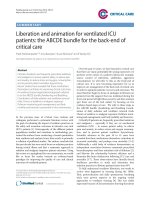
as soon after acetaminophen ingestion as possible. The nomogram shown in Figure 45.1 helps to guide treatment based on the serum acetaminophen
level when a single ingestion occurred at a known time. However, in the setting of ALF, treatment with NAC should be initiated if the serum
acetaminophen is elevated at any level, as significant liver injury can result from multiple relatively small doses over time. If ingestion is known to have
occurred within 4 hours of presentation, activated charcoal lowers the plasma acetaminophen level more effectively than does gastric lavage or ipecac,
and is typically given as a single dose (1 g/kg). The efficacy of NAC is not reduced by prior treatment with activated charcoal. Patients with
acetaminophen toxicity should be treated with NAC even if they present to medical care after a significant delay. A retrospective study including
patients who began NAC 10 to 36 hours after overdose showed improved outcomes in this group, compared to those receiving no antidote. Refer to
Algorithm 45.1 for PO and IV NAC dosing. The route of administration (oral or intravenous) has not been shown to affect outcomes. Cochrane analysis
of one prospective, controlled trial of NAC for acetaminophen-related ALF showed reduced mortality (Peto odds ratio 0.29) in patients treated with
NAC.
TABLE 45.1 Diagnosis and Causes of Acute Liver Failure
1. Acute hepatic injury <26 wks without evidence of
pre-existing liver disease
2. Encephalopathy
3. Coagulopathy (INR ≥1.5)
↓
Etiology
History and Physical Examination
Diagnostic Evaluation
Acetaminophen
History of ingestion
Acetaminophen level (short half-life–low serum level does not rule out ingestion), use
nomogram when time of ingestion known
Drug toxicity
New medications, antibiotics, NSAIDs,
Serum drug levels
anticonvulsants, psychiatric history, herbals; unlikely
if >1 yr on medication
Other toxins
Mushroom ingestion, cocaine or MDMA use
Viral
Viral syndrome, pregnancy, recent travel, skin lesions, Hepatitis B surface antigen, hepatitis B core IgM, Hepatitis A IgM, hepatitis E antibody,
immunocompromised state
hepatitis C antibody, hepatitis C RNA, HIV antibody, HSV antibodies and DNA, VZV DNA;
consider evaluation for rare viral causes including parvovirus B19, adenovirus, and EBV
Shock liver
History of heart failure, cardiac arrest, volume
depletion, or substance abuse
BNP, lactate dehydrogenase, lactate, echocardiogram
Infiltrative malignancy
History of malignancy, hepatomegaly
If suspected, cross-sectional abdominal imaging and liver biopsy (if feasible)
Budd–Chiari syndrome
History of malignancy or other prothrombotic condition, Abdominal ultrasound with Doppler
including recent pregnancy or exogenous
estrogens; personal or family history of venous
thromboembolism; lymphadenopathy
Wilson’s disease
Patient <40 yrs old, history of neuropsychiatric
symptoms; Kayser–Fleischer rings on slit lamp
exam
Urine drug screen
Serum ceruloplasmin and copper, 24-hr urine copper, uric acid, hemolysis labs; if
suspected, liver biopsy (if feasible) for quantitative copper measurement
Acute fatty liver of pregnancy,
HELLP
Pregnancy
B-HCG in women of childbearing potential; if suspected, urinalysis to evaluate for
proteinuria. If liver biopsy performed, frozen section needed for oil red O stain (AFLP)
Autoimmune
Hepatitis
History of other autoimmune diseases
Antinuclear antibody, antismooth muscle antibody, anti-LKM1, serum immunoglobulins; if
suspected, liver biopsy (if feasible)
ALGORITHM 45.1
Treatment Algorithm for Acute Liver Failure
Figure 45.1. Acetaminophen toxicity nomogram. (Adapted from Rumack BH, Peterson RC, Koch GG, et al. Acetaminophen overdose: 662 cases with evaluation of oral
acetylcysteine treatment. Arch Intern Med. 1981;141:380.)
Non-Acetaminophen Etiologies
The benefit of NAC in acetaminophen toxicity has been demonstrated for decades, but its role in non-acetaminophen–related ALF has only recently been
established. A randomized placebo-controlled trial including 173 patients with non-acetaminophen–related ALF, demonstrated improved transplant-free
survival at 3 weeks and 1 year in patients who received 72 hours of NAC therapy. This benefit was seen only in patients with grade 1 to 2
encephalopathy, but not in those with more advanced grades. Given its minimal adverse reaction profile, NAC therapy should be initiated for all patients
with ALF presenting with grade 1 to 2 encephalopathy. Treatment with NAC should not delay transfer to a transplant facility.
For non-acetaminophen–related causes of liver damage, etiology-specific interventions are unlikely to be life-saving in the setting of ALF; rather,
the decision regarding need and eligibility for liver transplant is crucial. Nonetheless, when a specific etiology is identified, initiation of directed
therapy can be considered as outlined in Algorithm 45.1. Hepatitis B, with or without hepatitis D, accounts for more than half of viral causes of ALF.
Treatment with a nucleotide or nucleoside analog is generally recommended, though evidence is mixed with regard to its impact on clinical outcomes in
this setting; lamivudine (100 mg/day) has been used in the majority of reports. Hepatitis E is a more common viral cause of ALF in endemic countries
and should be considered in returning travelers or recent immigrants from these regions. Treatment for acute hepatitis A and E is supportive. Hepatitis C
alone rarely causes ALF; however, other viruses such as HSV, EBV, adenovirus, and parvovirus B19 have been reported.
MANAGEMENT OF SYSTEMIC COMPLICATIONS
Central Nervous System
Cerebral edema and increased intracranial pressure (ICP) are serious complications of ALF. The risk of cerebral edema increases with progression of
encephalopathy, with a >75% incidence in patients with grade 4 encephalopathy. Advanced cerebral edema can lead to uncal herniation and death.
Management of neurologic complications is outlined in Algorithm 45.2. Patients with any degree of encephalopathy should be transferred to a liver
transplant center. Patients with grade 3 to 4 encephalopathy should be intubated for airway protection. Peri-intubation, attempts should be made to avoid
coughing and paralysis is often used as part of the induction regimen. Frequent neurologic examinations are imperative, and findings such as systemic
hypertension, bradycardia, posturing, and decreased pupillary reflexes can suggest impending herniation.
ALGORITHM 45.2
CNS Complications of Acute Liver Failure
ICP monitoring should be considered for patients with rapidly progressive encephalopathy and those listed for liver transplantation. In the absence
of definitive evidence of a mortality benefit, the frequency with which ICP monitoring is used varies widely among liver transplant programs. ICP can
be measured with an epidural, subdural, parenchymal, or ventricular catheter. Epidural catheters generally have a lower complication rate, but are less
reliable. The most common complications include bleeding in the setting of coagulopathy, infection, and volume overload resulting from correction of
coagulopathy. Recombinant factor VIIa has been used in a small trial to aid with placement of ICP transducers with favorable results. The role of
noninvasive ICP monitoring (using transcranial Doppler) is not yet established. ICP should be maintained at a level <20 mm Hg, with a cerebral
perfusion pressure (mean arterial pressure [MAP] minus ICP) >50 mm Hg.
Once increased ICP or cerebral edema is present, aggressive measures should be undertaken to prevent herniation. Propofol sedation, avoidance of
sensory stimulation, and raising the head of the bed can be helpful. Therapies focused on decreasing cerebral edema include osmotic agents (mannitol or
hypertonic saline) or decreasing cerebral blood flow (hyperventilation or hypothermia).
Mannitol is administered as a bolus dose (0.5 to 1 g/kg of a 20% solution). The dose can be repeated twice, however, administration is limited by
maintaining a serum osmolality <320 mOsm/kg. If patients have concomitant renal failure, hemofiltration should be considered. Hyperventilation has
only a short-term benefit, but can be used with the goal of reducing PaCO2 to 25 mm Hg. An RCT demonstrated no benefit of prophylactic continuous
hyperventilation in ALF. A study of 30 patients with ICP monitoring randomized to 3% hypertonic saline with a goal serum sodium concentration of 145
to 155 mmol/L, showed a significant decrease in average ICP and episodes of increased ICP, but no survival benefit. Hypothermia (32° to 34°C) has
been associated with a beneficial effect in uncontrolled trials. Patients with ALF may have seizure activity, but prophylactic phenytoin has not proven to
be effective in improving survival. Despite an association between an arterial ammonia level of >200 mcmol/L and herniation, no benefit of gut
decontamination or lactulose has been demonstrated in ALF. Hemofiltration via CRRT can reduce ammonia levels, though its effect on ICP has not been
studied. Barbiturate coma can be attempted for refractory increased ICP, but requires close monitoring of MAP due to its association with hypotension.
Dexamethasone is not effective at prolonging survival.
Coagulopathy
The management of coagulopathy is outlined in Algorithm 45.3. Synthesis of coagulation factors I, II, V, VII, IX, and X is depressed in patients with
ALF. Sources of bleeding include procedure sites, stress ulcers, lungs, and the oropharynx. Proton pump inhibitors should be used for stress ulcer
prophylaxis. Platelets should only be transfused for counts <10,000/μL or in the face of active bleeding. Vitamin K is routinely given, but fresh-frozen
plasma should not be transfused unless there is active bleeding or a planned procedure. Packed red blood cells can be transfused for symptomatic
anemia or to replace blood loss secondary to hemorrhage.
The role of recombinant factor VIIa has been evaluated during the placement of ICP monitors. In an unblinded study comparing patients with ALF
given recombinant factor VIIa with a cohort of historic controls, patients receiving recombinant factor VIIa all had successful placement (7/7 vs. 3/8).
Patients receiving recombinant factor VIIa also had a significant decrease in mortality and anasarca from fluid overload.
Hypotension
Hypotension is multifactorial in patients with ALF, resulting from volume depletion, third spacing, infection, gastrointestinal bleeding, or as a result of
overall low systemic vascular resistance and a hyperkinetic cardiovascular state. Fluid resuscitation should be balanced with avoidance of volume
overload and the theoretical risk of increasing ICP. Maintenance fluid should be glucose based due to the hypoglycemia associated with liver failure.
Although not compared directly in trials, dopamine or norepinephrine can be used for vasopressor support with a MAP goal of 65 to 75 mm Hg. In a
small study, dopamine led to a significant increase in cardiac output, systemic oxygen delivery, and hepatic and splanchnic blood flow when used to
increase MAP by 10 mm Hg. Although systemic oxygen consumption was increased, splanchnic oxygen consumption was decreased. A small trial
evaluating the role of norepinephrine in ALF noted an increase in MAP, although it was not associated with an increase in cardiac index and actually
resulted in a decrease in systemic oxygen consumption. Resuscitation with colloid is theoretically better than crystalloid, given that albumin induces a
more effective expansion of the central blood volume, but no mortality benefit has been shown.
ALGORITHM 45.3
Management of the Complications of Fulminant Hepatic Failure
Infection
Infections are found in 80% of patients with ALF, with 25% of patients developing documented bacteremia and 33% developing systemic fungal
infections. Periodic surveillance cultures should be obtained to detect infections as early as possible. Although prophylactic antibiotics do not provide a
survival advantage, a low threshold for initiation of broad-spectrum coverage should be maintained. Infections and hyperthermia increase the risk of
hepatic encephalopathy, therefore a theoretical benefit of empiric antibiotic therapy exists for patients with worsening encephalopathy.
Renal Failure
Renal failure is multifactorial in patients with ALF because of the direct toxic effect of ingested substances, volume depletion, hypotension, acute tubular
necrosis, and/or the hepatorenal syndrome. In contrast to acute tubular necrosis, renal failure due to the hepatorenal syndrome is characterized by low
urinary sodium (<10 mEq/L), progressive hyponatremia, and a lack of improvement with volume expansion. Nephrotoxic agents such as
aminoglycosides and NSAIDs should be avoided and NAC should be used prior to intravenous contrast studies. When dialysis is needed, continuous
renal replacement therapy should be used over daily intermittent hemodialysis, due to its association with improved cardiovascular dynamics.
Metabolic Complications
Metabolic complications include hypoglycemia resulting from diminished glucose synthesis and lactic acidosis due to anaerobic glucose metabolism.
Patients benefit from glucose monitoring and treatment of hypoglycemia with dextrose-based solutions. Electrolytes such as phosphorus, potassium, and
magnesium are usually abnormal, and should be repleted as indicated. Enteral or parenteral nutrition should be initiated early and protein should not be
restricted. A recent Cochrane database review did not find convincing evidence of a beneficial role of branched chain amino acids in the treatment of
patients with hepatic encephalopathy.
PROGNOSTIC INDICATORS
The most important prognostic indicator in ALF is the etiology of hepatic damage. Acetaminophen toxicity, hepatitis A, ischemic liver injury, and
pregnancy-related liver failure portend a transplant-free survival of >50% while idiosyncratic drug reactions, hepatitis B, autoimmune hepatitis,
Wilson’s disease, and Budd–Chiari Syndrome carry a survival rate of <25% without transplantation. The timing of disease onset is also important, but
this data may be confounded by the etiology of ALF. An illness of <1 week in duration suggests ischemic hepatopathy or acetaminophen overdose and is
associated with improved survival, while an illness >4 weeks in duration suggests an indeterminate or viral etiology and indicates a poor transplant-free
survival. The degree of encephalopathy is another strong predictor of outcome (Table 45.2). Patients with grade 2 encephalopathy have a 65% to 70%
chance of survival, whereas patients with grade 3 or 4 have a 30% to 50% and a 20% chance of survival, respectively. The King’s College Criteria
(Table 45.3) are important indicators of outcome. In patients with non-acetaminophen–associated ALF, the presence of a single factor is associated with
a mortality rate of 80%, while the presence of all three factors is associated with 95% mortality. In patients with acetaminophen hepatotoxicity and ALF,
a single risk factor is associated with a mortality of 55%, and the presence of severe acidosis confers 95% mortality.
TABLE 45.2 West Haven Criteria for Semiquantitative Grading of Mental State
Grade 1
Trivial lack of awareness
Euphoria or anxiety
Shortened attention span
Impaired performance of addition
Grade 2
Lethargy or apathy
Minimal disorientation for time or place
Subtle personality change
Inappropriate behavior
Impaired performance of subtraction
Grade 3
Somnolence to semistupor, but responsive to verbal stimuli
Confusion
Gross disorientation
Grade 4
Coma (unresponsive to verbal or noxious stimuli)
Atterbury CE, Maddrey WC, Conn HO. Neomycin-sorbitol and lactulose in the treatment of acute portal-systemic encephalopathy. A controlled, double-blind clinical trial. Am J Dig Dis.
1978;23(5):398–406.
TABLE 45.3 King’s College Hospital Criteria for Liver Transplantation in FHF
Acetaminophen-induced disease
Arterial pH <7.30
OR
Prothrombin time >100 s AND
Creatinine >3.4 mg/dL AND
Grade III or IV encephalopathy
Nonacetaminophen-induced disease
Prothrombin time >100 s (regardless of encephalopathy grade)
OR
Any three of the following (regardless of encephalopathy grade): Age <10 yrs or >40 yrs
Etiology: non-A, non-B hepatitis, halothane hepatitis, or idiosyncratic drug reaction
Duration of jaundice before onset of encephalopathy >7 days
Prothrombin time >50 s
Serum bilirubin >18 mg/dL
O’Grady JG, Alexander GJ, Kayllar KM, et al. Early indicators of prognosis in fulminant hepatic failure. Gastroenterology. 1989;97(2):439–445.
LIVER TRANSPLANTATION
Liver transplantation is a proven treatment for ALF, although limited by the prompt availability of donors. Posttransplant survival rates are as high as
80% to 90%. The decision to pursue transplantation versus continuing medical therapy (such as NAC) is difficult. Factors to consider include the
possibility of spontaneous recovery, the feasibility of transplantation, and assessment of contraindications to transplantation. Prognostic models such as
the King’s College Criteria (Table 45.3) and the Acute Physiology and Chronic Health Evaluation (APACHE) II score help in determining the need for
liver transplantation. For patients with acetaminophen-associated ALF, a recent meta-analysis reported that the King’s College Criteria had a sensitivity
of 0.59 and specificity of 0.92 in determining the need for transplantation. An APACHE II score of >15 was associated with a specificity of 0.81 and
sensitivity of 0.92 in determining the need for transplantation. The APACHE II score had a higher positive likelihood ratio of 16.4 and negative
likelihood ratio of 0.19 (one study) versus the King’s criteria, with a positive and negative likelihood ratio of 12.33 and 0.29, respectively, based on six
pooled studies.
SUGGESTED READINGS
Brok J, Buckley N, Gluud C. Interventions for paracetamol (acetaminophen) overdose. Cochrane Database Syst Rev. 2006;CD003328.
This meta-analysis provides a comprehensive review of proven and unproven therapies for the leading cause of fulminant hepatic failure.
Hoofnagle JH, Carithers RL, Shapiro C, et al. Fulminant hepatic failure: summary of a workshop. Hepatology. 1995;21(11):240–252.
This paper summarizes issues in the management of fulminant hepatic failure.
Kulkarni S, Cronin DC. Fulminant hepatic failure. In: Hall JB, Schidt GA, Wood LD, eds.Principles of Critical Care. 3rd ed. New York: McGrawHill Professional; 2005:1279–1288.
This chapter provides an excellent overview of the pathophysiology and management issues in fulminant hepatic failure.
Lee WM, Larson AM, Stravitz RT. AASLD position paper: the management of acute liver failure: update 2011.
/>This paper provides guidelines by the American Association for the Study of Liver Diseases on the management of fulminant hepatic failure.
O’Grady J. Acute liver failure. In: Feldman M, Friedman LS, Brandt LJ, eds.Sleisenger & Fordtran’s Gastrointestinal and Liver Disease. 10th ed.
Philadelphia, PA: Saunders; 2016:1591–1602.
This chapter provides an excellent overview of the pathophysiology and management issues in fulminant hepatic failure.
Raghavan M, Marik PE. Therapy of intracranial hypertension in patients with fulminant hepatic failure. Neurocrit Care. 2006;4(2):179–189.
This paper provides an excellent overview of treatment for intracranial hypertension and reviews the current understanding of the mechanisms
leading to this life threatening complication.
46
Hyperbilirubinemia
Yeshika Sharma and Jeffrey S. Crippin
PHYSIOLOGY
Heme is a breakdown constituent of senescent erythrocytes. It is converted to biliverdin by heme oxygenase and further reduced by biliverdin reductase
to bilirubin in the reticuloendothelial system. Bilirubin, unconjugated and water insoluble at this point, is tightly bound to albumin and delivered to the
liver. It is transported into the hepatocytes by carrier-mediated mechanisms, transferred to the endoplasmic reticulum bound by cytosolic proteins, and
converted to a water soluble form with the addition of uridine diphosphate glucuronic acid, the conjugated form of bilirubin. An ATP-dependent export
pump, the rate limiting step in bilirubin transport, transfers conjugated bilirubin into the biliary canaliculi, where it is added to bile. Bile eventually
drains into the small intestine and is subsequently metabolized by ileal and colonic bacteria to urobilinogen. Eighty per cent of urobilinogen is excreted
in stool, while approximately 20% is reabsorbed in the small intestine and enters the portal circulation. The reabsorbed urobilinogen is subsequently
excreted in stool and urine.
Based on the above physiology, the bilirubin pathway can be divided into four steps: (1) bilirubin production, (2) hepatic bilirubin uptake, (3)
bilirubin conjugation, and (4) bilirubin excretion. Hyperbilirubinemia is classically divided into unconjugated and conjugated forms with disruption at
steps 1, 2, and 3, leading to unconjugated hyperbilirubinemia and disruption of step 4 causing conjugated hyperbilirubinemia. However, this division is
rarely absolute and clinicians may encounter a mixed picture.
Indirect Hyperbilirubinemia
Unconjugated hyperbilirubinemia occurs when indirect bilirubin is >80% of the total bilirubin. This may be caused by increased bilirubin production or
decreased hepatocyte uptake and conjugation. Hemolysis, extravasation of blood into tissues (resorption of internal bleeding or hematoma),
dyserythropoiesis (thalassemia, myelodysplasia, aplastic anemia, vitamin B12 and folate deficiency), and sepsis are frequent causes of unconjugated
hyperbilirubinemia. Hemolysis is frequently characterized by an elevated reticulocyte count, schistocytes or spherocytes on peripheral smear, a positive
Coombs test, an increased lactate dehydrogenase, and a decreased haptoglobin level. Physical examination may reveal splenomegaly. Unconjugated
hyperbilirubinemia can lead to formation of pigmented gallstones. A decrease in hepatocyte uptake and conjugation is the result of inhibition of uptake
mechanisms, inhibition of glucoronidation, or defects in conjugation. Competitive inhibition of bilirubin uptake may be caused by medications such as
rifampin and probenecid, while inhibition of glucuronidation can occur with hyperthyroidism and estradiol therapy. A common enzymatic defect,
decreased activity of bilirubin UDP-glucuronyl transferase, results in asymptomatic unconjugated hyperbilirubinemia, better known as Gilbert’s
Syndrome. A more severe quantitative defect in UDP-glucuronyl transferase leads to Crigler-Najjar types I and II. In addition, cardio-pulmonary failure
can lead to congestive hepatopathy that presents as indirect hyperbilirubinemia.
Direct Hyperbilirubinemia
Conjugated or direct hyperbilirubinemia is usually secondary to hepatocellular dysfunction, biliary obstruction, or biliary injury. Hepatocellular
dysfunction, whether acute or chronic, can cause reflux of conjugated bilirubin into the circulation. This is dependent largely on the fact that active
canalicular excretion of conjugated bilirubin is the rate-limiting step in the bilirubin pathway and extremely sensitive to liver dysfunction. Acute
hepatocellular dysfunction is characterized by an elevated bilirubin in association with elevated aminotransferases. Chronic dysfunction results in lower
aminotransferase levels, common causes including chronic viral hepatitis, alcoholic liver disease, and nonalcoholic steatohepatitis (NASH). Both acute
and chronic liver dysfunction may give rise to a mixed hyperbilirubinemia if a disease process causing unconjugated hyperbilirubinemia is
superimposed on hepatic dysfunction.
Biliary dysfunction may result from obstruction of the extrahepatic biliary ducts or nonobstructive injury of the intra- or extra-hepatic ducts. A
direct bilirubin fraction >50% of the total bilirubin suggests a hepatobiliary etiology and, if accompanied by an elevated alkaline phosphatase and
gamma-glutamyl transpeptidase (GGTP), favors biliary obstruction. Causes of intrinsic obstruction include choledocholithiasis, biliary strictures,
cholangiocarcinoma, primary sclerosing cholangitis, AIDS cholangiopathy, and parasitic infection (e.g., cryptosporidium). Extrinsic compression can be
secondary to pancreatic masses (tumor, fibrosis, pseudocyst, or abscess), or lymphadenopathy. Nonobstructive biliary disease also presents with an
elevated alkaline phosphatase, an elevated GGTP, and direct hyperbilirubinemia, but without imaging evidence of obstruction. Potential etiologies
include acute viral hepatitis, primary biliary cirrhosis, infiltrative diseases such as amyloidosis and sarcoidosis, drug toxicity, sepsis, total parenteral
nutrition, and paraneoplastic syndrome secondary to renal cell carcinoma. Other diseases such as Dubin-Johnson and Rotor syndrome can also cause
direct hyperbilirubinemia.
Diagnosis and Therapy
Imaging is required for diagnosis and guides therapy. Imaging modalities include ultrasound, computed tomography (CT), endoscopic retrograde
cholangiopancreatography (ERCP), percutaneous transhepatic cholangiography (PTC), and magnetic resonance cholangiopancreatography (MRCP)
(Algorithm 46.1). An abdominal ultrasound or CT, both with high specificity, can confirm an obstructive process. Ultrasound is a more sensitive
technique for detecting stones within the gallbladder, whereas both techniques are less apt to identify choledocholithiasis. An ultrasound is less helpful
in obese patients and when overlying bowel gas is present. If these studies fail to reveal the cause of biliary obstruction, an MRCP gives better
visualization of the intrahepatic ducts. If an obstructive process is confirmed, cholangiography can provide direct access to the biliary tree. An ERCP
gains access to the proximal biliary tree while PTC, starting at the peripheral bile ducts, allows visualization of the biliary tree. Either study allows
decompression of obstructive processes via sphincterotomy and stone retrieval, stricture dilation, or stent placement. Superimposed infection of an
obstructed biliary tract must promptly be treated with broad spectrum antibiotics and prompt decompression of the biliary tree. If no obstruction is found
and a cholestatic pattern still persists, cholangiography may be useful to delineate biliary anatomy. CT imaging can reveal infiltrative disease, and a
liver biopsy may be required to further define the amount and type of liver injury.
ALGORITHM 46.1
Evaluation and Management of Hyperbilirubinemia
SUGGESTED READINGS
Greenberger NJ, Paumgartner G. Diseases of the gallbladder and bile ducts. In: Kasper D, et al., eds.Harrison’s Principles of Internal Medicine. 16th
ed. New York: McGraw-Hill; 2005.
This chapter discusses common causes of biliary dysfunction and provides an approach to diagnosing biliary disease.
Lidofsky S. Jaundice. In: Feldman M, ed. Sleisenger & Fordtran’s Gastrointestinal and Liver Disease. 7th ed. Philadelphia, PA: Saunders; 2002.
This chapter provides a systematic approach to evaluating a patient with jaundice and compares the various imaging modalities to evaluate
biliary disease.
Pratt DS, Kaplan MM. Jaundice. In: Kasper D, et al., eds. Harrison’s Principles of Internal Medicine. 16th ed. New York: McGraw-Hill; 2005.
This chapter also provides a systematic approach to evaluating a patient with jaundice.
Roche SP, Kobos R. Jaundice in the adult patient. Am Fam Physician. 2004;69(2):299–304.
Summerfield JA. Diseases of the gallbladder and biliary tree. In: Warrell DA, Cox TM, Firth JD, et al., eds.Oxford Textbook of Medicine. 4th ed.
Oxford: Oxford University Press; 2003.
This source provides an excellent overview of investigations in biliary disease.
Wolkoff A. The hyperbilirubinemias. Kasper D, et al., eds. Harrison’s Principles of Internal Medicine. 16th ed. New York: McGraw-Hill; 2005.
This chapter provides a great review of the pathophysiology and disorders of the biliary system.
47
End-Stage Liver Disease
Kevin M. Korenblat
The shared outcome of most untreated, chronic liver diseases is the development of cirrhosis. The resulting liver disease is commonly referred to as
decompensated cirrhosis and is characterized by the signs and symptoms of both portal hypertension and hepatic synthetic dysfunction. These
complications typically coexist in patients with cirrhosis and are the major cause of liver disease-related morbidity and mortality. Common
complications of portal hypertension are ascites, portal hypertensive-related bleeding, hepatic encephalopathy, and acute kidney injury (AKI). Intensive
care unit (ICU) admissions for these complications are a frequent occurrence, and successful management depends on prompt diagnosis and treatment.
ASCITES
Ascites describes the pathologic accumulation of serous fluid in the peritoneal cavity. It is the most frequent manifestation of decompensated cirrhosis
and is associated with a median 2-year mortality rate of 50%. Cirrhotic ascites is identified by its low albumin content and >1.1 g/dL difference
between serum and ascites albumin concentrations (serum ascites-albumin gradient).
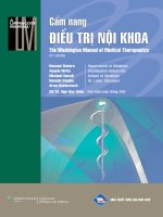
Paracentesis for sampling of the ascites is required in all patients with new-onset ascites or in those with a change in their clinical condition, such
as confusion, renal dysfunction, or gastrointestinal bleeding. Paracentesis (Fig. 47.1) is a safe procedure that can be done even in patients with
coagulopathy and thrombocytopenia. The right and left lower quadrants are the preferred site for paracentesis, and complications are unusual and mostly
limited to abdominal wall hematomas. The ascites should be analyzed for albumin, cell count with differential and the fluid inoculated directly into
blood culture media.
Although ascites is best managed with oral furosemide and spironolactone, diuretics may need to be withheld in ICU patients who frequently have
renal dysfunction, hypovolemia, or electrolyte disturbances. Intravenous (IV) diuretics for treatment of ascites and edema should be avoided in patients
with cirrhosis as they can precipitate renal failure. Repeated large-volume paracentesis is a valid strategy for the management of ascites refractory to
medical therapy. The administration of albumin at the time of paracentesis has been advocated to ameliorate the risk of post-paracentesis circulatory
dysfunction. In practice, 12.5 g of 25% albumin can be infused for every 2 L of ascites removed. The timing of administration has not been rigorously
studied, but owing to the long half-life of albumin in the circulation, the administration after completion of the paracentesis is likely to be sufficient. The
principle benefit of large-volume paracentesis is relief of symptoms; there is no evidence that those with large-volume cirrhotic ascites are at risk for
the abdominal compartment syndrome and, thus, paracentesis should not be expected to improve renal function.
Figure 47.1. Areas of dullness in both right and left lower abdominal quadrants are ideal sites for diagnostic paracentesis.
Hepatic hydrothorax occurs in as many as 13% of patients with ascites, is typically right-sided, and occurs as a result of defects in the diaphragm
that permit passage of ascites into the pleural space. This complication can be managed by thoracentesis, diuretics, and, when refractory to medical
therapy, transjugular intrahepatic shunt (TIPS). Tube thoracostomy should be avoided because volume losses can be substantial and precipitate renal
dysfunction.
SPONTANEOUS BACTERIAL PERITONITIS
The most common infectious complication of ascites is the development of spontaneous bacterial peritonitis (SBP). Infections, including SBP, are
associated with a fourfold increase in in-hospital mortality. Between 10% and 27% of those with cirrhotic ascites will have SBP at the time of
hospitalization. There is no typical presentation of SBP, and signs such as abdominal pain, fever, or leukocytosis are frequently absent. The diagnosis is
established by the finding of >250/mL polymorphonuclear cells in the ascites or the growth of organisms in a culture of ascites fluid. SBP should be
differentiated from secondary bacterial peritonitis as a consequence of bowel perforation or intra-abdominal abscess (Algorithm 47.1).
SBP should be treated with IV antibiotics. Second- and third-generation cephalosporins (cefotaxime 1 g IV q8h or ceftriaxone 1 g q24h) have
proven effective in the management of SBP; however, these antibiotics may not always be adequate. Infections with multidrug resistant (MDR) bacteria
are increasing. The rates of infection with MDR organisms, primarily SBP and urinary tract infections, can be as high as 47% in hospitalized patients
with ascites. Risk factors for MDR infections include nosocomial infections and exposure to systemic antibiotics within 30 days prior to an infection.
Exposure to oral, nonabsorbed antibiotics used in the management of encephalopathy has not been shown to be a risk factor.
ALGORITHM 47.1
Algorithm of the Assessment of Cirrhotic Ascites
Renal dysfunction occurs in as many as one-third of patients with SBP despite adequate antibiotic treatment. Discontinuation of diuretics and the
administration of IV albumin (25%) given at a dose of 1.5 g/kg body weight (day 1) and 1 g/kg (day 3) reduces rates of renal dysfunction. This
intervention should be strongly considered in all patients with SBP and particularly those with jaundice and renal insufficiency.
Antibiotic prophylaxis has been advocated in cirrhotic patients at high risk for bacterial complications. These include patients with variceal
hemorrhage, primary prevention of SBP in those with ascitic fluid protein <1.5 g/dL, and at least one of the following criteria: serum creatinine >1.2
mg/dL, BUN >25 mg/dL, Na <130 mEq/L, or bilirubin >3 mg/dL or secondary prevention in those with a prior episode of SBP. Balancing the benefits of
prophylactic antibiotics with the inevitable risks of selecting for antimicrobial resistance remains the central concern with antibiotic prophylaxis, and
additional studies will be required to define their optimal use.
ACUTE KIDNEY INJURY
AKI in decompensated cirrhosis can arise from changes in intravascular volume, parenchymal renal disease, medication-related injuries, and
disturbance to renal perfusion from the vascular dilation in mesenteric and systemic circulation that is the hallmark of decompensated cirrhosis.
A consensus definition of AKI in cirrhosis is an increase in serum creatinine (sCR) ≥0.3 mg/dL or increase in sCR ≥50% from baseline levels
within the past 7 days. The stage of AKI is defined by the magnitude of the change in sCR from baseline (Table 47.1).
Patients with stage 2 or 3 AKI who fail to respond to therapeutic interventions and who meet other consensus criteria (Table 47.2) are considered
to have the hepatorenal syndrome (HRS). The syndrome can be subdivided into a rapidly progressive (type 1) and a slower (type 2) form.
TABLE 47.1 AKI in Patients With Cirrhosis
Definition
Increase in sCR ≥0.3 mg/dL within 48 hours or increase in sCR ≥50% from baseline within the prior 7 days
Staging
Stage 1
Stage 2
Stage 3
Increase in sCR ≥0.3 mg/dL or increase in sCR ≥1.5–2-fold from baseline
Increase in sCR ≥2–3-fold from baseline
Increase in sCR >3-fold from baseline or sCR ≥4.0 mg/dL with an acute increase ≥0.3 mg/dL or initiation of renal replacement
therapy
Treatment Response
Partial response
Regression of AKI stage with a reduction of sCr ≥0.3 mg/dL above the baseline value
Return of sCR to a value within 0.3 mg/dL of the baseline value
Full response
TABLE 47.2 Diagnostic Criteria for the Hepatorenal Syndrome (HRS)
Diagnosis of cirrhosis and ascites
Diagnosis of AKI
No response after 2 consecutive days of diuretic withdrawal and plasma volume expansion with albumin 1 g/kg body weight
Absence of shock
No current or recent use of nephrotoxic drugs
No macroscopic signs of parenchymal kidney injury, defined as:
Absence of proteinuria (>500 mg/day)
Absence of microhematuria (>50 RBCs per high-power field)
Normal findings on renal ultrasonography
Treatment of HRS begins with intravascular volume expansion. Albumin (25%) is a particularly effective volume expander, and support for its role
is provided by the success of albumin in conjunction with vasoactive agents in improving HRS compared to vasoactive agents and saline. The
coadministration with vasoactive agents such as octreotide (100 to 200 mcg SC q8h) and midodrine (7.5 to 12.5 mg q8h) or terlipressin (a
vasoconstrictor not currently approved in the United States) has been studied for the treatment of HRS in clinical trials of varying quality. Liver
transplantation remains the most effective approach for the treatment of HRS.
ENCEPHALOPATHY
Early symptoms of hepatic encephalopathy are often subtle and can include changes in mood and insomnia that only later progress to agitation and coma.
The development of encephalopathy of any severity should prompt a search for precipitants that commonly include infection, gastrointestinal
hemorrhage, or medication exposures. The mediators of hepatic encephalopathy are unknown. Serum ammonia is a biomarker of hepatic encephalopathy,
but may not correlate well with the severity of the encephalopathy.
Treatment options include cathartics (lactulose 30 cc PO q2–8h or lactulose retention enemas) or nonabsorbable, oral antibiotics (neomycin 500
mg PO q6h or rifaximin 550 mg bid). In a randomized, double-blind placebo-controlled study in which 90% of patients were receiving lactulose, the
addition of rifaximin significantly reduced the risk of recurrent episodes of hepatic encephalopathy.
VARICEAL HEMORRHAGE
Variceal hemorrhage has an annual incidence rate of 20% in cirrhosis, and each episode carries a 20% to 40% mortality rate. There is no typical
presentation of variceal bleeding, and it should be suspected in those with known chronic liver disease and gastrointestinal hemorrhage. The initial steps
in the management of acute variceal bleeding involve treatment of shock and protection of the airway (Table 47.3). Volume resuscitation in the form of
packed red blood cells should be privileged to other blood products. Octreotide (50 mcg IV bolus followed by 50 mcg/hr IV infusion) should be started.
Diagnostic paracentesis should be performed, and prophylactic parenteral antibiotics should be given; the latter intervention is associated with
decreased risk of rebleeding.
TABLE 47.3 Guidelines for the Management of Variceal Hemorrhage
Resuscitate hypovolemic shock
Assessment of airway and intubation if airway protection necessary
Octreotide 50 mcg IV bolus followed by 50 mcg/hr IV infusion
Blood and urine culture; diagnostic paracentesis
Prophylactic parenteral antibiotics
Upper endoscopy
TIPS, BRTO, or Blakemore tube for variceal bleeding refractory to endoscopic management
Upper endoscopy should be performed promptly as both band ligation and sclerotherapy can result in effective hemostasis for esophageal varices.
TIPS is an option for esophageal variceal bleeding that is refractory to endoscopy or for bleeding gastric varices. Early TIPS placement (within 24 to 48
hours following hospitalization for variceal hemorrhage) in Child class B and C cirrhosis has also been advocated as a strategy that prolongs survival
based on randomized trials.
Balloon-occluded retrograde transvenous obliteration (BRTO) is another potential therapy that may be particularly helpful in the management of
bleeding gastric or ectopic varices. Balloon tamponade devices (Blakemore tube) can also be inserted temporary in cases where either TIPS or
endoscopy is delayed or unsuccessful. Nonselective beta blockers (e.g., propranolol, nadolol, or carvedilol) are effective at reducing the risk of initial
and recurrent variceal bleeding; however, they should be introduced only after acute bleeding is controlled and the patient is hemodynamically stable.
There are conflicting data on the safety of beta blockers in patients with decompensated cirrhosis. Until their role is clarified by further clinical
studies, a reasoned approach would be to continue the medication except for those with manifestations of extreme vasodilation such as those with
refractory ascites, AKI, hypotension (systolic blood pressure <90 mm Hg), or hyponatremia (serum sodium <130 mg/dL).
TRANSJUGULAR INTRAHEPATIC PORTOSYSTEMIC SHUNT (TIPS)
TIPS is a channel created between the hepatic vein and the intrahepatic portion of the portal vein. It is placed to reduce portal pressure in patients with
complications related to portal hypertension, most commonly variceal hemorrhage or refractory ascites. Contraindications to TIPS placement can
include pulmonary hypertension, right heart failure, severe encephalopathy, polycystic liver disease, or tumor within the path of the TIPS.
ACUTE ON CHRONIC LIVER FAILURE
An acute deterioration of liver function in patients with cirrhosis that results in the failure of one or more organs has been used to describe the category
of acute on chronic liver failure (ACLF). This is distinct from both acute liver failure anddecompensated liver disease. Not surprisingly, the
development of ACLF has been associated with alcohol consumption within 3 months and bacterial infections. In as many as 45% of cases, the cause is
unknown. The mortality risk increases with an increase in organ failure; for example, in the setting of the failure of three or more organs, the 28-day
transplant-free mortality risk was 78%.
SUGGESTED READINGS
Angeli P, Gines P, Wong F, et al. Diagnosis and management of acute kidney injury in patients with cirrhosis: revised consensus recommendations of the
international club of ascites. J Hepatol. 2015;62(4):968–974.
Revised definitions and diagnostic criteria for acute kidney injury by the International Ascites Club (IAC).
DellaVolpe JD, Garavaglia JM, Huang DT. Management of complications of end-stage liver disease in the intensive care unit.J Intensive Care Med.
2016;31(2):94–103.
A useful and readable summary of common complications encountered in the ICU care of patients with end-stage liver disease.
Jalan R, Fernandez J, Wiest R, et al. Bacterial infections in cirrhosis: a position statement based on the EASL special conference 2013.J Hepatol.
2014;60(6):1310–1324.
Moreau R, Arroyo V. Acute-on-chronic liver failure: a new clinical entity. Clin Gastroenterol Hepatol. 2015;13(5):836–841.
A summary of current research on acute on chronic liver failure, including definitions, risk factors, and outcomes.
Nadim MK., Durand F, Kellum JA, et al. Management of the critically ill patient with cirrhosis: a multidisciplinary perspective.J Hepatol.
2016;64(3):717–735.
Review of the multidisciplinary management of critically ill patients with cirrhosis.
Tandon P, Delisle A, Topal JE, et al. High prevalence of antibiotic-resistant bacterial infections among patients with cirrhosis at a US liver center. Clin
Gastroenterol Hepatol. 2012; 10(11):1291–1298.
Analysis on the frequency of multidrug resistant infections in hospitalized patients with cirrhosis.
Section XIII
GASTROINTESTINAL DISORDERS
48
Upper Gastrointestinal Bleeding
Jason G. Bill and C. Prakash Gyawali
Acute upper gastrointestinal bleeding (UGIB) is a common medical emergency that frequently results in emergency department evaluations and intensive
care unit admissions. The annual incidence of acute UGIB is estimated to range between 80 and 90 cases per 100,000 population, carrying a mortality
rate of 6% to 12%. In the past decade, the incidence of nonvariceal gastrointestinal hemorrhage has declined in all populations, possibly because of
lower Helicobacter pylori incidence and widespread use of proton pump inhibitors (PPIs). Common causes of acute UGIB are listed in Table 48.1.
One of the first assessments in any patient with acute gastrointestinal bleeding is determining the severity of the bleeding episode (Algorithm 48.1).
Bleeding is considered massive with loss of one-fifth to one-fourth of the circulating volume if a previously normotensive or hypertensive patient
develops resting hypotension. In the absence of resting hypotension, evidence of postural or orthostatic hypotension (drop of systolic blood pressure of
15 mm Hg or increase in heart rate of 20 beats per minute) indicates loss of 10% to 20% of the circulating volume. Bleeding is considered minor if
neither of these conditions is met, indicating loss of <10% of circulating volume. In all instances, two large-bore intravenous (IV) lines or a central line
must be urgently placed, and normal saline or lactated Ringer’s solution administered intravenously. Rapid repletion of circulating volume is crucial
when blood loss approaches massive, and transfusion of packed red blood cells needs to be arranged, requiring a blood draw for blood count,
metabolic profile, coagulation parameters, blood type, and cross-matching. When type-specific blood is not immediately available, O negative blood
may need to be transfused, using rapid infusing devices if necessary. Blood transfusions are performed with target hemoglobin ≥7 g/dL; higher
hemoglobin values may ultimately be necessary in patients with clinical evidence of intravascular volume depletion or comorbidities such as coronary
artery disease. In stable patients without comorbidities, transfusion is only required if hemoglobin is <7 g/dL. Oxygen is administered by nasal cannula
to improve oxygen-carrying capacity of the blood, and vital signs and urine output are constantly monitored.
TABLE 48.1 Etiology of Upper Gastrointestinal Bleeding
Peptic ulcer disease (accounts for ~50%)
Gastric ulcers
Duodenal ulcers
Gastric erosions and gastritis
Esophageal and/or gastric varices (accounts for 10%–20%)
Stress ulcers
Mallory–Weiss tear
Esophagitis and esophageal ulcers
Vascular abnormalities (angiodysplasia, Dieulafoy lesion, telangiectasia)
Portal hypertensive gastropathy
Neoplasms, benign and malignant
Hemobilia (bleeding into bile ducts)
Hemosuccus (bleeding into pancreatic ducts)
Aortoenteric fistula
ALGORITHM 48.1
Initial Management of Acute Gastrointestinal (GI) Bleeding
Factors propagating bleeding must be rapidly assessed during this initial evaluation. Patients receiving heparin infusion, thrombolytic therapy, or
newer antithrombotic agents (Algorithm 48.1) need to be assessed to determine if it is safe to temporarily discontinue these medications. Oral
anticoagulants are held, and the anticoagulation reversed with vitamin K and/or fresh-frozen plasma, if possible. As the use of novel oral anticoagulants
increases, reversal agents are under development. The only currently available reversal agent is idarucizumab (Praxbind), which has been approved for
patients with life-threatening hemorrhage while taking dabigatran (Pradaxa). Otherwise, prothrombin complex concentrates (PCC) may be considered in
patients with severe or life-threatening bleeding. Hemodialysis can be used to reduce the blood concentration of dabigatran, but not rivaroxaban and
apixaban, which are more tightly plasma protien bound.
Once the patient is stabilized hemodynamically, further evaluation can resume (Algorithm 48.1). A history of hematemesis or coffee-ground emesis
establishes the diagnosis of UGIB. Melena (passage of dark, tarry, sticky, and foul-smelling stool) typically indicates a proximal gut source for blood
loss, but melena can develop from bleeding sites as far distal as the proximal or even middle colon. Nevertheless, upper endoscopy is the first
investigation to be done in the presence of melena. Up to 10% to 11% of patients presenting with hematochezia and altered hemodynamic parameters are
found to have an upper gastrointestinal source for their bleeding. Therefore, in the presence of significant hemodynamic compromise, upper
gastrointestinal tract evaluation is indicated even if the bleeding presentation resembles lower gastrointestinal bleeding.
Aspiration of bloody gastric contents through a nasogastric tube establishes the diagnosis of UGIB and can be useful in identifying patients with
high-risk lesions who may benefit from emergent endoscopy. On the other hand, dark blood or coffee grounds that clear quickly on nasogastric tube
lavage may indicate that active bleeding has ceased, and elective endoscopy within 24 hours may be adequate (Table 48.2). The absence of a bloody
aspirate does not exclude active upper gastrointestinal bleeding, and bleeding can be present despite a negative aspirate in approximately 15% of cases.
Early indicators for the need for intensive care unit admission include massive bleeding, hemodynamic compromise, variceal bleeding, bleeding onset
while hospitalized for an unrelated illness, and the presence of factors that predict a poor outcome (Table 48.3). Hemoccult testing of nasogastric
aspirates has very little value in the assessment of acute UGIB. If the nasogastric aspirate is clear, or clears quickly with a tap water lavage, the
nasogastric tube can be removed; with bloody aspirates that do not clear, the nasogastric tube may provide an assessment of the acuity and ongoing
nature of bleeding.
Acute UGIB consists of two broad categories: acute variceal UGIB and acute nonvariceal UGIB Algorithm
(
48.2). These categories require
differing investigative and therapeutic approaches, and are associated with different short- and long-term morbidity and mortality. For instance, variceal
UGIB is associated with a higher rate of rebleeding (30% to 40% vs. 15% to 20% for nonvariceal bleeding), and a significantly higher mortality (20%
to 30% vs. 6% to 9%, respectively). Acute nonvariceal UGIB that develops in patients hospitalized for an unrelated illness is associated with worse
morbidity and mortality (estimated at 35%) compared with patients admitted through emergency departments for acute bleeding.
TABLE 48.2 Triage of Patients With Acute Upper Gastrointestinal Bleeding
Admission to Intensive Care Unit
Hypotension at presentation
Moderate-to-severe bleeding onset while admitted for an unrelated illness
Ongoing hemodynamic instability despite resuscitation
Absence of adequate hematocrit increase despite blood transfusion
Low initial blood count (hematocrit <25% with cardiopulmonary disease or stroke, <20% otherwise)
Bright or dark red NG tube aspirate, especially if it does not clear with lavage
Prolonged coagulation parameters (prothrombin time >1.2 times the control value)
Myocardial infarction, stroke, or other systemic complications of rapid blood loss
Any unstable comorbid disease, including altered mental status
Variceal bleeding
Evidence of active oozing, spurting, or visible vessel on endoscopy
Admission to Regular Hospital Floor
Stable hemodynamic parameters after initial resuscitation
Mild hematocrit drop (<5% from baseline and/or baseline hematocrit >30%)
Stable coagulation parameters
Coffee grounds on NG tube aspirate that clears with lavage
No systemic complications from blood loss
No bleeding source found on upper endoscopy
Nonvariceal bleeding source without active bleeding; bleeding lesion with a clean base or pigmented base
Emergent or Urgent Upper Endoscopy
Suspected or known variceal bleeding
Hemodynamic instability despite resuscitation
Bright red or dark red NG aspirate, especially if it does not clear with lavage
Absence of appropriate hematocrit increase despite blood transfusion
NG, nasogastric.
TABLE 48.3 Factors of Predicting Poor Outcome After Acute Upper Gastrointestinal Bleeding
Age >65 yrs
Comorbid medical illnesses (liver disease, COPD, renal failure, coronary artery disease, malignancy)
Variceal bleeding
Systolic blood pressure <100 mm Hg at presentation
Large peptic ulcers >3 cm
Active bleeding (spurting blood vessel) at endoscopy
Multiple units of blood transfusion
Onset of acute bleeding when hospitalized for unrelated illness
Need for emergency surgery for bleeding control
COPD, chronic obstructive pulmonary disease.
Initial management of acute variceal UGIB includes IV infusion of octreotide, while IV PPI administration is considered routine in nonvariceal
UGIB. Early clinical evaluation of acute UGIB should therefore include an assessment to determine which category the patient falls into, with the
understanding that such an assessment may not always be accurate or even possible.
The initial mode of therapy for acute UGIB is pharmacologic (Algorithm 48.2). Octreotide, a somatostatin analog, lowers splanchnic and portal
venous pressure in the short term, with slowing or cessation of variceal UGIB. Early administration of octreotide is encouraged (25 to 50 mcg bolus,
followed by 50 to 100 mcg/hr infusion) when acute variceal UGIB is suspected. Intravenous antibiotics with coverage of enteric pathogens are
administered for 7 to 10 days in patients with variceal bleeding to prevent infectious complications, particularly spontaneous bacterial peritonitis. In all
other instances, PPIs are administered to suppress gastric acid (Table 48.4) as clot formation and stabilization are facilitated in an alkaline environment.
Intravenous administration is recommended for the first 72 hours in patients with ongoing bleeding. Intravenous bolus administration (omeprazole, 40 mg
every 12 hours IV) has been shown to be equivalent to continuous infusion; however, IV bolus followed infusion (pantoprazole 80 mg then 8 mg/hr or
equivalent) has been advocated for rapid ongoing bleeding. Intravenous PPI therapy significantly decreases identification of high-risk stigmata of
bleeding on endoscopy, and has decreased the need for endoscopic therapy. In patients who do not undergo endoscopy, double-dose PPI (omeprazole,
40 mg or equivalent) administered two times daily has been demonstrated to reduce the likelihood of rebleeding or the need for surgery in acute peptic
ulcer bleeding. Stable patients without active ongoing bleeding can tolerate oral PPI administration, and double-dose two times daily may be beneficial
at least until endoscopy is performed; some centers administer this higher dose for 5 days. PPI therapy is also used in the prophylaxis of erosive upper
gut disease in predisposed patients on aspirin, nonsteroidal anti-inflammatory drugs (NSAIDs), especially when there are risk factors for peptic ulcer
disease. While the use of PPI with clopidogrel may result in decreased clopidogrel effect in vitro, large randomized placebo-controlled studies suggest
that the interaction does not appear to translate into worse vascular outcomes.
TABLE 48.4 Doses of Antisecretory Medication
Medication
Oral Therapy (mg)
Parenteral Therapy (mg)
Cimetidinea
300 qid
400 bid
800 qhs
300 q6h
Ranitidinea
150 bid
300 qhs
50 q8h
Famotidinea
20 bid
40 qhs
20 q12h
Nizatidinea
150 bid
300 qhs
Omeprazole
Esomeprazole
Lansoprazole
20 qd
40 qd
15–30 qd
Pantoprazole
20 qd
20–40 q24h
30 q12–24h
40 q12–24h or
80 IV, then 8 mg/hr infusion
aDosage adjustment required in renal insufficiency.
qid, four times daily; bid, two times daily; qhs, at bedtime; qd, daily.
ALGORITHM 48.2
Management of Acute Upper Gastrointestinal (GI) Bleeding
A crucial adjunct to pharmacologic therapy is endoscopy (Algorithm 48.2), both for definitive diagnosis of the bleeding lesion and for
administration of endoscopic therapy to lower the risks for rebleeding, surgery, and other morbidities or mortality. Timing of endoscopy depends on the
degree of bleeding, whether bleeding is ongoing, and the patient’s overall condition (Table 48.2). Urgent endoscopy is generally indicated in any patient
with significant or ongoing bleeding. Hemodynamic parameters should be in the process of being normalized when endoscopy is performed. Conscious
sedation can be administered when hemodynamic stability is achieved and the patient is no longer hypotensive. Rapid bleeding or the presence of blood
or clots within the upper gastrointestinal tract may preclude complete examination. Administration of a prokinetic agent such as metoclopramide (5 to 10
mg IV) or erythromycin (250 mg IV) is recommended to induce gastric-emptying and to allow a cleaner endoscopic field, thus reducing the need for
repeat endoscopy. Lavage using large-bore, double-lumen orogastric tubes can be performed to clear the stomach of blood and clots. Positioning the
patient so that the intraluminal blood pool is away from the area of interest during endoscopy can be useful. In some instances, especially if large
volumes of luminal blood or massive intraluminal clots are encountered, endoscopy may need to be repeated at a later time, or angiography used for
bleeding localization. Therapy administered during endoscopy can include variceal band ligation, sclerotherapy, glue injection for variceal UGIB,
epinephrine injection, thermal cautery, bipolar or monopolar electrocautery, and hemoclip deployment.
Short- and long-term outcomes of therapy depend on the etiology of the bleed. Rebleeding rates are typically high in variceal bleeding, to the order
of 30% to 40%. Nonselective beta-blocker therapy is initiated if the patient can tolerate. Repeat variceal band ligation or sclerotherapy can be
considered when bleeding recurs. When access to definitive therapy is not immediately available, placement of a Sengstaken–Blakemore or similar tube
can tamponade varices and temporarily stabilize the patient (Table 48.5). Rebleeding refractory to endoscopic therapy is managed by the placement of a
transjugular intrahepatic portosystemic shunt (TIPS). Gastric varices related to portal hypertension are likewise managed with TIPS or balloonoccluded retrograde transvenous occlusion (BRTO) earlier in the course, and those resulting from splenic vein thrombosis may require splenectomy for
successful management.
In nonvariceal bleeds, rebleeding rates approximate 15% to 20% when stratified by the presence or absence of stigmata of recent hemorrhage in the
case of peptic ulcer bleeding (Table 48.6). Rebleeding from peptic ulcers can be treated endoscopically, reserving angiographic measures (such as
embolization) or surgery for repeated endoscopic failures. Eradication of H. pylori accelerates healing of peptic ulcers (Algorithm 48.3, Table 48.7).
When aspirin or NSAIDs are the etiologic factors, discontinuation, substitution of a less toxic NSAID or a cyclo-oxygenase-2 inhibitor, continuous acid
suppression with a PPI, or addition of a mucosal protective agent such as misoprostol may reduce the risk for recurrence of bleeding. Bleeding from
neoplastic lesions responds poorly to endoscopic or angiographic hemostasis, and surgery is frequently required. Isolated vascular lesions such as
Dieulafoy lesion can be successfully treated endoscopically, angiographically, or surgically with low likelihood for recurrence. On the other hand,
angiodysplasia or telangiectasia can redevelop after endoscopic ablation, or may be present elsewhere in the luminal gut, and blood loss frequently
recurs.
TABLE 48.5 Balloon Tamponade for Variceal Bleeding
Indications
Temporary control of variceal bleeding (gastric, esophageal or both)
Access to endoscopic or radiologic therapy not immediately available, to stabilize patient for transport
Efficacy is thought to be better when combined with pharmacologic therapy (Algorithm 48.2)
Equipment
Sengstaken-Blakemore tube (three lumen), Minnesota tube (four lumen), Linton-Nachlas tube (gastric balloon alone) or similar tube
Nasogastric tube when three-lumen tube or gastric balloon alone is used
Soft restraints
Traction mechanism (typically a football helmet, weights, or orthopedic traction system)
Manometer
Tube clamps, surgical scissors
Topical anesthetic, tube connectors, syringes
Technique
Patient needs to be intubated and sedated, with soft restraints in place
Test balloons, check intraluminal pressures at full inflation using manometer
Gastric lavage till clear through nasogastric tube, which is then removed
Introduce lubricated tube through mouth
When gastric juice or blood is aspirated through gastric lumen, check tube position radiographically
With manometer attached to measuring port, fill gastric lumen with air in 100-mL increments to recommended volume for particular tube (typically 450–500 mL)
If rapid pressure increase noted on manometer, tube may have been inflated in esophagus; deflate immediately, advance tube, reinflate
Clamp air inlet for gastric balloon, pull back, secure to traction device
If esophageal balloon inflation is desired, inflate esophageal balloon to 30–45 mm Hg pressure as measured by manometer on measuring port
Further traction can be applied if bright red blood continues to be aspirated through gastric port
With three-lumen tubes, place nasogastric tube so the tip is 3–4 cm above esophageal balloon, and connect to intermittent suction
Deflate balloons for 5 min every 5–6 hrs to reduce risk of pressure necrosis
Keep balloons inflated for up to 24 hrs as needed
Efficacy is around 80% when correctly placed
Complications
Complications occur in 15%–30%; mortality rate is around 6%
Major complications include asphyxia, airway occlusion, esophageal rupture, esophageal and gastric pressure necrosis
Aspiration pneumonia, epistaxis, pharyngeal erosions are other complications
TABLE 48.7 Regimens for Helicobacter pylori Eradication
Medications
Dose
Commentsa
Clarithromycin
Amoxicillin
PPIb
500 mg bid
1 g bid
bid
First line
Pepto-Bismol
Metronidazole
Tetracycline
PPIb or H2RAc
524 mg qid
250 mg qid
500 mg qid
bid
First line in penicillin-allergic patients
Salvage regimen if three-drug regimen fails
Clarithromycin
500 mg bid
Alternate regimen, if four-drug therapy is
not tolerated
Metronidazole
PPIb
500 mg bid
bid
Levofloxacin
Amoxicillin
250 mg bid
1 g bid
Alternate salvage regimen
PPIb
bid
Rifabutin
Amoxicillin
PPIb
300 mg qd
1 g bid
bid
Alternate salvage regimen
aDuration of therapy: 10–14 days. When using salvage regimens after initial treatment failure, choose drugs that have not been used before.
bStandard doses for PPI: omeprazole, 20 mg; lansoprazole, 30 mg; pantoprazole, 40 mg, rabeprazole 20 mg, all twice daily. Esomeprazole is used as a single 40 mg dose once daily.
c Standard doses for H2RA: ranitidine, 150 mg; famotidine, 20 mg; nizatidine, 150 mg; cimetidine, 400 mg; all twice daily.
bid, twice daily; PPI, proton pump inhibitor; qid, four times daily; H2RA, H2-receptor antagonists.
TABLE 48.6 Outcome After Endoscopic Therapy of Peptic Ulcers
Endoscopic Finding
Risk for Rebleeding (%) (After Treatment)
Clean ulcer base
<5
Flat pigmented spot
10 (<1)
Adherent clot
22 (5)
Visible vessel
43 (15)
Active bleeding
55 (20)
Modified from Laine L, Petersen WL. Bleeding peptic ulcer. N Engl J Med. 1994;331:717–727.
ALGORITHM 48.3
SUGGESTED READINGS
Management of Peptic Ulcers
Mortality (%) (After Treatment)
2
3 (<1)
7 (<3)
11 (<5)
11 (<5)
Bhatt DL, Cryer BL, Contant CF, et al. Clopidogrel with or without omeprazole in coronary artery disease. N Engl J Med. 2010;363:1909–1917.
Cheung FK, Lau JY. Management of massive peptic ulcer bleeding. Gastroenterol Clin N Am. 2009;38:231–243.
Garcia-Tsao G, Sanyal AJ, Grace ND, et al. Prevention and management of gastroesophageal varices and variceal hemorrhage in cirrhosis.Hepatology.
2007;46(3):922–938.
Hwang JH, Fisher DA, Ben-Menachem T, et al. The role of endoscopy in the management of acute non-variceal upper GI bleeding.Gastrointest
Endosc. 2012;75(6):1132–1138.
Hwang JH, Shergill AK, Acosta RD, et al. Gastrointest Endosc. 2014;80(2):221–227.
Laine L, Jensen D. Management of patients with ulcer bleeding: ACG practice guidelines. Am J Gastroenterol. 2012;107:345–360.
Laine L, Peterson WL. Bleeding peptic ulcer. N Engl J Med. 1994;331:717–727.
Lanza FL, Chan FK, Quigley EM, et al. Guidelines for prevention of NSAID-related ulcer complications. Am J Gastroenterol. 2009;104:728–738.
Leontiadis GI, McIntyre L, Sharma VK, et al. Proton pump inhibitor treatment for acute peptic ulcer bleeding.Cochrane Database Syst Rev.
2004;3:CD002094.
Pollack C, Reilly P, Eikelboom J, et al. Idarucizumab for dabigatran reversal. N Engl J Med. 2015;373:511–520.
Villanueva C, Colomo A, Bosch A, et al. Transfusion strategies for acute upper gastrointestinal bleeding. N Engl J Med. 2013;368:12–21.
49
Lower Gastrointestinal Bleeding
Pierre Blais and C. Prakash Gyawali
Acute lower gastrointestinal bleeding (LGIB) was traditionally defined as recent onset bleeding originating distal to the ligament of Trietz. Advances in
endoscopic and radiologic diagnostic modalities, however, have given rise to the introduction of small bowel bleeding defined as blood loss between
the ampulla of Vater and the ileocecal valve. LGIB, then, is a term restricted to bleeding distal to the ileocecal valve. The new definitions reflect an
improved ability to accurately and promptly localize bleeding sites, but they also serve as useful clinical tools to stratify management based on the type
of bleed.
Incidence of acute LGIB in the literature ranges anywhere from 20 to 87 out of 100,000 in the population, making it one-fifth as frequent as acute
upper gastrointestinal bleed (UGIB). Nevertheless, the increase in clinical indications for use of antiplatelet and anticoagulation therapies, compounded
with refined guidelines for prophylaxis of peptic ulcers and nonsteroidal anti-inflammatory drug (NSAID)-induced enteropathies, likely has pushed the
total burden of disease toward a more even distribution between upper and lower sources. In contrast to UGIB, acute LGIB is associated with an overall
lower rate of hemodynamic compromise, fewer transfusions, and lower 1-year mortality (4.2%). Typically, acute LGIB is self-limiting, but bleeding can
be severe, and recurrence rates are higher than that for UGIB (46% at 5 years). Similar to acute UGIB, patients who develop acute LGIB while
hospitalized for any other indication have a worse outcome, with estimated mortality of 23% during admission. In recent years, advances in endoscopic
and radiologic therapies for actively bleeding patients (such as hemoclip use and superselective embolization of the bleeding vessel) have reduced the
need for emergent surgery.
Presentation can range from scant bright red blood around formed stool or on toilet tissue to massive uncontrolled bloody bowel movements with
hemodynamic collapse and shock. The color of bloody stool has been demonstrated to be a good predictor of the location of the bleeding source in
patients without hemodynamic compromise. Patients pointing to a bright red or a dark red color on a color card had the highest positive predictive value
for acute LGIB in one study, higher than physician reports of the same color data. Conversely, patients pointing to a black color on a color card
effectively ruled themselves out of a colonic or anorectal bleed.
Bloody diarrhea is sometimes interpreted as acute LGIB by patients, and a good clinical history usually distinguishes between the two. If the
presentation is bloody diarrhea rather than acute LGIB, stool culture, including culture for Escherichia coli O157:H7, and stool Clostridium difficile
toxin are ordered. Parasitic infestations such as amebiasis may need to be considered and ameba serology ordered, when relevant. In immunosuppressed
patients, cytomegalovirus colitis can present with bloody diarrhea. Bleeding presentations of inflammatory bowel disease (Crohn’s disease, ulcerative
colitis) are more often bloody diarrhea than acute LGIB. The upper gastrointestinal tract can also be the source for bright or dark red blood in the stool
if bleeding is brisk and massive. Small bowel bleeding may resemble UGIB or LGIB in its clinical presentation, but because the small intestine is the
least accessible portion of the bowel, initial work-up proceeds with an assessment for upper and lower sources first (Table 49.1). The small bowel is
then investigated if an alternate source is not identified elsewhere in the luminal gut. Therefore, the spectrum of acute LGIB is broad.
TABLE 49.1 Causes of Acute Lower Gastrointestinal Bleeding
Colonic Sources
Diverticulosis
Angiodysplasia
Neoplasia: includes large polyps and cancers
Post polypectomy bleeding
Colitis: includes inflammatory and infectious causes
Ischemia
Anorectal causes: hemorrhoids, anal fissure
Radiation proctopathy and colopathy
Aortoenteric fistula (rare)
Dieulafoy lesion (rare)
Rectal varices (rare)
Small Bowel Sources
Angiodysplasia
Neoplasia: includes cancers, stromal tumors, lymphoma
Enteritis: includes inflammatory and infectious causes
Radiation enteritis and enteropathy
Meckel diverticulum
Aortoenteric fistula (rare)
Initial resuscitation and early management of acute LGIB do not vary from that of acute UGIB (seeAlgorithm 48.1, Chapter 48). In addition to
adequate intravenous (IV) access (two 18-gauge or larger IV, central line, or introducer catheter—in the case of massive hemorrhage and shock),
volume expansion with normal saline, lactated Ringer’s solution, or blood products may be appropriate depending on severity of bleeding and acuity of
presentation. Anticoagulants, antiplatelet agents, and medications that affect the coagulation cascade are discontinued if possible. When clotting
parameters are significantly abnormal, infusions of fresh-frozen plasma, injections of vitamin K, and protamine are administered as indicated.
Several clinical and laboratory features at presentation help identify patients at risk for higher short-term morbidity or mortality. These include
recurrent bleeding, hemodynamic compromise, syncope, aspirin or anticoagulant use, more than two comorbid medical conditions, and continued
bleeding 4 hours after initial presentation. A prolonged prothrombin time (PT/INR) >1.2 times the control value and altered mental status have been
identified as additional predictors of poor outcome. These characteristics are useful in making triage decisions, especially in identifying patients who
could benefit from admission to an intensive care unit and patients who need urgent investigational procedures (Table 49.2).
TABLE 49.2 Triage of Patients With Acute Lower Gastrointestinal Bleeding
Admission to Intensive Care Unit
Hypotension (systolic blood pressure <115 mm Hg) at presentation
Moderate-to-severe bleeding onset while admitted for an unrelated illness
Ongoing hemodynamic instability despite resuscitation
Absence of adequate hematocrit increase despite blood transfusion
Low initial blood count (hematocrit <25% with cardiopulmonary disease or stroke, <20% otherwise)
Prolonged coagulation parameters (prothrombin time 1.2 times ≥ the control value)
Myocardial infarction, stroke, or other systemic complications of rapid blood loss
Any unstable comorbid disease, including altered mental status
Ongoing significant bleeding 4 hours after presentation
Evidence of active oozing, spurting, or visible vessel on endoscopy
Requirement of angiography for localization or control of bleeding
Admission to Regular Hospital Floor
Stable hemodynamic parameters after initial resuscitation
Mild hematocrit drop <5% from baseline and/or baseline hematocrit >30%)
Stable coagulation parameters
No systemic complications from blood loss
Absence of ongoing bleeding 4 hours after presentation
Emergent Upper Endoscopy in Patients with Bloody Stool
Bright red or dark red blood in stool with hemodynamic compromise
Bloody NG aspirate
Suspicion of aortoenteric fistula (distal duodenum needs to be evaluated)
NG, nasogastric.
An important decision point in the initial triage of patients with hematochezia is to rule out a brisk UGIB, since as many as 10% of
hemodynamically unstable patients with bright red blood per rectum may have a bleeding source within the reach of an upper endoscope. These patients
should be evaluated with the initial intent to exclude an upper source, as upper endoscopy is easier to perform than a colonoscopy in the setting of an
acute bleed. A nasogastric tube can be placed to assess for a bloody aspirate. However, only a bilious, nonbloody aspirate can reliably exclude UGIB.
Regardless of the aspirate appearance, if suspicion remains high, an upper endoscopic examination is indicated. Patients with acute unstable bleeding in
the setting of past aortic graft repair need an emergent upper endoscopy for evaluation of the distal duodenum, the most common location for an
aortoenteric fistula.
Further evaluation of the patient depends on several factors: the severity and acuity of bleeding, hemodynamic state of the patient, coagulation
parameters, and investigative facilities available at the institution. In patients with minimal bleeding with historical features suggesting a distal source
(red blood coating outside of formed stool, pain with defecation, tenesmus, passage of fresh clots), inspection of the perianal area, anal canal, rectum,
and sometimes the distal colon is often a useful initial step. This can be achieved with anoscopy and/or flexible sigmoidoscopy. However,
sigmoidoscopy rarely replaces full colonoscopy after bowel preparation, as a concurrent more proximal bleeding source cannot be excluded with this
approach alone.
In cases of rapid bleeding, hemodynamic instability, significantly impaired coagulation parameters, comorbid illnesses, or inability to tolerate a
bowel preparation, a tagged red blood cell (RBC) scan helps triage actively bleeding patients to more invasive procedures such as mesenteric
angiography and angiotherapy. In the research setting, bleeding rates as low as 0.1 to 0.5 mL/min are picked up by tagged RBC scans, but in the clinical
setting, only 45% of tagged RBC scans in patients with acute hematochezia will demonstrate extravasation. Rapidly positive scans have the highest
accuracy and predict the best likelihood of identification of the bleeding site at subsequent angiography. Delayed positive scans have a much lower
sensitivity in accurately localizing the bleeding source, as intestinal peristalsis may impact the reading. The test continues to be used as a screening tool
prior to more invasive testing, although some suggest that the test unnecessarily delays more definitive studies and reduces chances of early bleeding
localization. For example, studies suggest a 22% to 42% rate of false localization of bleeding sites in patients subsequently taken to surgery.
In patients with intact renal function and bleeds faster than 0.5 mL/min, multidetector computed tomography angiography (CT angiography) may
represent a faster and more convenient means to localize bleeding. Arterial-phase images may even demonstrate vascular abnormalities such as
angiodysplasia. It is less sensitive than the tagged RBC scan at detecting bleeds, but when the results are positive, it can more rapidly and accurately
localize the segment of bleeding bowel. Another new imaging modality that can be considered in refractory situations and in obscure gastrointestinal
bleeding is magnetic resonance enterography (MRE). Overall, both CT angiography and MRE have not been systematically studied as diagnostic
modalities for acute LGIB.
For patients with positive imaging findings for a bleeding source or for those too unstable to receive the aforementioned localizing studies,
selective angiography represents the final option in acute LGIB prior to surgery. If a rapidly bleeding site is identified on angiography, vasopressin can
be infused after selective catheterization of the bleeding vessel. This may induce vasoconstriction and cessation of bleeding. Alternatively, embolization
of the bleeding vessel can be attempted. Complications of angiography can be dye-related (renal failure), procedure-related (hematoma formation,
retroperitoneal bleeding, intestinal ischemia), or as a result of vasopressin infusion (arrhythmias, myocardial infarction).
All patients presenting with acute hematochezia eventually need a full colonoscopy for diagnosis of the bleeding source and biopsy of suspicious
lesions. For certain bleeding sources, therapeutic measures including epinephrine injection, thermal therapy, and mechanical therapy with hemoclips can
be attempted (Table 49.3). Still, it remains uncertain if urgent colonoscopy has any clinical benefit for patients. Although diagnostic rates for sources of
bleeding are higher in patients undergoing colonoscopy within 24 hours of presentation (45% to 90%), intervention is not indicated for many of the
etiologies of bleeding (Algorithm 49.1). To this end, two recent randomized clinical trials comparing urgent versus eventual colonoscopy for patients
with LGIB showed no change in mortality, hospital length of stay, rebleeding rates, or rates of receiving surgery. Ultimately, early colonoscopy is
preferable to maximize the diagnostic yield of the procedure, but the patient’s overall clinical picture will dictate the timing of colonsocopy.
TABLE 49.3 Management of Vascular Lesionsa
Initial Management
Endoscopic ablation when possible, using thermal cautery, argon plasma coagulation or laser:
Lesions accessible with conventional endoscopy
Double-balloon enteroscopy in specialized cases
Numerous lesions may not be amenable to endoscopic therapy
Iron repletion
Oral iron therapy, ferrous sulfate 325 mg tid or equivalent
Intravenous or parenteral iron repletion when oral iron is not tolerated or inadequate
Intermittent blood transfusions
Surgery: rare, only for isolated, discrete, limited vascular lesions such as hamartoma or Dieulafoy lesion
Refractory Situations
Continue above measures
Consider adding medications with anecdotal and limited evidence in decreasing blood loss:
Epsilon-amino caproic acid
Combination estrogen-progesterone hormone therapy
Danazol
Octreotide by subcutaneous injection
aVascular lesions include angiodysplasia, telangiectasia, hamartoma, arteriovenous malformation, nevus, Dieulafoy lesion.
tid, three times daily.
When blood is seen throughout the colon as well as within the terminal ileum, or if a potential colonic source is not evident despite a careful and
adequate examination, the bleeding source could reside in the small bowel. Capsule endoscopy is the recommended test to begin with in situations
where a small bowel source of bleeding is suspected following negative upper and lower endoscopy (Table 49.4). The drawbacks of capsule
endoscopy include the fact that real-time reading is not possible, accurate localization of findings is almost impossible, and no therapeutics can be
administered. Actively bleeding sources identified in the small bowel have required surgery in the past, either for surgical resection or for endoscopic
therapy during intraoperative enteroscopy. More recently, newer endoscopic techniques such as double-balloon enteroscopy have been developed that
allow reach of almost the entire small bowel for endoscopic therapy, but these may be associated with a higher degree of morbidity and complications
compared with routine endoscopic procedures. These studies can be considered in refractory situations or in obscure gastrointestinal bleeding.
Diverticulosis and angiodysplasia account for >50% of colonic bleeding sources. Diverticular bleeding is arterial bleeding, and therefore presents
with clinically significant painless episodes of bright red blood in the stool. Bleeding spontaneously ceases in >80% of patients, but one-quarter may
develop recurrent bleeding. Multiple recurrences of diverticular bleeding are an indication for resection of the offending segment of colon. Bleeding
from angiodysplasia can be slow and more persistent, and may be associated with iron deficiency anemia. Endoscopic ablation of bleeding lesions may
decrease the rate of bleeding, but patients typically require iron repletion and supplementation. In refractory situations, medications with anecdotal or
limited evidence can be considered, but these approaches can be associated with serious adverse effects including thrombotic complications (Table
49.3). Hemorrhoids account for 5% to 10% of episodes of acute LGIB, and are the most common cause of bright red blood in the stool or toilet tissue in
the ambulatory patient. Other causes are less common, and include neoplasia, colitis, Meckel diverticulum, and radiation proctopathy. Angiodysplasias
are the most common small bowel cause of acute LGIB. Other small bowel causes include stromal tumors, lymphoma, adenocarcinoma, inflammatory
disorders including Crohn’s disease, and ulcers/erosions from NSAID use (Table 49.1).
ALGORITHM 49.1
Investigation of Acute Lower Gastrointestinal Bleeding