Ebook Pediatric critical care medicine (Volume 1: Care of the critically ill or injured child - 2nd edition): Part 2
Bạn đang xem bản rút gọn của tài liệu. Xem và tải ngay bản đầy đủ của tài liệu tại đây (20.4 MB, 484 trang )
Part III
Resuscitation, Stabilization, and Transport of the
Critically Ill or Injured Child
Vinay Nadkarni
Post-resuscitation Care
25
Monica E. Kleinman and Meredith G. van der Velden
Abstract
Pediatric cardiac arrest is an infrequent but potentially devastating event. While return of
spontaneous circulation (ROSC) is the immediate objective, the ultimate goal is survival
with meaningful neurologic outcome. Once a perfusing rhythm is established, the pediatric
cardiac arrest victim requires expert critical care to optimize organ function, prevent secondary injury, and maximize the child’s potential for recovery. Common post-resuscitation
conditions include acute lung injury, myocardial dysfunction, hepatic and renal insufficiency, and hypoxic-ischemic encephalopathy. This constellation is described by the term
“post-cardiac arrest syndrome” and resembles the systemic inflammatory response seen in
sepsis or major trauma. Children may have single organ failure or multi-organ dysfunction,
and the need for critical care therapies may delay accurate evaluation of neurologic status
and limit prognostic ability. Pediatric post-resuscitation therapies are not typically evidencebased given the paucity of randomized trials and heterogeneous nature of the patient population. Goals of care include normalizing physiologic and metabolic status, preventing
secondary organ injury, and diagnosing and treating the underlying cause of the arrest.
Therapeutic hypothermia has been shown to mitigate the severity of brain injury for adults
following sudden arrhythmia induced cardiac arrest and neonates following resuscitation
from hypoxic-ischemic encephalopathy at birth, but the role of targeted temperature control
in pediatric post-arrest care is an area of active investigation. There is no single diagnostic
test or set of criteria to accurately predict neurologic outcome, providing a challenging situation for critical care specialists and families alike.
Keywords
Resuscitation • Cardiac arrest • Critical care • Organ dysfunction • Post-cardiac arrest
syndrome • Reperfusion • Brain injury
Introduction
M.E. Kleinman, MD (*)
Division of Critical Care Medicine,
Department of Anesthesiology, Children’s Hospital Boston,
300 Longwood Avenue, Bader 634, Boston, MA 02115, USA
e-mail:
M.G. van der Velden, MD
Department of Anesthesia, Children’s Hospital Boston,
300 Longwood Avenue, Bader 634, Boston, MA 02115, USA
e-mail:
D.S. Wheeler et al. (eds.), Pediatric Critical Care Medicine,
DOI 10.1007/978-1-4471-6362-6_25, © Springer-Verlag London 2014
The immediate objective of pediatric cardiopulmonary
resuscitation is return of spontaneous circulation (ROSC),
while the ultimate goal is survival with a favorable neurologic outcome. Once a perfusing rhythm is established, the
pediatric cardiac arrest victim requires critical care focused
to optimize organ function, prevent secondary injury, and
maximize the child’s potential for recovery. Common postresuscitation conditions include acute lung injury, myocardial dysfunction, hepatic and renal insufficiency, and
271
272
Post-cardiac Arrest Syndrome
Recent advances in the understanding of pathophysiologic events following return of circulation have led to
the description of the “post-cardiac arrest syndrome” [19].
This condition is characterized by myocardial dysfunction,
neurologic impairment, and endothelial injury that resemble inflammatory conditions such as sepsis (capillary leak,
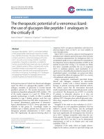
fever, coagulopathy, vasodilation). The series of events during reperfusion can be divided into four phases: (1) immediate (first 20 min after ROSC); (2) early post-arrest (20 min
through 6–12 h after resuscitation); (3) intermediate phase
(6–12 h through 72 h post-arrest); and (4) recovery phase
(beyond 72 h). Some experts have included a fifth phase, that
of rehabilitation after discharge from an acute care setting
(Fig. 25.1).
Pathophysiology of the Post-arrest
Reperfusion State
The post-cardiac arrest syndrome results from two distinct
but serial events – a period of ischemia, during which cardiac
output and oxygen delivery are profoundly compromised,
followed by a period of tissue and organ reperfusion. At the
time of cardiac arrest, oxygen extraction increases in an
effort to compensate for reduced delivery. As demand rapidly exceeds supply, tissue hypoxia triggers anaerobic
metabolism and lactate production. At the cellular level,
hypoxia limits oxidative phosphorylation and mitochondrial
ATP production. As a result, ATP-dependent membrane
functions such as maintenance of ion gradients begin to fail.
Phase
Goals
ROSC
Immediate
Intermediate
72 h
Recovery
Disposition
Rehabilitation
Prevent Recurrence
6–12 h
Prognostication
Early
Limit ongoing injury
Organ support
20 min
Rehabilitation
seizures/encephalopathy. The extent of neurologic injury
may be initially difficult to assess due to multi-organ system
failure following hypoxia-ischemia and reperfusion. In the
pediatric intensive care unit (PICU), the most common cause
of death following admission after cardiac arrest is hypoxicischemic encephalopathy [1, 2], which is also responsible for
the most significant morbidity in survivors.
Considerations for post-resuscitation care are impacted
by whether the resuscitation occurs out-of-hospital or inhospital, since the epidemiology and etiology for pediatric
cardiac arrest differ in these settings. Out-of-hospital arrest
is more likely to be asphyxial in origin, in which cardiac
arrest is the end result of progressive hypoxia and ischemia.
Multiple cohort studies of out-of-hospital pediatric cardiac arrests have found that most were of respiratory origin
[3–12]. A recent report from 11 North American sites participating in the Resuscitation Outcomes Consortium (ROC)
found that the incidence of non-traumatic out-of-hospital cardiac arrest in patients <20 years of age was 8.04 per 100,000
person-years, and was significantly higher among infants
than children or adolescents [5]. The initial cardiac rhythm
was asystole or pulseless electrical activity (PEA) in 82 %
of patients, and the most common etiology was an asphyxial event such as drowning or strangulation. In systematic
reviews, trauma and sudden infant death syndrome remain
the most common causes of pediatric out-of-hospital cardiac
arrest [3, 13]. Survival ranges from 6.4 to 12 %, with rates of
neurologically-intact survival of only 2.7–4 % [3–6, 13].
Pediatric cardiac arrest in the inpatient setting is more likely
to be witnessed or to occur in a monitored setting, but a high
proportion of patients have pre-existing co-morbidities [14].
Not surprisingly, the highest incidence of in-hospital pediatric
cardiac arrest is in the PICU, affecting 1–6 % of patients admitted [15, 16]. Regardless, the outcome from in-hospital arrest is
consistently better than for out-of-hospital events. A 2006
report of 880 pediatric inpatient arrests from a voluntary
national registry found survival to hospital discharge was 27 %,
while a 2009 review of 353 in-hospital cardiac arrests reported
a survival to discharge rate of 48.7 % [17, 18]. The etiology of
pediatric in-hospital arrest differs from out-of-hospital events
in that cardiac conditions (including shock) are as likely as
respiratory failure to be the immediate cause of the arrest (61–
72 %) [12, 17]. Asystole and PEA account for 24–64 % of the
initial cardiac rhythms. Interestingly, infants and children who
are resuscitated from inpatient cardiac arrest have a high likelihood of favorable neurologic outcome, with results ranging
from 63 to 76.7 % in two recent studies [17, 18].
M.E. Kleinman and M.G. van der Velden
Fig. 25.1 Phases of the post-cardiac arrest syndrome (Reprinted from
Neumar et al. [19]. With permission from Wolters Kluwer Health)
25
Post-resuscitation Care
The resultant depolarization permits opening of voltagedependent channels leading to entry of calcium, sodium, and
water into the cell. Cellular injury and death follow, with tissues demonstrating high oxygen consumption at most risk.
Lopez-Herce et al. described the progression of physiologic and biochemical changes occurring in an infant swine
model of asphyxial arrest [20]. At 10 min after discontinuation of mechanical ventilation, arterial pH had decreased
from a median of 7.40 to 7.09, PaO2 was unmeasurable, and
PaCO2 had increased from a median of 41 to 80 mmHg.
Lactate increased from 0.8 to 5.7 mmol/L. After transient
tachycardia and hypertension from increased systemic vascular resistance (SVR), progressive bradycardia and hypotension occurred with no measureable systemic blood
pressure by 10 min. After 10 min, subjects were resuscitated
with conventional CPR and one of four vasoconstrictor regimens (epinephrine alone, terlipressin alone, epinephrine +
terlipressin, or no medications). ROSC was achieved in just
over one-third of the animals within 20 min. Following
ROSC, there was an initial brief recovery of cardiac index,
SVR, and mean arterial pressure (MAP), followed by a progressive decline. Over the first 30 min after ROSC, arterial
and venous pH increased but did not return to baseline, and
lactate remained elevated.
Following ROSC, a complex cascade of biochemical
events occurs as blood flow and oxygen delivery are restored.
The major pathophysiologic processes include endothelial
activation and formation of oxygen-free radicals. Endothelial
activation by ischemia/reperfusion results in upregulation
of inflammatory mediators (e.g., leukocyte adhesion molecules, procalcitonin, C-reactive protein, cytokines, TNF-α
[alpha]) and downregulation of anti-inflammatory agents
such as nitric oxide and prostacyclin [21–23]. Coupled with
activation of the complement and coagulation cascades, this
systemic response leads to capillary leak, intravascular coagulation, and impaired vasomotor regulation.
Although restoration of oxygen delivery is one objective of cardiopulmonary resuscitation, the post-resuscitation
exposure of ischemic tissue to high concentrations of oxygen
can be injurious due to generation of oxygen-free radicals.
During ischemia, intracellular concentrations of hypoxanthine are increased; with the restoration of tissue oxygenation, hypoxanthine is converted to xanthine with oxygen
radicals produced as a byproduct. Furthermore, ischemic tissue becomes depleted of natural anti-oxidant defenses such
as nitric oxide, superoxide dismutase, glutathione peroxidase,
and glutathione reductase. In an infant rat model of asphyxial
arrest, animals resuscitated with 100 % oxygen during and
after CPR showed decreased hippocampal reduced glutathione, increased activity of manganese superoxide dismutase,
and increased cortical lipid peroxidation [24]. Reactive oxygen species have multiple negative effects and can modulate
signaling molecules including protein kinases, transcription
factors, receptors, and pro- and anti-apoptotic factors [25].
273
The use of anti-oxidant therapy or other inflammatory modulators to prevent or reduce the post-cardiac arrest syndrome
is a promising area of research, primarily in animal models.
Multiple agents have been studied including nitric oxide,
N-acetylcysteine, erythropoietin, steroids, cyclosporine,
ascorbic acid, trimetazidine and diazoxide [26–29].
Activation of the inflammatory cascade, with suppression
of anti-inflammatory defense mechanisms, interferes with
endothelial relaxation and promotes vasoconstriction and
microvascular thrombosis. At the vital organ level this results
in secondary ischemic injury [30]. In its most severe form,
ischemia-reperfusion injury results in multiple organ system
dysfunction (MODS), a common cause of delayed mortality
following resuscitation from cardiac arrest. Individually,
infants and children may demonstrate different patterns of
organ injury, with neurologic and cardiac dysfunction most
prevalent.
Post-resuscitation Care of the Respiratory
System
Oxygenation and ventilation are key components of resuscitation from pediatric cardiac arrest. In the out-of-hospital
setting, bag-mask ventilation is typically the initial airway
management technique. Advanced life support teams may be
trained and authorized to perform more invasive airway support such as supraglottic airway devices or tracheal intubation. Pre-hospital intubation for children with cardiac or
respiratory arrest is an area of ongoing controversy. A large
prospective randomized trial showed no difference in survival or neurologic outcome if children were intubated vs.
ventilated via bag and mask for respiratory failure, respiratory arrest, or cardiac arrest. In the intubation group there
was a high rate of failed intubations and unrecognized
esophageal intubations. Critics of this study note that the
EMS providers who participated in the study received 6 h of
classroom and mannequin training, suggesting that the
results could be partly attributed to lack of proficiency as
opposed to the intervention itself [31].
If the infant or child was tracheally intubated during
resuscitation, the first priority is to confirm appropriate tracheal tube position, patency, and security. Children who are
intubated at a referring hospital prior to transport are more
likely to have a right mainstem intubation than if they were
intubated in a tertiary PICU (13.4 % vs. 3.9 %) [32]. Prehospital providers frequently select tracheal tubes that are
either too large or too small for the patient’s size [33].
A tracheal tube that is too small for the child may hinder
adequate ventilation and oxygenation due to excessive glottic air leak and loss of tidal volume and end-expiratory pressure. Consideration should be given to reintubating the
patient with a larger and/or a cuffed tracheal tube. A tracheal
tube that is oversized, especially one with an inflated cuff,
274
can injure the tracheal mucosa and increase the risk of complications such as subglottic stenosis. In addition to deflating
the cuff, consideration should be given to replacing the tube
under controlled circumstances with an age-appropriate size,
weighing the risks and benefits of removing an existing airway. If a cuffed tracheal tube was used for intubation, cuff
pressures should be measured and adjusted to the recommended level of ≤20 cm H2O.
Regurgitation of gastric contents is common during cardiopulmonary resuscitation, leading to risk for aspiration.
Reflux of acidic gastric material into the pharynx can occur
even without active vomiting, especially when patients are
in the supine position and have loss of lower esophageal
sphincter tone. Regurgitation was reported in 20 % of adult
patients who survived cardiac arrest and received bystander
CPR, of whom 46 % had radiographic evidence of aspiration [34]. Aspiration of gastric contents or blood was documented on autopsy in 29 % of adult non-survivors after CPR
[35]. Specific circumstances such as near-drowning are associated with a high incidence of regurgitation. Computerized
tomography of the chest in drowning victims of multiple
ages revealed evidence of pulmonary aspiration in 60 % of
victims [36].
Bag-mask ventilations frequently result in gastric insufflation and distension and are the recommended initial technique for pediatric airway management during cardiac arrest
[37]. The use of cricoid pressure, often advocated to reduce
the risk of aspiration during positive-pressure ventilation and
tracheal intubation, may not be as effective as once believed.
Studies in anesthetized children suggest that the primary
effect of cricoid pressure is to prevent gastric insufflation
during mask ventilation, although there are no data about its
efficacy during pediatric cardiac arrest [38, 39].
Following cardiac arrest, children are at risk for development of acute lung injury (ALI) and acute respiratory distress syndrome (ARDS) as a result of reperfusion injury of
the lung and, potentially, pulmonary aspiration. ALI and
ARDS are clinical diagnoses and are distinguished by the
degree of impairment of oxygenation: a PaO2/FiO2 ratio of
<300 denotes ALI, while a PaO2/FiO2 ratio of <200 is used to
define ARDS [40]. ALI and ARDS are characterized by
decreased lung compliance and increased alveolar-capillary
permeability in the setting of a normal pulmonary arterial
occlusion pressure, resulting in surfactant deactivation, pulmonary edema and infiltrates, and hypoxemia. True cardiogenic pulmonary edema is more likely to occur in adults
after resuscitation from cardiac arrest, possibly related to the
common precipitating factor of coronary artery occlusion
and myocardial infarction with resultant depressed myocardial function.
The goals of mechanical ventilation in the pediatric
patient after cardiac arrest include provision of adequate
ventilation and oxygenation while minimizing the risk of
M.E. Kleinman and M.G. van der Velden
ventilator-induced lung injury (barotrauma or volutrauma).
Optimal ventilation and oxygenation parameters following
resuscitation from pediatric cardiac arrest are unknown. In
general, ventilation is considered acceptable if there is an
adequate pH (≥7.30). Hyperventilation should be avoided to
minimize the risk of further lung injury and to prevent secondary cerebral ischemia. Use of capnography for noninvasive assessment of ventilation may be misleading if there
is increased dead space related to reduced pulmonary blood
flow or parenchymal lung disease; in such situations arterial
blood gas measurement is a more accurate method to measure PaCO2.
Following return of spontaneous circulation, current
American Heart Association guidelines recommend that
the inspired oxygen concentration be progressively reduced
based on pulse oximetry [37]. In the presence of a normal
hemoglobin concentration, an arterial oxygen saturation of
>94 % is typically sufficient for the infant or child post-arrest.
In cases of severe anemia or hemorrhage, higher inspired
oxygen concentrations may be appropriate until adequate
oxygen carrying capacity is restored. Since an arterial oxygen saturation of 100 % could correspond with a PaO2 anywhere between ~80 and 500 mmHg, pediatric resuscitation
guidelines also recommend using 99 % as an upper limit for
arterial oxygen saturation. Because the use of 100 % oxygen
is a common default practice during intra- and interfacility
transfer, whenever possible, providers should be advised to
titrate FiO2 to achieve the goal arterial oxygen saturations.
Exposure to high concentrations of oxygen may result in
arterial hyperoxia, increasing the risk for oxygen free-radical
formation and oxidative injury during reperfusion. Evidence
from animal models and, more recently, human studies demonstrate that post-arrest hyperoxia worsens neurologic outcome [41–43]. Kilgannon et. al. reviewed >6,000 adult
non-traumatic cardiac arrest patients who survived to hospital
admission, and categorized them by the first PaO2 obtained
in the ICU. Patients who were hyperoxic, defined as a PaO2
>300 mmHg, had a higher in-hospital mortality compared
with patients who were normoxic (PaO2 60–300 mmHg) or
hypoxic (PaO2 <60 mmHg). Even after controlling for multiple confounders, hyperoxia was an independent risk factor
for mortality with an odds ratio of 1.8. The same investigators studied the relationship between post-resuscitation PaO2
as a continuous variable and in-hospital mortality.
Interestingly, the median post-resuscitation PaO2 was
231 mmHg with an interquartile range of 149–349 mmHg.
Using multivariable analysis, they demonstrated that for
each 100 mmHg increase in PaO2 during the first 24 h of
admission there was a 24 % increase in mortality.
For patients with acute lung injury or ARDS, a lung protective strategy is typically employed using pressure-controlled
ventilation. The components of a lung protective strategy
include: (1) low tidal volumes (5–6 mL/kg), (2) limited
25
Post-resuscitation Care
plateau pressures (≤30 cm H2O), (3) optimal PEEP to restore
and maintain functional residual capacity, and (4) exposure
to non-toxic concentrations of oxygen (FiO2 ≤ 0.6). A certain degree of respiratory acidosis is tolerated, an approach
termed permissive hypercapnea. The level of hypercarbia
that is acceptable may be influenced by other organ system
concerns such as cerebral edema from hypoxic-ischemic
brain injury. Cerebral blood vessel reactivity to carbon dioxide is preserved in comatose adult patients following cardiac
arrest, so extremes of hypo- and hyperventilation should be
avoided [44].
The initial maneuver for a patient with persistent hypoxemia is the escalation of PEEP in an effort to increase functional residual capacity and reduce intrapulmonary shunting.
Those who remain hypoxemic or develop extrapulmonary
air leak may be candidates for a trial of high frequency oscillatory ventilation (HFOV). Use of HFOV frequently requires
neuromuscular blockade, however, which hampers ongoing
neurologic assessment. Another therapeutic consideration is
surfactant replacement therapy, which was found to decrease
mortality in a recent meta-analysis of children with acute
respiratory failure [45]. The optimal dosing, frequency, and
duration of therapy have not been determined.
Post-resuscitation Care of the Cardiovascular
System
Except for very brief episodes of cardiac arrest, most patients
will demonstrate some impact of cardiac arrest on postresuscitation circulatory status. Initial assessment should
focus on the rate and rhythm, blood pressure, peripheral perfusion, and end-organ function (mental status, pupillary
exam, urine output). An inappropriately slow heart rate associated with hypotension requires urgent treatment to prevent
deterioration. Underlying causes of persistent bradycardia to
consider include hypothermia, hypoxia, acidosis, electrolyte
disturbances, hypoglycemia, toxins, or increased intracranial
pressure. Appropriate management is directed at treating the
suspected etiology and the use of pharmacologic agents to
increase heart rate, such as adrenergic agents or vagolytic
agents, or use of electrical pacing.
Tachycardia is commonly observed after resuscitation
from cardiac arrest and may be multifactorial, resulting from
use of β [beta]-adrenergic agents, early myocardial dysfunction, and cardiac rhythm disturbances. In general, tachycardia is well tolerated in infants and children, and treatment to
control rate is indicated only if the patient has a tachyarrhythmia that results in hemodynamic compromise.
Tachyarrhythmias should be managed according to the relevant treatment protocols depending on the type of rhythm
and the patient’s clinical status. Patients who are hypotensive
in the setting of supraventricular or ventricular tachycardia
275
should receive immediate synchronized cardioversion, with
or without sedation depending on the level of consciousness
[37]. If the patient is normotensive, pharmacologic therapy
can be attempted while closely monitoring the patient’s
hemodynamic status. Other causes for tachyarrhythmias
should also be considered, including central venous catheter
position, electrolyte or metabolic derangements, hyperpyrexia, and adverse effects of adrenergic agents. Increased
myocardial oxygen consumption associated with tachyarrhythmias may result in myocardial ischemia, and a 12-lead
ECG may identify ST-segment changes or pre-excitation that
could signal risk for further rhythm disturbances. Expert
consultation with a pediatric cardiologist is recommended
for guidance regarding anti-arrhythmic and other therapies.
Myocardial dysfunction occurs in most adults and children following resuscitation from cardiac arrest, a condition
known as “myocardial stunning.” Despite the restoration of
myocardial blood flow and oxygen delivery, echocardiographic evidence of myocardial dysfunction typically persists
for 24–48 h following resuscitation [46]. The pathophysiology of this reperfusion injury is characterized by cardiac tissue edema and decreased contractility with low cardiac index.
Hemodynamic studies of children following near-drowning
have demonstrated an increase in atrial and ventricular enddiastolic filling pressures as well as systemic and pulmonary
vascular resistance [47]. In animal models, the degree of myocardial dysfunction is correlated with the duration of cardiac
arrest and is more severe when cardiac arrest is due to ventricular fibrillation compared with asphyxia [48, 49]. Post-arrest
troponin levels are inversely correlated with ejection fraction
and survival in pediatric patients following resuscitation from
cardiac arrest [50]. Pediatric animal studies suggest that the
use of adult defibrillation doses leads to greater myocardial
dysfunction and higher levels of troponin leak than attenuated
pediatric doses [51].
The goals of hemodynamic support following resuscitation from cardiac arrest are to restore and maintain
end-organ perfusion and oxygen delivery. Those children
who are suspected of having inadequate preload due to
volume loss may receive isotonic fluids in small boluses
of 5–10 mL/kg, titrated to signs of improved hemodynamics such as resolving tachycardia and improved peripheral
perfusion. Frequent reassessment after each fluid bolus is
essential to avoid excessive increases in cardiac filling pressures that could lead to pulmonary edema and worsening gas
exchange. Patients resuscitated from cardiac arrest in the
setting of trauma or hemorrhage may benefit from resuscitation with blood products such as packed red blood cells
to replete low blood volume and increase oxygen-carrying
capacity. Optimization of preload is best accomplished using
invasive monitoring in the critical care setting. Placement of
a catheter with the tip in the SVC or IVC permits monitoring of central venous pressure to estimate right-sided filling
276
M.E. Kleinman and M.G. van der Velden
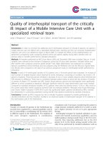
Table 25.1 Vasoactive agents for post-resuscitation myocardial dysfunction
Dopamine
Type
Endogenous
catecholamine
Receptors
Dopaminergic agonist
β [beta]-1 and -2 agonist
Dobutamine
Synthetic catecholamine
Epinephrine
Endogenous
catecholamine
α [alpha]-agonist
β [beta]-1 and -2 with intrinsic
α [alpha]-adrenergic agonist
and antagonist activity
β [beta]-1 and -2
α [alpha]-1 and -2 agonist
Levosimendan
Calcium-sensitizer
Milrinone
Phosphodiesterase type
III (PDE III) inhibitor
Norepinephrine Endogenous
catecholamine
Vasopressin
Increases cardiac myocyte
sensitivity to calcium; opens
potassium channels on vascular
smooth muscle
No receptor; PDE III enzyme
inhibition increases myocardial
cAMP and intracellular calcium
β [beta]-1 and -2 agonist
α [alpha]-1 and -2 agonist
Endogenous posterior
V1 (vascular smooth muscle),
pituitary peptide hormone V2 (renal)
pressures. Femoral venous lines in the infrahepatic IVC have
shown good correlation with right atrial filling pressures in
cohorts of pediatric cardiac patients and critically ill children
in the ICU setting even with changes in mean airway pressure and PEEP [52–55].
Inotropic and vasoactive infusions are the mainstay of
therapy for post-arrest myocardial dysfunction. These
agents improve cardiac output and oxygen delivery by
increasing myocardial contractility and by either increasing
or decreasing systemic vascular resistance. Despite the frequent use of vasoactive infusions for post-resuscitation
myocardial support, to date there is no data to establish that
such therapy improves patient outcome. The choice of
agent depends on the individual patient’s physiologic status
and the presence or absence of hypotension. Most children
with post-resuscitation myocardial dysfunction will have
low cardiac output and high systemic vascular resistance
and will benefit from medications that increase contractility and reduce afterload. If the patient is normotensive, inodilator drugs such as milrinone may improve cardiac output
and end-organ perfusion with less myocardial oxygen cost
compared with adrenergic inotropic agents. If the patient is
hypotensive, afterload reduction is not likely to be tolerated
and the use of agents with both inotropic and vasoconstrictive actions may be necessary to restore adequate end-organ
perfusion pressure.
Physiologic effect
Renal and splanchnic
vasodilation
Positive inotropy and
chronotropy
Vasoconstriction
Positive inotropy and
chronotropy; may cause
systemic vasodilation
Positive inotropy and
chronotropy; vasodilation
at low doses
At higher infusion rate
causes potent
vasoconstriction
Positive inotropy,
vasodilation
(“inodilator”)
Dose range
2–5 mcg/kg/min
5–10 mcg/kg/min
10–20 mcg/kg/min titrated to effect
2–20 mcg/kg/min titrated to effect
Low dose: 0.1–0.3 mcg/kg/min;
High dose: 0.3–1 mcg/kg/min
titrated to effect
Loading dose: 12–24 mcg/kg over
10 min; infusion: 0.1–0.2 mcg/kg/
min
Positive inotropy,
vasodilation
(“inodilator”)
Positive inotropy and
chronotropy;
vasoconstriction
Load: 50–75 mcg/kg over
10–60 min; infusion: 0.5–0.75 mcg/
kg/min
0.1–2 mcg/kg/min titrated to effect
Vasoconstriction,
anti-diuresis
0.17–10 milliunits/kg/min
(0.01–0.6 units/kg/h)
Medications used to manage post-arrest myocardial dysfunction are listed in Table 25.1, along with their primary
hemodynamic effects. Most of the vasoactive agents listed
have the potential to increase heart rate, either primarily or
secondarily, which may limit their benefit due to associated
increases in myocardial oxygen consumption. Among the
adrenergic agents, significant tachycardia is less likely with
norepinephrine. Use of milrinone may result in reflex tachycardia due to afterload reduction, which can generally be
managed with judicious volume administration. The exclusive use of pure vasoconstrictor agents such as phenylephrine and vasopressin is not recommended for post-arrest
myocardial dysfunction because these agents increase afterload without supporting contractility; however, for those
patients who demonstrate refractory vasodilation in the postarrest period, the use of vasopressin in conjunction with an
inotropic agent may be beneficial [56]. Patients who remain
hypotensive despite volume resuscitation and vasoactive
infusions should be evaluated for adrenal insufficiency,
which has been reported as a feature of the post-cardiac
arrest syndrome.
Levosimendan is a relatively new inotropic agent that
has been studied for treatment of congestive heart failure
in adults. The drug acts as an inodilator by increasing myocardial sensitivity to calcium and by activation of peripheral
vascular ATP-dependent potassium channels. Animal studies
25
Post-resuscitation Care
comparing levosimendan with dobutamine demonstrated
a greater increase in left ventricular ejection fraction with
levosimendan [57]. Several published case series of pediatric patients with post-cardiopulmonary bypass ventricular
dysfunction describe improvement in cardiac output and
decreased catecholamine requirements when levosimendan
was utilized [58, 59].
The physiologic endpoints for post-resuscitation myocardial support are not well established for pediatric patients.
Improved peripheral perfusion, normalization of heart rate,
normotension, and adequate urine output are accepted clinical signs of improving cardiac function. Serum or whole
blood lactate concentrations are laboratory markers of oxygen delivery and should improve as cardiac output normalizes unless there is impairment of oxygen utilization (as in
sepsis) or reduced lactate metabolism (as in acute hepatic
insufficiency). Echocardiography, while helpful in evaluating systolic function, is less reliable at demonstrating diastolic dysfunction and is of limited usefulness since it can
only be performed at discrete points in time.
Placement of a central venous catheter with its tip in the
superior vena cava allows the use of SVC oxygen saturations
to assess the adequacy of oxygen delivery to the tissues.
Proper measurement of SvcO2 requires that co-oximetry be
performed on a sample of venous blood from the SVC catheter to yield a measured (versus calculated) oxygen saturation. In the setting of normal arterial oxygen saturations and
an adequate hemoglobin concentration, SvcO2 reflects the
adequacy of cardiac output. Normal SvcO2 is between 70
and 80 %; an SvcO2 <60 % is evidence for excess oxygen
extraction in the setting of low cardiac output.
Patients with severe post-arrest myocardial dysfunction may also benefit from interventions to reduce oxygen
consumption such as temperature control, sedation and
analgesia, and neuromuscular blockade. If indicated by laboratory measurements, normalization of glucose, calcium,
magnesium and phosphorous may also support myocardial
contractility and prevent secondary cardiac arrhythmias [60].
Post-resuscitation Neurologic Management
Hypoxic-ischemic brain injury is one of the major factors
contributing to mortality after cardiac arrest [1] and arguably
the most important determinant of meaningful survival.
Despite improved survival rates compared to adults [17],
children resuscitated from cardiac arrest have a significant
risk of mortality with a majority of survivors having poor
neurological outcome [3–5, 12]. Post-cardiac arrest brain
injury has been designated to describe the spectrum of neurologic dysfunction observed after cardiac arrest [19], the
mitigation and management of which has become an intense
focus of basic and clinical research [61].
277
Pathophysiology
The mechanisms of post-cardiac arrest brain injury are complex [62] and are at interplay with the other components of
the post-cardiac arrest syndrome [19, 61]. However, despite
extensive knowledge of the molecular mechanisms involved
in hypoxic-ischemic injury, interventions to preserve affected
neuronal cells remain elusive. Furthermore, the degree of
injury itself depends on many factors including duration of
cardiac arrest and patient age [63].
Ischemic neurologic injury is known to involve a threepart process [61]. During the initial phase of cessation of
cerebral blood flow, oxygen, glucose and ATP are rapidly
depleted from cellular stores [61, 64, 65] and toxic metabolites accumulate [65]. As a result, there is disruption of
calcium homeostasis, glutamate release and neuronal hyperexcitability [61, 62, 66]. Elevation of intracellular calcium
activates multiple enzymatic pathways resulting in further
cell injury and death [61]. This occurs during conditions of
total ischemia observed in cardiac arrest, as well as during
the period of less severe ischemia accompanying effective
cardiopulmonary resuscitation. While restoration of cerebral blood flow remains the foremost goal in management of
cardiac arrest, there is compelling evidence that significant
injury occurs upon brain reperfusion, resulting in a second
phase of the injury process [63]. During the first few minutes
after return of circulation there is hyperemia of the cerebral
tissue [19], with associated lipid peroxidation, formation of
oxygen free radicals, inflammatory injury and ongoing disruption of calcium homeostasis, glutamate release and enzymatic pathway activation. Apoptosis is a major consequence
of injury during this stage [61]. Following the reperfusion
stage is a period of cerebral hypoperfusion that can last for
hours after resuscitation [67, 68]. Studies in adult patients
have shown impaired cerebral autoregulation during this
period [69, 70] with experimental pediatric animal models
confirming these findings [71]. As a result, cerebral blood
flow is dependent on systemic blood pressure so that avoidance of hypotension and efforts to minimize cerebral oxygen
demands (e.g. sedation, seizure control, temperature control)
are critical to avoid compounding neuronal injury [19, 69].
The exact cerebral blood flow required to optimize oxygen
delivery is difficult to determine for any individual patient
and likely changes over time [19]. Near-infrared spectroscopy (NIRS) is a non-invasive technology that has offered
promise in determining individualized optimal cerebral
blood flow to avoid cerebral hypoxia and ongoing neuronal
ischemia [65, 72, 73].
Cerebral edema is also known to compromise cerebral
oxygen delivery by elevating intracranial pressure [74] and
reducing cerebral perfusion pressure. Within hours after
the initial ischemic injury from cardiac arrest, the inflammatory process increases vascular permeability and disrupts the blood-brain barrier causing cerebral edema [62].
278
This pathophysiologic process, however, is not consistently
associated with an increase intracranial pressure in the postcardiac arrest patient [75, 76]. Furthermore, there is no data
to support the use of routine intracranial pressure monitoring
for management of the post-cardiac arrest patient [19].
Clinical Manifestations
Clinical manifestations of post-cardiac arrest brain injury in
the critical care setting include disorders of arousal and consciousness, myoclonus, movement disorders, autonomic
storms, neurocognitive dysfunction, seizures and brain death
[19, 61, 77–79]. Of these, seizures represent an important
manageable cause of secondary neuronal injury in the postcardiac arrest patient. Seizures are known to increase cerebral metabolic demand and subsequent ischemic injury [80].
Seizures may be partial, generalized tonic-clonic or myoclonic [61], the latter of which has been associated with more
severe cortical injury and worse prognosis [81, 82]. A prospective study of EEG monitoring in children undergoing
therapeutic hypothermia after cardiac arrest reported an
occurrence of electrographic seizures in 47 % of patients
[83]. Studies of critically ill pediatric patients at risk of seizures from multiple diagnoses undergoing long-term video
electroencephalography showed that seizures are relatively
common in these patients [84, 85]. Most of these seizures
were only detected by long-term EEG monitoring and missed
by beside caregivers [85] and many of the suspected seizures
by bedside staff were actually not epileptic seizures [84],
both advocating a lower threshold for obtaining long-term
EEG in patients at risk for seizures, including those in the
post-cardiac arrest state. This coincides with the American
Heart Association Guidelines recommending EEG evaluation in comatose adult patients after ROSC [86].
Management
Management of post-resuscitation brain injury involves therapies focused on preservation of cerebral blood flow and
oxygen delivery and prevention of secondary brain injury by
decreasing metabolic demand [62]. With regards to the former, the focus should be on avoidance of systemic arterial
hypotension, avoidance of significant hypoxia with target
oxygen saturation of 94 % or higher, ventilation to normocapnia, and management of cerebral edema [19, 62, 86]. Due
to its effect on cerebral perfusion, the use of intentional
hyperventilation should be reserved as temporizing rescue
therapy in the setting of impending cerebral herniation [37].
With regards to the management of global cerebral edema in
the post-cardiac arrest state, no trials exist to guide therapy in
this specific population. Standard therapy involves promotion of venous drainage by elevation of the head of the bed to
30° and midline head position, avoidance of hypotonic fluid
administration [87] and avoidance of hyperglycemia [62].
Animal models of cardiac arrest have demonstrated enhanced
M.E. Kleinman and M.G. van der Velden
cerebral blood flow after ROSC with use of hypertonic saline
compared to normal saline, however, this is yet to be
described in human studies [88].
Therapies directed at the prevention of secondary injury
by decreasing metabolic demand include seizure control,
analgesia, sedation and neuromuscular blockade, temperature control including therapeutic hypothermia and other
neuroprotective measures. Prompt and aggressive treatment
with conventional anti-convulsant regimens should be
employed for seizure management in the post-resuscitation
period. There have been no studies examining the role of
prophylactic anti-convulsants; however, clinical and subclinical seizures should be treated aggressively with standard
anti-convulsants such as benzodiazepines, fosphenytoin,
levetiracetam, valproate and barbiturates [61], the latter of
which may be needed for induction of pharmacologic coma
for refractory seizures. All anti-convulsants should be used
with vigilance towards managing the expected side effect of
systemic hypotension and reduction in cerebral perfusion
pressure.
There is no data to support routine use of sedation, analgesia or neuromuscular blockade to protect the brain from
secondary injury in the post-cardiac arrest patient; however,
some or all of the above may be required for safety and ease
of mechanical ventilation and/or to facilitate achievement of
therapeutic hypothermia (see below). Sedation and analgesia
may reduce cerebral oxygen consumption and metabolic
rate, improving matching of cerebral oxygen demand with
supply. Propofol is not recommended for routine use as an
anti-convulsant or sedative in pediatric patients due to the
risk of propofol infusion syndrome [89, 90]. Use of pediatric
sedation scales can be used to titrate sedative and analgesic
medications [91, 92]. When neuromuscular blockade is necessary, use of EEG monitoring should be considered in order
to detect masked seizure activity [19, 62].
Hyperthermia occurs commonly after neurological injury
in humans and is associated with worse neurological outcomes [93–100] likely related to increased cerebral oxygen
consumption and cellular destruction [101]. These findings
have been documented in pediatric patients as well with temperatures ≥38 °C in the first 24 h after ROSC with associated
unfavorable neurological outcome [102]. AHA guidelines
recommend aggressive fever control with antipyretics and
cooling devices in the post-resuscitation period [37, 86].
Beyond the clear recommendation for fever control in the
post-cardiac arrest pediatric patient comes the question of
use of therapeutic hypothermia. Therapeutic hypothermia
is believed to work by reducing cerebral metabolism, suppressing neurological excitotoxicity, suppressing inflammation and vascular permeability, mitigating cell destructive
enzymes and improving cerebral glucose metabolism
[62, 64]. Mild induced hypothermia has been shown to
improve neurological outcome in comatose adults after
25
Post-resuscitation Care
resuscitation from cardiac arrest associated with ventricular
fibrillation [103, 104]. Similar outcomes were observed with
hypothermia therapy in newborns with hypoxic-ischemic
encephalopathy [105, 106]. With regards to the pediatric
population, no prospective clinical trials have been published to date evaluating efficacy of therapeutic hypothermia
in survivors of cardiac arrest, although a large multi-center
trail is currently in progress [107–109]. A trial evaluating
effect of therapeutic hypothermia on outcome after traumatic brain injury in pediatric patients showed no improvement in outcome with a trend towards increased mortality in
the hypothermia group [110]. Retrospective studies of use
of hypothermia after pediatric cardiac arrest have shown no
benefit or harm, however, both called for a prospective, randomized trial to determine efficacy of therapeutic hypothermia after pediatric cardiac arrest [111, 112]. A feasibility trial
of therapeutic hypothermia using a standard surface cooling
protocol in pediatric patients after cardiac arrest showed feasibility and set the stage for future investigations of therapeutic hypothermia for cardiac arrest in children [113].
As therapeutic hypothermia is likely safe with temperatures in the range of 32–34 °C [114], the AHA recommends
consideration of this intervention for children who remain
comatose after resuscitation from cardiac arrest [87]. In
spite of these recommendations, a survey of pediatric critical
care providers demonstrated that therapeutic hypothermia
was not widely used in this population and that the methods
for utilization were variable [115]. Post-arrest hypothermia
protocols, when initiated, should involve rapid initiation of
cooling, continuous temperature monitoring and gradual
rewarming. Side effects may include shivering, hemodynamic complications, electrolyte derangements, hyperglycemia, mild coagulopathy and risk of infection [62].
Numerous pharmacologic neuroprotective strategies
have been proposed to improve neurological outcome after
ischemic injury. No benefit has been observed in human trials involving barbiturates, glucocorticoids, calcium channel
blockers, lidoflazine, benzodiazepines and magnesium sulfate [86, 116]. One trial showed improved survival and a trend
towards improved neurologic outcome when coenzyme Q10
was used as an adjunct to therapeutic hypothermia [117].
Prognosis
For survivors of cardiac arrest, neurological prognosis is one
of the most important factors guiding physicians and families
in determining the appropriate level of care for the patient.
Data that may be used when predicting outcome include
historical features, clinical examination, neuroimaging, neurophysiologic studies and biochemical markers [118, 119].
In a report of the Quality Standards Subcommittee of the
American Academy of Neurology, a practice parameter
was created after systematic review of available evidence
of neurological outcome in comatose adult survivors after
279
cardiopulmonary resuscitation for use in prognostication
in such patients. Pupillary light response, corneal reflexes,
motor responses to pain, myoclonic status epilepticus, serum
neuron-specific enolase, and somatosenory evoked potential
studies were shown to reliably assist in accurately predicting poor outcome. Notably, this practice parameter was not
derived from patients treated with therapeutic hypothermia [118]. No similar report has been created for pediatric
patients, however, a recent literature review of all available
evidence in domains used to provide prognostic information
in children with coma due to hypoxic ischemic encephalopathy, of which post-resuscitation brain injury would be
included, suggests that abnormal exam signs (pupil reactivity and motor response), absent N2O waves bilaterally on
somatosensory evoked potentials, electrocerebral silence
or burst suppression patterns on electroencephalogram,
and abnormal magnetic resonance imaging with diffusion
restriction in the cortex and basal ganglia are all individually
highly predictive of poor outcome and when used in combination are even more predictive. This predictive accuracy
can be improved by waiting 2–3 days after the event [119].
When evaluating prognostic indicators to predict neurologic
outcome, attention should be paid to confounding factors
that may affect the clinical neurological examination such
as renal failure, liver failure, shock, metabolic acidosis and
therapeutics such as sedatives, neuromuscular blockers and
induced hypothermia [118].
Blood Glucose Management
Blood glucose derangements are common in adults and
children after resuscitation from cardiac arrest. Studies in
adult survivors of cardiac arrest demonstrated an association
between post-arrest hyperglycemia and poor survival with
unfavorable neurological outcomes [120–123]. Adult studies of out-of-hospital cardiac arrest survivors also observed
worse outcomes with the administration of glucosecontaining fluids during cardiopulmonary resuscitation
[124]. A large retrospective registry report on adults with
in-hospital cardiac arrest found an association with mortality
if non-diabetic patients were either hyperglycemic or hypoglycaemic [125].
Recent studies in adults resuscitated from out-of-hospital
cardiac arrest indicate that post-cardiac arrest patients may
be treated optimally by maintaining blood glucose concentration below 8 mmol/L (144 mg/dL) [126–128]. Ninety survivors of out-of-hospital cardiac arrest due to ventricular
fibrillation were cooled and randomized into two treatment
groups: a strict glucose control group (SGC), with a blood
glucose target of 4–6 mmol/L (72–108 mg/dL), and a moderate glucose control group (MGC), with a blood glucose target of 6–8 mmol/L (108–144 mg/dL). Both groups were
280
treated with an insulin infusion for 48 h. Episodes of moderate hypoglycemia (<3.0 mmol/L or <54 mg/dL) occurred in
18 % of the SGC group and 2 % of the MGC group
(P = 0.008); however, there were no episodes of severe hypoglycemia (<2.2 mmol/L or <40 mg/dL). There was no difference in 30-day mortality between the groups (P = 0.846).
Strict control of blood glucose to 4.4–6.1 mmol/L (80–
110 mg/dL) with intensive insulin therapy reduced overall mortality in critically ill adults in a surgical ICU and
appeared to protect the central and peripheral nervous systems [129, 130]. In a subsequent medical ICU study, however, the overall mortality was similar in both the intensive
insulin and control groups [131]. Among those patients
with a longer ICU stay (≥3 days), intensive insulin therapy
reduced the mortality rate from 52.5 % (control group) to
43 % (P = 0.009). However, use of intensive insulin therapy
to maintain normoglycemia of 4.4–6.1 mmol/L (80–110 mg/
dL) was associated with more frequent episodes of hypoglycemia and some have cautioned against its routine use in the
critically ill [132, 133]. Finally, a large, multi-center trial of
critically ill adults (NICE-SUGAR) showed an increase in
90-day mortality for patients who received tight glycemic
control [134].
It is presently unknown if post-arrest hyperglycemia or
administration of glucose in the peri-resuscitation period
causes harm in children. A limited study in pediatric survivors of cardiac arrest demonstrated the occurrence of postarrest hyperglycemia (mean blood glucose concentrations
>150 mg/dL or >8.3 mmol/L) in more than two-thirds of
children within the first 24 h after the arrest. Limited retrospective studies in critically ill, non-diabetic children indicate that hyperglycemia frequently occurs in these children
and is independently associated with morbidity and mortality
[135–137], but it unknown if the observed hyperglycemia is
a surrogate marker of the severity of the child’s illness injury
rather than a cause of poor outcome. Two of these studies
additionally demonstrated that hypoglycemia and increased
glucose variability were also associated with higher mortality [137, 138].
To date there has been only one randomized controlled
trial of insulin management in critically ill pediatric patients
using a heterogenous group that was randomized to receive
intensive insulin therapy vs. insulin for a threshold level of
hyperglycemia [139]. The results of this study were favorable towards intensive insulin therapy, with shorter ICU stay,
lower rates of secondary infection, and lower unadjusted
30-day ICU mortality. In the absence of specific pediatric
data examining the efficacy and safety of intensive glycemic
control following cardiac arrest, current recommendations
are to target a normal range of blood glucose concentration.
Significant hyperglycemia is an indication for intravenous
insulin infusion, although there is no consensus on a specific
threshold for initiation of insulin. When using insulin in the
M.E. Kleinman and M.G. van der Velden
post-resuscitation period, intensive blood glucose monitoring is essential to avoid hypoglycemia. Hypoglycemia poses
a greater risk to the relatively immature pediatric brain compared with adults, especially in the setting of cardiac arrest
with ischemia/reperfusion injury. The use of therapeutic
hypothermia can further increase the risk for glucose
derangements.
Acid-Base and Electrolyte Management
Acid-base and electrolyte abnormalities are commonly seen
during and after recovery from cardiac arrest. These include,
but are not limited to, metabolic acidosis, hyperkalemia, ionized hypocalcemia, and hypomagnesemia. Severe acidosis
and other electrolyte disturbances may adversely affect cardiac function and vasomotor tone. Prompt recognition and
correction of acid-base and electrolyte abnormalities in the
post-arrest state is important to minimize the risk of arrhythmias and to support myocardial function.
Metabolic acidosis may be present prior to cardiac arrest
as a result of inadequate oxygen delivery and is further exacerbated by tissue hypoxia and ischemia occurring during the
low flow arrest state. Although metabolic acidosis may have
widespread effects on cellular and organ function, the use of
buffers during or immediately after pediatric cardiac arrest is
generally not recommended. The administration of sodium
bicarbonate leads to production of carbon dioxide and water;
rapid diffusion of carbon dioxide may result in intracellular
acidosis that is deleterious, especially to the brain. In addition, serum alkalosis shifts the oxyhemoglobin dissociation
curve to the left, inhibiting oxygen delivery to the tissues.
The use of sodium bicarbonate in adults experiencing outof-hospital cardiac arrest remains controversial [140–142].
While one large multi-center trial found that earlier and more
frequent use of sodium bicarbonate was associated with
higher early survival rates and better long-term outcome
[141], other studies have shown no benefit from administration of sodium bicarbonate during and after cardiac arrest
[143–145]. A prospective randomized controlled trial examined the use of buffer therapy (Tribonat) in the setting of cardiac arrest in adults and did not observe an improved outcome
compared with saline [146]. There have been no prospective
studies of the use of sodium bicarbonate during pediatric cardiac arrest, but two large retrospective studies of in and outof-hospital arrests found an association between bicarbonate
use and mortality [11, 18].
In general, management of post-arrest metabolic acidosis
caused by increased lactate and other metabolic acids consists of restoring adequate tissue perfusion and oxygen delivery, while assuring adequate ventilation. Oxygen delivery is
optimized by supporting cardiac output, as described in the
previous section, and ensuring adequate oxygen content. An
25
Post-resuscitation Care
anion gap acidosis that does not improve in response to supportive care suggests an ongoing source of acid production
such as ischemic bowel, or a respiratory chain disorder such
as cyanide poisoning. Patients with a non-anion gap metabolic acidosis following cardiac arrest may be hyperchloremic from the use of large volumes of normal saline during
resuscitation. Metabolic acidosis due to chloride administration is generally well tolerated and is associated with better
outcomes than other forms of acidosis in critically ill patients
[147, 148]. Treatment with bicarbonate is not usually indicated and the acidosis improves with restriction of chloride
intake. There is limited evidence to support the use of buffer
therapy in the post-resuscitation phase. Bicarbonate therapy
may be indicated to manage renal tubular acidosis, characterized by a non-anion gap acidosis with elevated urine pH.
There are specific conditions in which active correction of
acidosis may by beneficial, such as the patient with pulmonary hypertension or the child with certain toxic ingestions
(eg: tricyclic antidepressants). Continued alkalinization
may also be considered for treatment of associated conditions such as rhabdomyolysis, hyperkalemia, and tumor lysis
syndrome.
Prolonged cardiac arrest may be associated with ionized
hypocalcemia, which appears to be time-dependent and
perhaps related to intracellular sequestration of calcium
[149, 150]. Hypocalcemia may also result from the rapid
administration of blood products, which contain high concentrations of citrate that bind free calcium. Documented
ionized hypocalcemia is an indication for treatment with
exogenous calcium, as hypocalcemia negatively affects
myocardial contractility and can contribute to post-arrest
arrhythmias [151, 152]. Other indications for calcium
administration include cardiac arrest in the setting of suspected or documented hyperkalemia or calcium-channel
blocker overdose. Despite the potential benefits to of calcium for documented hypocalcemia, excess calcium administration may be harmful. During ischemia and reperfusion,
calcium channels become more permeable, allowing influx
of calcium. Increased intracellular calcium activates a
number of secondary messengers leading to apoptosis
and necrosis; indeed, intracellular calcium accumulation
is thought to be the final common pathway for cell death
[153]. A recent registry report of children experiencing inhospital pediatric cardiac arrest observed that calcium use
during resuscitation was associated with reduced survival
to discharge and unfavorable neurologic outcome [154].
Given the retrospective nature of the study it is not possible
to know if this association is based on effects of calcium
or the use of calcium for patients who are unresponsive to
other resuscitative measures. However, multiple adult studies, both randomized controlled trials and cohort studies,
showed no benefit of calcium administration during cardiac
arrest [155].
281
Magnesium is an important ion in cardiac conduction and
plays a role in smooth and skeletal muscle tone. Magnesium
is recommended for shock-refractory cardiac arrest due to
the ventricular arrhythmia torsades de pointes, but there are
conflicting data on its role in treating other rhythm disturbances. A pilot study of magnesium in adults with in-hospital
cardiac arrest who were unresponsive to other measures
demonstrated greater return of spontaneous circulation and
more favorable neurologic outcome [156]; however, other
studies have not demonstrated any difference in outcome
[157, 158]. One randomized trial of magnesium administration in the post-resuscitation period found no benefit [159].
There have been no studies evaluating magnesium use during or after pediatric cardiac arrest.
Hyperkalemia following cardiac arrest may be secondary
to metabolic acidosis as by hydrogen ions move intracellularly in exchange for potassium. This form of hyperkalemia
responds readily to correction of acidosis and typically does
not require other treatment. Hyperkalemia may also occur
due to muscle or tissue injury related to the underlying cause
of cardiac arrest such as trauma, prolonged seizures, or electrical shock. If life-threatening hyperkalemia requires treatment, the most effective methods to reduce serum
concentration are the use of sodium bicarbonate and the infusion of insulin and glucose. These measures temporarily
reduce extracellular potassium concentration but do not alter
total body potassium; refractory hyperkalemia may require
the use of hemodialysis for definitive correction. Resin binders and loop diuretics will also reduce potassium burden but
their onset of action is more gradual. Calcium may be used to
temporarily antagonize the adverse electrophysiologic
effects of hyperkalemia by stabilizing myocyte membranes.
Immunologic Disturbances and Infection
Evidence of a “systemic inflammatory response syndrome”
(SIRS) and endothelial activation triggered by whole-body
ischemia and reperfusion in patients successfully resuscitated
after cardiac arrest has been demonstrated in humans as early
as 3 h after cardiac arrest [160]. Biochemical changes include
a marked increase in plasma cytokines and soluble receptors
such as interleukin-1ra (IL-1ra), interleukin-6 (IL-6), interleukin-8 (IL-8) [161, 162], interleukin-10 (IL-10), and soluble
tumor necrosis factor receptor II, and were more pronounced
in nonsurvivors. Additionally, plasma endotoxin was noted
in about half of patients studied, possibly due to translocation through sites of intestinal ischemia and reperfusion damage [160, 163]. Studies have also shown increases in soluble
intracellular adhesion molecule-1, soluble vascular-cell adhesion molecule-1 and P and E selectins suggesting neutrophil
activation and endothelial injury [163–165], with additional
studies demonstrating direct evidence of endothelial injury
282
and inflammation with elevation of endothelial microparticles with the first 24 h after ROSC [166]. This inflammatory response from endothelial damage has been implicated
in the vital organ dysfunction often witnessed after cardiac
arrest [167]. Interestingly, in light of this immune activation,
hyporesponsiveness of circulating leukocytes has also been
noted in patients with cardiac arrest, a condition referred to as
endotoxin tolerance. While possibly protective against overwhelming inflammation, endotoxin tolerance may contribute
to immune paralysis with an increased risk of nosocomial
infection [163].
Along with the possible immune dysfunction mentioned
above, survivors of cardiac arrest have multiple risk factors
for infection, including prolonged ICU stays, organ dysfunction, and invasive procedures [168]. Infectious complications
in survivors of cardiac arrest are common [168–170] and
have been associated with increased duration of mechanical
ventilation and length of hospital stay [168, 169]. These
infections may be even more frequent after therapeutic hypothermia [168]. Pneumonia is the most commonly reported
infection [168–170] followed by bacteremia [168, 170] with
Staphylococcus aureus being the most commonly isolated
pathogen for all types of infection [168–170]. With regards
to bacteremia, several studies have shown a significant proportion to be of intestinal origin, suggesting bacterial translocation from gut ischemia as a source [171, 172]. While
there is no evidence to support the routine use of prophylactic antibiotics in critically ill survivors of cardiac arrest, vigilance for the possibility of infection and prompt evaluation
and treatment are necessary to minimize further morbidity in
this vulnerable population.
Coagulation Abnormalities
Studies in both animals and humans have shown marked activation of the coagulation cascade [173] without balanced
activation of anti-thrombotic factors or endogenous fibrinolysis following cardiac arrest [174, 175]. Specifically, the profile of systemic coagulation abnormalities includes increased
thrombin-antithrombin complexes, reduced antithrombin,
protein C and protein S, activated thrombolysis (plasminantiplasmin complex) and inhibited thrombolysis (increased
plasminogen activator inhibitor-1) [173]. In addition to alterations in the coagulation system, marked platelet activation
occurs during and after cardiopulmonary resuscitation as evidenced by elevation of tissue-factor levels as well as low levels of tissue factor pathway inhibitor [176–179]. These
hematologic derangements contribute to microcirculatory
fibrin formation and microvascular thrombosis resulting in
impairment of capillary perfusion and further organ and neurologic dysfunction [173, 179]. Furthermore, these changes
are more prominent in those dying from early refractory
shock and those with early inpatient mortality [173].
M.E. Kleinman and M.G. van der Velden
Therapeutic interventions directed at these hemostatic
disorders have been reported in the literature. Thrombolytic
therapy has been shown to improve cerebral microcirculatory perfusion in animal studies [180] and a meta-analysis
suggested that thrombolysis during cardiopulmonary resuscitation can improve survival rate to discharge and neurological outcome [181]. However, a recent randomized
clinical trial in adult patients showed no improvement in survival or neurological outcome with use of thrombolytic therapy in out-of-hospital cardiac arrest [182]. There are no
studies examining the effects of cardiac arrest on the coagulation system in pediatric patients, making it difficult to recommend the routine use of heparin or thrombolytic therapies
in this population.
Gastrointestinal Management
Gastrointestinal manifestations after cardiac arrest and cardiopulmonary resuscitation include those of a traumatic nature as
well as those related to ischemic injury to the visceral organs.
While traumatic injuries to the abdominal viscera following
chest compressions are rare, case reports have described bowel
injury [183], rupture and laceration of the liver [183, 184],
gastric rupture [185], esophageal injury [186], splenic laceration and rupture [187] and injury to the biliary tract [188].
Awareness of the possibility of these rare but critical injuries is
important in the post-cardiac arrest survivor.
With regards to ischemic injuries, the intra-abdominal
organs seem to tolerate longer periods of ischemia than the
heart and the brain [171]. With this in mind, however, mesenteric ischemia with injury to visceral organs has been well
described, attributed to periods of no or low cardiac output as
well as splanchnic vasoconstriction from use of vasoactive
agents during resuscitation [189]. Associated complications
include feeding intolerance, bacteremia related to bacterial
translocation [172] and need for therapeutic intervention
such as endoscopy [190] and bowel resection [191]. Reports
have described gut dysfunction, endoscopic evidence of
mucosal injury, transient hepatic dysfunction, colonic ischemia and necrosis, and acute pancreatitis, all of which may
be consequences of mesenteric ischemia [171, 190–192].
Management of these injuries is largely supportive; in particular, intestinal ischemia is likely to be diffuse rather than
focal, limiting the role for surgical intervention.
In addition to issues specifically related to cardiopulmonary resuscitation and the post-resuscitation syndrome,
attention to general issues concerning gastrointestinal management in critically patients remains important. Early gut
protection with proton pump inhibitors or H-2 blockers has
been shown to decrease the risk of bleeding complications in
critically ill adults [193] with less convincing evidence in
children [194], but may be considered as part of routine
intensive care in the post-cardiac arrest patient. Providing
25
Post-resuscitation Care
early enteral nutrition remains another important goal in the
critically ill child [195, 196] with vigilance towards signs of
feeding intolerance that may be related to gut dysfunction
from mesenteric ischemia. The same precautions that are
used for other critically ill patients with hypotension and
hemodynamic instability apply when considering enteral
nutrition in the post-cardiac arrest patient [197].
Acute Kidney Injury
Acute kidney injury (AKI) is common in adults following
cardiac arrest [198], especially in patients with postresuscitation cardiogenic shock [199]. Risk factors include
duration of cardiac arrest, administration of vasoconstrictor
agents, and pre-existing renal insufficiency [200, 201]. The
use of therapeutic hypothermia may transiently delay recovery of renal function, but does not increase the incidence or
renal failure or need for renal replacement therapy [202, 203].
There are no pediatric studies describing the incidence of
AKI or to examine the role of renal replacement therapies
following cardiac arrest. In general, the indications for renal
replacement therapy in cardiac arrest survivors are the same
as those used for other critically ill patients [204].
Endocrinologic Abnormalities
As the post-resuscitation state has been described as a
“sepsis-like” syndrome [160], multiple studies have looked
at the hormonal response to cardiac arrest. Relative adrenal
insufficiency has been well described in critically ill children
and adults, particularly those with systemic-inflammatory
syndrome and vasopressor-dependent shock [205–207] with
the dysfunction occurring at the level of the hypothalamus,
pituitary and/or adrenal gland [207]. While a consensus on
diagnostic criteria to define adrenal insufficiency in critical
illness is lacking [205], the presence of adrenal insufficiency
after cardiac arrest may be associated with poor outcome
[208–214]. In spite of this, relative adrenal insufficiency may
be under-evaluated in the post-cardiac arrest state in clinical
practice [215]. Management of relative adrenal insufficiency in all critically ill patients involves the consideration
of supplementation with corticosteroids. Studies evaluating
the use of corticosteroids in adults with septic shock and
relative adrenal insufficiency have been controversial
[216, 217]. In patients with cardiac arrest, two small studies,
one in animals and one in humans, demonstrated an improved
rate of return of spontaneous circulation (ROSC) when subjects were treated with hydrocortisone during resuscitation
[218, 219]. With regards to the post-resuscitation phase, a
single trial investigating steroid therapy with vasopressin
showed a survival benefit, however, interpretation of results
specific to steroids was not possible [220]. There have been
283
no trials performed evaluating the use of corticosteroids
alone in the post-resuscitation phase. Therefore, although
relative adrenal insufficiency likely commonly exists after
ROSC, there is not evidence to recommend routine use of
corticosteroids in this patient population. A special consideration may need be taken in patients who have received
etomidate as an induction agent prior to intubation, given its
known adrenally suppressive effects [221].
Abnormalities in thyroid function have also been well
described in critically ill patients following a variety of illnesses including trauma, sepsis, myocardial infarction, as
well as following cardiopulmonary bypass and in brain death
[222, 223]. These have been characterized as “euthyroid sick
syndrome” and “non-thyroidal illness syndrome” [222] indicating an etiologic condition other than the thyroid axis
itself. This state of abnormal thyroid homeostasis has also
been demonstrated after cardiac arrest in both animals and
humans [223–229] with alterations noted to be more pronounced after longer periods of resuscitation [226].
Controversy exists as to whether the thyroid function abnormalities noted in non-thyroidal illness syndromes, like cardiac arrest, represent an adaptive response that should be left
alone or a maladaptive response that needs to be treated. As
such, no convincing literature exists to support the restoration of normal serum thyroid hormone concentrations in
critically ill patients with non-thyroidal illness syndromes
[222]. In cardiac arrest specifically, animal studies have suggested that thyroid hormone replacement after cardiac arrest
may improve cardiac output, oxygen consumption [224, 229]
and neurologic outcome [225] with the type of thyroid hormone replacement being important [223], however, no
human evidence suggests that routine replacement of thyroid
hormone after cardiac arrest improves outcomes.
Conclusion
The relative infrequency of events and diverse etiologies
of pediatric cardiac arrest have hampered the performance of randomized, controlled trials to assess intra- and
post-cardiac arrest treatment strategies. For these reasons, many recommendations are based on animal studies, extrapolation from adult data, or expert consensus.
Fortunately, several multi-center trials are in progress, so
that post-resuscitation care guidelines are more likely to
be evidence-based in the future.
References
1. Laver S, Farrow C, Turner D, Nolan J. Mode of death after admission to an intensive care unit following cardiac arrest. Intensive
Care Med. 2004;30(11):2126–8.
2. Schindler MB, Bohn D, Cox PN, et al. Outcome of out-of-hospital
cardiac or respiratory arrest in children. N Engl J Med. 1996;
335(20):1473–9.
284
3. Donoghue AJ, Nadkarni V, Berg RA, et al. Out-of-hospital pediatric cardiac arrest: an epidemiologic review and assessment of current knowledge. Ann Emerg Med. 2005;46(6):512–22.
4. Young KD, Gausche-Hill M, McClung CD, Lewis RJ. A prospective, population-based study of the epidemiology and outcome
of out-of-hospital pediatric cardiopulmonary arrest. Pediatrics.
2004;114(1):157–64.
5. Atkins DL, Everson-Stewart S, Sears GK, et al. Epidemiology and
outcomes from out-of-hospital cardiac arrest in children: the
Resuscitation Outcomes Consortium Epistry-Cardiac Arrest.
Circulation. 2009;119(11):1484–91.
6. Kitamura T, Iwami T, Kawamura T, et al. Conventional and chestcompression-only cardiopulmonary resuscitation by bystanders for children who have out-of-hospital cardiac arrests: a
prospective, nationwide, population-based cohort study. Lancet.
2010;375(9723):1347–54.
7. Ong ME, Stiell I, Osmond MH, et al. Etiology of pediatric out-ofhospital cardiac arrest by coroner’s diagnosis. Resuscitation.
2006;68(3):335–42.
8. Sirbaugh PE, Pepe PE, Shook JE, et al. A prospective, populationbased study of the demographics, epidemiology, management, and
outcome of out-of-hospital pediatric cardiopulmonary arrest. Ann
Emerg Med. 1999;33(2):174–84.
9. Park CB, Shin SD, Suh GJ, et al. Pediatric out-of-hospital cardiac
arrest in Korea: a nationwide population-based study. Resuscitation.
2010;81(5):512–7.
10. Kuisma M, Suominen P, Korpela R. Paediatric out-of-hospital
cardiac arrests – epidemiology and outcome. Resuscitation.
1995;30(2):141–50.
11. Moler FW, Donaldson AE, Meert K, et al. Multicenter cohort study
of out-of-hospital pediatric cardiac arrest. Crit Care Med.
2011;39(1):141–9.
12. Moler FW, Meert K, Donaldson AE, et al. In-hospital versus out-ofhospital pediatric cardiac arrest: a multicenter cohort study. Crit
Care Med. 2009;37(7):2259–67.
13. Young KD, Seidel JS. Pediatric cardiopulmonary resuscitation: a
collective review. Ann Emerg Med. 1999;33(2):195–205.
14. Reis AG, Nadkarni V, Perondi MB, Grisi S, Berg RA. A prospective investigation into the epidemiology of in-hospital pediatric cardiopulmonary resuscitation using the international Utstein reporting
style. Pediatrics. 2002;109(2):200–9.
15. de Mos N, van Litsenburg RR, McCrindle B, Bohn DJ, Parshuram
CS. Pediatric in-intensive-care-unit cardiac arrest: incidence, survival, and predictive factors. Crit Care Med. 2006;34(4):1209–15.
16. Parra DA, Totapally BR, Zahn E, et al. Outcome of cardiopulmonary resuscitation in a pediatric cardiac intensive care unit. Crit
Care Med. 2000;28(9):3296–300.
17. Nadkarni VM, Larkin GL, Peberdy MA, et al. First documented
rhythm and clinical outcome from in-hospital cardiac arrest among
children and adults. JAMA. 2006;295(1):50–7.
18. Meert KL, Donaldson A, Nadkarni V, et al. Multicenter cohort
study of in-hospital pediatric cardiac arrest. Pediatr Crit Care Med.
2009;10(5):544–53.
19. Neumar RW, Nolan JP, Adrie C, et al. Post-cardiac arrest syndrome: epidemiology, pathophysiology, treatment, and prognostication. A consensus statement from the International Liaison
Committee on Resuscitation (American Heart Association,
Australian and New Zealand Council on Resuscitation, European
Resuscitation Council, Heart and Stroke Foundation of Canada,
InterAmerican Heart Foundation, Resuscitation Council of Asia,
and the Resuscitation Council of Southern Africa); the American
Heart Association Emergency Cardiovascular Care Committee; the
Council on Cardiovascular Surgery and Anesthesia; the Council
on Cardiopulmonary, Perioperative, and Critical Care; the Council
on Clinical Cardiology; and the Stroke Council. Circulation.
2008;118(23):2452–83.
M.E. Kleinman and M.G. van der Velden
20. Lopez-Herce J, Fernandez B, Urbano J, et al. Hemodynamic, respiratory, and perfusion parameters during asphyxia, resuscitation, and
post-resuscitation in a pediatric model of cardiac arrest. Intensive
Care Med. 2011;37(1):147–55.
21. Eltzschig HK, Collard CD. Vascular ischaemia and reperfusion
injury. Br Med Bull. 2004;70:71–86.
22. Niemann JT, Rosborough JP, Youngquist S, et al. Cardiac function
and the proinflammatory cytokine response after recovery from cardiac arrest in swine. J Interferon Cytokine Res. 2009;29(11):749–58.
23. Los Arcos M, Rey C, Concha A, Medina A, Prieto B. Acute-phase
reactants after paediatric cardiac arrest. Procalcitonin as marker of
immediate outcome. BMC Pediatr. 2008;8:18.
24. Walson KH, Tang M, Glumac A, et al. Normoxic versus hyperoxic
resuscitation in pediatric asphyxial cardiac arrest: effects on oxidative stress. Crit Care Med. 2011;39(2):335–43.
25. Gore A, Muralidhar M, Espey MG, Degenhardt K, Mantell LL.
Hyperoxia sensing: from molecular mechanisms to significance in
disease. J Immunotoxicol. 2010;7(4):239–54.
26. Incagnoli P, Ramond A, Joyeux-Faure M, Pepin JL, Levy P, Ribuot
C. Erythropoietin improved initial resuscitation and increased survival after cardiac arrest in rats. Resuscitation. 2009;80(6):696–700.
27. Minamishima S, Kida K, Tokuda K, et al. Inhaled nitric oxide
improves outcomes after successful cardiopulmonary resuscitation
in mice. Circulation. 2011;124(15):1645–53.
28. Charalampopoulos AF, Nikolaou NI. Emerging pharmaceutical
therapies in cardiopulmonary resuscitation and post-resuscitation
syndrome. Resuscitation. 2011;82(4):371–7.
29. Tsai MS, Huang CH, Tsai CY, et al. Ascorbic acid mitigates the
myocardial injury after cardiac arrest and electrical shock. Intensive
Care Med. 2011;37(12):2033–40.
30. Carden DL, Granger DN. Pathophysiology of ischaemiareperfusion injury. J Pathol. 2000;190(3):255–66.
31. Gausche M, Lewis RJ, Stratton SJ, et al. Effect of out-of-hospital
pediatric endotracheal intubation on survival and neurological outcome: a controlled clinical trial. JAMA. 2000;283(6):783–90.
32. Nishisaki A, Marwaha N, Kasinathan V, et al. Airway management
in pediatric patients at referring hospitals compared to a receiving
tertiary pediatric ICU. Resuscitation. 2011;82(4):386–90.
33. Easley RB, Segeleon JE, Haun SE, Tobias JD. Prospective study of
airway management of children requiring endotracheal intubation
before admission to a pediatric intensive care unit. Crit Care Med.
2000;28(6):2058–63.
34. Virkkunen I, Ryynanen S, Kujala S, et al. Incidence of regurgitation and pulmonary aspiration of gastric contents in survivors
from out-of-hospital cardiac arrest. Acta Anaesthesiol Scand.
2007;51(2):202–5.
35. Lawes EG, Baskett PJ. Pulmonary aspiration during unsuccessful cardiopulmonary resuscitation. Intensive Care Med.
1987;13(6):379–82.
36. Christe A, Aghayev E, Jackowski C, Thali MJ, Vock P. Drowning–
post-mortem imaging findings by computed tomography. Eur
Radiol. 2008;18(2):283–90.
37. Kleinman ME, Chameides L, Schexnayder SM, et al. Part 14:
pediatric advanced life support: 2010 American Heart Association
Guidelines for Cardiopulmonary Resuscitation and Emergency
Cardiovascular Care. Circulation. 2010;122(18 Suppl 3):
S876–908.
38. Moynihan RJ, Brock-Utne JG, Archer JH, Feld LH, Kreitzman TR.
The effect of cricoid pressure on preventing gastric insufflation in
infants and children. Anesthesiology. 1993;78(4):652–6.
39. Salem MR, Wong AY, Mani M, Sellick BA. Efficacy of cricoid
pressure in preventing gastric inflation during bag-mask ventilation
in pediatric patients. Anesthesiology. 1974;40(1):96–8.
40. Wheeler AP, Bernard GR. Acute lung injury and the acute
respiratory distress syndrome: a clinical review. Lancet. 2007;
369(9572):1553–64.
25
Post-resuscitation Care
41. Balan IS, Fiskum G, Hazelton J, Cotto-Cumba C, Rosenthal RE.
Oximetry-guided reoxygenation improves neurological outcome
after experimental cardiac arrest. Stroke. 2006;37(12):3008–13.
42. Kilgannon JH, Jones AE, Parrillo JE, et al. Relationship between
supranormal oxygen tension and outcome after resuscitation from
cardiac arrest. Circulation. 2011;123(23):2717–22.
43. Kilgannon JH, Jones AE, Shapiro NI, et al. Association between
arterial hyperoxia following resuscitation from cardiac arrest and
in-hospital mortality. JAMA. 2010;303(21):2165–71.
44. Buunk G, van der Hoeven JG, Meinders AE. Cerebrovascular reactivity in comatose patients resuscitated from a cardiac arrest.
Stroke. 1997;28(8):1569–73.
45. Duffett M, Choong K, Ng V, Randolph A, Cook DJ. Surfactant
therapy for acute respiratory failure in children: a systematic review
and meta-analysis. Crit Care. 2007;11(3):R66.
46. Kern KB, Hilwig RW, Rhee KH, Berg RA. Myocardial dysfunction
after resuscitation from cardiac arrest: an example of global myocardial stunning. J Am Coll Cardiol. 1996;28(1):232–40.
47. Hildebrand CA, Hartmann AG, Arcinue EL, Gomez RJ, Bing RJ.
Cardiac performance in pediatric near-drowning. Crit Care Med.
1988;16(4):331–5.
48. McCaul CL, McNamara P, Engelberts D, Slorach C, Hornberger
LK, Kavanagh BP. The effect of global hypoxia on myocardial
function after successful cardiopulmonary resuscitation in a laboratory model. Resuscitation. 2006;68(2):267–75.
49. Kamohara T, Weil MH, Tang W, et al. A comparison of myocardial
function after primary cardiac and primary asphyxial cardiac arrest.
Am J Respir Crit Care Med. 2001;164(7):1221–4.
50. Checchia PA, Sehra R, Moynihan J, Daher N, Tang W, Weil MH.
Myocardial injury in children following resuscitation after cardiac
arrest. Resuscitation. 2003;57(2):131–7.
51. Berg MD, Banville IL, Chapman FW, et al. Attenuating the defibrillation dosage decreases postresuscitation myocardial dysfunction
in a swine model of pediatric ventricular fibrillation. Pediatr Crit
Care Med. 2008;9(4):429–34.
52. Fernandez EG, Green TP, Sweeney M. Low inferior vena caval catheters for hemodynamic and pulmonary function monitoring in pediatric critical care patients. Pediatr Crit Care Med. 2004;5(1):14–8.
53. Chait HI, Kuhn MA, Baum VC. Inferior vena caval pressure reliably predicts right atrial pressure in pediatric cardiac surgical
patients. Crit Care Med. 1994;22(2):219–24.
54. Murdoch IA, Rosenthal E, Huggon IC, Coutinho W, Qureshi SA.
Accuracy of central venous pressure measurements in the inferior
vena cava in the ventilated child. Acta Paediatr. 1994;83(5):512–4.
55. Reda Z, Houri S, Davis AL, Baum VC. Effect of airway pressure on
inferior vena cava pressure as a measure of central venous pressure
in children. J Pediatr. 1995;126(6):961–5.
56. Mayr V, Luckner G, Jochberger S, et al. Arginine vasopressin in
advanced cardiovascular failure during the post-resuscitation phase
after cardiac arrest. Resuscitation. 2007;72(1):35–44.
57. Huang L, Weil MH, Tang W, Sun S, Wang J. Comparison between
dobutamine and levosimendan for management of postresuscitation
myocardial dysfunction. Crit Care Med. 2005;33(3):487–91.
58. Egan JR, Clarke AJ, Williams S, et al. Levosimendan for low cardiac
output: a pediatric experience. J Intensive Care Med. 2006;21(3):183–7.
59. Namachivayam P, Crossland DS, Butt WW, Shekerdemian LS.
Early experience with Levosimendan in children with ventricular
dysfunction. Pediatr Crit Care Med. 2006;7(5):445–8.
60. Yokoyama H, Julian JS, Vinten-Johansen J, et al. Postischemic
[Ca2+] repletion improves cardiac performance without altering
oxygen demands. Ann Thorac Surg. 1990;49(6):894–902.
61. Xiong W, Hoesch RE, Geocadin RG. Post-cardiac arrest encephalopathy. Semin Neurol. 2011;31(2):216–25.
62. Karanjia N, Geocadin RG. Post-cardiac arrest syndrome: update on
brain injury management and prognostication. Curr Treat Options
Neurol. 2011;13(2):191–203.
285
63. Neumar RW. Molecular mechanisms of ischemic neuronal injury.
Ann Emerg Med. 2000;36(5):483–506.
64. Safar P, Behringer W, Bottiger BW, Sterz F. Cerebral resuscitation potentials for cardiac arrest. Crit Care Med. 2002;30(4 Suppl):
S140–4.
65. Manole MD, Kochanek PM, Fink EL, Clark RS. Postcardiac arrest
syndrome: focus on the brain. Curr Opin Pediatr. 2009;21(6):745–50.
66. White BC, Sullivan JM, DeGracia DJ, et al. Brain ischemia and
reperfusion: molecular mechanisms of neuronal injury. J Neurol
Sci. 2000;179(S 1–2):1–33.
67. Snyder JV, Nemoto EM, Carroll RG, Safar P. Global ischemia in
dogs: intracranial pressures, brain blood flow and metabolism.
Stroke. 1975;6(1):21–7.
68. Kagstrom E, Smith ML, Siesjo BK. Local cerebral blood flow in
the recovery period following complete cerebral ischemia in the rat.
J Cereb Blood Flow Metab. 1983;3(2):170–82.
69. Sundgreen C, Larsen FS, Herzog TM, Knudsen GM, Boesgaard S,
Aldershvile J. Autoregulation of cerebral blood flow in patients
resuscitated from cardiac arrest. Stroke. 2001;32(1):128–32.
70. Nishizawa H, Kudoh I. Cerebral autoregulation is impaired in
patients resuscitated after cardiac arrest. Acta Anaesthesiol Scand.
1996;40(9):1149–53.
71. Manole MD, Foley LM, Hitchens TK, et al. Magnetic resonance
imaging assessment of regional cerebral blood flow after asphyxial
cardiac arrest in immature rats. J Cereb Blood Flow Metab.
2009;29(1):197–205.
72. Drayna PC, Abramo TJ, Estrada C. Near-infrared spectroscopy in
the critical setting. Pediatr Emerg Care. 2011;27(5):432–9; quiz
440–2.
73. Orihashi K, Sueda T, Okada K, Imai K. Near-infrared spectroscopy
for monitoring cerebral ischemia during selective cerebral perfusion. Eur J Cardiothorac Surg. 2004;26(5):907–11.
74. Paulson OB, Waldemar G, Schmidt JF, Strandgaard S. Cerebral circulation under normal and pathologic conditions. Am J Cardiol.
1989;63(6):2C–5.
75. Morimoto Y, Kemmotsu O, Kitami K, Matsubara I, Tedo I. Acute
brain swelling after out-of-hospital cardiac arrest: pathogenesis and
outcome. Crit Care Med. 1993;21(1):104–10.
76. Sakabe T, Tateishi A, Miyauchi Y, et al. Intracranial pressure
following cardiopulmonary resuscitation. Intensive Care Med.
1987;13(4):256–9.
77. Young GB. Clinical practice. Neurologic prognosis after cardiac
arrest. N Engl J Med. 2009;361(6):605–11.
78. Diamond AL, Callison RC, Shokri J, Cruz-Flores S, Kinsella LJ.
Paroxysmal sympathetic storm. Neurocrit Care. 2005;2(3):288–91.
79. Hawker K, Lang AE. Hypoxic-ischemic damage of the basal ganglia. Case reports and a review of the literature. Mov Disord.
1990;5(3):219–24.
80. Krumholz A, Stern BJ, Weiss HD. Outcome from coma after cardiopulmonary resuscitation: relation to seizures and myoclonus.
Neurology. 1988;38(3):401–5.
81. Hui AC, Cheng C, Lam A, Mok V, Joynt GM. Prognosis following postanoxic myoclonus status epilepticus. Eur Neurol.
2005;54(1):10–3.
82. Wijdicks EF, Parisi JE, Sharbrough FW. Prognostic value of myoclonus status in comatose survivors of cardiac arrest. Ann Neurol.
1994;35(2):239–43.
83. Abend NS, Topjian A, Ichord R, et al. Electroencephalographic
monitoring during hypothermia after pediatric cardiac arrest.
Neurology. 2009;72(22):1931–40.
84. Shahwan A, Bailey C, Shekerdemian L, Harvey AS. The prevalence
of seizures in comatose children in the pediatric intensive care unit:
a prospective video-EEG study. Epilepsia. 2010;51(7):1198–204.
85. Williams K, Jarrar R, Buchhalter J. Continuous video-EEG monitoring in pediatric intensive care units. Epilepsia. 2011;52(6):
1130–6.
286
86. Peberdy MA, Callaway CW, Neumar RW, et al. Part 9: postcardiac arrest care: 2010 American Heart Association Guidelines
for Cardiopulmonary Resuscitation and Emergency Cardiovascular
Care. Circulation. 2010;122(18 Suppl 3):S768–86.
87. Kleinman ME, Srinivasan V. Postresuscitation care. Pediatr Clin
North Am. 2008;55(4):943–67, xi.
88. Krep H, Breil M, Sinn D, Hagendorff A, Hoeft A, Fischer M.
Effects of hypertonic versus isotonic infusion therapy on regional
cerebral blood flow after experimental cardiac arrest cardiopulmonary resuscitation in pigs. Resuscitation. 2004;63(1):73–83.
89. Bray RJ. Propofol infusion syndrome in children. Paediatr
Anaesth. 1998;8(6):491–9.
90. Iyer VN, Hoel R, Rabinstein AA. Propofol infusion syndrome in
patients with refractory status epilepticus: an 11-year clinical
experience. Crit Care Med. 2009;37(12):3024–30.
91. Ista E, van Dijk M, Tibboel D, de Hoog M. Assessment of sedation levels in pediatric intensive care patients can be improved by
using the COMFORT “behavior” scale. Pediatr Crit Care Med.
2005;6(1):58–63.
92. Curley MA, Harris SK, Fraser KA, Johnson RA, Arnold JH. State
Behavioral Scale: a sedation assessment instrument for infants
and young children supported on mechanical ventilation. Pediatr
Crit Care Med. 2006;7(2):107–14.
93. Natale JE, Joseph JG, Helfaer MA, Shaffner DH. Early hyperthermia after traumatic brain injury in children: risk factors, influence
on length of stay, and effect on short-term neurologic status. Crit
Care Med. 2000;28(7):2608–15.
94. Takino M, Okada Y. Hyperthermia following cardiopulmonary
resuscitation. Intensive Care Med. 1991;17(7):419–20.
95. Hickey RW, Kochanek PM, Ferimer H, Graham SH, Safar P.
Hypothermia and hyperthermia in children after resuscitation
from cardiac arrest. Pediatrics. 2000;106(1 Pt 1):118–22.
96. Zeiner A, Holzer M, Sterz F, et al. Hyperthermia after cardiac
arrest is associated with an unfavorable neurologic outcome. Arch
Intern Med. 2001;161(16):2007–12.
97. Langhelle A, Tyvold SS, Lexow K, Hapnes SA, Sunde K, Steen
PA. In-hospital factors associated with improved outcome after
out-of-hospital cardiac arrest. A comparison between four regions
in Norway. Resuscitation. 2003;56(3):247–63.
98. Diringer MN, Reaven NL, Funk SE, Uman GC. Elevated body
temperature independently contributes to increased length of stay
in neurologic intensive care unit patients. Crit Care Med.
2004;32(7):1489–95.
99. Takasu A, Saitoh D, Kaneko N, Sakamoto T, Okada Y.
Hyperthermia: is it an ominous sign after cardiac arrest?
Resuscitation. 2001;49(3):273–7.
100. Greer DM, Funk SE, Reaven NL, Ouzounelli M, Uman GC. Impact
of fever on outcome in patients with stroke and neurologic injury:
a comprehensive meta-analysis. Stroke. 2008;39(11):3029–35.
101. Fink EL. Global warming after cardiac arrest in children exists.
Pediatr Crit Care Med. 2010;11(6):760–1.
102. Bembea MM, Nadkarni VM, Diener-West M, et al. Temperature
patterns in the early postresuscitation period after pediatric inhospital cardiac arrest. Pediatr Crit Care Med. 2010;11(6):723–30.
103. Mild therapeutic hypothermia to improve the neurologic outcome
after cardiac arrest. N Engl J Med. 2002;346(8):549–56.
104. Bernard SA, Gray TW, Buist MD, et al. Treatment of comatose
survivors of out-of-hospital cardiac arrest with induced hypothermia. N Engl J Med. 2002;346(8):557–63.
105. Gluckman PD, Wyatt JS, Azzopardi D, et al. Selective head cooling
with mild systemic hypothermia after neonatal encephalopathy:
multicentre randomised trial. Lancet. 2005;365(9460):663–70.
106. Shankaran S, Laptook AR, Ehrenkranz RA, et al. Whole-body
hypothermia for neonates with hypoxic-ischemic encephalopathy.
N Engl J Med. 2005;353(15):1574–84.
107. Baltagi S, Fink EL, Bell MJ. Therapeutic hypothermia: ready…
fire…aim? How small feasibility studies can inform large efficacy
trials. Pediatr Crit Care Med. 2011;12(3):370–1.
M.E. Kleinman and M.G. van der Velden
108. Sloniewsky D. Pediatric patients with out-of hospital cardiac
arrest: is therapeutic hypothermia for them? Crit Care Med.
2011;39(1):218–9.
109. Koch JD, Kernie SG. Protecting the future: neuroprotective strategies in the pediatric intensive care unit. Curr Opin Pediatr.
2011;23(3):275–80.
110. Hutchison JS, Ward RE, Lacroix J, et al. Hypothermia therapy
after traumatic brain injury in children. N Engl J Med. 2008;
358(23):2447–56.
111. Fink EL, Clark RS, Kochanek PM, Bell MJ, Watson RS. A tertiary
care center’s experience with therapeutic hypothermia after pediatric cardiac arrest. Pediatr Crit Care Med. 2010;11(1):66–74.
112. Doherty DR, Parshuram CS, Gaboury I, et al. Hypothermia therapy
after pediatric cardiac arrest. Circulation. 2009;119(11):1492–500.
113. Topjian A, Hutchins L, DiLiberto MA, et al. Induction and maintenance of therapeutic hypothermia after pediatric cardiac arrest:
efficacy of a surface cooling protocol. Pediatr Crit Care Med.
2011;12(3):e127–35.
114. Hutchison JS, Doherty DR, Orlowski JP, Kissoon N. Hypothermia
therapy for cardiac arrest in pediatric patients. Pediatr Clin North
Am. 2008;55(3):529–44, ix.
115. Haque IU, Latour MC, Zaritsky AL. Pediatric critical care community survey of knowledge and attitudes toward therapeutic
hypothermia in comatose children after cardiac arrest. Pediatr Crit
Care Med. 2006;7(1):7–14.
116. Weigl M, Tenze G, Steinlechner B, et al. A systematic review of
currently available pharmacological neuroprotective agents as a
sole intervention before anticipated or induced cardiac arrest.
Resuscitation. 2005;65(1):21–39.
117. Damian MS, Ellenberg D, Gildemeister R, et al. Coenzyme Q10
combined with mild hypothermia after cardiac arrest: a preliminary study. Circulation. 2004;110(19):3011–6.
118. Wijdicks EF, Hijdra A, Young GB, Bassetti CL, Wiebe S. Practice
parameter: prediction of outcome in comatose survivors after cardiopulmonary resuscitation (an evidence-based review): report of
the Quality Standards Subcommittee of the American Academy of
Neurology. Neurology. 2006;67(2):203–10.
119. Abend NS, Licht DJ. Predicting outcome in children with hypoxic
ischemic encephalopathy. Pediatr Crit Care Med. 2008;9(1):32–9.
120. Mullner M, Sterz F, Binder M, Schreiber W, Deimel A, Laggner
AN. Blood glucose concentration after cardiopulmonary resuscitation influences functional neurological recovery in human cardiac
arrest survivors. J Cereb Blood Flow Metab. 1997;17(4):430–6.
121. Longstreth Jr WT, Inui TS. High blood glucose level on hospital
admission and poor neurological recovery after cardiac arrest.
Ann Neurol. 1984;15(1):59–63.
122. Calle PA, Buylaert WA, Vanhaute OA. Glycemia in the postresuscitation period. The Cerebral Resuscitation Study Group.
Resuscitation. 1989;17(Suppl):S181–8; discussion S199–206.
123. Longstreth Jr WT, Diehr P, Cobb LA, Hanson RW, Blair AD.
Neurologic outcome and blood glucose levels during out-ofhospital cardiopulmonary resuscitation. Neurology. 1986;36(9):
1186–91.
124. Longstreth Jr WT, Copass MK, Dennis LK, Rauch-Matthews ME,
Stark MS, Cobb LA. Intravenous glucose after out-of-hospital cardiopulmonary arrest: a community-based randomized trial.
Neurology. 1993;43(12):2534–41.
125. Beiser DG, Carr GE, Edelson DP, Peberdy MA, Hoek TL.
Derangements in blood glucose following initial resuscitation
from in-hospital cardiac arrest: a report from the national registry of cardiopulmonary resuscitation. Resuscitation. 2009;80(6):
624–30.
126. Oksanen T, Skrifvars MB, Varpula T, et al. Strict versus moderate
glucose control after resuscitation from ventricular fibrillation.
Intensive Care Med. 2007;33(12):2093–100.
127. Sunde K, Pytte M, Jacobsen D, et al. Implementation of a standardised treatment protocol for post resuscitation care after outof-hospital cardiac arrest. Resuscitation. 2007;73(1):29–39.
25
Post-resuscitation Care
128. Losert H, Sterz F, Roine RO, et al. Strict normoglycaemic blood
glucose levels in the therapeutic management of patients within 12
h after cardiac arrest might not be necessary. Resuscitation.
2008;76(2):214–20.
129. Van den Berghe G, Schoonheydt K, Becx P, Bruyninckx F,
Wouters PJ. Insulin therapy protects the central and peripheral
nervous system of intensive care patients. Neurology.
2005;64(8):1348–53.
130. van den Berghe G, Wouters P, Weekers F, et al. Intensive
insulin therapy in the critically ill patients. N Engl J Med.
2001;345(19):1359–67.
131. Van den Berghe G, Wilmer A, Hermans G, et al. Intensive insulin
therapy in the medical ICU. N Engl J Med. 2006;354(5):449–61.
132. Marik PE, Varon J. Intensive insulin therapy in the ICU: is it now
time to jump off the bandwagon? Resuscitation. 2007;74(1):191–3.
133. Watkinson P, Barber VS, Young JD. Strict glucose control in the
critically ill. BMJ. 2006;332(7546):865–6.
134. Finfer S, Chittock DR, Su SY, et al. Intensive versus conventional glucose control in critically ill patients. N Engl J Med.
2009;360(13):1283–97.
135. Srinivasan V, Spinella PC, Drott HR, Roth CL, Helfaer MA,
Nadkarni V. Association of timing, duration, and intensity of
hyperglycemia with intensive care unit mortality in critically ill
children. Pediatr Crit Care Med. 2004;5(4):329–36.
136. Faustino EV, Apkon M. Persistent hyperglycemia in critically ill
children. J Pediatr. 2005;146(1):30–4.
137. Wintergerst KA, Buckingham B, Gandrud L, Wong BJ, Kache S,
Wilson DM. Association of hypoglycemia, hyperglycemia, and
glucose variability with morbidity and death in the pediatric intensive care unit. Pediatrics. 2006;118(1):173–9.
138. Hirshberg E, Larsen G, Van Duker H. Alterations in glucose
homeostasis in the pediatric intensive care unit: hyperglycemia
and glucose variability are associated with increased mortality and
morbidity. Pediatr Crit Care Med. 2008;9(4):361–6.
139. Vlasselaers D, Milants I, Desmet L, et al. Intensive insulin therapy
for patients in paediatric intensive care: a prospective, randomised
controlled study. Lancet. 2009;373(9663):547–56.
140. Bar-Joseph G, Abramson NS, Jansen-McWilliams L, et al.
Clinical use of sodium bicarbonate during cardiopulmonary resuscitation – is it used sensibly? Resuscitation. 2002;54(1):47–55.
141. Bar-Joseph G, Abramson NS, Kelsey SF, Mashiach T, Craig
MT, Safar P. Improved resuscitation outcome in emergency
medical systems with increased usage of sodium bicarbonate
during cardiopulmonary resuscitation. Acta Anaesthesiol Scand.
2005;49(1):6–15.
142. Vukmir RB, Katz L. Sodium bicarbonate improves outcome
in prolonged prehospital cardiac arrest. Am J Emerg Med.
2006;24(2):156–61.
143. Stiell IG, Wells GA, Hebert PC, Laupacis A, Weitzman BN.
Association of drug therapy with survival in cardiac arrest: limited
role of advanced cardiac life support drugs. Acad Emerg Med.
1995;2(4):264–73.
144. van Walraven C, Stiell IG, Wells GA, Hebert PC, Vandemheen K.
Do advanced cardiac life support drugs increase resuscitation
rates from in-hospital cardiac arrest? The OTAC Study Group.
Ann Emerg Med. 1998;32(5):544–53.
145. Dybvik T, Strand T, Steen PA. Buffer therapy during out-ofhospital cardiopulmonary resuscitation. Resuscitation. 1995;29(2):
89–95.
146. Bjerneroth G. Alkaline buffers for correction of metabolic acidosis during cardiopulmonary resuscitation with focus on Tribonat
– a review. Resuscitation. 1998;37(3):161–71.
147. Scheingraber S, Rehm M, Sehmisch C, Finsterer U. Rapid saline
infusion produces hyperchloremic acidosis in patients undergoing
gynecologic surgery. Anesthesiology. 1999;90(5):1265–70.
148. Brill SA, Stewart TR, Brundage SI, Schreiber MA. Base deficit
does not predict mortality when secondary to hyperchloremic acidosis. Shock. 2002;17(6):459–62.
287
149. Gando S, Igarashi M, Kameue T, Nanzaki S. Ionized hypocalcemia during out-of-hospital cardiac arrest and cardiopulmonary
resuscitation is not due to binding by lactate. Intensive Care Med.
1997;23(12):1245–50.
150. Urban P, Scheidegger D, Buchmann B, Barth D. Cardiac arrest and
blood ionized calcium levels. Ann Intern Med. 1988;109(2):110–3.
151. Niemann JT, Cairns CB. Hyperkalemia and ionized hypocalcemia
during cardiac arrest and resuscitation: possible culprits for postcountershock arrhythmias? Ann Emerg Med. 1999;34(1):1–7.
152. American Heart Association Guidelines for Cardiopulmonary
Resuscitation and Emergency Cardiovascular Care. Circulation.
2005;112(24 Suppl):IV1–203.
153. Cheung JY, Bonventre JV, Malis CD, Leaf A. Calcium and ischemic injury. N Engl J Med. 1986;314(26):1670–6.
154. Srinivasan V, Morris MC, Helfaer MA, Berg RA, Nadkarni VM.
Calcium use during in-hospital pediatric cardiopulmonary resuscitation: a report from the National Registry of Cardiopulmonary
Resuscitation. Pediatrics. 2008;121(5):e1144–51.
155. Neumar RW, Otto CW, Link MS, et al. Part 8: adult advanced
cardiovascular life support: 2010 American Heart Association
Guidelines for Cardiopulmonary Resuscitation and Emergency
Cardiovascular Care. Circulation. 2010;122(18 Suppl 3):S729–67.
156. Miller B, Craddock L, Hoffenberg S, et al. Pilot study of intravenous magnesium sulfate in refractory cardiac arrest: safety
data and recommendations for future studies. Resuscitation.
1995;30(1):3–14.
157. Allegra J, Lavery R, Cody R, et al. Magnesium sulfate in the treatment of refractory ventricular fibrillation in the prehospital setting. Resuscitation. 2001;49(3):245–9.
158. Thel MC, Armstrong AL, McNulty SE, Califf RM, O’Connor CM.
Randomised trial of magnesium in in-hospital cardiac arrest. Duke
Internal Medicine Housestaff. Lancet. 1997;350(9087):1272–6.
159. Longstreth Jr WT, Fahrenbruch CE, Olsufka M, Walsh TR,
Copass MK, Cobb LA. Randomized clinical trial of magnesium,
diazepam, or both after out-of-hospital cardiac arrest. Neurology.
2002;59(4):506–14.
160. Adrie C, Adib-Conquy M, Laurent I, et al. Successful cardiopulmonary resuscitation after cardiac arrest as a “sepsis-like” syndrome. Circulation. 2002;106(5):562–8.
161. Ito T, Saitoh D, Fukuzuka K, et al. Significance of elevated serum
interleukin-8 in patients resuscitated after cardiopulmonary arrest.
Resuscitation. 2001;51(1):47–53.
162. Mussack T, Biberthaler P, Kanz KG, et al. Serum S-100B and
interleukin-8 as predictive markers for comparative neurologic
outcome analysis of patients after cardiac arrest and severe traumatic brain injury. Crit Care Med. 2002;30(12):2669–74.
163. Adrie C, Laurent I, Monchi M, Cariou A, Dhainaou JF, Spaulding
C. Postresuscitation disease after cardiac arrest: a sepsis-like syndrome? Curr Opin Crit Care. 2004;10(3):208–12.
164. Geppert A, Zorn G, Karth GD, et al. Soluble selectins and
the systemic inflammatory response syndrome after successful cardiopulmonary resuscitation. Crit Care Med. 2000;28(7):
2360–5.
165. Gando S, Nanzaki S, Morimoto Y, Kobayashi S, Kemmotsu O.
Out-of-hospital cardiac arrest increases soluble vascular endothelial adhesion molecules and neutrophil elastase associated with
endothelial injury. Intensive Care Med. 2000;26(1):38–44.
166. Fink K, Schwarz M, Feldbrugge L, et al. Severe endothelial injury
and subsequent repair in patients after successful cardiopulmonary resuscitation. Crit Care. 2010;14(3):R104.
167. Adams JA. Endothelium and cardiopulmonary resuscitation. Crit
Care Med. 2006;34(12 Suppl):S458–65.
168. Mongardon N, Perbet S, Lemiale V, et al. Infectious complications
in out-of-hospital cardiac arrest patients in the therapeutic hypothermia era. Crit Care Med. 2011;39(6):1359–64.
169. Gajic O, Festic E, Afessa B. Infectious complications in survivors
of cardiac arrest admitted to the medical intensive care unit.
Resuscitation. 2004;60(1):65–9.
288
170. Tsai MS, Chiang WC, Lee CC, et al. Infections in the survivors of
out-of-hospital cardiac arrest in the first 7 days. Intensive Care
Med. 2005;31(5):621–6.
171. Cerchiari EL, Safar P, Klein E, Diven W. Visceral, hematologic
and bacteriologic changes and neurologic outcome after cardiac
arrest in dogs. The visceral post-resuscitation syndrome.
Resuscitation. 1993;25(2):119–36.
172. Gaussorgues P, Gueugniaud PY, Vedrinne JM, Salord F, Mercatello
A, Robert D. Bacteremia following cardiac arrest and cardiopulmonary resuscitation. Intensive Care Med. 1988;14(5):575–7.
173. Adrie C, Monchi M, Laurent I, et al. Coagulopathy after successful cardiopulmonary resuscitation following cardiac arrest: implication of the protein C anticoagulant pathway. J Am Coll Cardiol.
2005;46(1):21–8.
174. Bottiger BW, Motsch J, Bohrer H, et al. Activation of blood coagulation after cardiac arrest is not balanced adequately by activation of endogenous fibrinolysis. Circulation. 1995;92(9):2572–8.
175. Gando S, Kameue T, Nanzaki S, Nakanishi Y. Massive fibrin
formation with consecutive impairment of fibrinolysis in
patients with out-of-hospital cardiac arrest. Thromb Haemost.
1997;77(2):278–82.
176. Bottiger BW, Bohrer H, Boker T, Motsch J, Aulmann M, Martin
E. Platelet factor 4 release in patients undergoing cardiopulmonary resuscitation – can reperfusion be impaired by platelet activation? Acta Anaesthesiol Scand. 1996;40(5):631–5.
177. Gando S, Nanzaki S, Morimoto Y, Kobayashi S, Kemmotsu O.
Tissue factor and tissue factor pathway inhibitor levels during and after cardiopulmonary resuscitation. Thromb Res.
1999;96(2):107–13.
178. Gando S, Kameue T, Nanzaki S, Igarashi M, Nakanishi Y. Platelet
activation with massive formation of thromboxane A2 during
and after cardiopulmonary resuscitation. Intensive Care Med.
1997;23(1):71–6.
179. Bottiger BW, Martin E. Thrombolytic therapy during cardiopulmonary resuscitation and the role of coagulation activation after
cardiac arrest. Curr Opin Crit Care. 2001;7(3):176–83.
180. Fischer M, Bottiger BW, Popov-Cenic S, Hossmann KA.
Thrombolysis using plasminogen activator and heparin
reduces cerebral no-reflow after resuscitation from cardiac
arrest: an experimental study in the cat. Intensive Care Med.
1996;22(11):1214–23.
181. Li X, Fu QL, Jing XL, et al. A meta-analysis of cardiopulmonary
resuscitation with and without the administration of thrombolytic
agents. Resuscitation. 2006;70(1):31–6.
182. Bottiger BW, Arntz HR, Chamberlain DA, et al. Thrombolysis
during resuscitation for out-of-hospital cardiac arrest. N Engl J
Med. 2008;359(25):2651–62.
183. Ziegenfuss MD, Mullany DV. Traumatic liver injury complicating cardio-pulmonary resuscitation. The value of a major
intensive care facility: a report of two cases. Crit Care Resusc.
2004;6(2):102–4.
184. Meron G, Kurkciyan I, Sterz F, et al. Cardiopulmonary
resuscitation-associated major liver injury. Resuscitation. 2007;
75(3):445–53.
185. Reiger J, Eritscher C, Laubreiter K, Trattnig J, Sterz F, Grimm G.
Gastric rupture – an uncommon complication after successful cardiopulmonary resuscitation: report of two cases. Resuscitation.
1997;35(2):175–8.
186. Krischer JP, Fine EG, Davis JH, Nagel EL. Complications of cardiac resuscitation. Chest. 1987;92(2):287–91.
187. Stallard N, Findlay G, Smithies M. Splenic rupture following cardiopulmonary resuscitation. Resuscitation. 1997;35(2):171–3.
188. Ladurner R, Kotsianos D, Mutschler W, Mussack T. Traumatic
pneumobilia after cardiopulmonary resuscitation. Eur J Med Res.
2005;10(11):495–7.
M.E. Kleinman and M.G. van der Velden
189. Prengel AW, Lindner KH, Wenzel V, Tugtekin I, Anhaupl T.
Splanchnic and renal blood flow after cardiopulmonary resuscitation with epinephrine and vasopressin in pigs. Resuscitation.
1998;38(1):19–24.
190. L’Her E, Cassaz C, Le Gal G, Cholet F, Renault A, Boles JM. Gut
dysfunction and endoscopic lesions after out-of-hospital cardiac
arrest. Resuscitation. 2005;66(3):331–4.
191. Stockman W, De Keyser J, Brabant S, et al. Colon ischaemia and
necrosis as a complication of prolonged but successful CPR.
Resuscitation. 2006;71(2):260–2.
192. Piton G, Barbot O, Manzon C, et al. Acute ischemic pancreatitis
following cardiac arrest: a case report. JOP. 2010;11(5):456–9.
193. Cook DJ, Reeve BK, Guyatt GH, et al. Stress ulcer prophylaxis in
critically ill patients. Resolving discordant meta-analyses. JAMA.
1996;275(4):308–14.
194. Reveiz L, Guerrero-Lozano R, Camacho A, Yara L, Mosquera PA.
Stress ulcer, gastritis, and gastrointestinal bleeding prophylaxis in
critically ill pediatric patients: a systematic review. Pediatr Crit
Care Med. 2010;11(1):124–32.
195. Mehta NM. Approach to enteral feeding in the PICU. Nutr Clin
Pract. 2009;24(3):377–87.
196. Mehta NM, Compher C. A.S.P.E.N. Clinical Guidelines: nutrition
support of the critically ill child. JPEN J Parenter Enteral Nutr.
2009;33(3):260–76.
197. McClave SA, Chang WK. Feeding the hypotensive patient: does
enteral feeding precipitate or protect against ischemic bowel?
Nutr Clin Pract. 2003;18(4):279–84.
198. Domanovits H, Mullner M, Sterz F, et al. Impairment of renal
function in patients resuscitated from cardiac arrest: frequency,
determinants and impact on outcome. Wien Klin Wochenschr.
2000;112(4):157–61.
199. Chua HR, Glassford N, Bellomo R. Acute kidney injury after cardiac arrest. Resuscitation. 2012;83(6):721–7.
200. Domanovits H, Schillinger M, Mullner M, et al. Acute renal failure after successful cardiopulmonary resuscitation. Intensive Care
Med. 2001;27(7):1194–9.
201. Mattana J, Singhal PC. Prevalence and determinants of acute renal
failure following cardiopulmonary resuscitation. Arch Intern
Med. 1993;153(2):235–9.
202. Zeiner A, Sunder-Plassmann G, Sterz F, et al. The effect of mild
therapeutic hypothermia on renal function after cardiopulmonary
resuscitation in men. Resuscitation. 2004;60(3):253–61.
203. Knafelj R, Radsel P, Ploj T, Noc M. Primary percutaneous coronary intervention and mild induced hypothermia in comatose survivors of ventricular fibrillation with ST-elevation acute
myocardial infarction. Resuscitation. 2007;74(2):227–34.
204. Lameire N, Van Biesen W, Vanholder R. Acute renal failure.
Lancet. 2005;365(9457):417–30.
205. Annane D, Maxime V, Ibrahim F, Alvarez JC, Abe E, Boudou P.
Diagnosis of adrenal insufficiency in severe sepsis and septic
shock. Am J Respir Crit Care Med. 2006;174(12):1319–26.
206. Hebbar KB, Stockwell JA, Leong T, Fortenberry JD. Incidence of
adrenal insufficiency and impact of corticosteroid supplementation
in critically ill children with systemic inflammatory syndrome and
vasopressor-dependent shock. Crit Care Med. 2011;39(5):1145–50.
207. Menon K, Ward RE, Lawson ML, Gaboury I, Hutchison JS,
Hebert PC. A prospective multicenter study of adrenal function in critically ill children. Am J Respir Crit Care Med.
2010;182(2):246–51.
208. Pene F, Hyvernat H, Mallet V, et al. Prognostic value of relative
adrenal insufficiency after out-of-hospital cardiac arrest. Intensive
Care Med. 2005;31(5):627–33.
209. Schultz CH, Rivers EP, Feldkamp CS, et al. A characterization of
hypothalamic-pituitary-adrenal axis function during and after
human cardiac arrest. Crit Care Med. 1993;21(9):1339–47.
25
Post-resuscitation Care
210. Kim JJ, Hyun SY, Hwang SY, et al. Hormonal responses upon
return of spontaneous circulation after cardiac arrest: a retrospective cohort study. Crit Care. 2011;15(1):R53.
211. Kim JJ, Lim YS, Shin JH, et al. Relative adrenal insufficiency
after cardiac arrest: impact on postresuscitation disease outcome.
Am J Emerg Med. 2006;24(6):684–8.
212. Lindner KH, Strohmenger HU, Ensinger H, Hetzel WD, Ahnefeld
FW, Georgieff M. Stress hormone response during and after cardiopulmonary resuscitation. Anesthesiology. 1992;77(4):662–8.
213. Ito T, Saitoh D, Takasu A, Kiyozumi T, Sakamoto T, Okada Y. Serum
cortisol as a predictive marker of the outcome in patients resuscitated
after cardiopulmonary arrest. Resuscitation. 2004;62(1):55–60.
214. Hekimian G, Baugnon T, Thuong M, et al. Cortisol levels and
adrenal reserve after successful cardiac arrest resuscitation.
Shock. 2004;22(2):116–9.
215. Miller JB, Donnino MW, Rogan M, Goyal N. Relative adrenal
insufficiency in post-cardiac arrest shock is under-recognized.
Resuscitation. 2008;76(2):221–5.
216. Annane D, Sebille V, Charpentier C, et al. Effect of treatment with
low doses of hydrocortisone and fludrocortisone on mortality in
patients with septic shock. JAMA. 2002;288(7):862–71.
217. Sprung CL, Annane D, Keh D, et al. Hydrocortisone therapy for
patients with septic shock. N Engl J Med. 2008;358(2):111–24.
218. Smithline H, Rivers E, Appleton T, Nowak R. Corticosteroid
supplementation during cardiac arrest in rats. Resuscitation.
1993;25(3):257–64.
219. Tsai MS, Huang CH, Chang WT, et al. The effect of hydrocortisone on the outcome of out-of-hospital cardiac arrest patients: a
pilot study. Am J Emerg Med. 2007;25(3):318–25.
220. Mentzelopoulos SD, Zakynthinos SG, Tzoufi M, et al. Vasopressin,
epinephrine, and corticosteroids for in-hospital cardiac arrest.
Arch Intern Med. 2009;169(1):15–24.
289
221. Payen JF, Dupuis C, Trouve-Buisson T, et al. Corticosteroid after
etomidate in critically ill patients: a randomized controlled trial.
Crit Care Med. 2012;40(1):29–35.
222. Bello G, Paliani G, Annetta MG, Pontecorvi A, Antonelli M.
Treating nonthyroidal illness syndrome in the critically ill patient:
still a matter of controversy. Curr Drug Targets. 2009;10(8):
778–87.
223. Whitesall SE, Mayor GH, Nachreiner RF, Zwemer CF, D’Alecy
LG. Acute administration of T3 or rT3 failed to improve outcome
following resuscitation from cardiac arrest in dogs. Resuscitation.
1996;33(1):53–62.
224. D’Alecy LG. Thyroid hormone in neural rescue. Thyroid.
1997;7(1):115–24.
225. Facktor MA, Mayor GH, Nachreiner RF, D’Alecy LG. Thyroid
hormone loss and replacement during resuscitation from cardiac
arrest in dogs. Resuscitation. 1993;26(2):141–62.
226. Iltumur K, Olmez G, Ariturk Z, Taskesen T, Toprak N. Clinical
investigation: thyroid function test abnormalities in cardiac arrest
associated with acute coronary syndrome. Crit Care. 2005;9(4):
R416–24.
227. Longstreth Jr WT, Manowitz NR, DeGroot LJ, et al. Plasma thyroid hormone profiles immediately following out-of-hospital cardiac arrest. Thyroid. 1996;6(6):649–53.
228. Wortsman J, Premachandra BN, Chopra IJ, Murphy JE.
Hypothyroxinemia in cardiac arrest. Arch Intern Med.
1987;147(2):245–8.
229. Zwemer CF, Whitesall SE, Nachreiner RF, Mayor GH, D’Alecy
LG. Acute thyroid hormone administration increases systemic
oxygen delivery and consumption immediately following resuscitation from cardiac arrest without changes in thyroid-stimulating
hormone. Resuscitation. 1997;33(3):271–80.
Predicting Outcomes Following
Resuscitation
26
Akira Nishisaki
Abstract
Outcomes of pediatric resuscitation depend on the location, pre-arrest and arrest
variables including quality of CPR. Out-of-hospital cardiac arrest has poorer survival and
neurological outcomes due to the longer period of no-flow time, as well as the underlying
etiologies of out-of-hospital cardiac arrest that are themselves associated with poor
outcomes (e.g. Sudden Infant Death Syndrome or drowning). In contrast, more than 90 %
of in-hospital pediatric cardiac arrests are witnessed or monitored, and CPR is provided.
Half of in-hospital pediatric cardiac arrest victims are successfully resuscitated to return of
spontaneous circulation and a quarter will survive to discharge. Sixty-five percent of
survived children had favorable neurological outcomes. Pre-arrest and arrest variables are
highly associated with survival and neurological outcomes. However, these pre-arrest and
arrest variables tend to have a high false positive rate for predicting poor neurological
outcomes. There are no reliable predictors of outcome in children. High quality of CPR is
associated with short term survival outcomes. For post-arrest variables, absence of pupillary
exams after 48 h is predictive for poor neurological outcome when therapeutic hypothermia
is not induced. EEG finding with mild slowing and rapid improvement are associated with
good outcomes, while burst suppression, electrocerebral silence, and lack of reactivity are
associated with poor outcome. Somatosensory evoked potentials (SSEPs) are much less
influenced by drugs, and resistant to environmental noise artifacts in contrast to bedside
EEG. Bilateral absence of the N20 components in SSEPs is consistently associated with
poor neurological outcomes. Serum neuron-specific enolase (NSE) and S-100B protein
have been evaluated as prognostic indicators. NSE had higher discriminative ability for
poor neurological outcomes compared to S100-B protein. For patients receiving therapeutic
hypothermia, absence or extensor motor responses after achievement of normothermia is
predictive for poor neurological outcomes. Neurological finding during therapeutic
hypothermia is not reliable.
Keywords
Cardiac arrest • Outcome • Prediction • Pediatric cerebral performance scale (PCPC)
• Pediatric overall performance scale (POPC) • Electroencephalography (EEG) •
Somatosensory evoked potentials (SSEPs) • Neuron-specific enolase (NSE) • S-100B
• Therapeutic hypothermia
A. Nishisaki, MD, MSCE
Department of Anesthesiology and Critical Care Medicine,
The Children’s Hospital of Philadelphia,
34th Street and Civic Center Blvd., CHOP Main 8NE Suite 8566,
Philadelphia, PA, USA
e-mail:
D.S. Wheeler et al. (eds.), Pediatric Critical Care Medicine,
DOI 10.1007/978-1-4471-6362-6_26, © Springer-Verlag London 2014
291
292
Introduction
Outcomes of pediatric cardiac arrest are quite different in
out-of-hospital cardiac arrest versus in-hospital cardiac
arrest. Out-of-hospital cardiac arrest is associated with markedly worse outcomes, due to the longer period of no-flow
time, as well as the underlying etiologies of out-of-hospital
cardiac arrest that are themselves associated with poor outcomes (e.g. Sudden Infant Death Syndrome: SIDS, or drowning). In addition, many out-of-hospital cardiac arrests are
unwitnessed, and less than half of children suffering an outof-hospital cardiac arrest receive bystander Cardiopulmonary
resuscitation (CPR). In contrast, more than 90 % of inhospital pediatric cardiac arrests are witnessed or monitored,
and CPR is provided by healthcare providers.
Out-of-Hospital Cardiac Arrest
The overall incidence of non-traumatic pediatric out-ofhospital cardiac arrest reported in a recently published, multicenter, population-based study in the U.S. is approximately
8.04 per 100,000 pediatric person-years (95 % CI: 7.27–
8.81) [1]. The survival to hospital discharge in this study
was only 6.4 % of all arrests, which is significantly higher
than the reported rates of survival in adult out-of-hospital
cardiac arrests (adult: 4.5 %, p = 0.03). The age of the patient
at the time of cardiac arrest appears to play an important
role in outcome, as the survival rate for infants was 3.3 % –
significantly lower compared to both children (<12 years of
age, 9.1 %) and adolescents (12–19 years of age, 8.9 %) [1].
Another population-based study showed the rate of return of
spontaneous circulation among out-of-hospital pediatric cardiac arrest victims was 26 %, with a 1-month survival of 8 %
[2]. In this study, neurologically-favorable outcome, defined
as Glasgow-Pittsburgh Cerebral Performance Category scale
(1 = good performance, 2 = moderate disability, 3 = severe
cerebral disability, 4 = coma/vegetative state, 5 = death) of
1–2 or no change from baseline was observed in 3 % of all
non-traumatic out-of-hospital pediatric cardiac arrest victims. Finally, a meta-analysis showed 12 % of out-of-hospital
cardiac arrest victims survived to discharge, and only 33 %
of survivors had intact neurological survival at discharge
[3]. Collectively, these studies confirm the results of older
studies that out-of-hospital cardiac arrest remains associated
with poor (some investigators would even say “dismal”)
outcome.
In-Hospital Cardiac Arrest
Approximately half of in-hospital pediatric cardiac arrest
victims are successfully resuscitated to return of spontaneous circulation [4]. A report from the National Registry of
A. Nishisaki
Cardiopulmonary Resuscitation (NRCPR) showed that more
than 90 % of in-hospital cardiac arrests are witnessed or
monitored, and the majority (65 %) occurred in an intensive
care unit setting. The survival-to-discharge rate of inhospital pediatric cardiac arrest was higher in children than
adults (27 % vs. 18 %, adjusted Odds ratio = 2.29, 95 % CI,
1.95–2.68). Perhaps more importantly, 65 % of survived
children had favorable neurological outcomes [4].
Collectively, these studies suggest that while the outcome
from in-hospital cardiac arrest remains poor, it is much better compared to reported outcomes from out-of-hospital cardiac arrests [5, 6].
Classifying Outcomes Following Resuscitation
The outcome measures of pediatric cardiopulmonary arrest
are challenging. Currently, international consensus-based
reporting guidelines (the so-called Utstein templates) are
commonly used [7]. While the “gold standard” outcome is
the neurological function after hospital discharge, defining
and reporting this measure is often practically difficult and
expensive. Therefore, several surrogate outcome measures
have been used including Sustained ROSC (Return of
Spontaneous or Sustained Circulation), Survival after arrest
>24 h, Survival to hospital discharge, Functional outcome
(Pediatric Overall Performance Scale: POPC), Neurological
outcome (Pediatric Cerebral Performance Scale: PCPC)
(Table 26.1). Since many children who experience in-hospital
cardiac arrest have underlying neurological conditions, it is
practical to include a change in PCPC as a neurological outcome. Many studies define good neurological outcomes as
the PCPC 1 or 2, or no change in PCPC at the time of discharge compared to pre-arrest condition [5].
Predicting Outcomes Following Resuscitation
Prognostic tools for predicting outcome for children who
have suffered a cardiac arrest would be extremely helpful to
the bedside clinician. This information is used by family
and care providers to determine the appropriate level of care
offered and provided to each patient. For example, the family may opt not to pursue tracheostomy and limit or withdraw technological support if the child’s neurological
prognosis seems poor with vegetative state. Therefore
overly positive or negative prognostification should be
avoided. When the literature is reviewed, it is crucial to
examine the degree of informational bias (self-fulfilling
prophesy). This occurs when the neurological prognosis of
a patient is predicted poor and subsequently the life- sustaining therapy is withheld, while the true hypothetical outcome would have been otherwise. To minimize this effect,
we need to evaluate the diagnostic characteristics of each
26
Predicting Outcomes Following Resuscitation
Table 26.1 Pediatric cerebral performance category scale (PCPC)
Score Category
1
Normal
2
3
4
5
6
Description
Age-appropriate level of functioning;
preschool child developmentally appropriate;
school-age child attends regular classes
Mild disability Able to interact at an age-appropriate level;
minor neurological disease that is controlled
and does not interfere with daily functioning
(eg, seizure disorder); preschool child may
have minor developmental delays but more
than 75 % of all daily living developmental
milestones are above the 10th percentile;
school-age child attends regular school, but
grade is not appropriate for age, or child is
failing appropriate grade because of
cognitive difficulties
Moderate
Below age-appropriate functioning;
disability
neurological disease that is not controlled
and severely limits activities; most activities
of preschool child’s daily living
developmental milestones are below the 10th
percentile; school-age child can perform
activities of daily living but attends special
classes because of cognitive difficulties and/
or has a learning deficit
Severe
Preschool child’s activities of daily living
disability
milestones are below the 10th percentile, and
child is excessively dependent on others for
provision of activities of daily living;
school-age child may be so impaired as to be
unable to attend school; school-age child is
dependent on others for provision of
activities of daily living; abnormal motor
movements for both preschool and schoolage child may include nonpurposeful,
decorticate, or decerebrate responses to pain
Coma/
Coma; unawareness
vegetative
state
Death
Worst level of performance for any single criterion is used for categorizing. Deficits are scored only if they result from a neurological disorder.
Assessments are done from medical records or interview with caretaker
predictor (pre-arrest and arrest variables, neurological exam
findings, and neurological tests) closely.
List of Predictors (Pre-arrest, Arrest, Post-arrest)
From large pediatric studies, pre-arrest and arrest variables
are highly associated with survival and neurological outcomes. However, it should be noted that these pre-arrest
and arrest variables tend to have a high false positive rate
for predicting poor neurological outcomes, similar to adult
studies [8]. The current resuscitation literature clearly
states that there are no reliable predictors of outcome in
children [7–9]. For instance, while the duration of CPR is
highly associated with survival outcomes, several studies
have documented neurologically intact survival after prolonged in-hospital CPR [10, 11]. It is also important to
293
Table 26.2 Pre-arrest, arrest and post-arrest variables associated with
neurological outcomes
Pre-arrest
Age
Causes of cardiac
arrest
Arrest
First monitored
rhythm
Location of
cardiac arrest
Witnessed?
Bystander CPR
Duration of no
flow time (time
from cardiac
arrest to the
initiation of chest
compression)
Quality of CPR
Duration of chest
compression
Doses of
epinephrine
Post-arrest Temperature
Blood pressure
Inotrope
Good outcome
Infant (only
in-hospital
cardiac arrest)
Respiratory
failure
Cardiac
(post-operative)
Ventricular
fibrillation
Pulseless
ventricular
tachycardia
Bradycardia
In-hospital
Poor outcome
Trauma
Septic shock
Hematological/
oncological
Asystole
Out-of-hospital
Witnessed
Performed
Short
Unwitnessed
Not performed
Long
Good
Short
Poor
Long
Equal or less
than two doses
More than two
doses
Hyperthermia
Hypotension
Requires inotrope
support
High
Hypertension
Serum lactate
level
Serum glucose
level
Hyperglycemia
emphasize that high quality of CPR is associated with short
term survival outcomes. There are several variables, then,
that can impact outcome. Table 26.2 shows pre-arrest and
arrest variables associated with survival and neurological
outcomes [5, 6, 12, 13].
Neurological Diagnostic Studies
for Prognostification
There are several limitations to using neurological studies to
determine prognosis after cardiac arrest. Sedation and paralysis are often required for management of post-cardiac arrest
patients for physiologic stability or cerebral protection by
preventing agitation or shivering which increases cerebral
metabolic rate. This iatrogenic sedation and paralysis affects
both the accuracy and reproducibility of the neurologic exam-
294
ination and the bedside EEG. Cerebral imaging studies such
as CT or MRI require transport of unstable patients to radiology suites and are often not feasible. In addition, there is limited data correlating findings on these studies with long-term
neurologic outcome. More recently therapeutic hypothermia
is more widely accepted after cardiac arrest, and the effect of
induced hypothermia on neurological exam or neurophysiologic studies (EEGs, SSEPs) are still under investigation.
Neurologic Exam
In large adult studies with prospective data collection,
absence of pupillary response [Likelihood ratio 10.2 (95 %
CI: 1.8–48.6)], absence of corneal reflex [Likelihood ratio
12.9 (95 % CI: 2.0–68.7)] at 24 h after the cardiac arrest were
highly predictive for poor neurological outcome defined by
Cerebral performance categories 3 or higher (severe cerebral
disability, coma, vegetative state or death). Absence of a
motor response [Likelihood ratio 9.2 (95 % CI: 2.1–49.4)] at
72 h was also highly predictive for poor neurological outcome [14]. Each component of coma assessment has moderate to substantial, but not complete level of agreement among
raters regardless of disciplines. Glasgow coma scale or combined various neurological findings have not yielded additional predictive value in most studies. It is noteworthy that
the motor component of the GCS score is more useful and
accurate than the GCS sum score. The American Academy
of Neurology published a practice parameter in 2006 [8] that
stated the prognosis is invaluably poor in comatose patients
with absent pupillary or corneal reflexes from 1 to 3 days
after cardiac arrest, or absent or extensor motor responses
(Motor component of the GCS less than 3) 3 days after cardiac arrest. Myoclonus status epilepticus, defined as spontaneous, repetitive, unrelenting, generalized multifocal
myoclonus involving the face, limbs, and axial musculature
in comatose patients) is associated with poor outcome with a
0 % (95 % CI: 0–8.8 %) false positive rate on day 1.
There are significantly fewer pediatric studies available.
One small study with 57 consecutive children with hypoxic
ischemic encephalopathy showed absence of pupillary
response at 24 h and absence of spontaneous ventilation at
24 h were both 100 % predictive for poor neurological outcome – defined as severe disability, vegetative state or death
(Positive predictive value = 100 %) [15]. In another pediatric
study with 102 children with severe brain injury including
both traumatic brain injury and HIE, the initial pupillary
exam in the ICU had limited predictive value for neurological outcomes [16]. Specifically, the presence of initial pupillary response was 67 % (95 % CI: 53–78 %) predictive for
favorable neurological outcome, and bilaterally absent
pupillary response was 78 % (95 % CI: 58–91 %) predictive
for unfavorable outcomes. In their subset of HIE patients
(n = 36), the absence of bilateral pupillary exam at the
last exam in the ICU (from 48 h up to 9 days after ICU
A. Nishisaki
admission, the majority of examinations were performed on
day 3–7) was 100 % predictive for unfavorable outcomes.
The motor responses, however, had limited predictive value.
Absence of bilateral motor responses was 93 % sensitive,
50 % specific for poor neurological outcomes defined as
severe disability, vegetative or death.
Neurophysiologic Studies
In adult studies, the EEG literature is confounded by different classification systems and variable intervals of recordings after CPR. In general, generalized suppression to ≤20
microvolts, burst-suppression pattern with generalized epileptiform activity, or generalized periodic complexes on a
flat background are associated with outcomes no better than
persistent vegetative states [8]. In pediatric studies, similar
findings are documented in a series of in-hospital cardiac
arrest patients who survived at least 24 h [13, 15, 17]. In
general, mild slowing and rapid improvement are associated
with good outcomes, while burst suppression, electrocerebral silence, and lack of reactivity are associated with poor
outcome. In one study, discontinuous activity defined as
intervals of very low amplitude activity and bursts, spikes
and epileptiform discharges had 100 % (95 % CI: 56–100 %)
positive predictive value for poor neurological outcomes
(severe disability, vegetative state or death) [15]. Another
study, however, reported one infant who had burst and suppression pattern on EEG and had favorable outcomes [17].
Several other studies evaluated the reactivity of EEG to stimulation and identified it has moderate positive predictive
value. The absence of reactivity in EEG, however, does not
consistently indicate poor outcomes [15]. The prognostic
accuracy (i.e. false positive rate) has not been established for
those ‘malignant’ EEG patterns in both adults and children.
Furthermore EEG is sensitive to drugs often administered to
critically ill children after cardiac arrest. This limits the clinical use of EEG findings as prognosticators.
Somatosensory evoked potentials (SSEPs) are much less
influenced by drugs, and resistant to environmental noise
artifacts. Sufficient data in adults have demonstrated that
absence of the N20 components are consistently associated
with poor neurological outcomes. In one large adult multicenter study with 301 patients comatose at 72 h after CPR,
136 (45 %) had at least one bilateral absence of N20 on
SSEPs. All of those had poor neurological outcomes (persistent coma or death at 1 month), with positive predictive value
of 100 % (97 % CI: 97–100 %) [18]. In children, this finding
is consistent across several studies: i.e. no children with
hypoxic ischemic encephalopathy with absence of bilateral
N20 had good neurological outcomes [15, 19–21]. However,
it is important to note that the sensitivity is much lower: i.e.
the presence of bilateral N20 does not predict favorable neurological outcomes. One study demonstrated high specificity
for both good and poor neurological outcomes when SSEPs
26
Predicting Outcomes Following Resuscitation
are used in combination with motor examination [20].
Brainstem auditory evoked potentials (BAEPs) and visual
evoked potentials (VEPs) have been also evaluated in the
past, however, their role in predicting neurological outcomes
are less clear. In summary there is good evidence for abnormal SSEPs (absence of bilateral N20) being highly specific
for poor neurological outcome. Prediction is best achieved
by combining motor examination and SSEPs together.
Biomarkers
Serum neuron-specific enolase (NSE), S-100B protein, and
creatine kinase brain isoenzyme (CKBB) in CSF (cerebrospinal fluid) have been evaluated as prognostic indicators for
patients after CPR. NSE is a gamma isomer of enolase
located in neurons and neuroectodermal cells. Elevation of
NSE indicates neuronal injury. S-100B protein is a calciumbinding astroglial protein. CKBB is present in both neurons
and astrocytes. The existing adult literature documents high
positive predictive value of NSE within 72 h (100 %, 95 %
CI: 97–100 %) for poor neurological outcome in a large
cohort of patients using a priori defined cutoff (>33 mcg/L)
[18]. An abnormal level was most commonly seen after 48 h
of cardiac arrest. In the same study, an elevated serum
S-100B protein level with cutoff of 0.7 mcg/L within 72 h
also showed high positive predictive value for poor neurological outcome, but not 100 % (98 %, 95 % CI:93–99 %).
CKMB in CSF had only a modest positive predictive value
(median 85 %).
Topjian et al. evaluated the predictive value of the serum
NSE and S-100B protein in children after cardiac arrest [22].
Poor neurological outcome was defined as PCPC change ≥2
from pre-arrest to post-arrest discharge. NSE level was significantly higher among children with poor neurological outcomes at 48, 72 and 96 h after arrest. A level of 51 mcg/L or
higher at 48 h had 50 % sensitivity and 100 % specificity for
poor outcomes. Interestingly, S-100B level was not different
between good and poor outcome groups at any time points
up to 96 h. Consistent with adult studies, NSE had higher
discriminative ability for poor neurological outcomes compared to S100-B protein.
Neuroimaging
Neuroimaging has been explored as a modality for prognostication after cardiac arrest. There is general consensus that
computed tomography (CT) may take 24 h to develop findings consistent with HIE (cerebral edema identified by poor
gray white matter differentiation). The prognostic value of
CT scan for poor neurological outcome is not well defined in
both adult and children after cardiac arrest. Magnetic resonance imaging (MRI) has been identified useful especially
when diffuse cortical signal changes on diffusion-weighted
imaging (DWI) or fluid-attenuated inversion recovery
(FLAIR) are used.
295
In children with hypoxemic coma, abnormal brain MRI
with DWI and FLAIR showed high sensitivity but moderate
specificity for poor neurological outcome. In one study with
children with hypoxic coma from various etiologies, the positive predictive value of the abnormal MRI for poor neurological outcome was 82 % from their initial MRI studies.
The false negative rate was 4 %, indicating that small number of patients with normal MRI results may still experience
poor neurological outcome [23]. MRIs obtained during
4–7 days after the injury demonstrated higher accuracy with
positive predictive value for poor prognosis (92 %) and negative predictive value (100 %), compared to MRI during
1–3 days (positive predictive value 100 %, negative predictive value 50 %). A similar finding was observed in children
after drowning [24]. MR Spectroscopy to detect tissue cerebral hypoxia by measuring elevation of lactate, glutamine
and glutamate and decrease in N-acetylaspartate (NAA) may
also have capability to predict outcomes, however, we currently have insufficient evidence [13, 24]. In summary, a normal MRI after 3 days is a reasonably accurate predictor of
good neurological outcome. Abnormal MRI results, however, do not necessarily indicate poor outcomes.
Other Important Considerations
The use of therapeutic hypothermia for cerebral protection
presents yet another challenge for predicting neurological
outcomes of CPR survivors. A typical therapeutic hypothermia protocol involves sedatives and paralytic use to prevent
shivering that can potentially increase cerebral metabolic
rate. Abend and colleagues recently published the predictive
value of motor and pupillary responses in children treated
with therapeutic hypothermia after cardiac arrest [25]. In
their study, children who had return of spontaneous circulation had therapeutic hypothermia for 24 h followed by
12–24 h of rewarming to normothermia (36.5°C). Poor neurological outcome was defined as Pediatric Cerebral
Performance Category score of 4–6. The positive predictive
value of the absent motor function for poor neurological outcomes reached 100 % at 24 h after normothermia was
achieved. The positive predictive value of absent pupillary
response for poor neurological outcomes reached 100 % by
the end of hypothermia phase. The earlier exams soon after
the resuscitation or at 1 h after achievement of hypothermia
(<34°C) had lower positive predictive value. This finding is
consistent with an adult study demonstrating that the poor
neurological exam (absence of brainstem reflex, motor
response, or presence of myoclonus) are not as specific for
poor neurological outcomes in patients with induced hypothermia after cardiac arrest [26].
Hypothermia suppresses EEG activities and increase
latencies of the cortical responses in SSEPs. Two adult