Ebook The biology of cancer (2/E): Part 2
Bạn đang xem bản rút gọn của tài liệu. Xem và tải ngay bản đầy đủ của tài liệu tại đây (9.4 MB, 611 trang )
Chapter 9
p53 and Apoptosis: Master
Guardian and Executioner
To examine the causes of life, we must first have recourse to death.
Mary Shelley, Frankenstein, 1831
There cannot however be the least doubt, that the higher organisms,
as they are now constructed, contain within themselves the germs of
death.
August Weissmann, philosopher of biology, 1889
M
etazoan organisms have a vital interest in eliminating defective or malfunctioning cells from their tissues. Responding to this need, mammals have implanted
a loyal watchman in their cells. Within almost all cells in mammalian tissues, the p53
protein serves as the local representative of the organism’s interests. p53 is present onsite to ensure that the cell keeps its household in order.
If p53 receives information about metabolic disorder or genetic damage within a cell,
it may arrest the advance of the cell through its growth-and-division cycle and, at the
same time, orchestrate localized responses in that cell to facilitate the repair of damage. If p53 learns that metabolic derangement or damage to the genome is too severe
to be cured, it may decide to emit signals that awaken the cell’s normally latent suicide
program—apoptosis. The consequence is the rapid death of the cell. This results in
the elimination of a cell whose continued growth and division might otherwise pose a
threat to the organism’s health and viability.
The apoptotic program that may be activated by p53 is built into the control circuitry
of most cells throughout the body. Apoptosis consists of a series of distinctive cellular changes that function to ensure the disappearance of all traces of a cell, often
within an hour of its initial activation. The continued presence of a latent but intact
apoptotic machinery represents an ongoing threat to an incipient cancer cell, since
Movies in this chapter
9.1 p53 Structure
9.2 Apoptosis
331
332
Chapter 9: p53 and Apoptosis: Master Guardian and Executioner
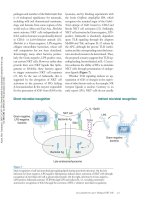
Figure 9.1 Large T antigen in SV40transformed cells Antibodies that bind
the SV40 large T (LT) antigen can be
used to detect LT in the nuclei of SV40transformed tumor cells. In the present
case, such antibodies were used to stain
human mammary epithelial cells (MECs)
that were transformed by introduction
of the SV40 early region plus two other
genes. A similar image would be seen if
such antibodies were used to stain SV40transformed mouse cells. LT was detected
by linking these antibody molecules to
peroxidase enzyme, which generated
the dark brown spots. In this image of a
tumor xenograft, the transformed MECs
form ducts (seen in cross section), which
are surrounded by normal stromal cells
(light blue nuclei). (Courtesy of T.A. Ince.)
this machinery is poised to eliminate cells that are en route to becoming neoplastic.
This explains why p53 function must be disabled before a clone of pre-malignant cells
gains a sure and stable foothold within a tissue. Without a clear description of p53
function and apoptosis, we have no hope of understanding a fundamental component
of the process that leads to the creation of virtually all types of human tumors.
9.1 Papovaviruses lead to the discovery of p53
TBoC2 B9.01,n9.100/9.01
When murine cells that have been transformed by the SV40 DNA tumor virus are
injected into a mouse of identical genetic background (that is, a syngeneic host), the
immune system of the host reacts by mounting a strong response; antibodies are
made that react with a nuclear protein that is present in the virus-transformed cells
and is otherwise undetectable in normal mouse cells (Figure 9.1). This protein, the
large tumor (large T, LT) antigen, is encoded by a region of the viral genome that is also
expressed when this virus infects and multiplies within monkey kidney cells—host
cells that permit a full infectious (lytic) cycle to proceed to completion (see Section
3.4).
Large T is a multifunctional protein that the SV40 virus uses to perturb a number
of distinct regulatory circuits within infected and transformed cells. Indeed, large T
was cited in the previous chapter because of its ability to bind and thus functionally
inactivate pRb (see Section 8.5). Anti-large T sera harvested from mice and hamsters
bearing SV40-induced tumors were used in 1979 to analyze the proteins in SV40transformed cells. The resulting immunoprecipitates contained both large T and an
associated protein that exhibited an apparent molecular weight of 53 to 54 kilodaltons
(Figure 9.2A). Antisera reactive with the p53 protein were found to detect this protein
in mouse embryonal carcinoma cells and, later on, in a variety of human and rodent
tumor cells that had never been infected by SV40. However, monoclonal antibodies
that recognized only large T immunoprecipitated the 53- to 54-kD protein in virusinfected but not in uninfected cells.
Taken together, these observations indicated that the large T protein expressed in
SV40-transformed cells was tightly bound to a novel protein, which came to be called
p53 (see Figure 9.2B). Antisera that reacted with both large T and p53 detected p53
in certain uninfected cells, notably tumor cells that were transformed by non-viral
mechanisms, such as the F9 embryonal carcinoma cells analyzed in Figure 9.2A. The
latter observations indicated that p53 was of cellular rather than viral origin, a conclusion that was reinforced by the report in the same year that mouse cells transformed
by exposure to a chemical carcinogen also expressed p53.
These various lines of evidence suggested that the large T oncoprotein functions,
at least in part, by targeting host-cell proteins for binding. (The discovery that large
Papovaviruses lead to the discovery of p53
(A)
(B)
3T3
SV40
–
F9
SV40
p53
–
T N T N T N T N
90°
94 kD
54 kD
large T hexamer
T antigen is also able to bind pRb, the retinoblastoma protein, came seven years later.)
In the years since these 1979 discoveries, a number of other DNA viruses and at least
one RNA virus have been found to specify oncoproteins that associate with p53 or perturb its function (Table 9.1). (As we will discuss later in this chapter, and as is apparent
from this table, these viruses also target pRb and undertake to block apoptosis.)
Table 9.1 Tumor viruses that perturb pRb, p53, and/or apoptotic function
Virus
Viral protein
targeting pRb
Viral protein
targeting p53
SV40
large T (LT)a
large T (LT)a
Adenovirus
E1A
E1B55K
HPV
E7
E6
Polyomavirus
large T
large T?
Herpesvirus saimiri
V cyclind
HHV-8 (KSHV)
K cyclind
LANA-2
v-Bcl-2,e v-FLIPf
Human
cytomegalovirus
(HCMV)
IE72g
IE86
vICA,h pUL37i
HTLV-I
Taxj
Tax
Epstein–Barr
EBNA3C
EBNA-1k
aSV40
TBoC2 b9.02/9.02
Viral protein targeting
apoptosis
E1B19Kb
middle T (MT)c
v-Bcl-2e
LMP1k
LT also binds a number of other cellular proteins, including p300, CBP, Cul7, IRS1, Bub1,
Nbs1, and Fbw7, thereby perturbing a variety of other regulatory pathways.
bFunctions like Bcl-2 to block apoptosis.
cActivates PI3K and thus Akt/PKB.
dRelated to D-type cyclins.
eRelated to cellular Bcl-2 anti-apoptotic protein.
fViral caspase 8 (FLICE) inhibitory protein; blocks an early step in the extrinsic apoptotic cascade.
gInteracts with and inhibits p107 and possibly p130; may also target pRb for degradation in
proteasomes.
hBinds and inhibits procaspase 8.
iInhibits the apoptotic pathway below caspase 8 and before cytochrome c release.
jInduces synthesis of cyclin D2 and binds and inactivates p16INK4A.
kLMP1 facilitates p52 NF-κB activation and thereby induces expression of Bcl-2; EBNA-1 acts via a
cellular protein, USP7/HAUSP, to reduce p53 levels. EBNA3C interferes with p53 function.
Figure 9.2 The discovery of p53 and
its association with SV40 large T
(A) Normal BALB/c 3T3 mouse fibroblasts
(3T3) transformed by SV40, as well as
F9 mouse embryonal carcinoma cells,
were exposed to 35S-methionine, and
resulting cell lysates were incubated
with either normal hamster serum (N)
or hamster antiserum reactive with
SV40-transformed hamster cells (T). The
anti-tumor serum immunoprecipitated
a protein of 94 kD from virus-infected
but not uninfected 3T3 cells. In
addition, a second protein running
slightly ahead of the 54-kD marker
was immunoprecipitated from SV40transformed 3T3 cells but not from
normal 3T3 cells. Moreover, this same
protein could be immunoprecipitated
from F9 cells, whether or not they had
been exposed to SV40 (arrow). These
particular data, on their own, did not
prove a physical association of SV40
large T (the 94-kD protein) with p53, but
they did show that p53 was a cellular
protein that was present in elevated
amounts in two types of transformed
cells. Moreover, they suggested that
the elevated levels of p53 in SV40transformed hamster cells cause the
hamster immune system to mount an
immune response against both large
T and the hamster’s own p53. (B) As
revealed by X-ray crystallography,
SV40 large T molecules assemble into
homohexamers, each subunit of which
binds and thereby sequesters a single
molecule of p53. (A, from D.I. Linzer and
A.J. Levine, Cell 17:43–52, 1979. B, from
D. Li et al., Nature 423:512–518, 2003.)
333
334
Chapter 9: p53 and Apoptosis: Master Guardian and Executioner
Figure 9.3 Effects of p53 on cell
transformation A cDNA encoding
a ras oncogene was co-transfected
with several alternative forms of a p53
cDNA into rat embryo fibroblasts. In
the presence of a p53 dl mutant vector,
which contains an almost complete
deletion of the p53 reading frame (left),
a small number of foci were formed.
In the presence of a p53 point mutant
(middle), a large number of robust foci
were formed. However, in the presence
of a p53 wild-type cDNA clone (right),
almost no foci were formed. (Courtesy of
M. Oren; from D. Michalovitz et al., Cell
62:671–680, 1990.)
ras + p53
deletion mutant
ras + p53 val-135
point mutant
ras + p53
wild type
9.2 p53 is discovered to be a tumor suppressor gene
The initial functional studies of p53 involved a substantial scientific detour: transfection of a p53 cDNA clone into rat embryo fibroblasts revealed that this DNA could
collaborate with a co-introduced ras oncogene in the transformation of these rodent
cells. Such activity suggested that the p53 gene (which is sometimes termed Trp53 in
mice and TP53 in humans) might operate as an oncogene, much like the myc oncogene, which had previously been found capable of collaborating with the ras oncogene
in rodent cell transformation (see Section 11.10). Like myc, the introduced p53 cDNA
seemed to contribute certain growth-inducing signals that resulted in cell transformation in the presence of a concomitantly expressed ras oncogene.
But appearances deceived. As later became
the p53 cDNA had originally
TBoC2 apparent,
b9.03/9.03
been synthesized using as template the mRNA extracted from tumor cells (rather
than normal cells). Subsequent manipulation of a p53 cDNA cloned instead from the
mRNA of normal cells revealed that this p53 cDNA clone, rather than favoring cell
transformation, actually suppressed it (Figure 9.3). Comparison of the sequences of
the two cDNAs revealed that the two differed by a single base substitution—a point
mutation—that caused an amino acid substitution in the p53 protein. Hence, the initially used clone encoded a mutant p53 protein with altered function.
These results indicated that the wild-type allele of p53 really functions to suppress cell
proliferation, and that p53 acquires growth-promoting powers when it sustains a point
mutation in its reading frame. Because of this discovery, the p53 gene was eventually
categorized as a tumor suppressor gene.
By 1987 it became apparent that such point-mutated alleles of p53 are common in the
genomes of a wide variety of human tumor cells. Data accumulated from diverse studies indicated that the p53 gene is mutated in 30 to 50% of commonly occurring human
cancers (Figure 9.4). Indeed, among all the genes examined to date in human cancer
cell genomes, p53 is the gene found to be most frequently mutated, being present in
mutant form in the genomes of almost one-third of all human tumors.
Further functional analyses of p53, conducted much later, made it clear, however,
that p53 is not a typical tumor suppressor gene. In the case of most tumor suppressor
genes, when the gene was inactivated (that is, “knocked out”) homozygously in the
mouse germ line (using the strategy of targeted gene inactivation described in Supplementary Sidebar 7.7), the result was, almost invariably, a disruption of embryonic
development due to deregulated morphogenesis in one or more tissues. These tumor
suppressor genes seemed to function as negative regulators of proliferation in a variety of cell types; their deletion from the regulatory circuitry of cells led, consequently,
to inappropriate proliferation of certain cells and thus to disruption of normal development.
In stark contrast, deletion of both p53 gene copies from the mouse germ line had no
significant effect on the development of the great majority of p53–/– embryos. Therefore, p53 could not be considered to be a simple negative regulator of cell proliferation
during normal development. Still, p53 was clearly a tumor suppressor gene, since mice
lacking both germ-line copies of the p53 gene had a short life span (about 5 months),
dying most often from lymphomas and sarcomas (Figure 9.5). This behavior provided
Mutant p53 acts as a dominant-negative
TP53 mutation prevalence by tumor site
Figure 9.4 Frequency of mutant p53
alleles in human tumor cell genomes
As indicated in this bar graph, mutant
alleles of p53 are found frequently in
commonly occurring human tumors.
This data set includes 26,597 somatic
mutations of p53 and 535 germ-line
mutations that had been reported by
November 2009. The bars indicate the
percentage of each tumor type found
to carry a mutant p53 allele. More
recent research indicates that virtually all
(119/123) of high-grade ovarian serious
carcinomas carry mutant p53 alleles.
(From International Agency for Research
on Cancer, TP53 genetic variations in
human cancer, IARC release R14, 2009.)
ovary
colorectum
head & neck
esophagus
lung
skin
pancreas
stomach
liver
bladder
brain
breast
uterus
soft tissues
lymph nodes
prostate
endocrine glands
bones
kidney
hematop. system
cervix
0
5
10
15
20
25
30
35
% of tumors with p53 mutation
40
45
50
the first hints that the p53 protein does not operate to transduce the proliferative and
anti-proliferative signals that continuously impinge on cells and regulate their proliferation. Instead, p53 seemed to be specialized to prevent the appearance of abnormal
cells, specifically, those cells that were capable of spawning tumors.
9.3 Mutant versions of p53 interfere with normal
p53 function
The observations of frequent mutation of the p53 gene in tumor cell genomes suggested that many incipient cancer cells must perturb or eliminate p53 function before
they can thrive. This notion raised the question of precisely how these cells succeed
in shedding p53 function. Here, another anomaly arose, because the p53 gene did
not seem to obey Knudson’s scheme for the two-hit elimination of tumor suppressor
genes. For example, the finding that a cDNA clone encoding a mutant version of p53
was able to alter the behavior of wild-type rat embryo fibroblasts (as described above)
ran directly counter to Knudson’s model of how tumor suppressor genes should operate (see Section 7.3).
TBoC2 b9.04/9.04
According to the Knudson scheme, an evolving pre-malignant cell can only reap substantial benefit once it has lost both functional copies of a tumor suppressor gene that
has been holding back its proliferation. In the Knudson model, such gene inactivation
events are caused by mutations that create inactive (“null”) and thus recessive alleles.
p53 + / +
100
90
% survival
80
p53 + / –
70
60
50
40
30
p53 – / –
20
10
0
0
100
200
300
age (days)
400
500
Figure 9.5 Effects of mutant p53
alleles in the mouse germ line This
Kaplan–Meier plot indicates the percent
of mice of the indicated genotype that
survived (ordinate) as a function of
elapsed lifetime in days (abscissa). While
the absence of p53 function in the p53–/–
mice (carrying two p53 null alleles) had
relatively little effect on their embryonic
development and viability at birth, it
resulted in a greatly increased mortality
relatively early in life, deriving largely
from the development of sarcomas
and leukemias. All p53–/– homozygotes
succumbed to malignancies by about
250 days of age (red line), and even
p53+/– heterozygotes (blue line) began to
develop tumors at this time, while wildtype (p53+/+) mice (green line) showed
virtually no mortality until almost 500
days of age. (Adapted from T. Jacks et
al., Curr. Biol. 4:1–7, 1994.)
335
336
Chapter 9: p53 and Apoptosis: Master Guardian and Executioner
Therefore, a pre-malignant cell may benefit minimally from inactivation of one copy
of a tumor suppressor gene—due to the halving of effective gene function—or not at
all, if the residual activity specified by the surviving wild-type gene copy suffices on its
own to mediate normal function. As we learned in Chapter 7, substantial change in
cell phenotype usually occurs only when the function of a suppressor gene is eliminated through two successive inactivating mutations or through a combination of an
inactivating mutation plus a loss-of-heterozygosity (LOH) event (see Section 7.4).
Knudson’s model was hard to reconcile with the observed behavior of the mutant p53
cDNA introduced into rat embryo fibroblasts (see Figure 9.3). The mutant p53 cDNAs
clearly altered cell phenotype, even though these embryo fibroblast cells continued
to harbor their own pair of wild-type p53 gene copies. This meant that the introduced
mutant p53 cDNA could not be functioning as an inactive, recessive allele. It seemed,
instead, that the point-mutated p53 allele was actively exerting some type of dominant
function when introduced into these rat embryo cells.
Another clue came from sequence analyses of mutant p53 alleles in various human
tumor cell genomes. These analyses indicated that the great majority of tumor-associated, mutant p53 alleles carry point mutations in their reading frames that create missense codons (resulting in amino acid substitutions) rather than nonsense codons
(which cause premature termination of the growing polypeptide chain). To date, more
than 26,000 tumor-associated p53 alleles originating in human tumor cell genomes
have been sequenced, 74% of which have been found to carry such missense mutations (Figure 9.6A). Furthermore, deletions of sequences within the reading frame of
the p53 gene are relatively uncommon. Consequently, researchers came to the inescapable conclusion that tumor cells can benefit from the presence of a slightly altered
p53 protein rather than from its complete absence, as would occur following the creation of null alleles by nonsense mutations or the outright deletion of significant portions of the p53 gene.
A solution to the puzzle of how mutant p53 protein might foster tumor cell formation
arose from two lines of research. First, studies in the area of yeast genetics indicated
that mutant alleles of certain genes can be found in which the responsible mutation
inactivates the normal functioning of the encoded gene product. At the same time,
this mutation confers on the mutant allele the ability to interfere with or obstruct the
ongoing activities of the surviving wild-type copy of this gene in a cell. Alleles of this
type are termed variously dominant-interfering or dominant-negative alleles.
A second clue came from biochemical and structural analyses of the p53 protein,
which revealed that p53 was a nuclear protein that normally exists in the cell as a
homotetramer, that is, an assembly of four identical polypeptide subunits (see Figure
9.6B and C). Together with the dominant-negative concept, this observed tetrameric
state suggested a mechanism through which a mutant allele of p53 could actively
interfere with the continued functioning of a wild-type p53 allele being expressed in
the same cell.
Assume that a mutant p53 allele found in a human cancer cell encodes a form of the
p53 protein that has lost most normal function but has retained the ability to participate in tetramer formation. If one such mutant allele were to coexist with a wild-type
allele in this cell, the p53 tetramers assembled in such a cell would contain mixtures
of mutant and wild-type p53 proteins in various proportions. The presence of only a
single mutant p53 protein in a tetramer might well interfere with the functioning of the
tetramer as a whole. Figure 9.7A illustrates the fact that 15 out of the 16 equally possible combinations of mutant and wild-type p53 monomers would contain at least one
mutant p53 subunit and might therefore lack some or all of the activity associated with
a fully wild-type p53 tetramer. Consequently, only one-sixteenth of the p53 tetramers
assembled in this heterozygous cell (which carries one mutant and one wild-type p53
gene copy) would be formed purely from wild-type p53 subunits and retain full wildtype function.
In an experimental situation in which a mutant p53 cDNA clone is introduced by
gene transfer (transfection) into cells carrying a pair of wild-type p53 alleles (see Figure 9.3), the expression of this introduced allele is usually driven by a highly active
p53 alterations largely affect DNA binding
transcriptional promoter, indeed, a promoter that is far more active than the gene
promoter controlling expression of the native p53 gene copies. As a consequence, in
such transfected cells, the amount of mutant p53 protein expressed by the introduced
(A)
9%
2%2%
4%
8%
54%
56%
74%
4% 9%
frameshift
2%
5%
ATM (n = 617)
in frame
deletions/insertions
missense
30%
28%
4%
APC (n = 15,451)
p53 (n = 26,597)
11%
14%
32%
51%
(B)
BRCA1 (n = 3,703)
nonsense
silent
splice site
(C)
transactivation
sequence-specific DNA binding
proline rich
tetramerization
175
distribution of mutations
248
245
249
C-terminal
273
282
COOH
H2N
1.7%
95.1%
tetramerization
3.2%
flexible linker
DNA
DNA-binding
transactivation
tetramerization
Taz2 co-activator
DNA binding
and the tetramerization domain are shown below. (C) The overall
Figure 9.6 Nature of p53 mutations (A) As indicated in these
structure of the DNA-bound p53 tetramer is shown here. The four
pie charts, point-mutated alleles of p53 leading to amino acid
DNA-binding domains are shown in green and blue, while the four
substitutions (green) represent the great majority of the mutant
tetramerization domains are seen as red and dark red α-helices
p53 alleles found in human tumors, while other types of mutations
are seen relatively infrequently. In contrast, the mutations striking
(above). The DNA double helix is shown in yellow. Each of the four
other tumor suppressor genes (APC) or “caretaker” genes involved
DNA-binding domains associates with half of a binding site in the
in maintenance of the genome (ATM, BRCA1) represent readingDNA; two copies of the binding site are present in the DNA with
frame shifts (yellow) or nonsense codons (blue) in the majority of
a small number of base pairs separating them (see Figure 9.12B).
cases; both of these types of mutation disrupt protein structure,
Each of the four transactivating domains (dark pink) is shown
TBoC2 b9.06,n9.101/9.06
usually by creating truncated versions of proteins that are often
interacting with the Taz2 domain of the p300 co-activator (light
degraded rapidly in cells. (B) The locations across the p53 reading
purple), which functions to stimulate transcription through its
frame of the point mutations causing amino acid substitutions
ability to acetylate histones and p53 itself. The C-terminal domain
are plotted here (above). As is apparent, the great majority of p53
(yellow) plays important roles in regulating transcription.
mutations (95.1%) affect the DNA-binding domain of the p53
(A, from International Agency for Research on Cancer, TP53 genetic
protein. The numbers above the figure indicate the residue numbers
variations in human cancer, IARC release R14, 2009; and A.I. Robles
of the amino acids that are subject to frequent substitution
et al., Oncogene 21:6898–6907, 2002. B, from K.H. Vousden and
in human tumors. The transactivation domain enables p53 to
X. Lu, Nat. Rev. Cancer 2:594–604, 2002; and A.C. Joerger and
interact physically with a number of alternative partners, including
A.R. Fersht, Annu. Rev. Biochem. 77:557–582, 2008. C, from
the p300/CBP transcriptional co-activator and Mdm2, the p53
A.C. Joerger and A.R. Fersht, Cold Spring Harb. Perspect. Biol.
antagonist. The detailed structures of the DNA-binding domain
2:000919, 2010.)
337
338
Chapter 9: p53 and Apoptosis: Master Guardian and Executioner
(A)
wild-type
p53 subunit
(B)
p53 function
point mutation
mutant
p53 subunit
Figure 9.7 p53 structure and p53
function as a dominant-negative
allele (A) In cells bearing a single mutant
p53 allele, the mutant protein usually
retains its ability to form tetramers but
loses its ability to function normally
because of a defective DNA-binding
domain. Consequently, mixed tetramers
composed of differing proportions of
wild-type (blue) and mutant (red) p53
subunits may form, and the presence of
even a single mutant protein subunit
may compromise the functioning of
the entire tetramer. Therefore, in a cell
that is heterozygous at the p53 locus,
fifteen-sixteenths of the p53 tetramers
may lack fully normal function.
(B) Perhaps the most direct
demonstration of the dominant-negative
mode of p53 action has come from
“knocking in” (see Supplementary
Sidebar 7.7) mutant p53 alleles into the
genome of mouse embryonal stem (ES)
cells. In cells in which a point mutation
in the DNA-binding domain (above) was
knocked into one p53 gene copy, almost
all p53 function was lost. In contrast,
when one p53 gene copy was completely
inactivated (yielding a null allele), p53
function was almost normal.
p53wt/wt
knock-in
mutations
wild-type
embryonic
stem cell
deletion
wt/pt.mut.
p53
p53wt/null
almost none
almost normal
gene will be vastly higher than the amount of normal protein produced by the cells’
endogenous wild-type p53 gene copies. Therefore, far fewer than one-sixteenth of the
p53 tetramers in such cells will be formed purely from wild-type p53 subunits. This
explains how an introduced mutant p53 allele can be highly effective in compromising
virtually all p53 function in such cells.
The above logic might suggest that many human tumor cells, which seem to gain some
advantage by shedding p53 function, should carry one wild-type and one mutant p53
b9.07b,n9.102/9.07
allele. Actually,TBoC2
in the great
majority of human tumor cells that are mutant at the p53
locus, the p53 locus is found to have undergone a loss of heterozygosity (LOH; see
Section 7.4), in which the wild-type allele has been discarded, yielding a cell with two
mutant p53 alleles. Thus, in such a cell, one copy of the p53 gene is initially mutated,
followed by elimination of the surviving wild-type copy through some type of loss-ofheterozygosity mechanism.
It is clear that an initial mutation resulting in a mutant, dominant-negative (DN) allele
is far more useful for the incipient tumor cell than one resulting in a null allele, which
causes total loss of an encoded p53 protein (see Figure 9.7B). The dominant-negative
allele may well cause loss of fifteen-sixteenths of p53 function, while the null allele will
result, at best, in elimination of one-half of p53 function. (Actually, if the levels of p53
protein in the cell are carefully regulated, as they happen to be, then this null allele
will have no effect whatsoever on a cell’s overall p53 concentration, since the surviving
wild-type allele will compensate by making more of the wild-type protein.)
Why, then, is elimination of the surviving wild-type p53 allele even necessary? The
answer seems to lie in the residual one-sixteenth of fully normal p53 gene function;
even this little bit seems to be more than most tumor cells care to live with. So, being
most opportunistic, they jettison the remaining wild-type p53 allele in order to proliferate even better. The observations described in Figure 9.7B of genetically altered
embryonic stem (ES) cells provide further evidence for p53’s dominant-negative
mode of action.
9.4 p53 protein molecules usually have short lifetimes
Long before the DNA-binding domain of p53 was discovered, the nuclear localization
of this protein in many normal and neoplastic cells suggested that it might function
as a transcription factor (TF). At least three mechanisms were known to regulate the
activity of transcription factors. (1) Concentrations of the transcription factor in the
nucleus are modulated. (2) Concentrations of the transcription factor in the nucleus
are held constant, but the intrinsic activity of the factor is boosted by some type of
covalent modification. (3) Levels of certain collaborating transcription factors may be
modulated. In some instances, all three mechanisms cooperate. In the case of p53, the
first mechanism—changes in the level of the p53 protein—was initially implicated.
Measurement of p53 protein levels indicated that they could vary drastically from
one cell type to another and, provocatively, would increase rapidly when cells were
exposed to certain types of physiologic stress.
These observations raised the question of how p53 protein levels are modulated by the
cell. Many cellular protein molecules, once synthesized, persist for tens or hundreds
of hours. (Some cellular proteins, such as those forming the ribosomal subunits in
p53 normally turns over rapidly
exponentially growing cells, seem to persist for many days.) Yet other cellular proteins
are metabolically highly unstable and are degraded almost as soon as they are assembled. One way to distinguish between these alternatives is to treat cells with cycloheximide, a drug that blocks protein synthesis. When such an experiment was performed
in cells with wild-type p53 alleles, the p53 protein disappeared with a half-life of only
20 minutes. This led to the conclusion that p53 is usually a highly unstable protein,
being broken down by proteolysis soon after it is synthesized.
This pattern of synthesis followed by rapid degradation might appear to be a “futile
cycle,” which would be highly wasteful for the cell. Why should a cell invest substantial
energy and synthetic capacity in making a protein molecule, only to destroy it almost
as soon as it has been created? Similar behaviors have been associated with other cellular proteins such as Myc (see Section 6.1).
The rationale underlying this ostensibly wasteful scheme of rapid protein turnover
is a simple one: a cell may need to rapidly increase or decrease the level of a protein in response to certain physiologic signals. In principle, such modulation could
be achieved by regulating the level of its encoding mRNA or the rate with which this
mRNA is being translated. However, far more rapid changes in the levels of a critical
protein can be achieved simply by stabilizing or destabilizing the protein itself. For
example, in the case of p53, a cell can double the concentration of p53 protein in 20
minutes simply by blocking its degradation.
Under normal conditions, a cell will continuously synthesize p53 molecules at a high
rate and rapidly degrade them at an equal rate. The net result of this is a very low
“steady-state” level of the protein within this cell. In response to certain physiologic
signals, however, the degradation of p53 is blocked, resulting in a rapid increase of p53
levels in the cell. This finding led to the further question of why a normal cell would
wish to rapidly modulate p53 levels, and what types of signals would cause a cell to
halt p53 degradation, resulting in rapidly increasing levels of this protein.
9.5 A variety of signals cause p53 induction
During the early 1990s, a variety of agents were found to be capable of inducing rapid
increases in p53 protein levels. These included X-rays, ultraviolet (UV) radiation, certain chemotherapeutic drugs that damage DNA, inhibitors of DNA synthesis, and
agents that disrupt the microtubule components of the cytoskeleton. Within minutes of exposing cells to some of these agents, p53 was readily detected in substantial
amounts in cells that previously had shown only minimal levels of this protein. This
rapid induction occurred in the absence of any marked changes in p53 mRNA levels
and hence was not due to increased transcription of the p53 gene. Instead, it soon
became apparent that the elevated protein levels were due entirely to the post-translational stabilization of the normally labile p53 protein.
In the years that followed, an even greater diversity of cell-physiologic signals were
found capable of provoking increases in p53 levels. Among these were low oxygen
tension (hypoxia), which is experienced by cells, normal and malignant, that lack
adequate access to the circulation and thus to oxygen borne by the blood. Still later,
introduction of either the adenovirus E1A or myc oncogene (see Sections 8.5 and 8.9)
into cells was also found to be capable of causing increases in p53 levels.
By now, the list of stimuli that provoke increases in p53 levels has grown even longer.
Expression of higher-than-normal levels of the E2F1 transcription factor, widespread
demethylation of chromosomal DNA, and a deficit in the nucleotide precursors of
DNA all trigger p53 accumulation. Exposure of cells to nitrous oxide or to an acidified
growth medium, depletion of the intracellular pool of ribonucleotides, and blockage
of either RNA or DNA synthesis also increase p53 levels.
These various observations made it clear that a diverse array of sensors are responsible for monitoring the integrity and functioning of various cellular systems. When
these sensors detect damage or aberrant functioning, they send signals to p53 and its
regulators, resulting in a rapid increase in p53 levels within a cell (Figure 9.8).
339
Chapter 9: p53 and Apoptosis: Master Guardian and Executioner
Figure 9.8 p53-activating signals and
p53’s downstream effects Studies
of p53 function have revealed that a
variety of cell-physiologic stresses can
cause a rapid increase in p53 levels.
The resulting accumulated p53 protein
then undergoes post-translational
modifications and proceeds to induce
a number of responses. A cytostatic
response (“cell cycle arrest,” often
called “growth arrest”) can be either
irreversible (“senescence”) or reversible
(“return to proliferation”). DNA repair
proteins may be mobilized as well as
proteins that antagonize blood vessel
formation (“block of angiogenesis”). As
an alternative, in certain circumstances,
p53 may trigger apoptosis.
lack of
UV
ionizing oncogene
blockage of
hypoxia
nucleotides radiation radiation signaling
transcription
p53
cell cycle DNA
block of
apoptosis
arrest
repair angiogenesis
OR
senescence
return to
proliferation
The same genotoxic (that is, DNA-damaging) agents and physiologic signals that provoked p53 increases were already known from other work to act under certain conditions in a cytostatic fashion, forcing cells to halt their advance through the cell cycle,
a response often called “growth arrest.” In other situations, some of these stressful
signals might trigger activation of the apoptotic (cell suicide) program. These observations, when taken together, showed a striking parallel: toxic agents that induced
growth arrest or apoptosis were also capable of inducing increases in p53 levels.
TBoC2
b9.08/9.08
Because such observations were
initially
only correlations, they hardly proved that
p53 was involved in some fashion in causing cells to enter into growth arrest or apoptosis following exposure to toxic or stressful stimuli.
Figure 9.9 p53 and the radiation
response Exposure of cells to X-rays
serves to strongly increase p53
levels. (A) Once it is present in higher
concentrations (8, 24 hours) and
is functionally activated via various
covalent modifications (not measured
here), p53 induces expression of the
p21Cip1 protein (see Section 8.4). p21Cip1
acts as a potent CDK inhibitor of the
cyclin–CDK complexes that are active in
late G1, S, G2, and M phases and can
thereby halt further cell proliferation at
any of these phases of the cell cycle.
The actin protein is included in all three
samples as a “loading control” to
ensure that equal amounts of protein
were added to the three gel channels
prior to electrophoresis. (B) Thymocytes
(leukocytes derived from the thymus)
of wild-type mice show an 80% loss
of viability relative to untreated control
cells during the 25 hours following
X-irradiation (green), while thymocytes
from p53+/– heterozygous mice (with
one wild-type and one null allele) show
almost as much loss of viability (red).
In contrast, thymocytes prepared from
p53–/– homozygous mutant mice exhibit
less than a 5% loss of viability during
this time period (blue). In all cases,
the loss of viability was attributable to
apoptosis (not shown). (A, courtesy of
K.H. Vousden. B, from S.W. Lowe et al.,
Nature 362:847–849, 1993.)
The definitive demonstrations of causality came from detailed examinations of p53
functions. For example, when genotoxic agents, such as X-rays, evoked an increase in
cellular p53 levels, the levels of the p21Cip1 protein (see Section 8.4) increased subsequently; this induction was absent in cells expressing mutant p53 protein. This suggested that p53 could halt cell cycle advance by inducing expression of this widely
acting CDK inhibitor (Figure 9.9A). Indeed, the long-term biological responses to
irradiation were often affected by the state of a cell’s p53 gene. Thus, cells carrying
mutant p53 alleles showed a greatly decreased tendency to enter into growth arrest
or apoptosis when compared with wild-type cells that were exposed in parallel to this
stressor (see Figure 9.9B).
These various observations could be incorporated into a simple, unifying mechanistic
model: p53 continuously receives signals from a diverse array of surveillance systems.
If p53 receives specific alarm signals from these monitors, it calls a halt to cell proliferation or triggers the apoptotic suicide program (see Figure 9.8).
(A)
(B)
100
p53
p53 – / –
p21Cip1
actin
0 8 24
hours post
radiation
viability (% of untreated)
340
80
60
p53 + / –
40
20
0
p53 + / +
0
5
10
15
time (h)
20
25
p53 processes many different signals
In fact, these cytostatic and pro-apoptotic powers of p53 represent a major threat to
incipient cancer cells that are advancing toward the malignant growth state. A number
of stresses, including hypoxia, genomic damage, and imbalances in the signaling
pathways governing cell proliferation, are commonly experienced by cancer cells during various stages of tumor development. In the presence of any one of these stresses,
an intact, functional p53 alarm system threatens the viability of would-be cancer cells.
Consequently, p53 activity must be blunted or even fully eliminated in these cells if
they are to survive and prosper.
This explains why most and perhaps all human tumor cells have partially or totally
inactivated their p53 alarm response. Without p53 on duty, cancer cells are far more
able to tolerate hypoxia, extensive damage to their genomes, and profound dysregulation of their growth-controlling circuitry. Once a cell acquires resistance to these
normally debilitating factors, the road is paved for it and its descendants to continue
their march toward a highly malignant growth state. In the same vein, normal cells
must also avoid excessive p53 activity, since it threatens to end their lives and thereby
cause depletion of the cells needed to maintain normal bodily functions (Sidebar 9.1).
9.6 DNA damage and deregulated growth signals cause
p53 stabilization
Three well-studied monitoring systems, two of which have already been cited, send
alarm signals to p53 in the event that they detect damage or signaling imbalances. The
first of these responds to double-strand breaks (DSBs) in chromosomal DNA, notably
those that are created by ionizing radiation such as X-rays. Indeed, a single dsDNA
break occurring anywhere in the genome seems sufficient to induce a measurable
increase in p53 levels. The identities of the proteins that detect such breaks are slowly
being resolved; it is known that these sensors of dsDNA breaks transfer signals to the
ATM kinase (Figure 9.10). (As described in Section 12.12, a deficiency of ATM leads
to the disease of ataxia telangiectasia and to hypersensitivity of cells to X-irradiation.)
ATM, in turn, transfers its signals on to the Chk2 kinase, which is able to phosphorylate
p53 itself; this phosphorylation of p53 protects it from destruction by a protein known
as Mdm2, discussed in the next section (see Figure 9.12).
A second signaling pathway is activated by single-strand DNA (ssDNA), which develops at stalled replication forks, often because DNA polymerases encounter bases that
have been altered by a wide variety of DNA-damaging agents, including certain chemotherapeutic drugs and UV radiation. ssDNA sensors activate ATR kinase, which
acts via the Chk1 kinase, to phosphorylate p53, again protecting it from degradation.
A third pathway leading to p53 activation is triggered by aberrant growth signals, notably those that result in deregulation of the pRb–E2F cell cycle control pathway. As we
will see below, this pathway does not depend upon kinase intermediates to induce
increases in p53 levels and signaling. The mechanisms by which other physiologic
stresses or imbalances, such as hypoxia, trigger increases in p53 levels remain poorly
understood.
These converging signaling pathways reveal a profound vulnerability of the mammalian cell. Through the course of evolution, a single protein—p53—has become
entrusted with the task of receiving signals from lookouts that monitor a wide variety of important physiologic and biochemical intracellular systems (see Figure 9.8).
The funneling of these diverse signals to a single protein would seem to represent an
elegant and economic design of the cellular signaling circuitry. But it also puts cells
at a major disadvantage, since loss of this single protein from a cell’s regulatory circuitry results in a catastrophic loss of the cell’s ability to monitor its own well-being
and respond with appropriate countermeasures in the event that certain operating
systems malfunction.
In one stroke (actually, the two strokes that cause successive inactivation of the two
p53 gene copies), the cell becomes blind to many of its own defects. It thereby gains
the ability to continue proliferation under circumstances that would normally cause
341
Chapter 9: p53 and Apoptosis: Master Guardian and Executioner
other target genes
OR
M
M
M
2
dm
2
dm
dm
2
M
p53
dm
2
mdm2
NUCLEUS
CYTOPLASM
U
bi
bi
U
U
U
bi
bi
U
bi
U
U
bi
bi
U
bi
U
bi
bi
U
mRNA
bi
U
bi
AAAA
mdm2
cytoplasmic
proteasomes
Mdm2
2
dm
M
Figure 9.10 Control of p53 levels by
Mdm2 After p53 concentrations increase
in response to certain physiologic signals
(not shown), the p53 tetramers bind to
the promoters of a large constituency
of target genes whose transcription
they induce (above), including the gene
encoding Mdm2; this results in a large
increase in mdm2 mRNA and Mdm2
protein (right). The Mdm2 molecules
then bind to the p53 protein subunits
and initiate their ubiquitylation, resulting
in export to the cytoplasm, further
ubiquitylation (not shown), resulting
in degradation in proteasomes. This
negative-feedback loop ensures that p53
levels eventually sink back to a low level
and, in undisturbed cells, helps to keep
p53 levels very low.
U
342
Md
m2
protein
Mdm2
it to call a halt to proliferation or to enter into apoptotic death. In addition, as we will
learn shortly, loss of the DNA repair and genome-stabilizing functions promoted by
p53 will make descendants of a p53–/– cell more likely to acquire further mutations
and advance more rapidly down
the road
of malignancy (Sidebar 9.2).
TBoC2
b9.11/9.11
9.7 Mdm2 destroys its own creator
The diverse alarm signals that impinge on p53 have a common effect—causing a rapid
increase in the levels of the p53 protein. Researchers have begun to understand how
this dramatic change is achieved. Like a wide array of other cellular proteins, p53 protein molecules are degraded by the ubiquitin–proteasome system (see Supplementary Sidebar 7.5). Proteins that are destined to be degraded by this system are initially
tagged by the covalent attachment of polyubiquitin side chains, which causes the proteins to be transported to proteasomes, in which they are digested into oligopeptides.
The critical control point in this process is the initial tagging process.
The degradation of p53 in normal, unperturbed cells is regulated by a protein termed
Mdm2 (in mouse cells) and MDM2 (in human cells). This protein recognizes p53 as a
target that should be ubiquitylated shortly after its synthesis and therefore marked for
rapid destruction (Figure 9.12). Mdm2 was initially identified as a protein encoded by
double-minute chromosomes present in murine sarcoma cells (hence mouse double
minutes). Subsequently, the human homolog of the mouse gene (HDM2) was discovered to be frequently amplified in sarcomas. In many human lung tumors, Mdm2 (as
we will call it) is overexpressed through mechanisms that remain unclear.
Mdm2 destroys its own creator
Sidebar 9.1 Too much of a good thing: a hyperactive p53 protein causes premature aging The descriptions of p53 actions
in this chapter include ample evidence that the p53 alarm,
once activated, provides important protection against the development of cancer. What would happen if a p53 allele that
encodes a constitutively active form of the protein were inserted into the mouse germ line? This has occurred through an
experimental accident, in which the first six exons of the p53
gene were inadvertently deleted during attempts to replace a
germ-line wild-type p53 allele with a point-mutated allele. The
p53 protein encoded by this truncated allele behaves as if it
were continuously active, even without the alarm signals that
are normally required to activate it. Mice heterozygous for this
allele are totally protected from the lymphomas, osteosarcomas, and soft-tissue (non-bone) sarcomas that commonly afflict wild-type mice late in life. And fibroblasts from these mice
are more resistant than their wild-type counterparts to transformation in vitro by an introduced ras oncogene. These outcomes, among many others, show that p53 activity does indeed
protect tissues from spawning tumors, ostensibly by eliminating cancerous cells before they have had a chance to proliferate
into tumors of a significant size.
However, rather than living longer lives, these “cancerresistant” mice showed a premature aging syndrome, their
lifespan being reduced by some 20%. The accelerated aging
included changes frequently observed in aged humans, such
(B)
(A)
1.00
cumulative survival
wild-type
mutant
as development of a hunched spine, retarded wound healing, reduced replacement of lost white blood cells, plus losses
of weight, vigor, muscle mass, bone density, and hair (Figure
9.11A). In many tissues, there was a widespread depletion of
cells, suggesting a loss of self-renewing stem cells.
These phenotypes are consistent with the idea that p53
plays a role in the aging process, but they hardly prove it. An
experiment of nature seems to address these issues more directly: a naturally occurring polymorphism in the human gene
pool places a proline residue in place of an arginine in residue
72 of p53. In vitro experiments show that the Arg72 protein is as
much as fivefold more effective in triggering apoptosis than the
Pro72 form of the protein. An epidemiologic study carried out
in the Netherlands of a population over 85 years of age demonstrated that those with a p53Pro/Pro genotype had a 2.54-fold
increased risk of cancer (compared with those of a p53Arg/Arg
genotype). However, those with a p53Pro/Pro genotype showed
an overall 41% greater survival during the period studied (see
Figure 9.11B). Moreover, deaths from general exhaustion and
frailty were observed in 21% of the Arg/Arg subjects but in only
6% of the Pro/Pro individuals. Observations like these add
weight to the notion that increased activity of p53 affords greater protection against cancer but at the same time accelerates
the age-dependent deterioration of tissues. Hence, too much
vigilance by the p53 watchman may incur a heavy price on the
organism as a whole.
Arg/Arg
Arg/Pro
Pro/Pro
0.75
0.50
0.25
0.00
85
90
age in years
95
subjects in the Netherlands followed their survival in the years
Figure 9.11 Premature aging induced by an overly active
after their enlistment. As is apparent from this Kaplan–Meier
p53 The p53 protein may afford protection against cancer and
plot, those with a Pro/Pro allele (affecting residue 72 of p53)
at the same time accelerate the aging process. (A) Attempts
exhibited a markedly improved survival in the decade that
at altering a germ-line copy of the p53 gene in mice resulted,
followed compared with heterozygotes or those with an Arg/
inadvertently, in the creation of an allele of p53 that specifies
Arg allele. (While consistent with a key role of p53 in governing
a constitutively active p53 protein. As seen here, the resulting
longevity, this striking, statistically significant association does
mutant mice (below, show many of the attributes of aging
not definitively prove such a role, since it remains formally
seen in humans, including muscle atrophy and a hunching
of supp02/9.10
TBoC2
possible that these alleles are tightly linked with other loci on
the back, when compared with wild-type mice of similar age
Chromosome 17 that are the actual determinants of longevity.)
(above); both mice were skinned. In addition, mutants exhibited
(A, from S.D. Tyner et al., Nature 415:45–53, 2002. B, from
osteoporosis, atrophy of numerous organs, and diminished
D. van Heemst et al., Exp. Gerontol. 40:11–15, 2005.)
tolerance to stress. (B) A prospective study of 1226 85-year-old
As is the case with other oncogenes, it seemed at first that amplification of the mdm2
gene (indicated by the presence of many double-minute chromosomal particles in
tumor cells; see Figure 1.12B) afforded tumor cells some direct, immediate proliferative advantage. Only long after the Mdm2 protein was first identified did its role as
the agent of p53 destruction become apparent. In fact, the detailed effects exerted by
Mdm2 on p53 are slightly more complex than indicated above.
343
344
Chapter 9: p53 and Apoptosis: Master Guardian and Executioner
Sidebar 9.2 Sunlight, p53, and skin cancer The p53 protein
stands as an important guardian against skin cancer induced
by sunlight. In the event that the genome of a keratinocyte in
the skin has suffered extensive damage from ultraviolet-B (UVB) radiation, p53 will rapidly trigger its apoptotic death. One
manifestation of this is the extensive scaling of skin several
days after a sunburn. At the same time, UV-B exposure may
cause the mutation and functional inactivation of a p53 gene
within a keratinocyte. This is indicated by the fact that mutant
p53 alleles found in human squamous cell carcinomas of the
skin often occur at dipyrimidine sites—precisely the sites at
which UV-B rays induce the formation of pyrimidine–pyrimidine cross-links (see Section 12.6). Such mutant p53 alleles
can also be found in outwardly normal skin that has suffered
chronic sun damage. Once p53 function is compromised by
these mutations, keratinocytes may be able to survive subsequent extensive exposures to UV-B irradiation, because apoptosis will no longer be triggered by their p53 protein. Moreover,
loss of p53 results in a diminished ability to repair subsequent
UV-B–induced DNA lesions. Hence, p53-mutant cells may subsequently acquire additional mutant alleles that enable them,
together with the mutant p53 alleles, to form a squamous cell
carcinoma.
Human papillomaviruses (HPVs) are increasingly implicated as co-factors in many of these squamous cell carcinomas; a
key function of the E6 virus–encoded oncoprotein may explain
the synergistic actions of UV-B radiation and HPV in the pathogenesis of these relatively common tumors: E6 tags p53 for destruction by ubiquitylation and degradation in proteasomes,
thereby phenocopying mutational inactivation of the chromosomal p53 gene (Supplementary Sidebar 9.1). Interestingly,
mice that lack functional p53 gene copies in all cells also respond to UV-B exposure by developing uveal melanomas—tumors of pigmented cells in the front of the eye; similar tumors
are suspected to be caused in humans by UV exposure.
Of additional interest, p53 operating in keratinocytes has
another totally unrelated effect that illustrates its diverse functions. In response to the DNA damage created by UV radiation, p53 causes these cells to release melanocyte-stimulating
hormone (αMSH); the latter proceeds to stimulate nearby skin
melanocytes to produce melanin pigment and to transfer resulting melanin granules back to the keratinocytes (see Figure
12.19), resulting in the increased pigmentation of the skin that
creates suntan!
As we will learn below, p53 operates by acting as a transcription factor; Mdm2 binding
to p53 immediately blocks the ability of p53 to function in this role. [In more detail,
Mdm2 succeeds in shutting down p53-driven transcription by (1) preventing the
binding to p53 of p300/CBP, which activate transcription by acetylating histones; and
(2) by actively recruiting yet other enzymes that block p53-mediated transcription by
methylating histones (see Section 1.8).] Thereafter, Mdm2 directs the attachment of a
ubiquitin moiety to p53 and the export of p53 from the nucleus (where p53 does most
of its work) to the cytoplasm; subsequent polyubiquitylation of p53 ensures its rapid
degradation in cytoplasmic proteasomes. The continuous, highly efficient actions of
Mdm2 ensure the short, 20-minute half-life of p53 in normal, unstressed cells.
While the present discussion and Figure 9.11 represent Mdm2 as a monomeric protein, it actually often forms heterodimeric complexes with its close cousin, MdmX (also
called Mdm4). This complex may be responsible for much of the ubiquitylation activity that drives p53 degradation. Indeed, there is evidence that without the presence of
MdmX, Mdm2 loses the ability to drive p53 degradation. When expressed on its own,
however, MdmX seems to be limited to blocking p53-mediated transcriptional activation. (Moreover, MdmX differs in another important respect from its Mdm2 cousin: its
expression is not regulated by p53, a process that is described below.)
In some circumstances—specifically, when cells are suffering certain types of stress
or damage—p53 protein molecules must be protected from their Mdm2 executioner
so that they can accumulate to functionally significant levels in the cell. This protection is often achieved by phosphorylation of p53, which blocks the ability of Mdm2
to bind p53 and trigger its ubiquitylation. More specifically, phosphorylation of p53
on amino acid residues in its N-terminal domain (see Figure 9.12) by kinases such as
ATM, Chk1, and Chk2 (which become activated in response to DNA damage, as was
described in Section 9.6) alters the domain of p53 that is normally recognized and
bound by Mdm2, and in this way prevents the association of Mdm2 with p53. At the
same time, the DNA damage–activated ATM kinase can phosphorylate Mdm2 in a way
that causes its functional inactivation and destabilization. As a consequence of this
phosphorylation of both p53 and Mdm2, Mdm2 fails to initiate ubiquitylation of p53,
p53 escapes destruction, and p53 concentrations in the cell increase rapidly (Figure
9.13). Once it has accumulated in substantial amounts, p53 is then poised to evoke a
series of downstream responses, to be discussed in detail later.
Mdm2 destroys its own creator
Note that Mdm2 operates here as an oncoprotein, but one whose mechanism of
action is very different from those of the various oncoproteins that we encountered
in Chapters 4, 5, and 6. The latter function as components of mitogenic signal cascades and thereby induce cell proliferation by mimicking the signals normally triggered by the binding of growth factors to their receptors. Mdm2, in contrast, operates
by antagonizing p53 and thereby prevents entrance of a cell into cell cycle arrest, into
the nongrowing state known as senescence, or into the apoptotic suicide program.
The final outcome is, however, the same: the actions of oncoproteins and Mdm2 both
favor increases in cell number.
The activity and levels of the Mdm2 protein are affected by yet other positive and negative signals. The signaling pathway that favors cell survival through activation of the
PI3 kinase (PI3K) pathway leads, via the Akt/PKB kinase, to Mdm2 phosphorylation
(at a site different from that altered by the ATM kinase described above) and to the
resulting translocation of Mdm2 from the cytoplasm to the nucleus, where it is poised
to attack p53 (see Figure 9.13A). Because PI3K itself is activated by Ras and growth
factor receptors, we come to realize that the mitogenic signaling pathway does indeed
influence Mdm2 and thereby p53, albeit indirectly. At the same time, activation of the
mitogenic Ras → Raf → MAPK signaling pathway leads, via the Ets and AP-1 (Fos +
Jun) transcription factors, to greatly increased transcription of the mdm2 gene, yielding higher levels of mdm2 mRNA and protein (see Figure 9.13A). These elevated levels
of Mdm2 protein amplify the phosphorylation-induced activation of Mdm2 achieved
by the PI3K → Akt/PKB signaling pathway. Ultimately, all these effects converge on
suppressing p53 protein levels.
(A)
(B)
Mdm2-binding
transactivation
p53
transcription
regulation,
regulation of
DNA binding
p53 domains
sequence-specific DNA binding
proline rich
tetramerization
H 2N
COOH
NLS
Mdm2
NLS NLS
sites of phosphorylation
(C)
1
2
3
4
5
6
7
8
9
10
11 12 13 14 15 16 17 18 19 20
Figure 9.12 Specialized domains of p53 (A) The structure of
the interface where p53 and Mdm2 interact has been revealed
by X-ray crystallography. The interacting domain of p53 is shown
as a yellow space-filling model that includes p53 residues 18
through 27, while the surface of the complementary pocket of
Mdm2 is shown as a blue wire mesh. (B) The interaction of p53
with Mdm2 (see Figure 9.10) occurs in a small domain near its
N-terminus, where the transactivation domain of p53 is also
located. The phosphorylation of p53 amino acid residues in this
region (red lollipops; not all are indicated) blocks Mdm2 binding
and thus saves p53 from ubiquitylation and degradation. The
nearby proline-rich domain (salmon) contributes to p53’s proapoptotic functions. Its tetramerization domain is located toward
its C-terminus (see Figure 9.6). Nearby are nuclear localization
signals (NLS), which allow import of recently synthesized p53 into
the nucleus, as well as amino acid sequences that regulate its
DNA binding. (C) p53–DNA complexes present in the chromatin
of human cells can be immunoprecipitated by anti-p53 antibodies
(the ChIP procedure; see Supplementary Sidebar 8.3). In one
such experiment, sequence analyses of DNA fragments in the
precipitates led to the identification of 1546 sites in the human
genome to which p53 bound after cells were stressed by exposure
to the drug actinomycin, a potent inhibitor of transcription. The
consensus DNA sequence to which p53 bound is shown here,
where the relative size of each letter indicates how frequently a
DNA base was found at the indicated position in the binding site.
Interestingly, the majority of these 1546 sites could also be bound
by p53’s cousins, p63 and p73, to be described later in this chapter.
(A, from P.H. Kussie et al., Science 274:948–953, 1996. B, from
D.E. Fisher, ed., Tumor Suppressor Genes in Human Cancer. Totowa,
NJ: Humana Press, 2000. C, from L. Smeenk et al., Nucleic Acids
Res. 36:3639–3654, 2008.)
345
Chapter 9: p53 and Apoptosis: Master Guardian and Executioner
346
(A)
mitogenic and
cell survival signals
AP-1
other target genes
Ets
P
P
AP-1
mdm2
activated
p53
P
P
P
P
P
kinases
Chk1/2
bi
U
ATR
RPA
DNA
damage
ATM
M/R/N
dsDNA
P Mdm2
P Mdm2 inactivated
Mdm2
P
bi
U
sensors
P
P
U
bi
bi
U
P
Ets
ssDNA
OR
p53
P
NUCLEUS
U
bi
U
bi
bi
U
fluorescence (AU)
2
bi
U
bi
U
bi
U
U
bi
bi
U
dm
U
bi
bi
U
Mdm2
0.4
0.2
400 600
time (min)
Mdm2
M
U
bi
bi
U
p53
200
cell survival
signals
Md
m2
Yet another mechanism that affects Mdm2 has been revealed through the discovery
of an Mdm2 antagonist, which is termed p19ARF in mouse cells and p14ARF in human
cells. Astute sequence analysis led to the discovery of ARF, as we will call it hereafter.
Its encoding gene was originally uncovered in mouse cells as a gene whose sequences
are intertwined with those specifying p16INK4A, the important inhibitor of the CDK4
and CDK6 kinases that initiate pRb phosphorylation (see Section 8.4).
Through use of a transcriptional promoter located 13 kilobases upstream of the
p16INK4A promoter and an alternative splicing program, an mRNA is assembled that
encodes, in an alternative reading frame, the structure of the ARF protein (Figure
9.14). Forced expression of an ARF-encoding cDNA in wild-type rodent cells was
found to cause a strong inhibition of their proliferation. However, this inhibition was
not observed when the ARF cDNA was expressed in cells that lacked wild-type p53
function. This indicated that the growth-inhibitory powers of ARF depend absolutely
on the presence of functional p53 in these cells.
1
0
0
AAAA
mdm2
Akt/
PKB
Mdm2
(B)
0.6
mRNA
OR
Mdm2
activated Mdm2
cytoplasmic
proteasome
0.8
P
Mdm2
CYTOPLASM
P
800 1000
Further investigation revealed that in wild-type cells, the expression of ARF causes
a rapid increase in p53 levels. We now understand the molecular mechanisms that
explain how this response works. ARF binds to Mdm2 and inhibits its action, either
by sequestering Mdm2 in the nucleolus—the nuclear structure that is largely devoted
TBoC2 b9.13/9.13
to manufacturing ribosomal subunits—or by inhibiting Mdm2 in the nucleoplasm
(Figure 9.15A). Once Mdm2 is diverted from interacting with p53, the latter escapes
p53 phosphorylation saves it from destruction
Figure 9.13 Control of p53 levels by various kinases (A) The cycle of p53 synthesis
and destruction indicated in Figure 9.10 can be modulated by a series of regulators. DNA
damage-sensors, such as RPA (replication protein A) and the M/R/N (MRE11/Rad50/Nbs1)
complex, detect either extensive ssDNA regions/replication-blocking DNA lesions or dsDNA
breaks (DSBs) and proceed to activate two kinases, ATR and ATM, respectively. These kinases
act directly on p53, or indirectly via Chk1 and Chk2, to phosphorylate p53 (center) in its
N-terminal domain (see Figure 9.12B), thus preventing the binding of Mdm2 (gold, left
center). At the same time, phosphorylation of Mdm2 molecules by these kinases inactivates
them, blocking their ability to associate with p53 (bottom center). These alterations save p53
from Mdm2-mediated binding, ubiquitylation, and destruction in proteasomes (lower left).
Acting in an opposing manner, certain survival signals (such as those conveyed by mitogenic
growth factors), acting through the AP-1 and Ets transcription factors, collaborate with p53
to promote expression of the mdm2 gene (top right), resulting in increases in mdm2 mRNA
and Mdm2 protein synthesis (lower right). These survival signals also activate the Akt/PKB
kinase, which activates already-synthesized Mdm2 molecules by phosphorylating them
at another site (bottom). The activated Mdm2 then proceeds to bind p53 and trigger its
ubiquitylation and proteasome-mediated destruction. Not illustrated is the Mdm2-driven
destruction of pRb by ubiquitylation in physiologically stressed cells. (B) The mutually inhibitory
interactions between p53 and Mdm2, which form a reciprocal negative-feedback loop
(see Figure 9.10), result in oscillations of the levels of the two proteins. These levels can be
monitored in individual cells using proteins labeled with different-colored fluorescent tags.
Following DNA-damaging X-irradiation, the height and duration of each pulse are not affected
by radiation dose, but the number of successive pulses, which form a digital clock, increases
with increasing radiation dose, perhaps continuing until the DNA is repaired or the cell dies.
The fluorescence intensities (reflective of the levels of the two proteins) are presented in
arbitrary units (AU). (B, from G. Lahav et al., Nat. Genet. 36:147–150, 2004.)
Mdm2-mediated ubiquitylation and resulting destruction and therefore accumulates
rapidly to high levels in the cell. The enemy of an enemy is a friend: ARF can induce
rapid increases in p53 levels because it kidnaps and inhibits p53’s destroyer, Mdm2.
Importantly, in normal, unstressed cells, Mdm2 must be allowed to perform its normal role of keeping p53 levels very low, as is highlighted by the results of inactivating both mdm2 gene copies in the genomes of mouse embryos. These embryos die
very early in embryogenesis, ostensibly because p53 levels increase to physiologically
intolerable levels, preventing the normal proliferation of embryonic cells or causing
them to die. (That the profoundly disruptive effects of Mdm2 gene inactivation are
due to runaway p53 activity is made clear by studies in which both genes in a mouse
embryo are inactivated homozygously, yielding the Mdm2–/– p53–/– genotype. Once
p53 is eliminated from the embryonic cells, the loss of Mdm2 becomes tolerable and
embryonic development occurs normally!)
exons
1β
1α
13 kb
2
3
p16INK4A
p14ARF
Figure 9.14 The gene encoding p16INK4A and p14/p19ARF Analysis of the p16INK4A gene
(red) has revealed that it shares its second exon with a second gene encoding a 19-kD protein
in mice and a 14-kD protein in humans. The p14/p19 gene uses an alternative transcriptional
promoter (blue arrow, left) located more than 13 kilobases upstream of the one used by
p16INK4A (red arrow, center). Because translation of its mRNA uses an alternative reading
frame (green bracket) in exon 2 (red, blue), the resulting protein and thus gene came to be
called p19ARF (or in humans p14ARF). The patterns of RNA splicing are indicated by the carets
connecting the various exons of the two intertwined genes. The boxes indicate exons, while
the filled areas within each exon indicate reading
frames.
(From C. Sherr, Genes Dev.
TBoC2
b9.14/9.14
12:2984–2991, 1998.)
347
348
Chapter 9: p53 and Apoptosis: Master Guardian and Executioner
Sidebar 9.3 Have mammalian cells placed too many eggs in one basket? The discovery of the p16INK4A/p14ARF genetic locus, which is inactivated through one mechanism
or another in about half of all human tumors, raises a provocative question: Why have
mammalian cells invested a single chromosomal locus with the power to encode two
proteins regulating the two most important tumor suppressor pathways, those of pRb
and p53? Deletion of this single locus results in the simultaneous loss of normal regulation of both pathways. As was the case with p53 itself, enormous power has been concentrated in the hands of a single genetic locus.
Placing two such vital eggs in a single genetic basket seems foolhardy for the mammalian cell, as it causes the cell to be vulnerable to two types of deregulation through
loss of a single gene. To make matters even worse, the gene encoding p15INK4B, another
important regulator of pRb phosphorylation (see Section 8.4), is closely linked to the
p16INK4A/p14ARF locus, indeed so close that all of these genetic elements are often lost
through the deletion of only about 40 kb of chromosomal DNA. We have yet to discern
the underlying rationale of this genetic arrangement. Maybe there is none, and perhaps
mammalian evolution has produced a less-than-optimal design of its control circuitry.
The series of mutual antagonisms indicated in Figure 9.15 makes ARF an ally of p53
and, like p53, a tumor suppressor protein. In many human tumors, inactivation of the
p16INK4A/p14ARF locus by genetic mutation or epigenetic promoter methylation can
be demonstrated. Once a cell has lost ARF activity, it loses the ability to block Mdm2
function. As a consequence, Mdm2 is given a free hand to drive p53 degradation, and
the cell is deprived of the services of p53 because the latter can never accumulate to
functionally significant levels.
Since ARF has a central role in increasing p53 levels in many cellular contexts, this
means that the p14ARF gene, like the gene encoding its p53 target, is an extremely
important tumor suppressor gene. Moreover, it seems likely that many of the human
cancer cells that retain wild-type p53 gene copies have eliminated p53 function by
inactivating their two copies of the gene encoding ARF. Finally, we should note that
the co-localization of the p16INK4A and p14ARF genes (see Figure 9.14) represents yet
another concentration of power that creates additional vulnerability for normal cells
(Sidebar 9.3).
9.8 ARF and p53-mediated apoptosis protect against
cancer by monitoring intracellular signaling
The influential role of ARF in increasing p53 levels raises the question of how ARF itself
is regulated. In this instance, we learn something highly relevant from our discussion
in Chapter 8 of the pRb pathway, and from the fact that mammalian cells are very
sensitive to higher-than-normal levels of E2F1 activity (see Figure 9.15B). In fact, a cell
seems to monitor the activity level of this particular transcription factor (together with
those of E2F2 and E2F3) as an indication of whether its pRb circuitry is functioning
properly; excessively high levels of active E2F transcription factors provide a telltale
sign that pRb function has gone awry.
Evolution has created several ways to eliminate cells that carry too much E2F activity
and, by implication, have lost proper pRb control (Figure 9.16). Runaway E2F1 activity drives expression of a number of genes encoding proteins that directly participate
in the apoptotic program. Included among these are genes encoding caspases (types
3, 7, 8, and 9), pro-apoptotic Bcl-2–related proteins (Bim, Noxa, PUMA), Apaf-1, and
p53’s cousin, p73; these proteins collaborate to drive cells into apoptosis. We will learn
more about them later.
In addition, the p53-dependent apoptotic program is often triggered by elevated E2F
activity. It turns out that the p14ARF gene carries an E2F recognition sequence in its
promoter. In a way that is still incompletely understood, unusually high levels of E2F1,
E2F2, or E2F3A activity induce transcription of p14ARF mRNA. The ARF protein soon
ARF monitors intracellular signaling
(A)
NUCLEOPLASM
Md
m2
E2F DP
1/2/3 1/2
ARF
p14ARF
ARF
m2
Mdm2
NUCLEOLUS
Md
ARF
(
(C)
Ras
OR
100
80
ARF
Mdm2
m2
Mdm2
60
40
cell cycle
arrest
apoptosis
0
0
5
10
weeks
15
bi
U
ARF +/GFP
U
20
bi
p53
Md
U
% survival
E2F
Mdm2
ARF +/+
p53
bi
c-Myc
U
E1A
bi
(B)
accumulates)
20
NUCLEUS
CYTOPLASM
bi
U
bi
U
bi
U
bi
U
bi
U
bi
bi
U
U
bi
U
This pathway, working together with the several p53-independent pro-apoptotic signals cited above, accomplishes the goal of eliminating cells that lack proper pRb function. Such E2F-initiated apoptosis seems to explain why mouse embryos that have
been deprived of both copies of their Rb gene die in mid-gestation due to the excessive
bi
p53 → apoptosis
U
Mdm2
bi
E2F → ARF
U
pRB
bi
TBoC2
b9.15/9.15
appears on the scene and blocks Mdm2 action. p53 then
accumulates
and triggers, in
turn, apoptosis (see Figure 9.15B), leading to a signaling cascade configured like this:
U
Figure 9.15 Control of apoptosis by ARF (A) The human
protein
in mice),
termed ARF in the figure (light blue, top), associates with and inactivates Mdm2 (gold); it
remains unclear whether this interaction occurs only in the nucleoplasm of cells or whether
ARF drags Mdm2 into the nucleolus for sequestration (top right). Once it is neutralized,
Mdm2 can no longer attack p53 and tag it for destruction by ubiquitylation (lower right).
Hence, elevated ARF levels cause increases in the levels of p53. As shown here and in panel B,
transcription of the p14ARF gene is driven by E2F transcription factors composed of E2F1/2/3
and their DP1/2 partners (see Figure 8.23). (B) A variety of oncogenic signals favor apoptosis
through their ability to induce E2F activity (see Section 8.7), which leads, in turn, to increased
ARF expression. Included among these are the adenovirus E1A, the Myc (= c-Myc), and the
Ras oncoproteins. This suggests that this signaling pathway evolved to eliminate cells with,
among other defects, overly active E2F signaling. (C) One copy of the p19ARF-encoding gene
was inactivated in a mouse germ line by replacing it with a GFP (green fluorescent protein)encoding sequence. These mice (ARF+/GFP) and wild-type mice (ARF+/+) were mated with others
carrying an Eμ-myc transgene known to cause B-cell lymphomas (also described in Figure
9.23). The ARF+/GFP Eμ-myc mice (red line) developed fatal tumors far more rapidly than mice
carrying only the Eμ-myc transgene (green line), and cells in these tumors shed their remaining
wild-type ARF allele. Hence, in the absence of ARF function, the pro-apoptotic effects of a myc
oncogene (B) are largely lost, allowing its proliferative effects to dominate and drive tumor
formation. (B, courtesy of P.J. Iaquinta and J.A. Lees. C, from F. Zindy et al., Proc. Natl. Acad.
Sci. USA 100:15930–15935, 2003.)
bi
(p19ARF
U
p14ARF
proteasome
349
Chapter 9: p53 and Apoptosis: Master Guardian and Executioner
proliferation and concomitant apoptosis of certain critical cell types, including those
involved in erythropoiesis (formation of red blood cells) and in placental function.
The discovery of the critical role of ARF in the control of p53 function suggested the
possibility that ARF function is eliminated by a variety of molecular strategies during
tumor formation. Such elimination may well confer on cancer cells the same benefits as those resulting from mutation of the p53 gene itself. In fact, regulation of transcription of the p14ARF gene is quite complex and therefore susceptible to disruption
through a variety of alterations (Sidebar 9.4).
In sum, because loss of pRb control within a cell represents a grave danger to the surrounding tissue, cells are poised to trigger apoptosis whenever E2F1 deregulation
occurs. These connections between E2F1 activity and apoptosis suggest another idea,
still speculative: the great majority of cells that suffer loss of pRb control never succeed
in generating clones of pre-neoplastic or neoplastic descendants, because these cells
suffer apoptosis as soon as they lose this important control mechanism.
Consistent with this logic are some of the known properties of the E1A and myc oncogenes. Both deregulate pRb control, and both are highly effective in inducing apoptosis. Recall that the adenovirus E1A oncoprotein binds and effectively sequesters pRb
and its cousins. Myc, for its part, pulls regulatory levers in the cell cycle clock that
ensure that pRb is inactivated through phosphorylation (see Sections 8.5 and 8.9).
Many studies of myc oncogene function indicate that this gene exerts both potent
mitogenic and pro-apoptotic functions. Indeed, the pro-apoptotic effects of the myc
oncogene are so strong that it is highly likely that most cells that happen to acquire
a myc oncogene are also rapidly eliminated through apoptosis. On occasion, the
(A)
(B)
600
without OHT
–OHT
counts
480
360
240
2.44%
0
50 100 150 200 250
fluorescence intensity
(cell size →)
200
150 32.06%
100
ER
DNA
phospho Erk
Arf
E2F1
50
0
cytoplasmic
sequestration,
E2F1 inactive
with OHT
250
+OHT
ER
E2F1
120
counts
350
50 100 150 200 250
fluorescence intensity
(cell size →)
transport to
nucleus,
E2F1 active
genes, among them those that have pro-apoptotic effects. As a
Figure 9.16 Induction of apoptosis and ARF expression by
result, a significant proportion (32.06%) of the cells now show a
E2F1 and Ras. (A) The apoptotic state of cells can be monitored
size smaller than that of normal healthy cells, indicative of their
by fluorescence-activated cell sorting (FACS), which in this case is
having fragmented during apoptosis. (B) In a transgenic mouse
used to measure the size of individual cells or subcellular fragments
model of lung adenocarcinoma development, signaling by Ras
(abscissa) and the number of cells of a given size (ordinate). In this
oncoprotein and its activated downstream effector, phosphorylated
experiment, the E2F1 transcription factor (red) has been fused to
Erk/MAPK (see Figure 6.14), increases as tumors progress from
the estrogen receptor (ER) protein (green), making E2F1 activity
lower-grade adenomas to higher-grade adenocarcinomas. As
dependent on the presence of tamoxifen (OHT), a ligand of the
seen here, in those cancer cells in which phosphorylated (and thus
ER. In the absence of tamoxifen (upper panel), the E2F1 factor is
TBoC2 b9.16,n9.103/9.16
activated) Erk (red) is apparent in the cytoplasm, there are clusters
sequestered in the cytoplasm; under this condition almost all cells in
of ARF (green) in the nucleus; conversely, in some lower-grade
such a population have a size of roughly 100 (arbitrary) units (with
cancer cells, neither of these markers is apparent and only the DNA
2.44% having a smaller size). However, when tamoxifen is added
stain (blue) can be seen. (A, courtesy of K. Helin, from T. Hershko
to these cells (lower panel, purple ball), a nuclear localization signal
and D. Ginsberg, J. Biol. Chem. 279:8627–8634, 2004. B, from
(NLS) is exposed and the E2F1-containing fusion protein is imported
D.M. Feldser et al., Nature 468:572–575, 2010.)
into the nucleus, where it induces the expression of a cohort of
ARF monitors intracellular signaling
Sidebar 9.4 Elimination of ARF (and thus p53) often occurs
through alterations that affect the transcription of the ARF
gene ARF function is often eliminated in cancer cells either
through mutation of the encoding DNA sequences or through
methylation (see Section 7.8) of the p14ARF promoter. The direct consequence is suppression of p53 levels by Mdm2. An
alternative and ostensibly equally effective way of suppressing
ARF expression is used by the tumor cells in childhood acute
lymphocytic leukemia (ALL) and in acute myelogenous leukemia (AML) of adults. A frequently observed chromosomal
translocation is seen in the leukemic cells of these patients,
in which a gene termed variously AML1 or Runx1 is fused to
a second gene termed ETO, resulting in a gene that specifies
an AML1–ETO fusion protein. AML1 is normally capable, on
its own, of activating transcription of the ARF gene. However,
when it becomes fused to ETO—a protein normally involved in
transcriptional repression—the resulting fusion protein is directed by its AML1 DNA-binding portion to the ARF promoter,
whereupon the associated ETO portion of the protein represses transcription of the p14ARF gene. This results in a failure to
express ARF protein and thereby liberates pre-leukemic cells
from p53 function. Similarly, TBX2, a repressor of p14ARF transcription, is overexpressed in some human breast cancers.
In mice infected by murine leukemia viruses (MLVs), expression of the Bmi-1 gene is often activated by MLV provirus
insertion (see Section 3.11), leading to overexpression of its
product, which also functions as a repressor of ARF transcription. Yet another gene, termed Dmp-1, encodes a transcription
factor that serves as an activator of ARF transcription; its deletion from the mouse germ line leads to cancer susceptibility,
indicating that it functions as a tumor suppressor gene.
These various mechanisms help to explain why the p53
gene is often found in wild-type configuration in the genomes
of many human malignancies, since repression of ARF expression represents a highly effective alternative strategy for eliminating p53 protein and thus p53 function in cancer cells.
apoptotic program may be blunted or inactivated, and only then can the mitogenic
actions of myc become apparent.
As an example, when a myc oncogene becomes activated in the lymphoid tissues of a
mouse, it prompts a substantial increase in cell proliferation. However, there is no net
increase in cell number, since the newly formed cells are rapidly lost through apoptosis. If one of the myc oncogene–bearing cells happens subsequently to inactivate its
p53 gene copies, then myc-induced apoptosis is diminished and the cell proliferation
driven by myc leads to a net increase in the pool of mutant lymphoid cells. As might be
expected from the organization of the pathway drawn in Figure 9.15B, a similar effect
operates in mice that carry only a single functional p19ARF gene (see Figure 9.15C).
These discussions suggest that E2F-induced apoptosis functions solely as an anticancer mechanism designed to eliminate unwanted, pre-neoplastic cells. However,
research with genetically altered mice provides evidence that normal physiologic
mechanisms also depend on E2F-induced apoptosis to weed out extra cells that are
not required for the development of a normal immune system (Sidebar 9.5).
While we have focused here on the role of the pRb–E2F axis in activating ARF, at
least one other major oncogenic signaling pathway can also contribute significantly to increased ARF expression and thus to p53 activation: the signaling pathway
Sidebar 9.5 E2F1-induced apoptosis appears to participate
in normal lymphoid development The depiction of the pRb
→ E2F1 → ARF pathway might suggest that this pathway is
activated only in response to the type of pathologic deregulation occurring in cancer cells. But there are indications that
mammals exploit this pathway as well during normal development. This is suggested by surprising results coming from the
inactivation of both copies of the E2F1-encoding gene in the
mouse germ line. The resulting loss of E2F1 activity should, by
all rights, lead to loss of its growth-promoting function, since
this transcription factor normally ushers the cell into S phase
by activating genes required for DNA replication (see Section
8.7). However, in genetically altered mice deprived of both
germ-line copies of the gene specifying E2F1, one observes,
instead, reasonably normal development and, after birth, a
hyperproliferation of their lymphocytes; some cells in this
population may actually progress to form lymphomas months
later.
A clue to explaining this paradoxical finding comes from
the known behavior of lymphocytes during the development
of the immune system: 99% of the cells that are initially formed
are subsequently discarded because they have acquired undesirable reactivity against the body’s own antigens or are otherwise dysfunctional (see Section 15.5). The immune system uses
apoptosis as the means of jettisoning these unwanted cells.
These diverse phenomena—really pieces of a puzzle—can
now be put together in the following, speculative model: In
order to eliminate some of these unwanted lymphocytes, the
immune system intentionally causes hyperactivation of E2F1
function in these cells. However, E2f1–/– mice, whose lymphocytes are incapable of activating this apoptotic pathway,
are fated to accumulate vast numbers of these lymphocytes,
which predispose them to the subsequent development of
lymphomas. Similar dynamics seem to operate in other tissues,
as evidenced by the hemangiosarcomas and lung adenocarcinomas that these mutant mice exhibit at elevated rates.
351
352
Chapter 9: p53 and Apoptosis: Master Guardian and Executioner
downstream of Ras. Hyperactive signaling by Ras or its immediate downstream partners, Raf or B-Raf (see Figure 6.14), drives increased ARF expression (see Figure 9.16B)
and, acting via Erk/MAPK, the expression of p53. This imposes great selective pressure
on incipient cancer cells that experience intense Ras oncoprotein signaling, forcing
them to either neutralize p53 function or confront rapid elimination by p53-dependent apoptosis.
9.9 p53 functions as a transcription factor that halts
cell cycle advance in response to DNA damage and
attempts to aid in the repair process
The p53 protein has a DNA-binding domain with an affinity for binding a sequence
motif composed of the sequence Pu-Pu-Pu-C-A/t-T/a-G-Py-Py-Py repeated twice in
tandem (where Pu represents the purine nucleotides A or G while Py represents the
pyrimidine nucleotides C or T; A/t represents a site at which A occurs more frequently
than T; and T/a denotes a site where T occurs more frequently than A). Between 0
and 13 nucleotides of random sequence are found to separate these two tandemly
arrayed recognition sequences (see Figure 9.12C). This sequence motif is present in
the promoters or initial introns of a number of the downstream target genes whose
expression is induced (or repressed) by p53.
Actually, the transcription-activating powers of p53 depend on more than its ability
to recognize and bind this sequence within a promoter. In addition, a complex array
of covalent modifications of p53 must occur, many affecting its C-terminal domain
(see Figure 9.6C). These include acetylation, glycosylation, phosphorylation, ribosylation, and sumoylation (involving respectively attachment of acetyl, sugar, phosphate,
ribose, and sumo groups, the latter being a ubiquitin-like peptide that appears to
target proteins for localization to specific intracellular sites, often in the nucleus; see
Figure 9.39). These modifications are likely to affect the ability of p53 to interact physically with other factors that modulate its transcriptional powers. [For example, phosphorylation of p53’s N-terminal transactivation domain can increase its ability to bind
the Taz2 domain of the p300 co-activator (see Figure 9.6C); the latter then contributes
to transcriptional activation by acetylating nearby histones H3 and H4 as well as p53
itself.] Indeed, it seems likely that combinatorial interactions of p53 with other transcription factors determine the identities of the specific target genes that are activated
in various circumstances by p53.
Significantly, as indicated in Figure 9.6B, the great majority (>90%) of the mutant p53
alleles found in human tumor cell genomes encode amino acid substitutions in the
DNA-binding domain of p53. The resulting defective p53 proteins, being unable to
bind to the promoters of downstream target genes, have therefore lost the ability to
mediate most of p53’s multiple functions.
As described earlier, one key target of the p53 transcription factor is the Mdm2 gene.
Consequently, when active as a transcription factor, p53 encourages the synthesis of
Mdm2—the agent of its own destruction (see Figure 9.10). This creates a negativefeedback loop that usually functions to ensure that p53 molecules are degraded
soon after their synthesis, resulting in the very low steady-state levels of p53 protein
observed in normal, unperturbed cells.
The operations of this p53–Mdm2 feedback loop explain a bizarre aspect of p53 behavior. In human cancer cells that carry mutant, defective p53 alleles, the p53 protein is
almost invariably present in high concentrations (for example, see Figure 9.17), in
contrast to its virtual absence from normal cells. At first glance, this might appear paradoxical, since high levels of a growth-suppressing protein like p53 would seem to be
incompatible with malignant cell proliferation.
The paradox is resolved by the fact, mentioned above, that the great majority of the
mutations affecting the p53 gene cause the p53 protein to lose its transcription-activating powers. As a direct consequence, p53 is unable to induce Mdm2 transcription
and thus Mdm2 protein synthesis. In the absence of Mdm2, p53 escapes degradation
p53 exerts multiple functions
Figure 9.17 Accumulation of p53
in p53-mutant cells This microscope
section of ovarian tissue has been stained
with an anti-p53 antibody coupled to
the peroxidase enzyme, resulting in
the blackened nuclei seen here. Large
patches of epithelial cells in an ovarian
carcinoma (above) are composed of
cells that have high levels of p53; a
patch of dysplasia (left middle) is also
p53-positive. Stromal cells (small black
nuclei, pink matrix, below) are unstained,
as is a patch of normal ovarian surface
epithelium (OSE, below, right). (Courtesy
of R. Drapkin and D.M. Livingston.)
tumor
dysplasia/tumor
stroma
OSE
and accumulates to very high levels. This means that many types of human cancer
cells accumulate high concentrations of essentially inert p53 molecules.
This logic explains why the presence of readily detectable p53 in a population of tumor
cells, usually revealed by immunostaining (see Figure 9.17), is a telltale sign of the
presence of a mutant p53 allele in the genome of these cells. (Such a conclusion cannot be drawn, however, from analyzing human tissue that has recently been irradiated, since radiation can also evoke
the widespread
TBoC2
b9.17/9.17expression of p53 throughout a
tissue for days, even weeks after radiotherapy.) The identical logic explains the large
amounts of p53 protein in SV40-infected or SV40-transformed cells, in which sequestration of p53 by the viral large T (LT) antigen prevents p53-induced expression of the
Mdm2 gene and resulting p53 degradation (see Figure 9.1). (According to one measurement, when SV40 LT is expressed in cells, the half-life of p53 increases from 20
minutes to 24 hours.)
Mdm2 is only one of a large cohort of genes whose expression is induced by p53 (Table
9.2). As mentioned earlier, another highly important target gene is p21Cip1 (sometimes
called Cdkn1a in the mouse), which we encountered previously as a widely acting
CDK inhibitor (see Section 8.4). Its induction explains the cytostatic (rather than proapoptotic) actions of p53. In fact, the gene encoding p21Cip1 was originally discovered by a molecular search strategy designed to uncover genes whose expression is
increased by p53. Soon after its discovery, it became apparent that p21Cip1 functions as
an important inhibitor of a number of the cyclin-dependent kinases (CDKs). Thus, the
ability of p21Cip1 to inhibit two CDKs—CDK2 and CDC2—that are active in the late G1,
S, G2, and M phases of the cell cycle (see Figure 8.8) explains how p53 is able to block
forward progress at multiple points in this cycle.
This information also provides us with insight into the physiologic roles played by p53
in the life of a cell. For example, if the chromosomal DNA of a cell should suffer some
damage during the G1 phase of the cell cycle, p53 will become activated, both by rapid
increases in its concentration and by post-translational modifications that enable it
to function effectively as a transcription factor. p53 will then induce p21Cip1 synthesis,
and p21Cip1, in turn, will halt further cell proliferation.
At the same time, components of the cellular DNA repair machinery will be mobilized
to repair the damage. Some of these are directly induced by p53. This is suggested by
observations that certain DNA repair proteins are mobilized far more effectively in
cells carrying wild-type p53 alleles than in those with mutant p53 alleles. For example,
cells lacking functional p53 are unable to efficiently repair the DNA lesions caused by
benzo[a]pyrene (a potent carcinogen present in tars) and the cyclobutane pyrimidine
dimers caused by ultraviolet (UV) radiation. In addition, DNA polymerase β, which
plays a critical role in reconstructing DNA strands after chemically altered bases have
353
354
Chapter 9: p53 and Apoptosis: Master Guardian and Executioner
Table 9.2 Examples of p53 target genes according to function The expression of genes in this table is induced by p53 unless
otherwise indicated.
Class of genes
Name of gene
Function of gene product
p53 antagonist
MDM2/HDM2
induces p53 ubiquitylation
Growth arrest genes
p21Cip1
inhibitor of CDKs, DNA polymerase
Siah-1
aids β-catenin degradation
14-3-3σ
sequesters cyclin B–CDC2 in cytoplasm
Reprimo
G2 arrest
p53R2
ribonucleotide reductase—biosynthesis of DNA precursors
XPE/DDB2
global NER
XPC
global NER
XPG
global NER, TCR
GADD45
global NER [?]
DNA pol κ
error-prone DNA polymerase
BAX
mitochondrial pore protein
PUMA
BH3-only mitochondrial pore protein
NOXA
BH3-only mitochondrial pore protein
p53AIP1
dissipates mitochondrial membrane potential
Killer/DR5
cell surface death receptor
PIDD
death domain protein
PERP
pro-apoptotic transmembrane protein
APAF1
activator of caspase 9
NF-κB
transcription factor, mediator of TNF signaling
FAS/APO1
death receptor
PIG3
mitochondrial oxidation/reduction control
PTEN
reduces levels of the anti-apoptotic PIP3
Bcl-2
repression of anti-apoptotic protein expression
IGF-1R
repression of anti-apoptotic protein expression
IGFBP-3
IGF-1–sequestering protein
TSP-1 (thrombospondin)
antagonist of angiogenesis
DNA repair genes
Regulators of apoptosis
Anti-angiogenic proteins
Abbreviations: NER, nucleotide excision repair; TCR, transcription-coupled repair.
been excised by DNA repair proteins, is much less active in p53-negative cells than
in their wild-type counterparts. We will return to these DNA repair proteins and their
mechanisms of action in Chapter 12.
In the event that the DNA is successfully repaired, the signals that have protected p53
from destruction (see Figure 9.13A) will disappear. The consequence is that the levels
of p53 collapse and p21Cip1 follows suit. This allows cell cycle progression to resume,
enabling cells to enter S phase, where DNA replication now proceeds.
The rationale for this series of steps is a simple one: by halting cell cycle progression in G1, p53 prevents a cell from entering S phase and inadvertently copying stillunrepaired DNA. Such copying, if it occurred, would cause a cell to pass mutant DNA
p53 exerts multiple functions
sequences on to one or both of its daughters. The importance of these cytostatic
actions of p21Cip1 can be seen from the phenotype of genetically altered mice in which
both germ-line copies of the p21Cip1 gene have been inactivated. Although not as
tumor-prone as p53-null mice, they show an increased incidence of tumors late in life.
This milder phenotype is what we might expect, since p21Cip1 mediates some, but not
all, of the tumor-suppressing activities of p53.
If a cell suffering DNA damage has already advanced into S phase and is therefore in
the midst of actively replicating its DNA, the p21Cip1 induced by p53 can engage the
DNA polymerase machinery at the replication fork and halt its further advance down
DNA template molecules. [It does so by binding PCNA (proliferating cell nuclear antigen), which interacts, in turn, with the key DNA polymerases δ and ε, thereby blocking
further advance of replication forks.] Once again, the goal here is to hold DNA replication in abeyance until DNA damage has been successfully repaired.
The p53 protein uses yet other genes and proteins to impose a halt to further cell cycle
advance. For example, Siah-1, the product of another p53-induced gene, drives the
degradation of β-catenin; the latter helps to induce cyclin D1 synthesis and thus progression through most of the G1 phase of the cell cycle (see Figure 8.11B; Section 8.3).
The loss of β-catenin may also cause a decrease in transcription of the myc gene, which
in turn may slow progression through several phases of the cell cycle in addition to its
effects on G1 advance (see Section 8.9).
Two other genes that are activated by p53 encode the 14-3-3σ and Reprimo proteins
(see Table 9.2), which help to govern the G2/M transition. The 14-3-3σ protein, for
its part, sequesters the cyclin B–CDC2 complex in the cytoplasm, thereby preventing
it from moving into the nucleus, where its actions are needed to drive the cell into
mitosis. This mechanism holds mitosis in abeyance until the chromosomal DNA is in
good repair.
These various actions of p53 have caused some to portray this protein as the “guardian of the genome.” By preventing cell cycle advance and DNA replication while chromosomal DNA is damaged and by inducing expression of DNA repair enzymes, p53
can reduce the rate at which mutations accumulate in cellular genomes. Moreover,
in the event that severe DNA damage has been sustained (for example, damage that
exceeds a cell’s ability to repair its DNA), p53 may trigger apoptosis, thereby eliminating mutant cells and their damaged genomes. This contrasts with the behavior of cells
that have lost p53 function: they may replicate their damaged, still-unrepaired DNA,
and this can cause them to exhibit relatively mutable genomes, that is, genomes that
accumulate mutations at an abnormally high rate per cell generation.
In one particularly illustrative experiment, pregnant p53+/– mice that had been bred
with p53+/– males were treated with the highly mutagenic carcinogen ethylnitrosourea
(ENU). In all, 168 offspring were born. Of these, 70% of the p53–/– pups (which had
been exposed in utero to this carcinogen) developed brain tumors, 3.6% of the p53+/–
pups did so, and none of the p53+/+ offspring gave evidence of brain tumor formation.
Hence, in the absence of p53 function, fetal cells that had been mutated by ENU could
survive and spawn the progeny forming these lethal tumors.
The absence of p53 results in the accumulation of genomic alterations more far-reaching than the point mutations caused by ENU. For example, when mouse fibroblasts
are deprived of p53 function, they show greatly increased rates of chromosomal loss
and duplication (ascribable, at least in part, to the loss of G2/M checkpoints) and also
exhibit increased numbers of interstitial deletions, that is, deletions involving the loss
of a microscopically visible segment from within the arm of a chromosome.
9.10p53 often ushers in the apoptotic death program
As mentioned repeatedly above, under certain conditions p53 can opt to provoke a
response that is far more drastic than the reversible halting of cell-cycle advance. In
response to massive, essentially irreparable genomic damage, anoxia (extreme oxygen deprivation), or severe signaling imbalances, p53 will trigger apoptosis. We now
begin to explore the apoptotic program in more detail.
355