The therapeutic role of 5-HT2A receptor in depression and effects of escitalopram on the firing rate of 5-HT neurons in the central nervous system
Bạn đang xem bản rút gọn của tài liệu. Xem và tải ngay bản đầy đủ của tài liệu tại đây (1.44 MB, 8 trang )
Journal of military pharmaco-medicine no7-2017
THE THERAPEUTIC ROLE OF 5-HT2A RECEPTOR IN
DEPRESSION AND EFFECTS OF ESCITALOPRAM
ON THE FIRING RATE OF 5-HT NEURONS
IN THE CENTRAL NERVOUS SYSTEM
Nguyen Thanh Hai*; Bruno P. Guiard**
SUMMARY
Objectives: To study the therapeutic role of 5-HT2A receptor in the regulation of the serotonergic
system, its functional interactions with 5-HT1A autoreceptor and effects of escitalopram (a selective
serotonin (5-HT) reuptake inhibitor, SSRI) on the firing rate of 5-HT neurons in the central
nervous system. Methods: The present study was examined by using in vivo electrophysiological
experiments. Results: In 5-HT2A-/- knock-out (KO) mice, a significant increase in the firing
activity of dorsal raphe 5-HT neurons was observed to suggest a tonic inhibitory influence of this
receptor upon serotonergic neurons. Consistent with this hypothesis, the preferential 5-HT2A
receptor agonist DOI reduced the firing activity of 5-HT neurons in wild-type mice while these
effects were completely blunted in 5-HT2A-/- mutants. Interestingly, in wild-type mice the lesion
of the noradrenergic system attenuated the inhibitory effects of DOI on 5-HT neurons. The study
showed that the inhibitory effects of the escitalopram (SSRI) currently described in wild-type
mice, persisted in 5-HT1A-/- KO mice, probably in relation with hypersensitivity of 5-HT2A
receptor in these mutants. Conclusion: This study clearly indicates that 5-HT2A receptor acts in
concert to maintain an inhibitory influence on the serotonergic system, particularly in response
of effects of the escitalopram on the increased levels of endogenous 5-HT.
* Keywords: 5-HT2A receptor; 5-HT neurons; Escitalopram; Central nervous system.
INTRODUCTION
Selective serotonin (5-HT) reuptake
inhibitors (SSRIs) are indirect-acting 5-HT
receptor agonists since they block the
reuptake of 5-HT, increase 5-HT extracellular
concentrations in various brain regions
and finally activate multiple 5-HT receptor
types. In the central nervous system (CNS),
5-HT1A and 5-HT2A receptors are the target
for effective drugs in the treatment of
psychiatric disorders such as major
depression and schizophrenia, respectively
[7]. Both receptors draw particular attention
because of their close association with
negative feedback on serotonergic neurons
in response to SSRIs. 5-HT1A autoreceptor
is known to play a role in mood disorders
and their treatments [10]. However,
studies on the therapeutic role of 5-HT2A
receptor that activation of 5-HT2A receptors
may attenuate the therapeutic effects
of SSRIs even in absence of 5-HT1A
autoreceptors, have not been clarified.
This study used electrophysiological
* Hanoi University of Pharmacy
**University of Paris Sud 11
Corresponding author: Nguyen Thanh Hai ()
Date received: 12/06/2017
Date accepted: 07/08/2017
27
Journal of military pharmaco-medicine No7-2017
experiment to further examine the reciprocal
interactions between 5-HT1A and 5-HT2A
receptors and combined escitalopram
(SSRI) by using deficient mice for these
receptors or wild-type mice treated either
with the selective agonist/antagonist of
5-HT1A (8-OH-DPAT/WAY100635) or
5-HT2A (DOI/MDL100907) receptors.
MATERIALS AND METHODS
1. Animals.
Male wild-type (WT) and 5-HT1A (-/-)/5HT2A (-/-) receptors deficient mice, 6 - 8
weeks old, weight 24 - 32 g, were used in
this study. Mice that were maintained at
Weill Medical College of Cornell University,
were transferred to our laboratory in order
to grow a stable colony in the animal facility
of the Faculté de Pharmacie, University of
Paris 11 (France). Experimental animals
were housed in our animal care facility in
groups of 3 - 6 and kept under standard
conditions (room temperature of 22 - 23°C,
12:12 light - dark cycle, free access to
food and water). Mice were tested between
9.00 a.m and 5.00 p.m during the light
phase.
2. Drugs and administration.
Escitalopram oxalate was provided by
Lundbeck (Denmark) and dissolved in
NaCl 0.9% NaCl/NaCl/dimethylsulfoxide
(80/20). It was administered by the
subcutaneous (s.c.) route at a dose of
4 mg/kg, which was known to produce an
increase in extracellular levels of 5-HT,
without affecting dopaminergic and
noradrenergic neurotransmissions in the
frontal cortex [8]. 1.2,5-dimethoxy-4iodoamphetamine (DOI) is known as a
28
selective
5-HT2A
receptor
agonist;
alpha-(2.3-dimethoxyphenyl)-1-(2-(4fluorophenylethyl))-4-piperidine methanol
(MDL 100907) is known as a selective
5-HT2A receptor antagonist; 8-hydroxy-2(di-n-propylamino) tetralin hydrobromide
(8-OHDPAT) is known as a selective
5-HT1A receptor agonist and (N-{2-[4
(2-methoxyphenyl)-1-piperazinyl]ethyl}-N(2-pyridinyl) cyclohexanecarboxamide trihydrochloride (WAY100635) is known as
a selective 5-HT1A receptor antagonist, were
obtained from Sigma-Aldrich (L’Isle d’Abeau,
France) and dissolved in distilled water or
beta-cyclodextrin. These pharmacological
compounds
were
administered
subcutaneously (s.c) at the doses of 1 10 mg/kg (DOI); 2 mg/kg (MDL100907);
0.1 mg/kg (8-OHDPAT) and 0.5 mg/kg
(WAY100635). N-(2-Chloroethyl)-N-ethyl2-bromobenzylamine)
(DSP-4)
was
dissolved in NaCl (0.9%) and injected
intraperitoneally (i.p) at the dose 25 and
50 mg/kg.
3. In vivo
recordings.
electrophysiological
Mice were anaesthetized with chloral
hydrate (400 mg/kg i.p) and placed in a
stereotaxic frame (using the David Kopf
mouse adaptor) with the horizontally
positioned skull. The extracellular recordings
were performed using single glass
micropipettes (R&D Scientific Glass, USA)
for recordings in the dorsal raphe (DR).
Micropipettes were preloaded with fibre
glass strands to promote capillary filling
with a 2 M NaCl solution. Single glass
micropipettes pulled on a pipette puller
Journal of military pharmaco-medicine no7-2017
(Narishige, Japan) with impedances ranging
from 2.5 to 5 mV, were positioned 0.2 0.5 mm posterior to the interaural line on
the midline and lowered into the DR, usually
attained at a depth of 2.5 - 3.5 mm from
the brain surface. The DR 5-HT neurons
were then identified according to the
following criteria: a slow (0.5 - 2.5 Hz) and
regular firing rate and a long duration,
potential positive action by the
electrophysiological
signal
recording
system (R&D Scientific, USA). Identity of
all recorded cells was confirmed by
visualization of the biocytin-filled cells.
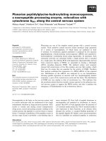
Immunohistochemical identification of each
neuron recorded in patch-clamp configuration
was completed as previously described
[10] (figure 1).
Figure 1: Model of in vivo electrophysiological signal recording system.
4. Data statistical analysis.
Electrophysiological data obtained
from recordings in the DR were
expressed as means ± S.E.M. of the
firing rate of 5-HT neurons. A one- or twoway analysis of variance (ANOVA) with
repeated measures (when appropriate) on
pre-treatment, treatment and/or genotype
(5-HT2A+/+ vs. 5-HT2A-/-) factors was
performed to compare the experimental
groups. When ANOVA were statistically
significant, pair wise comparisons were
performed using Fisher protected least
significance difference post hoc test
with the computer software StatView
4.02 when they were appropriate. The
level of statistical significance was set
(p < 0.05).
RESULTS
1. Effects of the genetic inactivation
of the 5-HT2A receptor on the
spontaneous firing rate of 5-HT
neurons in the dorsal raphe.
One-way ANOVA on the basal firing
rate of DR 5-HT neurons revealed a
significant effect of genotype factor
[F(1.92) = 5.6, p < 0.05].
29
Journal of military pharmaco-medicine No7-2017
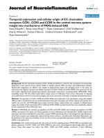
Figure 2: Effects of the genetic inactivation of the 5-HT2A receptor on the spontaneous
firing rate of 5-HT neurons in the dorsal raphe.
(*p < 0.05: significantly different from 5-HT2A +/+ wild-types (WT, n = 5 mice per group)
Figure 2 showed that 5-HT2A-/- displayed a higher spontaneous firing rate of DR 5HT neurons than their wild-type littermates, raising the possibility that 5-HT2A receptor
exerted a tonic receptor inhibitory influence on serotonergic neuronal activity.
2. Effects of DOI, a selective 5-HT2A receptor agonist on the firing rate of 5-HT
neurons in the dorsal raphe of 5-HT2A+/+ and 5-HT2A-/- mice.
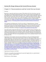
Figure 3: Effects of DOI on the firing rate of 5-HT neurons in the dorsal raphe of
5-HT2A+/+ and 5-HT2A-/- mice.
(A. Percent of inhibition of basal DR 5-HT firing rate in 5-HT2A+/+ and 5-HT2A-/- mice
administrated with DOI (1-10 mg/kg s.c.). B. Percentage of inhibition of basal DR 5-HT
firing rate in WT induced by DOI in DSP-4 (25; 50 mg/kg i.p.) pre-treated mice. In these
experiments, DSP-4 was injected 7 days before the electrophysiological recordings)
(*p < 0.05; **p < 0.01 and ***p < 0.001: significantly different from baseline of the
corresponding group. *p < 0.05: significantly different from and DSP-4 pre-treated mice
(n = 5 mice per group)
30
Journal of military pharmaco-medicine no7-2017
5-HT2A and 5-HT2A-/- mice were then
administered with increasing doses of
DOI (1-10 mg/kg s.c.). A two-way ANOVA
with repeated measures on the firing rate
of DR 5-HT neurons indicated a
significant effect of treatment [F(1.36) =
11.6, p < 0.01] and genotype factors
[F(1.36) = 25.3, p < 0.001). DOI induced a
dose-dependent inhibition of DR 5-HT
neuronal activity in 5-HT2A+/+ with a
maximal decreased observation at the
highest dose test (10 mg/kg; s.c.). In a
marked contrast, this inhibitory effect was
completely blunted in 5-HT2A-/- mice thus
confirming the inhibitory role of 5-HT2A
receptor on the serotonergic system and
demonstrating the selectivity of DOI
towards 5-HT2A receptor in this response
(figure 3A).
In order to identify the brain region
involved in the inhibitory effect of DOI, we
then examined the effect of this selective
5-HT2A receptor agonist in 5-HT2A+/+ mice
pre-treated with the neurotoxin DSP-4
at the doses of 25 and 50 mg/kg i.p.
One-way ANOVA with repeated measures
on the firing rate of DR 5-HT neurons
indicated a significant effect of pretreatment factor [F(2, 48) = 7.06, p < 0.05).
The inhibitory effect of DOI on DR 5-HT
neuronal activity was significantly attenuated
in DSP-4 (25 mg/kg; i.p.) pre-treated mice
whereas no greater electrophysiological
effects were observed by increasing the
dose of DSP-4 at 50 mg/kg; i.p (figure 3B).
Since, it was previously reported that
DSP-4 (25 mg/kg; i.p.) resulted in a selective
lesion of the noradrenergic system whereas
DSP-4 (50 mg/kg; i.p.) produced the
ablation of both the noradrenergic and
dopaminergic systems (Cassano et al,
2009) [5], the present data strongly
suggested that the inhibitory effect of DOI
on DR 5-HT neuronal activity involved, at
least in part, NE neurons.
3. Effects of escitalopram (SSRI) on the firing rate of 5-HT neurons in the
dorsal raphe of 5-HT1A+/+ and 5-HT1A-/- mice.
31
Journal of military pharmaco-medicine No7-2017
Figure 4: Effects of the SSRI escitalopram on the firing rate of 5-HT neurons in the
dorsal raphe of 5-HT1A+/+ and 5-HT1A-/- mice.
(A. Firing rate of DR 5-HT neurons in 5-HT1A+/+ mice injected with vehicle (baseline),
escitalopram (ESC 4 mg/kg s.c) or escitalopram + WAY100635 (0.5 mg/kg; s.c.). B, C.
Firing rate of DR 5-HT neurons in the DR 5-HT1A-/- mice injected with vehicle
(baseline), 8-OHDPAT (0.2 mg/kg; s.c.), escitalopram (ESC 4 mg/kg s.c), escitalopram
+ MDL100907 (2 mg/kg; s.c.) or escitalopram + WAY 100635 (0.5 mg/kg; s.c.), when
administrating esc 4 mg/kg s.c. or esc + WAY 100635 or esc + MDL 100907)
(**p < 0.01; ***p < 0.001: significantly different from baseline. *p < 0.05; **p < 0.01:
significantly different escitalpram injected mice; ns: not significant (n = 5 mice per group)
Having established that 5-HT2A receptor
exerted an inhibitory effect on DR 5-HT
neuronal activity, we hypothesized that
the inhibitory action of SSRIs might persist
in mice lack of the 5-HT1A autoreceptor. In
5-HT1A+/+ and 5-HT1A-/- mice, separate
one-way ANOVA on the firing rate of DR
5-HT neurons indicated a significant effect
of treatment factor [F(2,15) = 9.1, p < 0.01],
[F(2,17) = 14.6, p < 0.01] and [F(3,21)
= 6.2, p < 0.05]. All dorsal raphe 5-HT
neurons tested in 5-HT1A+/+ and 5-HT1A-/mice were inhibited by escitalopram
(4 mg/kg s.c.). In 5-HT1A+/+ mice, the
inhibitory effect of escitalopram was reversed
by the subcutaneous administration of
32
WAY 100635 (figure 4A). In 5-HT1A-/- mice,
the inhibitory effect of escitalopram was
reversed by MDL100907 but not by
WAY100635 (figures 4B-C).
DISCUSSION
5-HT1A autoreceptor plays a role in
mood disorders and their treatments.
Indeed, an increase in somatodendritic 5HT1A autoreceptor density in the dorsal
raphe (DR) attenuates the therapeutic
activity of selective serotonin reuptake
inhibitors (SSRIs), whereas their functional
desensitization promotes activation of
brain serotonergic transmission, thereby
representing an adaptive change relevant
Journal of military pharmaco-medicine no7-2017
to their therapeutic effect [10]. Nevertheless,
multiple source of evidence suggests that
other serotonegic receptors including
5-HT2A type may also exert inhibitory
effects on the DR 5-HT neurons raising
the possibility that the functional inactivation
of 5-HT1A autoreceptor would be necessary,
but not sufficient for SSRIs to exert their
optimal antidepressant activity. The present
study examined the role of 5-HT2A receptor
in the regulation of the serotonergic
system and more particularly its functional
interaction with 5-HT1A autoreceptor.
Our electrophysiological data demonstrated
that the inactivation or the stimulation of
5-HT2A receptor increased or reduced
respectively the firing activity of DR 5-HT
neurons. These findings concur with
previous studies in rats showing that the
administration of the 5-HT2A receptor
agonist DOI produced an inhibitory impact
upon the serotonergic system [9].
Nevertheless, since the selectivity of DOI
for 5-HT2A receptor remains somewhat
equivocal [2], our study examined whether
its electrophysiological effects were
altered in 5-HT2A-/- mice or not. The
observation that DOI-induced decrease in
DR 5-HT neuronal activity was completely
blunted in 5-HT2A-/- mice confirmed the
involvement of 5-HT2A in this response.
The possibility that DOI might have acted
through another receptor such as 5-HT2C
could not be ruled out. In order to identify
the brain region involved in the inhibitory
effect of DOI, we tested the effect of this
selective 5-HT2A receptor agonist in mice
displaying a lesion of the noradrenergic
or noradrenergic/dopaminergic systems.
Indeed, it was reported that depending on
the dose, the neurotoxin DSP-4 could
trigger one or both systems [5]. Interestingly,
we showed that the loss of noradrenergic
neurons attenuated the inhibitory effects
of DOI on the activity of DR 5-HT neurons
while the combined lesion of the
noradrenergic and the dopaminergic
systems failed to produce additional
attenuation. These results strongly
suggested that the control of serotonergic
neurons by 5-HT2A receptor specifically
involved noradrenergic neurons, which are
anatomically and functionally connected
to 5-HT neurons in the DR [6]. This
conclusion is consistent with previous
data from Dr Blier’s group reporting that
SSRI treatment enhances a tonic inhibitory
influence by 5-HT on Locus Coeruleus
neurons through postsynaptic 5-HT2A
receptors that are located on local
GABAergic interneurons [3]. Having
shown that 5-HT2A receptor exerts a tonic
inhibitory effect in the DR of wild-type
mice, we next asked whether this receptor
could maintain or enhance his negative
influence in mice lacking the 5-HT1A
receptor. Interestingly, we found that the
ability of DOI to decrease DR 5-HT
neuronal activity was potentiated in 5HT1A -/- mice indicating 5-HT2A receptor
hypersensitivity in these mutants. This
observation was corroborated by data,
showing an increased expression of 5HT2A receptor in 5-HT1A -/- mice. In this
context, we tested the effect of the
escitalopram (SSRI) on the firing rate of
DR 5-HT neurons in 5-HT1A +/+ and 5HT1A -/- mice. As expected and previously
shown, escitalopram decreased the firing
rate of serotonergic neurons in wild-type
mice. More surprisingly, this response
persisted in 5-HT1A -/- and was reversed
33
Journal of military pharmaco-medicine No7-2017
by the 5-HT2A receptor antagonist
MDL100907. This stands in contrast with
a previous electrophysiological study
showing that fluoxetine failed to decrease
to neuronal activity of 5-HT neurons in 5HT1A -/- mice [1] but this lack of response
can result from the 5-HT2A/2C antagonistic
activity of the SSRI. Thus, one of the most
remarkable result obtained in this study
was the observation that 5-HT2A receptors
maintain an inhibitory influence on the
serotonergic system, particularly in
response to increased levels of endogenous
5-HT induced by SSRI. The genetic
inactivation of 5-HT1A was compensated
by an over-expression of 5-HT2A receptor
thereby maintaining an inhibitory effect on
the serotonergic system. Such functional
interaction between both receptors has
been reported previously. In particular, it
has been shown that sustained administration
of SSRI produced a functional desensitization
of 5-HT1A receptor while increasing that of
5-HT2A receptor [4].
CONCLUSION
This study clearly indicates that 5-HT2A
receptor acts in concert to maintain an
inhibitory influence on the serotonergic
system, particularly in response of effects
of the escitalopram to increased levels of
endogenous 5-HT.
REFERENCES
1. Amargós-Bosch M, Bortolozzi A, Puig
MV et al. Co-expression and in vivo interaction
of serotonin1A and serotonin2A receptors in
pyramidal neurons of prefrontal cortex. Cereb
Cortex. 2004, 14 (3), pp.281-99.
2. Banas S.M, Doly S, Boutourlinsky K et
al. Deconstructing antiobesity compound action:
requirement of serotonin 5-HT2B receptors
34
for dexfenfluramine anorectic effects.
Neuropsychopharmacology. 2010, 36 (2),
pp.423-33.
3. Blier P, Szabo S.T. Potential
mechanisms of action of atypical antipsychotic
medications in treatment-resistant depression
and anxiety. J Clin Psychiatry. 2005, 66 (8),
pp.30-40.
4. Cadogan A.K, Marsden C.A, Tulloch I et
al. Evidence that chronic administration of
paroxetine or fluoxetine enhances 5-HT2
receptor function in the brain of the guinea
pig. Neuropharmacology. 1993, 3, pp.249-56.
5. Cassano T, Gaetani S, Morgese M.G et
al. Monoaminergic changes in locus coeruleus
and dorsal raphe nucleus following noradrenaline
depletion. Neurochem Res. 2009, 34 (8),
pp.1417-26.
6. Guiard B.P, El Mansari M, Blier P.
Prospect of a dopamine contribution in the
next generation of antidepressant drugs: the
triple reuptake inhibitors. Curr Drug Targets.
2009, 10 (11), pp.1069-84.
7. Li J.X, Crocker C, Koek W et al. Effects
of serotonin (5-HT)1A and 5-HT2A receptor
agonists on schedule-controlled responding in
rats: drug combination studies. Psychopharmacology
(Berl). 2011, 213 (2-3), pp.489-97.
8. Nguyen H.T, Guiard B.P, Bacq A et al.
Blockade of the high-affinity noradrenaline
transporter (NET) by the selective 5-HT
reuptake inhibitor escitalopram: an in vivo
microdialysis study in mice. Br J Pharmacol.
2013, 168 (1), pp.103-16.
9. Rahman W, Bannister K, Bee LA et al. A
pronociceptive role for the 5-HT2 receptor on
spinal nociceptive transmission: an in vivo
electrophysiological study in the rat. 2011,
1382, pp.29-36.
10. Richardson-Jones J.W, Craige C.P,
Guiard B.P et al. 5-HT1A autoreceptor levels
determine vulnerability to stress and response
to antidepressants. Neuron. 2010, 65 (1),
pp.40-52.