Ebook Sports emergency care (3/E): Part 2
Bạn đang xem bản rút gọn của tài liệu. Xem và tải ngay bản đầy đủ của tài liệu tại đây (18.39 MB, 134 trang )
Abdominal
and Pelvic Injuries
David A. Middlemas, EdD, ATC, CCISM
You have been assigned to provide the medical care for a high school ice hockey
tournament involving 15- to 18-year-olds. During one of the games, a player is
checked hard into the boards. After the collision, the player is kneeling on the ice
for about 30 seconds. He slowly gets up, shakes it off, and finishes his shift. About 2
minutes later, at the end of the shift, the player slowly skates to the bench. The coach
calls you to the bench because the player is doubled over with abdominal pain
and has just vomited. You approach the athlete to begin your assessment. What is
wrong? How bad is it? What do you do?
Injuries to the abdominal and pelvic regions are not uncommon in sports. Athletes and others participating in exercise are subject to pain and discomfort resulting from injuries or illness
involving the internal organs of the abdomen. Although potentially life-threatening abdominal
injuries are not everyday occurrences, the sports emergency care team members need to be aware
of the potential causes of abdominal problems in athletes, the signs and symptoms, and the importance of recognizing the nature and extent of injury so the athlete can be referred for appropriate
medical care.
Many sports and physical activities involve intentional and unintentional collisions with other
athletes, impact with sports implements, and high-velocity movement and twisting. The ability of
the sports emergency care provider to recognize and interpret how exercise and sports affect the
internal organs of the abdomen is essential in determining the extent of injury and the need for
immediate action. This chapter will provide the reader with an overview of the anatomy of the
abdominopelvic region, assessment of abdominal injuries, and medical conditions and guidelines
for immediate care.
135
Rehberg RS, Konin JG.
Sports Emergency Care: A Team Approach,
Third Edition (pp 135-151).
© 2018 SLACK Incorporated.
136
Chapter 10
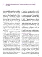
Figure 10-1. The abdominopelvic cavity.
(Illustration by Joelle Rehberg, DO.)
RUQ
RLQ
LUQ
LLQ
REVIEW OF CLINICALLY RELEVANT ANATOMY
The abdominal cavity is defined as the area below the thoracic cavity that contains many
of the body’s internal organs. It is separated from the thorax by the diaphragm and lined with a
membrane called peritoneum. The lower portion of the abdominal cavity surrounded by the pelvis,
vertebra, and sacrum is called the pelvic region (Figure 10-1).
The location of the organs in the abdomen and pelvis is usually described by dividing the
abdomen into 4 quadrants. The abdominal quadrants are defined by drawing a vertical and horizontal line through the navel. The quadrants and the structures located within them are shown
in Figure 10-1. The quadrants are called the left upper quadrant (LUQ ), right upper quadrant
(RUQ ), left lower quadrant (LLQ ), and right lower quadrant (RLQ ). The quality of communication between medical professionals and the accuracy of injury records is improved when everyone involved in the care of the injured athlete uses the same terminology.
The liver, gallbladder, spleen, pancreas, and digestive organs (stomach, small intestine, and
large intestine) are contained in the abdominal cavity. The urinary bladder and female reproductive organs are in the pelvic region, with male genitalia being external. It is important to note
that the kidneys are not within the abdomen. They are located outside the peritoneum behind the
abdominal cavity, covered by the muscles of the back and protected by the lower ribs.
To assist in understanding the nature of emergencies in the abdominopelvic region and their
implications, it is important to understand the basic structure and functions of the organs in this
region. It is helpful to divide the organs into 2 categories: hollow organs and solid organs (Table
10-1).
Hollow organs either allow materials to pass through them, as in the stomach and intestines,
or serve as holding tanks for materials until they are needed or expelled from the body, as in the
gallbladder or urinary bladder. As a rule, hollow organs tend to be injured less in sports and physical activity because they are at significantly less risk when they are empty. The best way to prevent
injuries to the hollow organs is to have them as empty as possible when participating in sports or
Abdominal and Pelvic Injuries
137
Table 10-1
CATEGORIES OF ORGANS OF THE ABDOMINAL AND PELVIC CAVITIES
Solid Organs
Hollow Organs
Reproduction
Liver
Spleen
Pancreas
Kidney
Stomach
Small intestine
Large intestine
Gallbladder
Urinary bladder
Female: ovaries, uterus, and vagina
Male: scrotum, testes, and penis
exercise. Such things as not eating immediately before competition and urinating before a game or
practice significantly reduce the risk of injury to digestive organs and the urinary bladder.
Solid organs do not have cavities inside them to hold or store fluids. They tend to have significant blood supplies that are necessary to complete their functions. The solid organs include the
liver, spleen, pancreas, kidneys, ovaries, and testes. The very fact that these organs will not easily
compress during a collision, combined with their ample blood supply, place them at a higher risk
of bruising or tearing with potentially life-threatening bleeding.
The liver, primarily located in the RUQ , is the largest solid organ of the body. It has many
functions, including making bile, converting glucose to glycogen for storage, producing urea, and
storing multiple substances for the body. As a result of these critical functions, it has a very rich
blood supply. Injuries to the liver can result in serious bruising or significant bleeding into the
abdominal cavity.
The spleen is located in the LUQ of the abdomen. Its job is to filter blood and to store red
blood cells and platelets. It has a plentiful blood supply and is at risk for injury from blows to the
upper abdomen. It is also important to note that the spleen swells in individuals who have had
mononucleosis, thus increasing the risk of injury from contact or collision.
Although the kidneys are located outside the abdominal cavity, their function of producing
urine is critical to the body. The kidneys, which are on the back of the body, are somewhat protected by the ribs. The process of filtering waste products from the blood produces urine. It then
flows through the ureters to the urinary bladder, which is located in the lower abdominal cavity.
Because the kidneys are the primary filters that remove waste from the bloodstream, they have
a very rich blood supply. Although the lower ribs cover the kidneys, blows to the back over the
kidneys can cause significant injuries.
The majority of reproductive organs in women are within the abdominal cavity. The ovaries,
uterus, fallopian tubes, and vagina are internal, placing them at significantly less risk for injury
than the male’s external reproductive anatomy. The male reproductive anatomy is more likely to
be injured from a direct blow or collision due to the fact that it is external. The penis, which has
a rich blood supply, and the testes, which are solid, have little protection.
AVOIDING INJURY
Preventing abdominal injuries in athletes is very important and requires the efforts of many
individuals. The sports emergency care personnel, coaches, officials, parents, and even the athlete
can be essential to preventing or reducing the occurrence of abdominal trauma in sports. By working together, everyone can ensure that athletes have the proper equipment, learn and use correct
sports techniques, and ensure that rules are appropriately taught and enforced.
138
Chapter 10
Protective equipment for the abdominal region includes such items as baseball and softball
chest protectors and extensions for shoulder pads in sports such as football and ice hockey, sometimes called flak jackets. To get the best protection possible, the coach and sports emergency care
team must work together to ensure that protective equipment is in good repair, meets required
standards, and fits the athlete properly. The athlete is a critical link in helping to keep his or her
equipment safe. It is very important to take the time to educate athletes about how to care for
their equipment and how to recognize potential problems in need of repair. Reporting damaged or
ill-fitting equipment allows for immediate repair or adjustment of any problems before an injury
occurs.
Proper technique in sports where contact and collision are part of the game is essential to
reducing injury. Coaches and officials can work together to reduce the occurrence of injury by
teaching proper methods of contact and collision and to appropriately penalize those who abuse
the rules.
Finally, there are times where the best method for preventing a potentially devastating situation is to disqualify an individual from participation in certain activities where the potential for
injury is unacceptable for that person. Examples of situations in which a physician might disqualify
an athlete from participation in collision or contact sports include absence of a paired organ, such
as a kidney or eye, or a medical condition that could place the athlete in danger. It may be appropriate in these situations to substitute an activity with lower risk of injury for the involved athlete.
EVALUATION AND RECOGNITION OF ABDOMINAL INJURIES
Many sports-related injuries can be assessed by directly visualizing and touching the injured
tissue. However, evaluation of injuries and medical conditions in the abdominal region requires
the practitioner to apply knowledge and skills that will allow him or her to recognize emergencies without the ability to directly access the affected organ or tissue. This section will help the
caregiver to understand the use of vital signs to recognize illnesses and injuries requiring indirect
methods of evaluation.
We begin our discussion with an explanation of the concept of indirect methods of evaluation.
Unlike such things as open wounds or bruising, injuries to internal organs and structures require
the caregiver to evaluate the status of an affected body part by looking at something else. Usually
that something else is one or more of the vital signs. When assessing someone who has been
participating in exercise or sports, it is important to remember that he or she will likely have vital
signs that are different from someone who was resting immediately before the injury occurs. These
differences, which may be interpreted as abnormal for the average person, are the norm or baseline
for determining the extent of injury in someone who was physically active at the time he or she
was hurt. It is important for the emergency caregiver to be familiar with these differences as he or
she begins the assessment (Table 10-2). A summary of the differences is presented in Chapter 3.
In athletic situations, injuries to the abdomen usually involve a collision with another athlete,
running into an object such as a wall or fence, or being struck by an athletic implement like a bat
or stick. These impacts often occur during the course of play, and the injured athlete may or may
not appear to be injured immediately after the incident. The primary concern in these situations is
that of internal bleeding from damaged internal organs, especially those with ample blood supply,
like the liver, spleen, and kidneys. Unrecognized injuries to these structures have the potential to
be life threatening and may require surgery. It is important for the sports emergency care provider
to assess the injured athlete as quickly and efficiently as possible in situations where abdominal
trauma may be present. Decisions relating to the possible extent of injury and immediate course of
care will depend on the caregiver’s ability to assess the situation and get the athlete to appropriate
medical care in a timely fashion.
Abdominal and Pelvic Injuries
139
Table 10-2
EXAMPLES OF CHANGES IN DIAGNOSTIC SIGNS AND
WHAT THEY MAY INDICATE
Diagnostic Sign
Change
Possible Cause
Blood pressure
Below normal
Internal bleeding
Pulse
Weak, rapid
Shock
Internal bleeding
Respirations
Rapid
Internal injury
Internal bleeding
Pain
Skin color
Pale
Bruising
Shock
Internal bleeding
Evidence of direct blow
Abdominal palpation
Rigidity
Internal bleeding
Guarding
Pain
Injury to internal organ
In the ideal situation, abdominal injury assessment begins with observation of the events
leading up to the injury and the mechanism of injury. For example, a running back in football
who is struck in the middle of the back with another player’s helmet may have a kidney injury, or
a lacrosse player who gets the butt of another player’s stick thrust into the LUQ of the abdomen
might have ruptured the spleen. To gain the most information from observing the events leading up to an injury, the caregiver must have an understanding of the anatomy of the injured body
region and the possible injuries that can result from the event causing the injury.
It is not unusual for the sports emergency care provider to be called to the location of an
injury after it has occurred. The disadvantage in these situations is that he or she was not able to
witness the mechanism of injury. Information about how the injury occurred must be gathered by
observing the injured athlete and surroundings as one approaches and by asking questions of the
athlete, coaches, officials, and other players to determine how the accident happened. It is usually
best to take the history using a structured interview format such as the SAMPLE history (signs/
symptoms, allergies, medications, past medical history, last oral intake, events leading to injury
illness; see Chapter 3 for more details). The information collected is extremely important in helping one determine the extent of any possible injuries.
Like any emergency situation, the first concern of the caregiver is to assess the injured athlete
for the presence of severe or potentially devastating injuries or conditions. When life-threatening
problems such as absence of breathing or pulse or severe bleeding are present, the sports emergency
care provider should take the appropriate actions to immediately deal with the problem. When
the injured athlete is determined to be in no immediate danger, a more thorough examination, or
secondary survey, that can focus on the potential abdominal injury, should take place.
Understanding what caused the injury is particularly helpful when dealing with internal injuries because the provider must make decisions about injured organs that cannot be directly seen or
touched. The care provider should ask the patient about where and how the blow to the abdomen
took place and what the patient felt immediately at the time of injury. Questions about the nature
140
Chapter 10
A
B
Figure 10-2. (A, B) Referred pain patterns. (Reprinted with permission from O’Connor
DP, Fincher AL. Clinical Pathology for Athletic Trainers: Recognizing Systemic Disease. 3rd ed.
Thorofare, NJ: SLACK Incorporated; 2015.)
and intensity of any pain, lightheadedness or dizziness, nausea, and any other abnormal feelings
or sensations at the time of injury and afterward will help the rescuer get an overall understanding
of the possibility of internal injury to the athlete.
After determining the mechanism of injury, one of the first concerns in assessing abdominal
injuries is the location and nature of the patient’s pain. Generally, the injured athlete will have
pain at the location of the injury. For example, if a hockey player has an injury to the liver after
being checked into the boards, one would expect pain in the RUQ of the abdomen; if the spleen is
ruptured after being hit in the abdomen with a lacrosse stick, one would expect pain in the LUQ
of the abdomen, and so on. Victims of internal organ injuries may have pain or soreness at places
away from the injured structure in addition to pain at the location of the injury. This phenomenon
is called referred pain. Referred pain is a condition in which pain from an injury or illness in one
part of the body presents in another location of the body. One example is Kehr’s sign, which is a
referred pain pattern for an injury to the spleen in which the patient will have pain or soreness in
the left shoulder. Some referred pain patterns are presented in Figure 10-2.
Questions about lightheadedness, nausea, and changes in sensations around the abdomen
provide information about whether there might be internal bleeding from injured structures in the
abdomen. Because any bleeding from abdominal injuries cannot be directly observed, the caregiver
must look for signs and symptoms that indicate the presence of secondary conditions caused by the
internal bleeding. A secondary condition is one that occurs as a result of an injury or illness existing in the body. The most significant secondary condition when it comes to suspecting the possibility of internal bleeding is shock, of which lightheadedness, dizziness, and nausea are symptoms.
Abdominal and Pelvic Injuries
141
Remember that a comprehensive patient history will collect information from the athlete,
other players in the area, officials, and coaches about the causes of the injury and the patient’s
condition. The answers to questions about what happened, the presence and nature of any pain,
and other feelings or sensations help the caregiver understand the potential severity of the injury
and set the basis for the hands-on portion of the patient assessment.
After taking a thorough history, the sports emergency care provider will conduct a physical
assessment of the patient. The physical assessment is done to verify what was learned in the history and to collect additional information to help pinpoint the specific structures that may have
been injured. The physical examination should assess appropriate vital signs and include palpation
of the abdomen.
A primary concern when caring for patients with potential internal bleeding from injuries
to solid internal organs, like the liver and spleen, is the onset of shock. The sports emergency
care provider should be prepared to assess the rate and quality of the athlete’s pulse and respirations. It is also important to assess the victim’s blood pressure. As with any other bleeding injury,
changes in vital signs provide information about the patient’s current status and the stability of
his or her condition. Vital sign assessment should focus on changes that indicate the possibility of
internal bleeding, such as a weak, rapid pulse; changes in rate and quality of breathing; a drop in
blood pressure; pale skin; and sweating. Patients with significant blood loss may also present with
changes in their level of consciousness consistent with those of patients in shock.
Injuries to hollow organs can present additional problems when their contents leak into the
abdominal cavity. The presence of such things as urine or bowel contents in the abdominal cavity creates the additional dangers of significant infection in the abdominal region, inflammation,
and irritation of the lining of the cavity. This is called peritonitis. The sports emergency care team
member may find elevated body temperature, elevated skin temperature, and severe abdominal
pain. These conditions may require surgery and/or the administration of antibiotics by the physician, and, if not treated promptly, may be life threatening.
Palpation of the abdomen can be very helpful in determining the nature and extent of injuries to the region (Figure 10-3). Abdominal assessment should include the ability to recognize
guarding, abdominal rigidity, and rebound tenderness. Guarding occurs when the athlete tightens
the muscles of the abdominal wall when the sport emergency care team member applies pressure
to the abdomen at a point where the athlete has pain. Guarding can be an indication of acute
abdominal pain and/or inflammation to internal organs and serves as an attempt to protect the
area from additional aggravation. Abdominal rigidity presents as contraction of the muscular walls
of the abdomen so that the abdomen feels firm or hard to the touch of the evaluator. It can indicate
swelling in the abdomen, possibly related to bleeding, abdominal pain, or patient apprehension
about being touched. Pain upon quickly releasing the abdominal wall after slow pressure is called
rebound tenderness. It is an indicator of pain in the abdominal lining and happens in response to
the rapid stretching of the irritated tissue after pressure. It is a sign commonly found in individuals
with acute appendicitis.
When you assess someone for abdominal injury, remember to complete the following:
Take a thorough history.
Determine the events leading up to the injury and what actually happened.
Take and record the patient’s vital signs.
Take them again frequently to look for any changes that may indicate a change in the patient’s
status.
Palpate the abdomen. Note any rigidity or guarding.
142
Chapter 10
Figure 10-3. Palpation of the
abdomen.
ABDOMINAL AND PELVIC INJURIES
Direct blows to the abdomen can result in injuries ranging from surface contusions and
muscle bruises to significant internal organ damage. This section will present some common
abdominal injuries, their common causes, and how they usually present.
Blows to the anterior surface of the abdomen tend to cause injuries to the organs and structures in the abdominal cavity where the impact took place. Because solid organs such as the liver
and spleen are located in the upper 2 quadrants of the abdomen, internal bleeding is of particular
concern when the athlete is struck at that location. Staying with the classification of internal injuries into those involving either solid or hollow organs, let us first look at how injuries to some of
the solid organs might present themselves.
SOLID ORGAN INJURIES
The spleen is located under the stomach in the LUQ of the abdomen. Contusions or rupture
of the spleen can occur as a result of a direct blow to the LUQ. Athletic activities that might
result in injury to the spleen include such things as tackling in football, collisions or checking in
ice hockey, or being struck in the abdomen with a sports implement such as a stick or bat. The
victim will have pain in the LUQ. In addition, spleen injuries may present with Kehr’s sign. If the
spleen is ruptured, there will be internal bleeding, which may be delayed by the organ’s ability to
splint itself. When this happens, internal bleeding, and hence the signs and symptoms of shock,
begin sometime after the injury takes place. Patient evaluation will often reveal tenderness in
the LUQ , along with the possibility of rebound tenderness, nausea, and signs and symptoms of
shock. Athletes in contact and collision sports with medical conditions such as mononucleosis are
at increased risk of spleen injury due to enlargement of the organ. Physician clearance should be
obtained before these athletes return to their sports activities.
The liver is the largest solid organ in the body. It occupies the majority of the RUQ and is susceptible to contusion or laceration from direct blows to the abdomen. Like the spleen, it is highly
vascularized, and injuries have the potential to bleed into the abdomen relatively quickly. Victims
of a lacerated liver may have pain on deep palpation, rebound tenderness, and nausea, and they
can develop signs and symptoms of shock fairly quickly. Referred pain may present in the center
of the chest and under the left arm.
Blows to the back can cause injury to the kidneys. Contusions or lacerations to the kidneys can
result in internal bleeding. Often an injury to the kidney will present with localized pain over the
Abdominal and Pelvic Injuries
143
flank that may be intense and burning. Palpation of the back in the area of the kidneys may elicit
tenderness. The victim of a kidney contusion or laceration might also have a burning sensation
while urinating, blood in his or her urine (hematuria), loss of the ability to urinate, and/or referred
pain in the lower abdominal region.
HOLLOW ORGAN INJURIES
Injuries to hollow organs like the urinary bladder, stomach, and intestines can usually be
prevented by having them as empty as possible before activities with the potential for collisions or
contact. Although some bleeding can occur with injuries to these organs, the main concern is the
spilling of contents into the abdominal cavity, causing inflammation, infection, and peritonitis.
Generally speaking, victims will present with abdominal pain, tenderness on palpation, abdominal
guarding, and signs and symptoms of inflammation and infection, including fever and soreness.
There may also be nausea and vomiting.
An injury to the urinary bladder can occur from a direct blow to the midline in the pelvic
region. Spilling of urine into the abdominal cavity can cause severe pain and inflammation in the
lower abdomen.
Open wounds in the abdominal cavity or those involving penetrating objects present the
possibility of internal bleeding and infection. Open abdominal injuries can occur from sports
implements such as the javelin or a ski pole or collisions with equipment such as metal fence posts.
Injuries to the genitalia can occur in sports in which there is the possibility of being struck
in the groin area by a ball or sports implement or in a collision with another athlete. Because the
majority of female reproductive organs are internal, genital injuries in female athletes are not very
common in sports. Direct blows to the genital area can cause contusions or lacerations, which the
sports emergency care provider can care for using ice or appropriate bandaging. Care should always
be taken to protect the privacy of the victim at all times by moving to a private area or covering
the athlete with a blanket or other available item. Males, on the other hand, have a higher risk
of genital injury because the anatomy is outside the abdominal cavity. Injuries to male genitalia
include contusions to the scrotum, testes, and penis; testicular torsion; and laceration or entrapment of anatomy in clothing or equipment. Athletes participating in activities in which there is a
risk of injury to the external genitalia should be required to wear a cup protector.
Blows to the groin area can result in painful injuries to the external anatomy in males. It is not
uncommon for contusions and lacerations to happen as a result of being hit by another athlete, a
ball, or a sports implement. Lacerations to the penis are of concern because of the rich blood supply in the area, and thus they have the potential to bleed freely. Lacerations to the scrotum can be
superficial or deep enough to expose and damage the testicle. Superficial wounds that are bleeding
can be treated the same as any other laceration, taking care to preserve the victim’s privacy. Deeper
lacerations involving the penis or scrotum should be considered emergent, and the athlete should
be transported by ambulance to the emergency room.
Closed injuries to the male genitals can be very serious. A direct blow to the groin can result
in deep contusion or fracture of a testicle or tearing of a blood vessel in the scrotum. In either case,
the situation is an emergency. Disruption of blood supply to the testicle can possibly result in loss
of the organ if not cared for by a physician immediately and properly. These sorts of injuries present with significant pain in the scrotal area accompanied by significant swelling in the scrotum,
and they require immediate transportation to the emergency room.
Testicular torsion is a medical emergency that can result in loss of blood supply and possibly
result in loss of the testicle. In this condition, the testicle can rotate in the scrotum. When this
happens, the blood supply can be cut off. The patient complains of sudden pain and swelling on
one side of the scrotum or in one of the testes. Testicular torsion is often the result of a predisposing situation in which the testicle is not adequately attached to the inside of the scrotum. This
144
Chapter 10
condition is seen most frequently in boys but has been seen in adults. The condition must be
addressed promptly with surgery to restore the blood supply.
EMERGENCY CARE OF ABDOMINAL AND PELVIC INJURIES
When suspecting abdominal injury, it is important to continue monitoring the patient’s vital
signs for changes that would indicate the possibility of internal bleeding. The sports emergency
care provider should evaluate the injured athlete’s pulse, respirations, skin color and temperature,
and, when possible, blood pressure. Weak, rapid pulse; rapid, shallow breathing; pale, cool, and
clammy skin; and decreased blood pressure are all indicators of internal bleeding that will send the
patient into shock. The injured athlete may also complain of nausea and dizziness and may vomit.
Once an abdominal injury is suspected, the following steps should be taken:
Activate the emergency action plan.
Place the victim in a comfortable position. The recovery position will assist in
maintaining a patent airway in the event the patient is nauseated or vomits.
Treat for shock.
If the victim does not have a spinal or head injury, elevate the feet and legs.
Maintain the athlete’s body temperature by using a blanket, jacket, or some other covering
when necessary.
It is important that the victim’s vital signs be assessed for changes at regular intervals while
waiting for the ambulance and during transportation to the hospital. Do not give the injured athlete anything to eat or drink because internal injuries may require surgery. Because it is not possible
to control internal bleeding directly, it is important to be prepared to provide basic life support in
the event the patient’s condition should worsen significantly.
There are times when an athlete may suffer an abdominal injury from an impaled object. One
example of this would be an individual struck in the abdomen with a javelin. As with all injuries
involving impaled objects, it is important to leave the object in place, pad it, and bandage it where
it is. The caregiver must continue to be aware that the visible injury is complicated by the possibility that the javelin (or other object) is also penetrating an internal organ and that moving it could
result in significant internal bleeding.
An additional consideration with an impaled sports implement like a javelin is that it may
not fit into the back of the ambulance. In rare cases, the sports emergency care team may need to
summon rescue personnel for assistance in cutting the impaled object to a length that will allow
the victim to be safely transported with it bandaged in place. Professional rescue personnel will
have access to specialized equipment such as the Jaws of Life (Hurst, Shelby, NC), which can cut
the post or implement with as little movement as possible.
COMMON MEDICAL EMERGENCIES IN THE
ABDOMEN AND PELVIS
There will be times when athletes will have abdominal pain or discomfort that is not a result
of an injury or collision. Although the sports emergency care provider cannot directly treat the
cause of the problem, assessment and recognition of medical conditions in the abdomen can prevent significant problems. Timely awareness of potentially serious illness will allow the athlete to
be referred to a physician for rapid diagnosis and treatment.
Abdominal and Pelvic Injuries
145
Table 10-3
SUGGESTED OUTLINE FOR STRUCTURED INTERVIEW FOR
ABDOMINAL INJURIES OR CONDITIONS
Abdominal Injury
Illness
What happened? (Were you hit? Was there a
collision?)
Describe the problem.
Where were you hit?
Have you eaten anything you do not
usually eat?
What did you feel at the time of injury?
Please list the symptoms.
Have you had this problem before?
Are you nauseous? Have you vomited?
Does it hurt?
O
Onset
When did the problem begin? What caused it?
P
Provokes/
palliates
What makes it better? What makes it worse?
Q
Quality
Describe your pain (ie, is the pain sharp, dull, achy, burning?).
R
Region/radiates
Where does it hurt? Does the pain move or spread?
S
Severity
Rate your pain on a scale from 1 to 10.
T
Timing of the
pain
Has it been constant? Does it come and go? How long has the
pain been there?
The patient is said to have an acute abdomen when he or she suddenly develops abdominal
pain. Conditions that can lead to abdominal pain or discomfort can be relatively minor or severe.
A physician will be able to determine whether the pain can be alleviated through medication and
conservative treatment or whether the patient requires more invasive care, such as surgery.
EVALUATING AND RECOGNIZING
MEDICAL CONDITIONS IN THE ABDOMEN
The sports emergency care team member should observe the patient for signs indicating the
presence, location, and intensity of pain. Facial expression, sweating, and posture provide information about the severity of the pain. The athlete may be lying on his or her side with knees drawn
up to try to alleviate the pain. It is also important to take a history focusing on the abdomen in
order to identify the possible causes of the pain.
The primary focus in taking a history for a person reporting abdominal pain is the location,
nature, and intensity of the pain (Table 10-3). The sports emergency care provider can easily
remember what to ask the patient by using OPQRST described in Chapter 3. This mnemonic
device serves as a reminder to ask about the onset (the start of the problem), provocation and palliation (what makes it feel better or worse), quality (sharp, dull, ache, burning), region (where it
hurts), severity (how much it hurts), and the timing (when it happens, how often it happens, and
146
Chapter 10
Figure 10-4. Assessing bowel
sounds.
how long it lasts) of the pain. Information about nausea and vomiting, diarrhea, constipation, and
fever often provides additional details that identify the cause of the problem.
The patient’s answers from the history will guide the sports emergency care provider in performing a physical exam concentrating on the abdomen. Take the patient’s vital signs. The steps
in the assessment process should be explained to the patient to reduce stress and apprehension.
The 4 quadrants of the abdomen should be palpated. Begin away from the suspected location of
the pain and work toward it. Gently press on the regions of the abdomen, feeling for rigidity and/
or guarding. Ask the patient if he or she can relax the abdomen. When the location of the pain is
identified, check for rebound tenderness. Note the results of the assessment and record the information so it can be communicated to the physician.
The sports emergency care team member can also quickly check to see if the patient’s bowel
sounds are present (Figure 10-4). The absence of normal bowel sounds can indicate the possibility
of such problems as bowel obstruction or significant abdominal injury or illness. Place the head
of the stethoscope on the anterior abdomen. Listen to all 4 quadrants of the abdomen. Normal
bowel sounds include a combination of squeaking and gurgling sounds, indicating that intestinal
contents are being moved through the digestive system. If the sounds are diminished or absent, the
information should be recorded in the patient notes and communicated to the physician.
REDUCING THE LIKELIHOOD OF ABDOMINAL PAIN
Many of the nontraumatic causes of abdominal pain, such as acute appendicitis, gall or kidney stones, and kidney or bladder infections, result from medical conditions or emergencies that
cannot be predicted by the patient. There are no effective prevention strategies that target these
sorts of conditions. Basic common-sense lifestyle choices, such as a well-balanced diet, adequate
hydration, and close attention to bodily changes, can help reduce the chances of many medically
related problems.
EMERGENCY CARE OF THE ACUTE ABDOMEN
The need for emergency transportation and treatment for an individual with abdominal pain
would be dictated by the onset and severity of the pain, the possible underlying cause, and the stability of the patient’s vital signs. Individuals with moderate to severe abdominal pain accompanied
by vital sign changes such as altered pulse or blood pressure, fever, chills, nausea, vomiting, and/
Abdominal and Pelvic Injuries
147
or signs of shock should be made as comfortable as possible and monitored while awaiting transportation to a hospital. The location and nature of the pain may provide the sports emergency care
provider with clues as to its possible cause, but definitive diagnosis and treatment by a physician
or other appropriate health care provider are essential for these patients. In situations like this, the
patient should be given nothing to eat or drink while waiting for the ambulance because it may
aggravate the condition or make it more difficult in the event surgery is required.
In many cases, teenagers and adults with relatively minor episodes of abdominal pain or
discomfort may have had it before. Such conditions as indigestion, irritable bowel syndrome, or
menstrual cramps may be significant enough to affect an athlete’s ability to exercise or compete,
but they do not usually require emergency transportation and treatment. An athlete who does
not have a history of abdominal discomfort should stop the activity, be made comfortable, and be
referred to his or her physician for diagnosis and appropriate treatment. Those who have recurrent
or chronic episodes of minor abdominal conditions may have already been advised by their health
care provider on how to care for discomfort or minor pain when it occurs. In these situations,
it is appropriate to assist the athlete in following the instructions he or she has been given by
the doctor.
The most effective method of determining the patient’s knowledge regarding the abdominal
discomfort or pain is by taking a comprehensive history related to the abdominal discomfort.
Asking the athlete about when the pain started, the severity of the pain, and factors that worsen
or lessen the pain can verify whether the episode is a recurrence of an existing problem or something new. Listening carefully to the patient’s answers to questions can help the sports emergency
care team member to identify whether the athlete is familiar with the problem. In any situation in
which the athlete has had to stop participation due to abdominal pain or discomfort, it is appropriate to make sure a qualified medical professional has assessed him or her before returning to play.
In situations in which the athlete is a minor, it is imperative that the parent or guardian be advised
of the situation. In many cases, reviewing the options for follow-up with a physician provides the
parent and athlete with information they need and a degree of comfort.
OTHER MEDICAL CONDITIONS OF THE ABDOMEN AND PELVIS
This section presents the signs and symptoms for some common medical conditions in the
abdomen. This information can help the sports emergency care provider decide the potential
severity of the problem and the type of assistance that is needed.
Some causes of abdominal discomfort or pain are relatively minor and may resolve with little
medical treatment. Other illnesses or conditions causing abdominal pain can be significant and
may be life threatening if not diagnosed and treated properly. The role of the caregiver is to recognize signs and symptoms in the athlete that indicate potential abdominal illness and facilitate getting the patient to the appropriate medical professional in a timely fashion. Signs and symptoms of
medical conditions in the abdomen are presented to provide background information for the sports
emergency care team that helps them recognize the athlete’s need for medical care.
Problems with the organs of the digestive system often give the patient abdominal pain. The
pain can be burning, sharp, dull, or intense.
Dyspepsia is a term that describes pain in the upper abdomen that may come and go but
is usually present the majority of the time. Common causes of dyspepsia are gastroesophageal
reflux disease (GERD) and stomach ulcers. GERD is a condition in which acid from the stomach
splashes out of the upper valve onto the walls of the esophagus. The patient will complain of burning pain in the mid-upper abdomen and/or heartburn. The pain may be constant but is sometimes
relieved when the patient eats or takes an antacid. Occasional heartburn may not be a significant
problem, but recurrent burning pain in the upper abdomen may be a sign of GERD, which has the
potential to cause long-term damage to the esophagus. Stomach ulcers are wounds in the lining of
148
Chapter 10
Figure 10-5. Palpation of McBurney’s
point.
the stomach. They may be caused by stress, a virus, or dietary concerns. Ulcers also present with
abdominal pain, burping, nausea, and/or heartburn. The potential for significant bleeding exists
if ulcers go untreated because they are open wounds in the stomach lining. A physician should
evaluate persistent upper abdominal pain and burning in order to provide proper treatment.
Generalized abdominal pain can result from a number of conditions in the intestinal tract.
Intestinal gas can cause significant pain in the abdomen that might be strong enough to cause an
athlete to double over. Often gas pains are accompanied with increased bowel sounds, or gurgling,
and will resolve themselves.
Irritable bowel syndrome is a term used to describe conditions that cause abdominal pain,
diarrhea, and significant discomfort in the abdominal region. The term includes conditions like
Crohn’s disease and ulcerative colitis. Abdominal pain can also be caused by pockets or folds in the
walls of the intestines, called diverticula, that become infected or inflamed, causing pain, nausea,
vomiting, fever, and changes in bowel habits. This condition is called diverticulitis. A physician
should properly diagnose and treat an athlete with frequent instances of abdominal pain that persist for a prolonged period of time.
Infection and inflammation of the appendix can cause significant abdominal pain, nausea,
vomiting, diarrhea, and fever. Acute appendicitis is often identified by pain in the RLQ of the
abdomen, referred pain to the area of the navel, and rebound tenderness at the location of the
appendix, called McBurney’s point (Figure 10-5). Failure to recognize the signs and symptoms of
appendicitis can allow the problem to progress as the infected appendix continues to swell and fill
with pus. If left untreated, the appendix will eventually rupture, spreading the infection’s contents
into the abdomen. When this happens, the patient has a potentially life-threatening condition
that causes inflammation to the peritoneal lining and serious infection to the abdominal cavity.
There are medical conditions that do not present as emergencies, but the sports emergency
care personnel may be the first person to whom the athlete reports the onset of symptoms relating to the illness. Listening to the pattern of symptoms and performing an initial assessment to
determine the potential severity of the condition can be essential to preventing the progression of
a condition to a serious problem.
An athlete with discomfort or pain in the RUQ with referred pain to the right shoulder may
be suffering from an inflamed gallbladder (cholecystitis) or gallstones. The pain can be aggravated
by fatty foods because bile is essential to their digestion. The individual may also have nausea and
vomiting, depending on the severity of the condition.
An individual with unexplained abdominal pain, joint ache, fever, loss of appetite, nausea or
vomiting, and fatigue may have contracted hepatitis. Hepatitis is a disease that affects the liver and
Abdominal and Pelvic Injuries
149
is most often caused by a virus. There are 5 types of hepatitis. Hepatitis type A is the most common in the United States, but cases of type B and C are not uncommon. Hepatitis is contagious
and is spread through such routes as unsanitary conditions, blood, feces, and sexual contact. The
cause of the symptoms and the proper course of care must be determined by the physician after
proper diagnostic testing.
Medical conditions of the urinary tract involve the kidneys, ureters, and bladder. Infections in
the urinary tract can present with pain in the lower abdominal region and pubic area. Athletes with
kidney infections can have low back soreness or pain, fever, and difficulty urinating. Infections in
the urinary bladder, ureters, and/or urethra can cause pain or burning during urination.
The development of kidney stones can cause pain in the flank region of the back that radiates
to the genital area. The pain can become severe and even disabling. Abnormal urinary habits and
painful urination often occur in patients with kidney stones. Physician intervention is necessary to
resolve the problem using one or more of many available treatment methods.
Abdominal pain may present in the female athlete as part of her normal menstrual cycle.
Pain in the lower middle portion of the abdominopelvic region may occur in the middle of the
menstrual cycle, which is associated with release of the egg from the ovary, or may occur with
cramping during the menstrual period. The severity of the pain and cramping varies with the
individual. When assessing a female athlete with lower abdominal pain, she is usually able to provide information relating to her normal pattern of pain and cramping during the menstrual cycle.
Sometimes abdominal pain in girls or women is due to medical conditions requiring the attention of their general physician or gynecologist. Patients who develop ovarian cysts can have severe
pain in the abdominal or pelvic region and may also present with vaginal bleeding, nausea, and
fever. Athletes who suddenly develop these symptoms should be treated as a medical emergency.
Ectopic pregnancy occurs when the fertilized egg implants in the wall of the fallopian tube
outside the uterus. Women with a possible ectopic pregnancy can become dizzy and faint, develop
low blood pressure, and have vaginal bleeding. It is important to ask female patients whether they
may be pregnant during the history portion of the examination to rule out the possibility of gynecological causes for abdominal pain or symptoms.
We would be remiss in not providing a short discussion of the possibility of sexually transmitted diseases (STD) in the athletic population. The likelihood that sexually active individuals
will be seeking advice and treatment from sports emergency care professionals they trust supports
the need to recognize the signs of a potential STD. When the athlete communicates the onset
of lesions, sores, or unusual skin problems on the genitals; unusual discharges from the penis or
vagina; or pain during urination or intercourse, he or she may be communicating the presence
of symptoms of STD. The sports emergency care team member should maintain the confidence
and dignity of the athlete while strongly encouraging or requiring him or her to seek appropriate medical care for the condition. Because STDs are contagious, strongly encouraging medical
follow-up and care provides appropriate care for the athlete and anyone with whom he or she has
intimate contact.
CONCLUSION
When dealing with emergencies in the abdomen and pelvic regions, the role of the sports
emergency care team or other emergency responder is to identify the potential causes of the athlete’s problem and select the appropriate course of immediate care and referral for medical treatment. In order to be able to provide the best on-site care for the athlete, one should possess the
ability to assess victims of both abdominal trauma and those whose abdominal pain may be due
to medical conditions. The ability of the sports emergency care provider to recognize the signs of
significant abdominal injury or illness provides the basis for sound decision making and access to
prompt emergency care.
150
Chapter 10
The potential effects of internal bleeding or infection due to such conditions such as a ruptured appendix can be minimized by rapid identification of the problem’s cause through effective
assessment and immediate access to medical care. Daily contact between the athlete and the sports
emergency care team or other emergency care provider can play the most important role in early
recognition of significant abdominal injury or illness by providing the athlete with a trusted professional to whom he or she can go immediately when discomfort, pain, or injury occur.
SUMMARY OF KEY POINTS
Evaluation of injuries and medical conditions in the abdominal region requires the practitioner to apply knowledge and skills that will allow him or her to recognize emergencies without
the ability to directly access the affected organ or tissue.
Proper protective equipment and proper technique are essential in reducing injury.
Victims of internal organ injuries may have pain or soreness at places away from the injured
structure in addition to pain at the location of the injury. This phenomenon is called referred pain.
Shock is a primary concern of the sports emergency care team member when caring for
patients with potential internal bleeding from injuries to solid internal organs, such as the
liver and spleen.
Abdominal assessment should include the ability to recognize guarding, abdominal rigidity,
and rebound tenderness.
Direct blows to the abdomen can result in injuries ranging from surface contusions and
muscle bruises to significant internal organ damage.
Blows to the anterior surface of the abdomen tend to cause injuries to the organs and structures in the abdominal cavity where the impact took place.
Injuries to hollow organs like the urinary bladder, stomach, and intestines can usually be prevented by having them as empty as possible before activities with the potential for collisions
or contact.
Injuries to the genitalia can occur in sports in which there is the possibility of being struck in
the groin area by a ball or sports implement or in a collision with another athlete.
When suspecting abdominal injury, it is important to continue monitoring the patient’s vital
signs for changes that would indicate the possibility of internal bleeding.
The victim’s vital signs should be assessed for changes at regular intervals while waiting for
the ambulance and during transportation to the hospital.
As with all injuries involving impaled objects, it is important to leave the object in place, pad
it, and bandage it where it is.
The sports emergency care provider can easily remember what to ask the patient by using the
OPQRST acronym.
The need for emergency transportation and treatment for an individual with abdominal pain
is dictated by the onset and severity of the pain, the possible underlying cause, and the stability of the patient’s vital signs.
The most effective method of determining the patient’s knowledge regarding the abdominal
discomfort or pain is by taking a comprehensive history related to the abdominal discomfort.
Generalized abdominal pain can result from a number of conditions in the intestinal tract.
Some abdominal pain in girls or women is due to medical conditions requiring the attention
of their general physician or gynecologist.
Abdominal and Pelvic Injuries
151
REVIEW QUESTIONS
1.
2.
3.
4.
5.
What conditions might cause abdominal rigidity and guarding?
A severe blow to the RUQ of the abdomen might produce what kind of injury?
Why is a splenic rupture considered a medical emergency?
Describe proper care for a patient with an acute abdomen.
What are some causes of severe abdominal pain specific to women?
BIBLIOGRAPHY
American Red Cross. Emergency Medical Response. Yardley, PA: Staywell Publishing; 2011.
American Urological Association Urology Care Foundation. What is testicular torsion? />urology/index.cfm?article=34. Accessed October 1, 2012.
Barrett C, Smith D. Recognition and management of abdominal injuries at athletic events. Int J Sports Phys Ther.
2012;7(4):448-451.
Booher JM, Thibodeau GA. Athletic Injury Assessment. 4th ed. New York, NY: McGraw Hill; 2000.
Cuppett M, Walsh K. General Medical Conditions in the Athlete. 2nd ed. St. Louis, MO: Mosby; 2011.
Finch R, Banting SW. Commentary: modern management of splenic injury. ANZ J Surg. 2004;74(7):513.
Klepac SR, Samett EJ. Spleen trauma imaging. Accessed
September 30, 2012.
Kluger Y, Paul DB, Raves JJ, et al. Delayed rupture of the spleen—myths, facts, and their importance: case reports and
literature review. J Trauma. 1994;36(4):568-571.
Limmer D, O’Keefe M, Dickinson EV, Grant H, Murray B, Bergeron JD. Emergency Care. 10th ed. New York, NY:
Prentice Hall; 2005.
Krin C, Brohi K. Penetrating abdominal trauma: guidelines for evaluation. Trauma.org. />php/main/article/414/. Published August 9, 2004. Accessed October 1, 2012.
Pollak AN, ed. Emergency Care and Transportation of the Sick and Injured. 10th ed. Boston, MA: Jones and Bartlett
Publishers; 2011.
Prentice WE. Arnheim’s Principles of Athletic Training: A Competency-Based Approach. 12th ed. New York, NY: McGrawHill; 2006.
Tamparo CD, Lewis MA. Diseases of the Human Body. 3rd ed. Philadelphia, PA: FA Davis; 2000.
Wright JA. Seven abdominal assessment signs every emergency nurse should know. J Emerg Nurs. 1997;23(5):446-450.
Fractures and
Soft Tissue Injuries
Michael A. Prybicien, MA, ATC, PES, CES and Louis Rizio III, MD
A 15-year-old volleyball player is participating in drills during practice. When a
teammate spiked the ball over the net, she dove to reach it and landed on an
outstretched arm. You arrive to evaluate the athlete, who is complaining of severe
pain in the shoulder. She is guarding the arm by holding it against her side. You note
an obvious deformity at the acromioclavicular joint. The area was point
tender, but no crepitus was noted. What would you do?
Fractures, dislocations, and soft tissue injuries are among the most common injuries sustained
in sports. This chapter aims to provide a straightforward approach to understanding injuries to
bone and soft tissue, and the initial evaluation and management of such injuries. Proper initial
evaluation and management are critical to ensure the athlete receives the proper medical attention,
is transferred to the hospital for further evaluation when appropriate, and, most importantly, is
protected from further harm.
REVIEW OF CLINICALLY RELEVANT ANATOMY
BONE
This chapter will focus on bones of the extremities. Information on spinal anatomy can be
found in Chapter 6. The bones of the arms and legs are long bones, each composed of an epiphyseal, metaphyseal, and diaphyseal segment (Figure 11-1). The epiphyseal segment is the portion
of the bone that forms one side of a joint and is typically covered with articular cartilage. The
metaphyseal segment is adjacent to the epiphyseal segment. The epiphyseal and metaphyseal
segments fuse together once the individual reaches skeletal maturity. In childhood, bone growth
occurs at the growth plate, which is between the epiphyseal, metaphyseal, and diaphyseal segments. The diaphyseal segment is the shaft of the long bone and is very strong.
153
Rehberg RS, Konin JG.
Sports Emergency Care: A Team Approach,
Third Edition (pp 153-170).
© 2018 SLACK Incorporated.
154
Chapter 11
Figure 11-1. Bone. (Illustration by
Joelle Rehberg, DO.)
Diaphyseal bone is composed of cortical bone, which is very strong and supports the body’s
weight. Metaphyseal bone tends to be wider and less tubular in appearance and is the portion of
the long bone that forms one end of a joint. This metaphyseal bone is composed of cancellous bone
and is not as strong as cortical bone.
JOINTS
The joints of the extremities are called synovial joints. The joint is formed by the proximal
end of one bone and the distal end of another bone and is held together by a capsule and ligaments. The ends of each bone are covered with articular cartilage, which provides a low-friction
surface for motion and a cushion for shock absorption. The connection of the 2 bones in this type
of arrangement allows for motion of the joint; the ligaments and capsule provide stability (Figure
11-2). The capsule of the joint can be divided into a fibrous (outer) layer and synovial membrane
(inner) layer. The ligaments that hold the joint stable are often thickenings of the fibrous layer
made of dense collagen. The synovial layer makes synovial fluid that bathes and nourishes the
cartilage surfaces of the bones forming the joint.
SOFT TISSUE
Soft tissue is a broad term that can be used to describe many tissues in the musculoskeletal
system. Although the skin can be considered soft tissue and will be covered in the wound management section of this chapter, for the purposes of this section soft tissue refers to ligaments,
tendons, and muscle. All of these structures are composed predominantly of collagen, but the type
of collagen varies between the tissues. These soft tissues are critical for the normal functioning and
action of joints. These structures allow for motion and stability of the joints they cross.
Fractures and Soft Tissue Injuries
155
Figure 11-2. Synovial joint.
(Illustration by Joelle Rehberg,
DO.)
Ligaments usually attach on either side of the joint and connect one bone to another. Their
major function is to provide stability to the joint it crosses. Injury to a ligament is termed a sprain.
It is a good idea to keep terminology accurate, especially when communicating with other members of the health care system; this avoids confusion and will hopefully convey the message most
effectively.
Tendons are the connection between bone and muscle. The tendon attachment to bone allows
a muscle to move a joint. Muscle tissue shortens (contracts) under voluntary control to produce
movement. Injury to the tendon or muscle is termed a strain. Tearing of a tendon can lead to
inability to move an extremity or joint, especially if completely torn.
FRACTURES
“Is it broken or just fractured?” There is no distinction between breaks and fractures; they are
one and the same. The disruption of the bone’s continuity is what defines this injury. Fracture can
occur from a direct blow or a rotational (twisting) injury without contact.
EVALUATION
The typical signs of a fracture are pain, swelling, and tenderness over the area. Movement of
the extremity will aggravate the athlete’s symptoms, and he or she often cannot bear weight on the
lower extremity or move the upper extremity due to discomfort. Loss of function of the extremity
is usually apparent.
Initial assessment of an injured and potentially fractured extremity includes a careful inspection of the limb, especially the skin. The clothing should be removed around the injured limb for
complete inspection. Any wounds over the painful area should be considered indicative of an open
(compound) fracture. Deformity may be present, indicating severe malalignment or displacement
of the fractured ends (Figure 11-3). Tenderness over the bone is usually present, and sometimes
motion can be felt between the fractured ends; this is highly suspicious of a fracture.
156
Chapter 11
Figure 11-3. Immobilization of wrist and
forearm injuries using a structural aluminum
malleable (SAM) splint (Sam Medical
Products).
A careful assessment of vascular supply and nerve function distal to the injury is vital. Sensory
function is assessed grossly by determining the athlete’s ability to feel the examiner’s touch. This
should be done on all surfaces of the limb circumferentially. In addition, an assessment of muscle
function below the injury level is performed to determine motor nerve function. For example, ability to move all the toes or fingers up and down can give a gross estimate of nerve function. Any
loss of sensation or movement below the injury needs to be documented prior to any splinting or
immobilization.
Vascular status or circulation is evaluated as well. Pulses should be felt below the level of the
injury. In addition, a cold, very white (pallor), or blue extremity signals severe injury to the blood
supply of the extremity. Capillary refill is not a reliable method of determining adequacy of the
blood supply to the limb. All pulses felt or not felt need to be documented prior to transfer or
immobilization.
INITIAL TREATMENT
If a fracture is suspected of the lower extremity, carrying the athlete off the field or assisting with ambulation to prevent weight bearing on the injured extremity is necessary. A splint or
immobilization device is utilized to protect the injured extremity from undue motion. Typically,
it is best to immobilize on the field as far above and below the area in question as possible. The
athlete should be sent for confirmatory radiographs. Examples of basic extremity splinting will be
presented at the end of the chapter.
FRACTURE EMERGENCIES
An open (compound) fracture is an orthopedic emergency, and the athlete should be transported to a hospital for immediate treatment, which includes thorough operative irrigation and
removal (debridement) of dirt, debris, or foreign material (eg, clothing pieces); stabilization; and
antibiotics by intravenous administration. Initial management of an open fracture is listed in
Table 11-1.
Loss of circulation to a limb is uncommon, but it needs to be corrected as soon as possible.
When severe deformity exists to a limb and the circulation is compromised, straight traction on
the limb may reduce pressure on a blood vessel from a displaced bone end or remove a kink in the
vessel from the angulated position of the limb. Traction should be applied gently, slowly, and in
line with the limb; never should an attempt be made to forcibly reduce the fracture. Documenting
circulation before and after this maneuver is critical so that the treating emergency department
will have the information. Also, transportation to the hospital should not be delayed in order to
try to get circulation to return while the athlete is on the field. Splinting is then performed with
the traction being held; this will improve the chances the limb will remain straight after splint
application.
Compartment syndrome can occur following fracture due to rapid swelling in the closed compartments of the leg and forearm. The lower leg (below the knee) and forearm (elbow to hand) are
the most common locations where compartment syndrome can develop; however, it should never
Fractures and Soft Tissue Injuries
157
Table 11-1
EMERGENCY MANAGEMENT OF AN OPEN FRACTURE
Cover the wound with Betadine (iodine)- or alcohol-soaked gauze bandage.
Immobilize the limb.
Transfer the patient to the hospital immediately. (Infection risk increases if not treated
within the first 6 hours!)
be assumed it cannot occur anywhere else (eg, thigh, foot, hand). The classic signs of compartment
syndrome are remembered as the 5 Ps: pain, pallor (whiteness), paresthesia (numbness or tingling),
pulselessness, and paralysis.
Severe damage may have already occurred to the limb when one symptom progresses to the
others. The pain with compartment syndrome is usually severe, unresponsive to splinting and
medication, and out of proportion to what one might expect to see from an injury. Bandages
or compression wraps can make symptoms worse and should be loosened; this alone sometimes
relieves the pain. If the loosening of the bandage or wrap relieves the pain, it is likely that fullblown compartment syndrome has not yet occurred. If there is any question, immediate transfer
to the hospital is required. Surgery is usually the only treatment for this syndrome.
A closed fracture is a break or crack in the bone that does not come through the skin but
sometimes causes injury to tissues in the area. A closed fracture can vary in severity, depending on
what bone is affected and the size of the crack or break.
Displaced and nondisplaced refer to the way the bone breaks. In a displaced fracture, the bone
snaps into 2 or more parts and moves so that the 2 ends are not lined up straight. If the bone is in
many pieces, it is called a comminuted fracture.
An avulsion fracture is an injury to the bone in a location where a tendon or ligament attaches
to the bone. When an avulsion fracture occurs, the tendon or ligament pulls off a piece of the
bone. Avulsion fractures can occur anywhere in the body, but they are more common in a few
specific locations, with the ankle being the most common.
With all fractures, documenting circulation before and after this maneuver is critical information for the treating emergency department. Also, transportation to the hospital should not be
delayed. Splinting is then performed, and, for lower extremity injuries, the person should be kept
nonweight bearing until seen by the appropriate health care provider.
DISLOCATIONS
A dislocation is the most severe form of ligament and/or joint capsule injury. The normal
relationship between the 2 bones forming the joint is lost; basically, the ball is out of the socket.
EVALUATION
Dislocations can occur at any joint. There is an obvious injury in most cases, and the individual may have heard a pop or felt the joint slide out of place. Pain is usually severe, and motion
is virtually impossible.
Attempts to passively range the joint are unsuccessful; there is a block to motion from the
abnormal relationship of the 2 ends of the joint to one another. The ends are overlapping, creating
a block to motion. The athlete is typically holding the injured limb to protect him- or herself from
158
Chapter 11
painful attempts at moving the joint (commonly known as splinting). A deformity is usually more
obvious with a superficial joint, such as the fingers.
As with any extremity injury, careful evaluation of nerve function below the injury level is
critical. Document all nerve function prior to any attempts at reducing the joint. Vascular status
should similarly be evaluated and documented. The signs of nerve and vascular injury, as noted
previously for fractures, apply to dislocations as well.
INITIAL TREATMENT
A trained member of the medical team can attempt a gentle reduction, or popping the joint
back in place. Forceful attempts to reduce the joint should never be made because there can be
a tendon, ligament, or piece of bone trapped in the joint, preventing reduction. Also, a forceful
reduction can cause a fracture or make an associated fracture worse. Always follow local protocol
regarding attempted reduction of dislocations.
If the initial reduction attempt is successful, there will usually be a much more fluid motion
to the joint, and the athlete will be nearly pain free. In this scenario, the athlete can be placed
in a splint or immobilizer (depending on the joint involved) and sent for radiographs that day or
evening. It is important to always get radiographs to rule out a fracture and ensure there has been
an adequate reduction. Often, an athlete can tell if the joint is reduced or not; when told by an
athlete that the joint is “not in,” this should be taken seriously.
In the event that a trained and qualified person to reduce the joint is not available, the athlete
should be transported to the local emergency room for radiographs and reduction there. Also, any
signs of nerve or vascular injury require immediate transfer to the hospital, even if a successful
reduction has been performed.
EMERGENCIES
As with fractures, any open dislocations require immediate attention. Also, any nerve or
vascular injuries should be considered emergencies. As stated previously, a joint that cannot be
reduced should also be considered an emergency.
PRINCIPLES OF SPLINTING
Splinting of fractures, dislocations, or other extremity injuries has a number of benefits and
should be included in the initial emergency management. Splinting benefits the injured athlete in
the following ways:
Reduces pain and swelling
Prevents further blood vessel and nerve injury from sharp fracture ends
Prevents sharp fracture ends from piercing the skin (turning a closed fracture to an open one)
Decreases further contamination of open wounds
GENERAL PRINCIPLES
Sports emergency care personnel should follow these guidelines whenever splinting a fracture
or dislocation:
Remove clothing around the suspected injury to make sure there are no open wounds or
deformities.
Check pulse and nerve function below level of injury prior to splinting.
Fractures and Soft Tissue Injuries
159
Table 11-2
SPLINTING MATERIALS
Splint
Material
Padding
Required
Water
Required
Reusable
Heat
Required
Plaster
Yes
Yes
No
No
Fiberglass
Yes
Yes
No
No
Aluminum
No
No
Often
No
Plastic
Sometimes
Sometimes
Yes
Yes
Pneumatic
No
No
Yes
No
Cover wounds with sterile dressing as noted previously (see Table 11-1).
Splint should immobilize above and below area of injury.
Pad splint well to avoid pressure points from rigid splints.
Hold extremity immobile until splint hardens in desired position.
If a deformity cannot be straightened by gentle, continuous traction, splint the limb in the
position of deformity.
MATERIALS
There are a variety of options when it comes to splinting, and all have their own pros and
cons. It is beyond the scope of this chapter to critically analyze each type of splint, but general
principles will be addressed. Splints come in plaster, fiberglass, moldable thermoplastic material,
metal (usually aluminum for easy molding), and pneumatic (air splints) options. In addition, there
are numerous preshaped splints; however, the do-it-yourself molding types are usually the most
versatile. The advantage of prefabricated splints is they do not require water or heat to work. In
general, most items can be used for a variety of extremity and joint injuries. The athletic trainer
should sample several different splints and splinting materials to decide which he or she is most
comfortable using. Proper preparation before an injury occurs will decrease the chance the trainer
is on the field with an emergency and does not have the proper tools. What is presented here is an
example of different, available materials and is by no means all-inclusive. See Table 11-2 for some
basic points on material types. Figure 11-4 shows examples of different materials commonly used.
The sports emergency care team should keep several different types and sizes of splinting
material on hand. Cast padding of various sizes should be on hand for use when using plaster or
fiberglass splints. Padding will decrease pressure from the splint and protect the skin. Padding,
like splinting material, comes in a variety of sizes (typically 1 to 6 inches) in order to accommodate most joints and extremities. A bucket to fill with water is useful as well because plaster and
fiberglass need to be wet in order to shape and to set or harden. A good pair of scissors to cut the
material is essential as well. Gloves should be used when utilizing plaster and especially fiberglass
to protect the user’s hands. Several sizes of elastic bandages are required to hold the splint in place.