The changes in serum tumor necrosis factor alpha in patients with rheumatoid arthritis
Bạn đang xem bản rút gọn của tài liệu. Xem và tải ngay bản đầy đủ của tài liệu tại đây (141.33 KB, 7 trang )
Journal of military pharmaco-medicine n08-2017
THE CHANGES IN SERUM TUMOR NECROSIS FACTOR ALPHA
IN PATIENTS WITH RHEUMATOID ARTHRITIS
Nguyen Huy Thong*; Doan Van De*; Nguyen Dang Dung**
SUMMARY
Objectives: To evaluate serum levels of tumor necrosis factor (TNF)-α in rheumatoid arthritis
(RA) patients and to assess the correlations of this cytokine with clinical and laboratory
parameters. Subjects and methods: 86 patients with RA and 30 healthy volunteers were
enrolled in the study. Disease activity was determined by disease activity score (DAS28) in
patients with RA. The serum levels of TNF-α cytokine was measured by fluorescence covalent
microbead immunosorbent assay (FCMIA). Results: Serum TNF-α levels was significantly
decreased in RA patients comparing with controls (p < 0.001). Serum TNF-α showed no
significant correlations with mesurements of disease activity. Conclusions: Patients with RA had
a significantly lower TNF-α cytokine than that of healthy controls, and serum TNF-α cytokine
was not associated with disease activity mesurements. However, further follow-up studies
involving larger samples are required to clarify precise role of this cytokine in development and
progress of disease.
* Keywords: Rheumatoid arthritis; TNF-α; Biomarkers.
INTRODUCTION
Rheumatoid arthritis (RA) is a chronic
inflammatory disease characterized by joint
swelling, joint tenderness, and destruction
of synovial joints, leading to severe
disability and premature mortality [1].
Cytokine networks play a fundamental
role in the processes that cause inflammation,
articular destruction of RA [2]. TNF-α is a
key cytokine in the pathogenesis of RA
that involved in chronic synovial inflammation
and articular destruction.
TNF-α induces the production of other
proinflammatory cytokines, including IL-1
and IL-6. It also induces the production
and release of chemokines, hepcidine,
acute phase response as well as endothelial
cell activation, angiogenesis, activation of
chondrocyte of metalloproteinase production,
osteoclast activation [2], thus it may be
related to disease activity of RA.
Several disease activity indices based
on different clinical, laboratory, and physical
measures have been introduced. Most of
these, including the Disease Activity Score
(DAS), the modified DAS in 28 joints
(DAS28), the Simplified Disease Activity
Index (SDAI), the Clinical Disease Activity
Index (CDAI), rely on either quantitative
joint counts, patient-reported outcomes or
both, and erythrocyte sedimentation rate
(ESR) and serum CRP, those have some
limitations and can be influeced by aging,
sex and conditions other than RA (eg.,
osteoarthritis, fibromyalgia, anemia) [3, 4].
* 103 Military Hospital
** Vietnam Military Medical University
Corresponding author: Nguyen Huy Thong (bsthong103(@gmail.com)
Date received: 10/07/2017
Date accepted: 26/09/2017
180
Journal of military pharmaco-medicine n08-2017
The aim of this study was: To evaluate
serum levels of TNF-α in RA patients and
its role in assessing disease activity.
SUBJECTS AND METHODS
1. Subjects.
* Patients:
This study was carried out at Department
of Rheumatology and Endocrinology of 103
Military Hospital between May 2012 and
June 2015.
Eighty six patients, 75 women and
11 men, with the diagnosis of RA fulfilled
the ACR/EULAR 2010 RA classification
criteria [1]. Before entering study, 43 and
4 patients were taking glucocorticoids and
conventional synthetic disease-modifying
antirheumatic drugs (DMARDs), respectively.
Patients with concomitant other rheumatic
disease, severe infection, chronic autoimmune
disease, and/or taking bio-DMARDs which
may effect laboratory and cytokine profile
were excluded from the study.
* Healthy subject population:
Thirty sex-matched healthy controls
(age, mean 41.60 ± 4.57; range, 35 - 50
years, 26 women and 4 men) were included
in the study.
2. Methods.
* Clinical assessment:
Disease activity was assessed by the
28-joint disease activity score C-reactive
protein (DAS28 CRP) [5] in RA patients.
Based on the DAS28 CRP, the patients
were subdivided into 2 subgroups: low
and moderate group (DAS28 ≤ 5.1), and
high group (DAS28 > 5.1). Patient global
assessment of disease activity and provider
global assessment of disease activity
were evaluated using a 10-cm horizontal
visual analog scale (VAS). We also calculated
SDAI (Simplified Disease Activity Index)
and CDAI (Clinical Disease Activity Index).
Erythrocyte sedimentation rate (ESR) and
C-reactive protein (CRP) were recorded.
* Laboratory analysis:
Blood samples of patients and controls
were collected and put in a sterile plain
tube and stored frozen at -80oC until
analysis. We used commercially available
human fluorescence covalent microbead
immunosorbent assay (FCMIA) kits for
IL-6, IL-17 and TNF-α (R&D systems MN,
USA). The procedure for the FCMIA method
was performed according to the instructions
provided by the manufacturer. The levels
of cytokines were recorded as a pg/mL.
* Statistical analysis:
All statistical analyses were performed
using the statistical package for the social
sciences (SPSS), version 18.0, for Windows
(SPSS, Chicago, IL, USA). Continuous
variables are presented as the mean ±
standard deviation or median. The normality
of the distribution for all variables was
assessed by the Kolmogorov-Smirnov
test. Intergroup comparisons were made
using the student’s t-test for normally
distributed variables and and MannWhitney U test for non-parametric variables.
To assess the correlations between
variables, Sperman’s rank or Pearson’s
correlation analysis were used according
to data distribution. Values of p < 0.05
were considered statistically significant.
181
Journal of military pharmaco-medicine n08-2017
RESULTS
1. Patients and demographic, clinical characteristics.
Table 1: Demographic and clinical characteristics of RA patients and control.
Mean age ± SD, min - max (years)
RA group (n = 86)
Control group (n = 30)
53.44 ± 7.30; 35 - 66
41.60 ± 4.57; 35 - 50
75/11
26/4
Sex, n (female/male)
Mean disease duration ± SD (years)
Mean tender joint count ± SD (range 0 - 28)
4.29 ± 5.34
14.13 ± 9.08; 13.00
Mean swollen joint count ± SD (range 0 - 28)
Mean morning stiffness ± SD (minutes)
10.52 ± 7.38; 9.0
37.25 ± 33.82; 30.00
Mean patient global assessment of disease
activity ± SD (cm)
7.16 ± 2.25
Mean provider global assessment of disease
activity ± SD (cm)
5.65 ± 1.92
Mean ESR ± SD (mm/h)
79.68 ± 44.37
7.63 ± 3.92
Mean plasma CRP ± SD (mg/L)
68.37 ± 47.24
0.52 ± 0.36
Mean, DAS28 CRP
DAS28 CRP
Pre-study
treatment
6.19 ± 1.36; 2.81 - 8.50
Low and moderate (n; %)
17; 20.5
High (n; %)
66; 79.5
Glucorticoids (n, %)
43 (50.6)
DMARDs (n, %)
4 (4.7)
(DAS28 (CRP) is missing in three patients)
(Abbreviations: Anti-CCP: Anti-cyclic citrulinated peptide; CRP: C-reative protein; DAS28:
Disease activity score; ESR: Erythrocyte sedimentation rate)
Patients and controls did not significantly differ in sex. The mean age of controls
was lower than RA patients. The mean disease duration in RA patients was 4.29 ± 5.34
years. The mean DAS28 CRP was 6.19 ± 1.36 (range, 2.81 - 8.50). Seventeen (20.5%)
and sixty six (79.5%) patients had low-moderate and high DAS28 CRP, respectively.
182
Journal of military pharmaco-medicine n08-2017
2. Comparison of laboratory parameters among patients and healthy subjects.
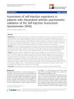
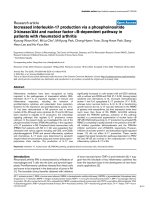
Figure 1: The comparison of serum TNF-α level between RA patients and controls
(p, test Mann - Whiney).
The mean and median of serum TNF-α of RA patients and controls was 2.37 ± 2.69;
1.68 and 3.87 ± 2.11; 3.69 pg/mL, respectively. Median of serum TNF-α concentrations
in RA patients was significantly lower than that in controls group (p < 0.001).
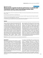
Figure 2: The correlation of serum IL-6 levels
and serum TNF-α levels in RA patients
(numbers are Spearman correlation coefficients).
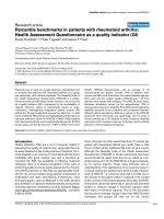
Figure 3: The correlation of serum TNF-α
levels and serum IL-17 levels in RA patients
(numbers are Spearman correlation coefficients).
Serum TNF-α had a possitive correlation with serum IL-6 and IL-17 in RA patients
(r = 0.233; p = 0.035 and r = 0.25; p = 0.023, respectively).
183
Journal of military pharmaco-medicine n08-2017
3. Correlation between serum TNF-α and clinical, laboratory variables in RA
patients group.
Table 2: The comparision of serum TNF-α based on measurements of disease activity.
Serum TNF-α levels (pg/ml)
Mean ± SD
Median
1 - 4 (n = 13)
2.72 ± 3.22
2.24
≥ 5 (n = 68)
2.33 ± 2.62
1.64
1 - 4 (n = 20)
2.34 ± 2.84
2.24
≥ 5 (n = 61)
2.41 ± 2.68
1.64
Low & moderate (n = 14)
2.51 ± 3.17
1.94
High (n = 65)
2.40 ± 2.65
1.72
Joint tender count 28
p
0.694
Joint swollen count 28
0.784
DAS28 CRP
0.944
Table 3: The correlation of serum TNF-α levels in RA patients with measurements of
disease activity.
TJC28
SJC28
MS
PtGA
PGA
CRP
ESR
r
0.105
0.040
0.050
-0.062
-0.131
-0.183
-0.065
p
0.352
0.725
0.664
0.585
0.245
0.102
0.600
Serum TNF-α
(Abbreviations: TJC: Tender joint count; SJC: Swollen joint count; MS: Morning stiffness;
PtGA: Patient global assessment of disease activity; PGA: Provider global assessement of
disease activity; r: Spearman’s correlation coefficient)
There were no differences according to joint tender count 28, joint swollen count 28
and DAS28 CRP.
Table 4: The correlation of serum TNF-α levels with composite indices in RA patients.
DAS28 CRP
DAS28 ESR
SDAI
CDAI
r
0.001
0.090
-0.009
0.024
p
0.995
0.472
0.938
0.832
Serum TNF-α
(Abbreviations: SDAI: Simplified disease activity index, CDAI: Clinical disease
activity index)
There were not associations between the serum TNF-α levels of RA patients with
measurements of disease activity.
184
Journal of military pharmaco-medicine n08-2017
DISCUSSION
In the present study, level of serum
TNF-α cytokine was evaluated in patients
with RA, and associations of its with clinical
and laboratory parameters.
In accordance with other study [6], we
found that serum TNF-α was significantly
lower in RA patients compared to healthy
subjects (figure 1). However, Kokkonen H
et al (2010) found serum TNF-α had no
differrences between RA patients and
healthy controls [7]. By contrast our results,
do Prado A.D et al (2016) observed serum
TNF-α increased in RA patients compared
to healthy controls (p < 0.001) [8]. This
condition may be caused by treatment
before, this study including 50.6% of patients
treated by glucocorticoid. Glucocorticoids
exert potent inhibitory effects on the
transcription and action of a large variety
of cytokines with pivotal importance in the
pathogenesis of RA. Most T helper type 1
(Th1) proinflammatory cytokines are inhibited
by glucocorticoids, including interleukin
(IL)-1β, IL-2, IL-3, IL-6, TNF, interferon-γ [9].
In the current study, serum TNF-α had
a significantly positive correlation with
serum IL-6 and serum IL-17 (figure 2 and
figure 3). In consistent of our observation,
Manicourt D.H et al (1993) [10] and Zhao
P.W et al (2014) [11] also reported that
serum TNF-α had a positive correlation
with serum IL-6 and serum IL-17. These
studies supports the concept that TNF-α
plays a key role in pathogenesis of RA by
stimulating pro-inflammation cytokines
including IL-6, IL-17 [2].
TNF-α is a key cytokine in the pathogenesis
of RA that involved in chronic synovial
inflammation and articular destruction,
thus it may influence disease activity of
RA patients. We assessed the change of
serum TNF-α according to measurements
of disease activity including TJC28,
SJC28 and DAS28 CRP, however we did
not found differences based on these
parameters. In the present study, we also
did not observe the correlation between
serum IL-6 with measurements of disease
activity such as TJC28, SJC28, morning
stiffness, PtGA, PGA, ESR, plasma CRP
levels as well as composite index DAS28
CRP, DAS28 ESR, SDAI and CDAI.
Consistantly with the present study, do
Prado A.D et al (2016) observed that
serum TNF-α had no associations with
joint tender count 28, joint swollen count
28, DAS28 CRP, DAS28 ESR [8]. Keiko
Shimamoto et al (2013) found serum
TNF-α was not related to DAS28 CRP
and DS28 ESR [12]. By contrast these
results, Reham Dwedar A.R.A et al (2015)
reported serum TNF-α had a negative
relation to DAS28 (r = -0.404, p = 0.045)
[13]. Thus, there are many controversial
studies regarding the relationship between
serum TNF-α as an assessing role of
disease activity and measurements of
disease activity in RA patiets, so we need
more studies with larger sample number
to discover this interesting correlation.
Our study has some limitations. The
sample size of patients was relatively small,
and the patients were on drug treatment
including glucorticoids DMARDs. In fact,
our study had a cross-sectional design,
and cytokines profile had wide range.
185
Journal of military pharmaco-medicine n08-2017
CONCLUSION
Our study demonstrated a significantly
lower of serum TNF-α in RA patients
comparing with healthy controls. However,
we did not find any associations between
serum TNF-α levels and measurements
of disease activity in RA patients.
REFERENCES
1. Aletaha D, T. Neogi, A.J Silman et al.
RA classification criteria: an American College
of Rheumatology/European League Against
Rheumatism collaborative initiative. Arthritis
Rheum. 2010, 62 (9), pp.2569-2581.
2. Brennan F.M, I.B. McInnes. Evidence
that cytokines play a role in RA. J Clin Invest.
2008, 118 (11), pp.3537-3545.
3. Gabay C, I. Kushner. Acute-phase
proteins and other systemic responses to
inflammation. N Engl J Med. 1999, 340 (6),
pp.448-454.
4. Pollard L.C, G.H. Kingsley, E.H. Choy et
al. Fibromyalgic RA and disease assessment.
Rheumatology. Oxford. 2010, 49 (5), pp.924-928.
5. Prevoo M.L, M.A. van 't Hof, H.H. Kuper
et al. Modified disease activity scores that
include twenty-eight-joint counts. Development
and validation in a prospective longitudinal
study of patients with rheumatoid arthritis.
Arthritis Rheum. 1995, 38 (1), pp.44-48.
6. Selaas O, H.H. Nordal, A.K. Halse et al.
Serum markers in RA: A longitudinal study of
patients undergoing infliximab treatment.
2015, p.276815.
186
7. Kokkonen H, I. Soderstrom, J. Rocklov
et al. Up-regulation of cytokines and chemokines
predates the onset of RA. Arthritis Rheum.
2010, 62 (2), pp.383-391.
8. do Prado A.D, M.C. Bisi, D.M. Piovesan
et al. Ultrasound power Doppler synovitis is
associated with plasma IL-6 in established
rheumatoid arthritis. Cytokine. 2016, 83.
Ultrasound power Doppler synovitis is
associated with plasma IL-6 in established
RA. pp.27-32.
9. Gary S. Firestein. Kelley’s Textbook of
th
Rheumatology. 9 ed. Glucocorticoid therapy,
ed. Johannes W.G. Jacobs. Johannes W.J.
Bijlsma. Elsevier Saunders. Philadelphia. 2013.
10. Manicourt D.H, R. Triki, K. Fukuda et
al. Levels of circulating tumor necrosis factor
alpha and interleukin-6 in patients with RA.
Relationship to serum levels of hyaluronan
and antigenic keratan sulfate. Arthritis Rheum.
1993, 36 (4), pp.490-499.
11. Zhao P.W, W.G. Jiang, L. Wang et al.
Plasma levels of IL-37 and correlation with
TNF-alpha, IL-17A, and disease activity
during DMARD treatment of RA. PLoS One.
2014, 9 (5), p.e95346.
12. Shimamoto K., T. Ito, Y. Ozaki et al.
Serum interleukin 6 before and after therapy
with tocilizumab is a principal biomarker in
patients with RA. J Rheumatol. 2013, 40 (7),
pp.1074-1081.
13. Reham Dwedar A.R.A, Hala A. Raafat.
Does novel IL-33 correlates with TNF-α in
RA and SLE?. Egyptian Journal of Medical
Microbiology. 2015, 24, pp.13-20.