Ebook Color atlas of anatomy - A photographic study of the human body (7th edition): Part 2
Bạn đang xem bản rút gọn của tài liệu. Xem và tải ngay bản đầy đủ của tài liệu tại đây (40.35 MB, 292 trang )
104750_S_243_264_Kap_4:_
05.01.2010
10:33 Uhr
Seite 243
243
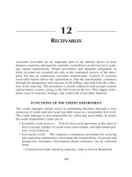
4 Thoracic Organs
1
2
3
4
5
The thoracic cavity contains
heart, lungs, and mediastinal
organs. The thorax protects all
organs but is still movable so
that respiration can occur. The
respiratory movements of the
lung depend on the pleura
covering, the thoracic wall,
and the surface of the lungs.
The mediastinal organs comprise the esophagus, trachea,
and the related nerves and
vessels, particularly the aorta,
superior vena cava, and the
thoracic duct. The thoracic
cavity is separated from the
abdominal cavity by the diaphragm.
6
7
Thoracic organs, heart, and lungs in situ (ventral aspect). Anterior thoracic wall, parietal pleura,
and pericardium have been removed.
1
2
1
2
3
4
5
6
7
Trachea
Ascending aorta
Left lung
Right coronary artery
Heart (right ventricle)
Costal arch
Liver
3
5
7
Position of lungs and heart within the thoracic
cavity (schematic drawing). The anterior part of
the thorax is not depicted.
104750_S_243_264_Kap_4:_
05.01.2010
10:34 Uhr
Seite 244
Position of the Thoracic Organs
244
1
2
3
4
5
6
7
8
9
10
11
12
13
14
15
16
17
18
19
20
21
22
23
24
25
26
27
28
29
30
31
32
33
34
35
36
37
38
39
40
41
42
43
44
Positions of thoracic organs. The anterior thoracic wall has been removed.
Arrow: horizontal fissure of right lung.
32
31
9
10
39
33
23
40
41
42
12
34
35
36
27
43
37
44
38
Horizontal section through the thorax at the level of the seventh thoracic vertebra (from below).
Cricothyroid muscle
Right internal jugular vein
Vagus nerve
Right common carotid artery
Right subclavian vein
Right brachiocephalic vein
Superior vena cava
Upper lobe of right lung
Right auricle
Middle lobe of right lung
Oblique fissure of right lung
Lower lobe of right lung
Diaphragm
Falciform ligament
Costal margin
Transverse colon
Thyroid gland
Trachea
Left internal jugular vein
Left cephalic vein
Left brachiocephalic vein
Pericardium (cut edge)
Upper lobe of left lung
Right ventricle
Left ventricle
Anterior interventricular sulcus
Lower lobe of left lung
Xiphoid process
Liver
Stomach
Pectoralis major muscle
Sternum
Left ventricle and bulb of aorta
Left main bronchus
Esophagus
Descending aorta
Spinal cord
Scapula
Right atrium
Right pulmonary vein
Right main bronchus
Azygos vein
Body of vertebra
Rib
104750_S_243_264_Kap_4:_
05.01.2010
10:34 Uhr
Seite 245
245
Position of the Thoracic Organs
1
1
10
2
2
3
4
11
5
7
5
Sagittal section through the thorax (MRI scan).
6
7
8
9
Sagittal section through the thorax, 2 cm lateral to the median
plane.
1
2
3
4
5
6
7
8
9
10
11
12
13
14
15
16
Aortic arch
Left atrium of the heart
Esophagus
Right atrium of the heart
Liver
Stomach
Abdominal aorta
Transverse colon (dilated)
Lumbar vertebral body
Pulmonary trunk
Left ventricle of the heart
Trachea
Ascending aorta
Right ventricle of the heart
Pericardium
Remaining parts of thymus gland
12
16
13
Parts of
mediastinum
Content
Superior
mediastinum
(yellow)
Trachea, brachiocephalic vein,
thymus, aortic arch, esophagus,
thoracic duct
Middle portion
of mediastinum
(light blue)
Heart, ascending aorta,
pulmonary trunk, pulmonary veins,
phrenic nerves
Posterior
mediastinum
(red)
Esophagus with vagus nerves,
descending aorta, thoracic duct,
sympathetic trunks
Anterior portion
of mediastinum
(light red)
Smaller vessels and nerves,
fat and connective tissue,
thymus (only in the child)
15
4
14
3
Regional anatomy of the thoracic cavity (midsagittal section).
The parts of the mediastinum are indicated by colors.
104750_S_243_264_Kap_4:_
246
05.01.2010
10:34 Uhr
Seite 246
Respiratory System: Bronchial Tree
1
2
3
4
5
6
7
8
9
10
11
12
13
14
15
16
17
18
19
20
21
22
23
24
25
26
27
28
29
30
31
Sphenoid sinus
Pharyngeal opening of auditory tube
Spinal cord
Dens of axis
Oropharynx (oropharyngeal isthmus)
Epiglottis
Entrance of larynx
Esophagus
Upper lobe of right lung
Azygos vein
Branches of pulmonary artery
Right main bronchus
Bifurcation of trachea
Tributaries of right pulmonary veins
Middle lobe of right lung
Lower lobe of right lung
Frontal sinus
Superior nasal concha
Middle nasal concha
Inferior nasal concha
Hard palate
Soft palate with uvula
Tongue
Vocal fold
Larynx
Trachea
Upper lobe of left lung
Left pulmonary artery
Left main bronchus
Left pulmonary veins
Lower lobe of left lung
To page 247:
Respiratory system. The lungs have been fixed in expiration and turned laterally.
Head bisected and turned laterally.
Bronchial tree (ventral aspect). The lung tissue has been removed.
The bronchopulmonary segments are numbered 1–10.
1
2
3
4
5
6
7
8
9
10
11
12
13
14
15
16
17
18
19
20
21
22
23
24
25
26
Nasal cavity
Pharynx
Larynx (thyroid cartilage)
Trachea
Upper lobe of right lung
Bifurcation of trachea
Right main bronchus
Horizontal fissure of right lung
Middle lobe of right lung
Oblique fissures of lungs
Lower lobe of right lung
Clavicle
Upper lobe of left lung
Left main bronchus
Bronchi supplying bronchopulmonary
segments
Lower lobe of left lung
Costal margin
Hyoid bone
Right superior lobe bronchus
Right middle lobe bronchus
Right inferior lobe bronchus
Left superior lobe bronchus
Left inferior lobe bronchus
Segmental bronchi
Branches of pulmonary arteries
Branches of pulmonary veins
104750_S_243_264_Kap_4:_
05.01.2010
10:34 Uhr
Seite 247
247
Respiratory System: Bronchial Tree
1
2
3
12
4
5
6
13
7
14
15
8
9
10
10
11
16
17
17
Larynx, trachea, and bronchial tree (anterior aspect).
Organization and positions of respiratory organs
(schematic drawing).
13
5
1
26
2
19
1-2
3
25
26
4
4
5
6
5
20
23
6
21
7
10
25
22
3
9
10
10
8
25
26
9
10
16
11
Mediastinal dissection of the bronchial tree, pulmonary veins, and pulmonary arteries of right lung (left) and left lung (right)
(medial aspect). The bronchopulmonary segments are numbered 1–10.
104750_S_243_264_Kap_4:_
248
05.01.2010
10:34 Uhr
Seite 248
Respiratory System: Projections of Lungs and Pleura
Surface projections of lungs and pleura on the thoracic wall. Left: anterior aspect; right: right-lateral aspect.
Red = margins of the lung; blue = margins of pleura. The numbers indicate ribs.
Surface projections of lungs and pleura on the thoracic wall. Left: posterior aspect; right: left-lateral aspect.
Red = margins of lung; blue = margins of pleura. The numbers indicate ribs.
1
2
3
4
5
Apex of lung
Upper lobe of right lung
Horizontal fissure of right lung
Middle lobe of right lung
Oblique fissures of lungs
6
7
8
9
10
Lower lobe of right lung
Upper lobe of left lung
Cardiac notch of left lung
Lower lobe of left lung
Infrasternal angle
11
12
13
14
Costal margin
Spine of scapula
First lumbar vertebra
Space between border of lung and pleura
(costodiaphragmatic recess)
104750_S_243_264_Kap_4:_
05.01.2010
10:34 Uhr
Seite 249
Respiratory System: Lungs
Right lung (lateral aspect).
Left lung (lateral aspect).
Right lung (medial aspect).
Left lung (medial aspect).
1
2
3
4
5
6
7
Apex of lung
Upper lobe of right lung
Horizontal fissure of right lung
Oblique fissure of right lung
Middle lobe of right lung
Lower lobe of right lung
Inferior border
8
9
10
11
12
13
14
Upper lobe of left lung
Impressions of ribs
Oblique fissure of left lung
Lower lobe of left lung
Groove of subclavian artery
Groove of azygos arch
Branches of right pulmonary artery
15
16
17
18
19
20
21
Bronchi
Right pulmonary veins
Pulmonary ligament
Diaphragmatic surface
Groove of aortic arch
Left pulmonary artery
Branches of left pulmonary veins
22
23
24
25
26
Left secondary bronchi
Groove of thoracic aorta
Groove of esophagus
Cardiac impression
Lingula
249
104750_S_243_264_Kap_4:_
250
05.01.2010
10:34 Uhr
Seite 250
Respiratory System: Bronchopulmonary Segments
5
Right lung (medial aspect).
Left lung (medial aspect).
Right lung (lateral aspect).
Left lung (lateral aspect).
The bronchopulmonary segments of the lungs are differentiated by the various colors. Notice that there is no
segment in the left lung that corresponds to the seventh
segment of the right lung. Compare with the schematic
drawing on the facing page.
104750_S_243_264_Kap_4:_
05.01.2010
10:34 Uhr
Seite 251
Respiratory System: Bronchopulmonary Segments
Distribution of bronchopulmonary segments of the lungs and their relation to the bronchial tree (after J. F. Huber).
The bronchopulmonary segments are morphologically and
functionally separate, independent respiratory units of the
lung tissue. Each segment is surrounded by connective
tissue that is continuous with the visceral pleura. The
segmental bronchi are centrally located in each segment
and are closely accompanied by branches of the pulmonary
Right lung
Left lung
1 Apical segment
2 Posterior segment
3 Anterior segment
·
4 Lateral segment
5 Medial segment
lobe
· Middle
bronchus
6
7
8
9
10
arteries, whereas the tributaries of the pulmonary veins run
between the segments. Thus, the veins serve two adjacent
segments that drain for the most part into more than one
vein. A bronchopulmonary segment is therefore not a
complete vascular unit, but segmentation is the result of a
specific architecture of the lung vasculature.
Upper lobe
bronchus
Superior (apical) segment
Medial basal segment
Anterior basal segment
Lateral basal segment
Posterior basal segment
·
Lower lobe
bronchus
1+2 Apicoposterior
segment
3 Anterior segment
·
Superior
division
4 Superior lingular segment
5 Inferior lingular segment
6
7
8
9
10
Superior (apical) segment
Absent
Anteromedial basal segment
Lateral basal segment
Posterior basal segment
Inferior
· division
·
·
Upper lobe
bronchus
Lower lobe
bronchus
251
104750_S_243_264_Kap_4:_
252
05.01.2010
10:34 Uhr
Seite 252
Heart
10
Heart of 30-year-old woman (anterior aspect).
1
2
3
4
5
6
7
8
Left subclavian artery
Left common carotid artery
Brachiocephalic trunk
Superior vena cava
Ascending aorta
Bulb of the aorta
Right auricle
Right atrium
Heart of 30-year-old woman (oblique-posterior view).
9
10
11
12
13
14
15
16
17
Position of heart and its vessels within the thorax
(schematic drawing).
Coronary sulcus
Right ventricle
Aortic arch
Ligamentum arteriosum
Left pulmonary veins
Left auricle
Pulmonary trunk
Sinus of pulmonary trunk
Anterior interventricular sulcus
18
19
20
21
22
23
24
25
Left ventricle
Apex of the heart
Left atrium
Epicardial fat overlying coronary sinus
Posterior interventricular sulcus
Right pulmonary artery
Right pulmonary veins
Inferior vena cava
1
2
3
4
5
6
7
8
9
10
11
12
13
14
Right brachiocephalic vein
Superior vena cava
Ascending aorta
Right atrium
Right ventricle
Inferior vena cava
Left internal jugular vein
Left common carotid artery
Left axillary artery and vein
Left brachiocephalic vein
Pulmonary trunk
Left auricle
Left ventricle
Descending aorta
104750_S_243_264_Kap_4:_
05.01.2010
10:34 Uhr
Seite 253
Heart
9
1
10
2
11
3
4
12
13
14
5
15
16
6
17
1
2
3
4
5
6
7
8
9
10
11
12
13
14
15
16
17
18
19
18
7
8
19
20
20
21
22
23
24
25
26
27
Brachiocephalic trunk
Right pulmonary artery
Superior vena cava
Right pulmonary veins
Ascending aorta
Right atrium
Right coronary artery
Right ventricle
Left common carotid artery and left
subclavian artery
Descending aorta (thoracic part)
Ligamentum arteriosum (remnant of
ductus arteriosus Botalli)
Left pulmonary artery
Aortic arch
Left pulmonary veins
Pulmonary trunk
Left atrium
Left coronary artery
Diagonal branch of left coronary artery
Interventricular branch of left coronary
artery
Left ventricle
Right brachiocephalic vein
Thoracic wall
Liver
Aortic valve
Chordae tendineae
Papillary muscles
Stomach
Heart with related vessels. Dissection of coronary arteries (anterior aspect,
systolic phase of heart action).
Coronal section through the thorax
at the level of the ascending aorta
(MRI scan, courtesy of Prof. W. Bautz and
R. Janka, M. D., University of Erlangen,
Germany).
253
104750_S_243_264_Kap_4:_
254
05.01.2010
10:34 Uhr
Seite 254
Heart
1
2
3
4
5
6
7
8
9
10
11
12
13
Ascending aorta
Right coronary artery
Right ventricle
Left atrium
Pulmonary trunk
Septal branch of left coronary artery
Diagonal branch
Anterior interventricular branch of left
coronary artery
Left ventricle
Aortic root
Superior vena cava
Circumflex branch of left coronary artery
Sternum
Human heart (3-D reconstruction of electron beam CT scans as “Shaded Surface
Display” 1).
1
Electron beam tomographic image of the human heart (axial section after
injection of contrast medium1).
Courtesy of Drs. W. Moshage, S. Achenbach,
and D. Ropers, Dept. of Internal Medicine II,
University of Erlangen-Nürnberg, Germany.
104750_S_243_264_Kap_4:_
05.01.2010
10:34 Uhr
Seite 255
Heart
Heart and related vessels in situ (anterior aspect). Anterior thoracic wall, pericardium, and
epicardium have been removed; trachea divided.
Heart in situ. Position of valves (anterior aspect). (Schematic drawing.)
1 Larynx (thyroid cartilage)
2 Sternocleidomastoid muscle
(divided)
3 Trachea (divided) and right
internal jugular vein
4 Vagus nerve
5 Right common carotid
artery and cephalic vein
6 Esophagus
7 Right axillary vein
8 Right and left
brachiocephalic veins
9 Superior vena cava
10 Right auricle
11 Right coronary artery
12 Right atrium
13 Diaphragm
14 Pericardium (cut edges)
15 Costal margin
16 Omohyoid muscle
17 Left common carotid artery
18 Left internal jugular vein
19 Clavicle (divided)
20 Left recurrent laryngeal
nerve
21 Subclavian vein
22 Pericardial reflection
23 Pulmonary trunk
24 Ascending aorta
25 Anterior interventricular
sulcus and anterior
interventricular branch of
left coronary artery
26 Right ventricle
27 Left ventricle
28 Aortic valve
29 Tricuspid or right
atrioventricular valve
30 Inferior vena cava
31 Pulmonary veins
32 Pulmonary valve
33 Left atrioventricular
(bicuspid or mitral) valve
255
104750_S_243_264_Kap_4:_
256
05.01.2010
10:34 Uhr
Seite 256
Heart
1
2
3
4
5
6
7
8
9
10
11
12
13
14
15
16
17
18
19
20
21
22
23
24
25
26
27
28
29
30
31
32
33
34
35
Brachiocephalic trunk
Superior vena cava
Sulcus terminalis
Right auricle
Right atrium
Aortic valve
Conus arteriosus (interventricular septum)
Right atrioventricular (tricuspid) valve
Anterior papillary muscle
Myocardium of right ventricle
Left common carotid artery
Left subclavian artery
Aortic arch
Ligamentum arteriosum (remnant of ductus
arteriosus)
Thoracic aorta (descending aorta)
Ascending aorta
Left pulmonary vein
Pulmonary trunk
Left auricle
Pulmonic valve
Anterior papillary muscle with chordae
tendineae
Myocardium of left ventricle
Posterior papillary muscle
Interventricular septum
Right and left brachiocephalic veins
Chordae tendineae
Papillary muscles of right ventricle
Left atrium
Infundibulum
Anterior papillary muscle of left ventricle
Left atrioventricular (bicuspid or mitral) valve
and chordae tendineae
Apex of heart
Inferior vena cava
Liver
Aorta (pars abdominalis)
Anterior aspect of the heart. Dissection of the four valves.
Circulation within the heart (anterior aspect).
Arrows = direction of blood flow.
MRI scan of the heart (coronal section at the level of the
left atrium; courtesy of Prof. W. Bautz and R. Janka, M. D.,
University of Erlangen, Germany).
104750_S_243_264_Kap_4:_
05.01.2010
10:34 Uhr
Seite 257
Heart: Myocardium
11
1
12
2
3
13
14
4
15
5
16
6
7
17
8
18
15
9
10
34
Heart (posterior aspect). The myocardium of the left ventricle has
been fenestrated to show the muscle fiber bundles of the deeper
layer with their more circular course.
Heart in situ. Myocardium and coronary arteries
(anterior aspect).
1
2
3
4
5
6
7
8
9
10
11
12
13
14
15
16
17
18
19
20
21
22
23
24
25
26
27
28
29
30
31
32
33
34
Internal jugular vein
Common carotid artery
Brachiocephalic trunk
Ascending aorta
Right lung
Right auricle
Right coronary artery
Myocardium of right ventricle
Diaphragm
Costal margin
Thyroid gland and internal jugular vein
Trachea and left common carotid artery
Left brachiocephalic vein
Left lung
Pericardium (cut edge)
Pulmonary trunk
Anterior interventricular artery
Myocardium of left ventricle
Muscular vortex (right ventricle)
Posterior interventricular sulcus
Anterior interventricular sulcus
Muscular vortex (left ventricle)
Aortic arch
Left atrium
Coronary sinus
Superior vena cava
Right pulmonary vein
Right atrium
Inferior vena cava
Coronary sulcus
Myocardium of left ventricle
Left pulmonary artery
Left pulmonary vein
Apex of heart
Vortex of cardiac muscle fibers (from below).
257
104750_S_243_264_Kap_4:_
05.01.2010
10:34 Uhr
Seite 258
Heart: Valves
258
1
2
3
4
5
6
7
8
9
10
11
12
13
14
15
16
17
18
19
20
21
22
23
24
25
26
27
28
29
30
Superior vena cava
Crista terminalis
Fossa ovalis
Opening of inferior vena cava
Opening of coronary sinus
Right auricle
Right coronary artery and coronary sulcus
Anterior cusp of tricuspid valve
Chordae tendineae
Anterior papillary muscle
Myocardium
Pulmonary trunk
Ascending aorta
Pulmonic valve
Conus arteriosus (interventricular septum)
Septal papillary muscles
Septomarginal trabecula or moderator band
Apex of heart
Left auricle
Aortic valve
Left ventricle
Pulmonary veins
Position of fossa ovalis
Left atrium
Left atrioventricular (bicuspid or mitral) valve
Right atrium
Pericardium
Posterior papillary muscle
Right ventricle
Interventricular septum
Right heart (anterior aspect). Anterior wall of right atrium and
ventricle removed.
12
13
13
12
22
19 22
23
19
24
25
20
27
9
28
26
9
9
9
21
21
10
30
29
Heart, left ventricle with mitral valve, papillary muscles, and
aortic valve (anterior portion of the heart removed).
Heart, left ventricle, and atrium (opened) showing the
posterior part of the mitral valve with papillary muscles.
104750_S_243_264_Kap_4:_
05.01.2010
10:34 Uhr
Seite 259
Heart: Valves
1
2
3
4
5
6
7
8
9
10
11
12
13
14
15
16
Valves of heart (superior aspect). Left and right atria removed. Dissection of coronary arteries.
Above: anterior wall of the heart.
Pulmonic and aortic valves (from above). Anterior wall of the
heart at the top. Both valves are closed.
17
18
19
20
21
22
23
24
Pulmonic valve
Sinus of pulmonary trunk
Left coronary artery
Great cardiac vein
Left atrioventricular (mitral) valve
Coronary sinus
Aortic valve
Right coronary artery
Right atrioventricular (tricuspid)
valve
Bulb of aorta
Anterior semilunar cusp of
pulmonic valve
Left semilunar cusp of pulmonic
valve
Right semilunar cusp of pulmonic
valve
Left semilunar cusp of aortic valve
Right semilunar cusp of aortic
valve
Posterior semilunar cusp of aortic
valve
Right atrium
Anterior cusp of tricuspid valve
Chordae tendineae
Trabeculae carneae
Interventricular septum
Septal cusp of tricuspid valve
Anterior papillary muscle
Myocardium of right ventricle
Right atrioventricular (tricuspid) valve (anterior aspect after
removal of the anterior wall of the right ventricle).
259
104750_S_243_264_Kap_4:_
260
05.01.2010
10:34 Uhr
Seite 260
Heart: Function
1
2
3
4
5
6
7
8
9
10
11
12
13
14
15
16
17
18
Ascending aorta
Superior vena cava
Right auricle
Right atrium
Coronary sulcus
Right ventricle
Pulmonary trunk
Left auricle
Anterior interventricular sulcus
Left ventricle
Right pulmonary artery
Sulcus terminalis with sinu-atrial node
Line indicating plane of position of valves
Myocardium of right atrium
Inferior vena cava
Valve of pulmonary trunk
Tricuspid valve
Myocardium of right ventricle
Heart, fixed in diastole (anterior aspect). The ventricles are relaxed, atria
contracted.
Heart, fixed in systole (antero-lateral aspect). The ventricles are contracted,
atria dilated.
Morphological changes during heart movements.
Note the changes in position of the valves (red arrows). Contracted portions of heart are indicated in
black.
A. Diastole: muscles of the ventricles relaxed,
atrioventricular valves open, semilunar valves closed.
B. Systole: muscles of ventricles contracted,
atrioventricular valves closed, semilunar valves open.
104750_S_243_264_Kap_4:_
05.01.2010
10:34 Uhr
Seite 261
Heart: Conducting System
Right ventricle, dissection of atrioventricular node, atrioventricular bundle (bundle of His), and right limb or bundle
branch (probes).
1
2
3
4
Superior vena cava
Sulcus terminalis
Bulb of aorta
Sinu-atrial node (arrows)
5
6
7
8
Muscle fiber bundles of right atrium
Coronary sulcus (with right coronary artery)
Aortic sinus
Entrance to left coronary artery
Right atrium, anterior wall, showing the location of the
sinu-atrial node (arrows).
Left ventricle, dissection of left limb or bundle branch of
conducting system (probes).
9
10
11
12
13
14
15
16
17
18
19
20
21
22
23
24
25
26
27
Aortic valve
Branches of left bundle branch
Purkinje fibers
Left auricle
Interventricular septum
Papillary muscles
Ascending aorta
Right atrium
Opening of coronary sinus
Atrioventricular node
Septal cusp of tricuspid valve
Pulmonary trunk
Atrioventricular bundle (bundle of His)
Bifurcation of atrioventricular bundle
Right bundle branch
Inferior vena cava
Left atrium
Left bundle branch
Papillary muscles with Purkinje fibers
Conducting system of the heart (schematic drawing).
261
104750_S_243_264_Kap_4:_
262
05.01.2010
10:34 Uhr
Seite 262
Heart: Arteries and Veins
Coronary arteries (anterior aspect). The epicardium and
subepicardial fatty tissue have been removed. The arteries
have been injected with red resin from the aorta.
Right coronary artery and veins of the heart (dorsal aspect).
The epicardium and subepicardial fatty tissue have been removed.
32
1
34
2
3
4
35
27
20
9
11
8
23
10
21
22
14
36
33
30
36
28
26
17
Vessels of the heart. Coronary arteries (red) and veins (blue)
of the heart (anterior aspect).
1 Ascending aorta
2 Aortic bulb and (in the above specimen) sinu-atrial branch
of right coronary artery
3 Right auricle
4 Right coronary artery
5 Right atrium
6 Coronary sulcus
7 Right ventricle
8 Left auricle
9 Pulmonary trunk
10 Circumflex branch of left coronary artery
11 Left coronary artery
12 Diagonal branch of left artery
13 Great cardiac vein
14 Anterior interventricular artery
15 Anterior interventricular sulcus
16 Left ventricle
17 Apex of heart
18 Right pulmonary vein
19 Left atrium
20 Left pulmonary veins
21 Oblique vein of left atrium (Marshall’s vein)
22 Coronary sinus
23 Great cardiac vein
24 Coronary sulcus (posterior portion)
25 Posterior vein of left ventricle
26 Middle cardiac vein
27 Left pulmonary artery
28 Inferior vena cava
29 Right atrium
30 Posterior interventricular branch of right coronary artery
31 Posterior interventricular sulcus
32 Superior vena cava
33 Right marginal branch
34 Branch of sinu-atrial node
35 Minimal cardiac veins
36 Small cardiac vein
104750_S_243_264_Kap_4:_
05.01.2010
10:34 Uhr
Seite 263
Heart: Arteries and Veins
7
1
8
2
9
10
11
3
12
4
13
14
5
15
6
1
2
3
4
5
6
7
8
9
10
11
12
13
14
15
16
17
18
19
20
21
22
23
24
25
26
Brachiocephalic trunk
Superior vena cava
Right atrium
Right coronary artery
Right ventricle
Connection of one of the bypass vessels with the
anterior interventricular artery
Left subclavian artery
Left common carotid artery
Ductus arteriosus (Botalli) (still open)
Ascending aorta with three bypass vessels implanted
Pulmonary trunk
Left atrium
Circumflex branch of left coronary artery
Anterior interventricular branch of left coronary artery
Left ventricle
Apex of heart
Sternum
Right ventricle
Liver
Spinal cord
Trachea
Aorta
Body of thoracic vertebrae
Pulmonary artery
Inferior vena cava
Hepatic vein
16
Heart, coronary vessels after implantation of three bypass
vessels (anterior aspect). The ductus arteriosus (9) is still open.
Sagittal section through the thoracic cavity (MRI scan,
courtesy of Prof. W. Bautz and R. Janka, M. D., University of
Erlangen, Germany).
Heart, coronary vessels after implantation of three
bypass vessels (yellow) (schematic drawing of the
specimen above).
263
104750_S_243_264_Kap_4:_
264
05.01.2010
10:34 Uhr
Seite 264
Regional Anatomy of the Thoracic Organs
Thoracic organs (ventral aspect). The left clavicle and ribs have been partially removed, and the right intercostal spaces have been opened
to show the internal thoracic vein and artery.
1
2
3
4
5
6
7
8
9
10
Right internal jugular vein
Omohyoid muscle
Sternohyoid muscle and external jugular vein
Clavicle
Thoraco-acromial artery
Right subclavian vein
Pectoralis major muscle
External intercostal muscle
Pectoralis minor muscle
Body of sternum
11
12
13
14
15
16
17
18
19
20
Right internal thoracic artery and vein
Fascicles of transversus thoracis muscle
Internal intercostal muscles
Serratus anterior muscle
Costal margin
External abdominal oblique muscle
Anterior sheath of rectus abdominis muscle
Sternocleidomastoid muscle
Left internal jugular vein
Transverse cervical artery
21
22
23
24
25
26
27
28
29
30
Brachial plexus
Vagus nerve
Left axillary vein
Left internal thoracic artery and vein
Ribs and thoracic wall (cut)
Costal pleura
Xiphoid process
Superior epigastric artery
Diaphragm
Rectus abdominis muscle
104750_S_265_290_Kap_4:_
05.01.2010
10:42 Uhr
Seite 265
Regional Anatomy of the Thoracic Organs
Thoracic organs, anterior mediastinum, and pleura (ventral aspect). Ribs, clavicle, and sternum have been partly removed.
Red = arteries; blue = veins; green = lymph vessels and nodes.
1 Sternothyroid muscle and its nerve
(a branch of the ansa cervicalis)
2 Right internal jugular vein
3 Right common carotid artery
4 Cephalic vein
5 Right subclavian vein
6 Right brachiocephalic vein
7 Pectoralis major muscle (divided)
8 Pectoralis minor muscle (divided)
9 Parasternal lymph nodes
10 Internal thoracic artery and vein
11 Anterior margin of costal pleura
12 Pericardium
13 Fifth and sixth ribs (divided)
and serratus anterior muscle
14 Costodiaphragmatic recess
15 External abdominal oblique muscle
16 Rectus abdominis muscle
17 Larynx (thyroid cartilage)
18 Thyroid gland
19 Trachea
20 Left vagus nerve
21
22
23
24
25
26
27
28
29
30
Left brachiocephalic vein
Left internal thoracic artery and vein
Thymus
Costal pleura
Costal margin
Superior epigastric artery
Margin of costal pleura
Diaphragm
Linea alba
Cut edge of anterior sheath of rectus
abdominis muscle
265
104750_S_265_290_Kap_4:_
266
05.01.2010
10:42 Uhr
Seite 266
Regional Anatomy of the Thoracic Organs: Thymus
1
2
3
4
5
6
The thymus above the heart, showing its position and size.
7
8
9
10
11
12
13
14
15
16
17
18
19
20
21
22
23
24
25
26
27
28
29
30
31
32
33
34
35
36
37
38
39
Larynx (thyroid cartilage)
Thyroid gland
Trachea
Internal jugular vein
Brachial plexus
Right brachiocephalic vein and common carotid
artery
Right phrenic nerve
Ascending aorta
Pectoralis minor muscle (divided)
Pulmonary trunk (covered by pericardium)
Costal pleura
Pericardium and heart
Serratus anterior muscle
Xiphoid process
Costal margin
External abdominal oblique muscle
Sternothyroid muscle (divided and reflected)
Vagus nerve
Left common carotid artery
Left sympathetic trunk
Left recurrent laryngeal nerve
Left internal thoracic artery and vein (divided)
Margin of costal pleura
Intercostal nerves and vessels
Superior epigastric artery
Rectus abdominis muscle
Diaphragm
Ansa cervicalis
Phrenic nerve and scalenus anterior muscle
External jugular vein (divided)
Right subclavian vein
Right brachiocephalic vein
Internal thoracic artery (divided)
Internal thoracic vein (divided)
Right lung
Cricothyroid muscle
Omohyoid muscle
Thymus
Left lung
Thoracic organs (ventral aspect). The internal thoracic
vessels have been removed, and the anterior margins of the
pleura and lungs have been slightly reflected to display the
anterior and middle mediastinum, including the heart and
great vessels.
104750_S_265_290_Kap_4:_
05.01.2010
10:42 Uhr
Seite 267
Regional Anatomy of the Thoracic Organs: Thymus
Thoracic organs (ventral aspect). The pleura has been opened and the lungs exposed. Remnants of the thymus and
pericardium are seen.
1
2
3
4
5
6
7
8
9
10
Right internal jugular vein
Phrenic nerve and scalenus anterior muscle
Clavicle (divided)
Right subclavian artery and vein
Internal thoracic artery
Right brachiocephalic vein
Brachiocephalic trunk
Thymus (atrophic)
Upper lobe of right lung
Horizontal fissure of right lung (incomplete)
11
12
13
14
15
16
17
18
19
Middle lobe of right lung
Oblique fissure of right lung
Lower lobe of right lung
Xiphoid process
Diaphragm
Thyroid gland
Left internal jugular vein
Brachial plexus
Left cephalic vein
20 Left common carotid artery and
vagus nerve
21 Left brachiocephalic vein
22 Internal thoracic artery and vein (divided)
23 Ascending aorta and aortic arch
24 Upper lobe of left lung
25 Pericardium
26 Oblique fissure of left lung
27 Lower lobe of left lung
28 Costal margin
267