Mutation analysis of EGFR and FGFR gene in glioblastoma patients in Vietnam
Bạn đang xem bản rút gọn của tài liệu. Xem và tải ngay bản đầy đủ của tài liệu tại đây (136.11 KB, 6 trang )
Journal of military pharmaco-medicine no1-2019
MUTATION ANALYSIS OF EGFR AND FGFR GENE IN
GLIOBLASTOMA PATIENTS IN VIETNAM
Kieu Dinh Hung1; Nguyen Thi Thom1; Tran Quoc Dat1; Dang Thi Ngoc Dung1
Tran Huy Thinh1; Tran Van Khanh1; Ta Thanh Van1
SUMMARY
Background: Glioblastoma is the most prevalence primary malignant brain tumor, which
takes up 16% of all primary brain and central nervous system malignancy. Molecular variations
or gene expression patterns have also been recognized in primary and secondary glioblastomas.
Genetic typical alterations for primary glioblastoma are epidermal growth factor receptor and
fibroblast growth factor receptors variations. Subjects and methods: We recruited 60 patients
diagnosed with primary glioblastoma in which biopsy samples were collected to assess for
FGFR and EGFR mutations. Results and conclusion: 6/60 patients (8.3%) were positive with
FGFR mutation (p.R576W, p.A575V, p.N546K). 8/60 patients (13.3%) were identified with
EGFR, a total of 7 mutations were identified p.P272S, p.G42D, p.T274M, p.K293X, p.L62I,
p.G42D, p.A289T. This is the first study on FGFR and EGFR mutation in glioblastoma patients
in Vietnam. The results would contribute to better understanding the pathological and molecular
mechanism of glioblastoma in Vietnam.
* Keywords: Glioblastoma; EGFR; FGFR; Mutation analysis.
INTRODUCTION
Glioblastoma (GBM) is the most
prevalence primary malignant brain tumor,
which take up 16% of all primary brain
and central nervous system malignancy
[1]. The average age-adjusted incidence
rate in the population is 3.2 per 100,000
[1]. GBMs were primary thought to be
resulting exclusively from glial cells; however,
recent studies suggest that they may
result from several cell types with neural
stem cell-like properties [2].
By the end of the genomic profiling and
the Cancer Genome Atlas project (Parsons
et al 2008), more than 600 genes were
profiled from more than 200 human tumor
samples, which revealed the complex
genetic profile of GBM and we were able
to characterize a set of three core signaling
pathways that are commonly affected
(i.e, the tumor protein p53 pathway, the
receptor tyrosine kinase/Ras/phosphoinositide
3-kinase signaling pathway, and the
retinoblastoma pathway) [3, 4]. Almost all
primary and secondary GBMs presented
abnormality in these pathways, allowing
uncontrolled cell growth and persistence
cell survival, while also letting the tumor
cell to escape programmed cell death and
1. Hanoi Medical University
Corresponding author: Kieu Dinh Hung ()
Date received: 20/10/2018
Date accepted: 29/11/2018
46
Journal of military pharmaco-medicine no1-2019
cell cycle checkpoint [5]. Molecular variations
or gene expression patterns have also
been recognized in primary and secondary
GBM. Genetic alterations typical for
primary GBM are epidermal growth
factor receptor (EGFR) and fibroblast
growth factor receptors (FGFRs) variations
[4].
EGFR is a trans-membrane glycoprotein
and belongs to the tyrosine kinase
superfamily receptor [6]. Gliomas are
tumors which emerge from glial cells,
which express a variety of aggressiveness
based on grade and stage. Many EGFR
gene mutations have been characterized
in gliomas, especially GBM. FGFR is a
family of gene, sub-family of receptor
tyrosine kinases (RTKs), it is comprised of
four closely related genes (FGFR1-4) [7].
FGFR abnormalities have been associated
with many cancers in human and play
significant roles in tumor development
and advancement [5, 7]. FGFRs activating
mutations and overexpression have been
linked with the development of various
cancers, such as bladder, ovarian, breast,
renal cell and more recently GBM [5, 8].
Up to now, there have been few studies to
characterize mutation of FGFR and EGFR
in Vietnamese patients with malignancy.
This study aims: To investigate the percentage
and characterizes EGFR and FGFR gene
alterations in GBM patients. The result will
help better understand of the pathological
and molecular characteristics of GMB in
Vietnamese population.
SUBJECTS AND METHODS
1. Subjects.
We recruited 60 patients diagnosed with
primary GBM. Patients with secondary
GBM or secondary tumor were excluded
from the study. Informed consents were
obtained from the patients prior to
participation in the study. Biopsies taken
from tumor-removing surgery were used
to confirm diagnosis of GBM and for
molecular investigation of FGFR and
EGFR genes.
2. Methods.
* DNA extraction from biopsy sample:
DNA was extracted from biopsy sample
using the phenol-cloroform-isoamyl method.
DNA concentration and purity were verified
using Nanodrop (ThermoFisher, US).
* FGFR and EGFR mutations analysis:
To identify point mutations in the FGFR
and EGFR genes, another PCR amplification
product (100 - 150 ng starting DNA) was
obtained for each sample. After agarose
gel discrimination, the PCR product was
purified with Gel Purification Kit followed
by sequencing using Big Dye Terminator
V3.1 on ABI 3500 genetic analyzers
(Applied Biosystems, CA, USA). Results
were analyzed by CLC Main Workbench
Software. Novel mutations were confirmed
by conducting search on online databases
(i.e. LOVD, 1000 Genomes, ExAC, and
Pubmed) and all previous publications on
FGFR or EGFR gene mutations. The
primers used are provided by the author
on reasonable request.
* In silico missense mutation analysis:
For novel missense variants, to predict
whether the mutation has direct impact on
EGFR or FGFR function, we utilized several
in silico tool: Mutation Taster which estimates
the pathogenic probability of DNA sequence
change and predict the functional
consequences of other non-coding
47
Journal of military pharmaco-medicine no1-2019
sequence or deletion/insertion mutations
[6]; polyphen-2, a method using prediction
models like HumVar and HumDiv for
predicting damaging missense mutations.
DUET to predict protein stability change
upon mutation, results were taken from
the mutation Cutoff Scanning Matrix
(mCSM) method which calculate the
mutated protein structure to be stabilizing
or destabilizing.
RESULTS
1. FGFR mutation.
Table 1: FGFR mutation detected in the study cohort of 60 GBM patients.
Patient ID
Exon
GB46
13
GB48
12
GB52
Nucleotid change
Amino acid change
Publication
p.Ala575Val
Novel
g.56504C>T
p.Asp546Lys
Previously reported by
Rand et al [9]
13
g.57837C>T
p.Arg576Try
Rand et al
GB53
13
g.57837C>T
p.Arg576Try
Rand et al
GB57
13
g.57837C>T
p.Arg576Try
Rand et al
g.57835C>T
Table 1 showed the result of FGFR mutation spectrum in 60 GBM patients in the
study’s cohort. After mutation analysis, 5/60 patients (8.3%) were positive with FGFR
mutation. Of these, 2 mutations were located on exon 13 (1 mutation had been
reported p.R576W, 1 with novel mutation p.A575V), 1 mutation located on exon 12
(p.N546K).
2. EGFR mutation.
Table 2: EGFR mutation detected in the study cohort of 60 GBM patients.
Patient ID
Exon
Nucleotid change
Amino acid change
Publication
GB6
7
c.814C>T
p.Pro272Ser
Rand et al
GB8
7
c.814C>T
p.Pro272Ser
Rand et al
GB10
7
c.814C>T
p.Per272Ser
Rand et al
GB23
2
c.124G>A
p.Gly42Asp
Rand et al
2
c.124G>A
p.Gly42Asp
Rand et al
GB24
7
c.820C>T
p.Thr274Met
Rand et al
7
c.877A>T
p.Lys293Stop
Rand et al
GB25
2
c.183C>A
p.Leu62Iso
Rand et al
2
c.124G>A
p.Gly42Asp
Rand et al
GB26
GB27
7
c.866G>A
p.Ala289Thr
Rand et al
7
c.866G>A
p.Ala289Thr
Rand et al
Table 2 showed the result of EGFR mutation identification in 60 GBM patients in the
study’s cohort. After mutation analysis, 8/60 patients (13.3%) were identified with EGFR.
48
Journal of military pharmaco-medicine no1-2019
A total of 7 mutations were identified p.P272S, p.G42D, p.T274M, p.K293X, p.L62I,
p.G42D, p.A289T. All mutations were previously reported in other studies.
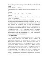
Figure 1: Molecular prediction model of novel mutation p.A575V.
Figure 1 showed the stimulated protein structure of FGFR with mutation
p.Ala575Val. Prediction models (MutationTaster, Polyphen2, DUET) showed the
mutation would cause altered FGFR activity thus contributes to the phenotype and
neoplasticity of GBM.
DISCUSSION
The current study investigated the
mutation spectrum of FGFR and EGFR in
Vietnamese GBM patients. The patients
had been enrolled and oncologists and
pathologists carried out clinical evaluation
to confirm the diagnosis of primary GBM.
Therefore, the cohort is well defined and
well suited for molecular study.
We identified FGFR mutation in
5/60 cases (8.3%), the mutation detection
rate is comparable with other study in
which FGFR mutations were identified in
which it is higher than previously reported.
Snuderl et al (2011) and Szerlip et al
(2012), found that, FGFR mutations were
found in 3 - 3.5% of cases [10]. The
difference may be due to the difference in
GBM staging between the cohort or the
genetics composition of Vietnam compared to
other population. The study identified 3 FGFR
mutations,
including
3
missenses
p.R576W, p.A575V, p.N546K. 2 mutations
(p.R576W and p.N546K) were previously
reported. We identified a novel mutation
p.A575V, we utilized prediction models
(MutationTaster,
Polyphen2,
DUET)
showed the mutation would cause altered
FGFR activity thus contributes to the
phenotype and neoplasticity of GBM.
However, further in vitro and in vivo
studies are needed to confirm the
mechanism in which this mutation affects
GBM pathogenicity.
49
Journal of military pharmaco-medicine no1-2019
We identified EGFR mutation in 8/60 cases
(13.3%). Many EGFR modifications in
gliomas have been reported in the
literature, some of which were specific to
GBM. EGFR amplification was seen in
0 - 4%, 0 - 33% and 34 - 64% of grade II,
III and IV astrocytomas, respectively.
44% of patients with EGFR amplification
had EGFR point mutations, mostly seen
in the extracellular domain - e.g, A289 or
R108 [11]. Other studies reported EGFR
amplification in GBMs, anaplastic
oligodendrogliomas (AOs) and anaplastic
oligoastrocytomas (AOAs). EGFR
overexpression was seen in 6 - 28%,
27 - 70% and 22 - 89% of grade II, III
and IV astrocytomas, respectively, and
represents an increase in gene
transcription independent of DNA
alterations. Half of the tumors with focal
amplification and/or mutation of PDGFRA
harbored concurrent EGFR alterations
(14/33 patients = 42.4%), as did the
majority of MET-altered tumors (3/4),
reflecting a pattern of intratumoral
heterogeneity that has been previously
documented by in situ hybridization.
FGFR and EGFR are both potent
oncogene; therefore, in many cases of
malignancy there exist some form of
mutation in these genes. The identification
of FGFR and EGFR mutation has become
routine in cancer management such as
non-small cell lung cancer. In GBM, these
genes have undergone extensive clinical
trial for targeted therapy and for prognostic
biomarkers [9]. FGFR mutation and fusion
are undergoing trials for targeted therapy
(TKI), and many mutation specific drugs
are being tested. Similarly, the mutations
50
have been linked with respond to erlotinib
(first generation EGFR TKI) with prolonged
survival and/or longer time to progression
[12]. It is clear that FGFR and EGFR have
been proven to be an independent factor
in gliomagenesis and play a role in tumor
formation. Although FGFR and EGFR status
as a clinical marker remains controversy,
more trails are needed to verify the clinical
implication of each mutation. Finally, the
need for larger study in Vietnam is required
to examine the prognostic significance of
FGFR/EGFR gene and protein status for
survival, treatment and other clinical factors
affecting the patient’s outcome and quality
of life.
CONCLUSION
This is the first study on FGFR and EGFR
mutation in GBM patients in Vietnam.
The results would contribute to better
understanding of the pathological and
molecular mechanism of GBM in Vietnam.
REFERENCE
1. Omuro A, DeAngelis L.M. Glioblastoma
and other malignant gliomas: A clinical review.
JAMA. 2013, 310 (17), pp.1842-1850.
doi:10.1001/jama. 2013, p.2803.
2. Brown T.J, Brennan M.C, Li M et al.
Association of the extent of resection with
survival in glioblastoma: A systematic review
and meta-analysis. JAMA Oncol. 2016, 2 (11),
pp.1460-1469. doi:10.1001/jamaoncol. 2016.
1373.
3. Inda M.M, Bonavia R, Mukasa A et al.
Tumor heterogeneity is an active process
maintained by a mutant EGFR-induced
cytokine circuit in glioblastoma. Genes Dev.
2010, 24 (16), pp.1731-1745. doi:10.1101/gad.
1890510.
Journal of military pharmaco-medicine no1-2019
4. Mellinghoff I.K, Wang M.Y, Vivanco I
et al. Molecular determinants of the response
of glioblastomas to EGFR kinase inhibitors.
N Engl J Med. 2005, 353 (19), pp.2012-2024.
doi:10.1056/NEJMoa051918.
adjuvant temozolomide versus radiotherapy
alone on survival in glioblastoma in a
randomized phase III study: 5-year analysis of
the EORTC-NCIC trial. Lancet Oncol. 2009,
10 (5), pp.459-466. doi:10.1016/S1470-2045 (09).
5. Singh D, Chan J.M, Zoppoli P et al.
Transforming fusions of FGFR and TACC
genes in human glioblastoma. Science. 2012,
337 (6099), pp.1231-1235. doi:10.1126/science.
1220834.
9. Verhaak Hoadley K.A, Purdom E et al.
Integrated genomic analysis identifies clinically
relevant subtypes of glioblastoma characterized
by abnormalities in PDGFRA, IDH1, EGFR,
and NF1. Cancer Cell. 2010, 17 (1), pp.98-110.
doi:10.1016/j.ccr.2009.12.020.
6. PTEN mutation, EGFR amplification,
and outcome in patients with anaplastic
astrocytoma and glioblastoma multiform.
Journal of the National Cancer. Institute
Oxford Academic. />article/93/16/1246/2519475. Accessed October
30, 2018.
7. Partanen J, Mäkelä T.P, Eerola E et al.
FGFR-4, a novel acidic fibroblast growth
factor receptor with a distinct expression
pattern. EMBO J. 1991, 10 (6), pp.1347-1354.
doi:10.1002/j.1460-2075.1991.tb07654.x.
8. Stupp R, Hegi M.E, Mason W.P et al.
Effects of radiotherapy with concomitant and
10. Rand V, Huang J, Stockwell T et al.
Sequence survey of receptor tyrosine kinases
reveals mutations in glioblastomas. Proc Natl
Acad Sci USA. 2005, 102 (40), pp.1434414349. doi:10.1073/pnas.0507200102.
11. Davis M.E. Glioblastoma: Overview of
disease and treatment. Clin J Oncol Nurs.
2016, 20 (5), S2-S8. doi:10.1188/16.CJON.S1.
pp.2-8.
12. Gan H.K, Cvrljevic A.N, Johns T.G. The
epidermal growth factor receptor variant III
(EGFRvIII): Where wild things are altered.
FEBS J. 2013, 280 (21), pp.5350-5370.
doi:10.1111/febs.12393.
51