Ebook The 4 stages of heart failure: Part 2
Bạn đang xem bản rút gọn của tài liệu. Xem và tải ngay bản đầy đủ của tài liệu tại đây (7.26 MB, 182 trang )
CHAPTER 7
Assessment of Stage C
Patients with HF-pEF
FAST FACTS
•
HF-pEF is associated with conditions that cause diastolic dysfunction:
– Hypertensive heart disease
– Coronary artery disease
– Hypertrophic cardiomyopathy (HCM)
– Restrictive cardiomyopathy
• Doppler echocardiography assesses left ventricular diastolic function.
an a family history of
,
7
ill have an
• n patients ith
•
•
i enti able genetic mutation of the sarcomere.
myloi osis is the most common i enti able cause of restrictive
cardiomyopathy.
ther causes of heart failure ithout left ventricular systolic ysfunction
include valvular disease, pericardial disease, and cor pulmonale.
• Heart failure associated with obstructive or central sleep apnea may be
improved by night-time use of continuous positive airway pressure (CPAP).
“In the heart, the velocity and extent of relaxation, in
other words, the ease with which the muscle stretches
under the distending force of venous pressure is probably quite as important a factor in the heart’s behavior
as the force and rapidity of the systolic contraction.”
–Yandell Henderson, 19231
The 4 Stages of Heart Failure © 2015 Brian E. Jaski. Cardiotext Publishing, ISBN: 978-1-935395-30-0.
135
136 • Chapter 7: Assessment of Stage C Patients with HF-pEF
Diagnosis of HF-pEF
Heart failure with preserved ejection fraction (HF-pEF) can be diagnosed
when clinical findings of congestion due to elevated pulmonary or systemic venous pressures present with no more than mild left ventricular
systolic dysfunction (EF > 40%). Although HF-pEF is associated with
echocardiography findings of left ventricular diastolic dysfunction, this is
not always the case (see Chapter 2). Similarly, left ventricular hypertrophy
by echocardiography is often present, but HF-pEF may still be present due
to coronary artery disease or other conditions without increased left ventricular wall thickness (Figure 7.1). Beyond pathologic processes that
directly affect myocardial structure, inflammation from noncardiac
comorbidities and increased arterial stiffness can indirectly contribute to
cardiomyocyte hypertrophy, interstitial fibrosis, and impaired left ventricular diastolic filling.2
Is left ventricular wall
thickness increased (>11 mm)?
YES
Hypertensive
Heart Disease
FIGURE 7.1
thickness.
YES
Hypertrophic, Infiltrative,
or Restrictive Cardiomyopathy
NO
R/O CAD, other
Causes of diastolic dysfunction and associated left ventricular wall
DIFFERENTIAL DIAGNOSIS
Infiltrates on chest x-ray and preserved systolic function by echocardiogram can also be seen with interstitial lung disease or non-cardiogenic
pulmonary edema. However, interstitial lung disease does not improve
with diuretics. Non-cardiogenic pulmonary edema (also called adult
respiratory distress syndrome) can occur in a patient with an acute severe
noncardiac systemic illness. In this setting, lung edema develops due to a
“capillary leak” despite normal left heart filling pressures. BNP may be
normal or mildly elevated secondary to right ventricular strain.
Diagnosis of HF-pEF • 137
In most patients, clinical findings and Doppler echocardiographic
assessment (see List 7.1) are adequate to distinguish these conditions
from HF-pEF. In some cases with indeterminate or overlapping findings,
right heart catheterization should be performed; pulmonary capillary
wedge pressure is high in HF-pEF and normal or low in primary pulmonary disorders.
LIST 7.1 Echo-Doppler Findings in Diastolic Dysfunction
• Left ventricular hypertrophy (wall thickness > 11 mm)
• Left atrial enlargement
• itral an pulmonary vein oppler flo abnormalities
• Increased pulmonary artery systolic pressure estimated from velocity of tricuspid
regurgitation
• atio of early iastolic mitral inflo to mitral annulus tissue velocities e′
ECHOCARDIOGRAPHIC FINDINGS WITH DIASTOLIC DYSFUNCTION
Diastolic filling of the left ventricle can be assessed by Doppler echocardiography. 3 Left atrial enlargement can indicate the presence of
longstanding structural heart disease.
Echocardiographic measurement of left atrial size by dimension or
calculated volume has been called the “hemoglobin A1C” of left atrial
pressure and, when increased, serve as an index of chronically elevated
left-sided heart filling pressures.4 In patients with symptomatic HF-pEF,
progressive shortening of the transmitral deceleration time (DT) and
increasing E/A ratio can be seen with decreasing ventricular compliance
and increasing left atrial pressure (Figure 7.2).5 Acute alterations in mitral
inflow velocities may occur in response to patient treatment or other
changes in hemodynamic status.
Tissue Doppler imaging (TDI) of mitral annulus motion can also
assess myocardial relaxation. With systolic ejection of blood, there is contraction of the left ventricle in part achieved by mitral annular descent
toward a relatively fixed apex. Following this, during ventricular filling,
the annulus returns towards its initial position (Figure 7.3). Tissue Doppler
imaging (TDI) displays the velocity profile of these movements. The velocity of the mitral annulus away from the left ventricular apex during early
diastole (e′) reflects the rate of myocardial relaxation and may be less
dependent on pressure gradients than transmitral blood flow velocity.3
138 • Chapter 7: Assessment of Stage C Patients with HF-pEF
LV
20
LA
mm Hg
0
E
80
A
E
E
A
A
cm/s
0
DT
Normal
40
Impaired Relaxation
Decreased Compliance
FIGURE 7.2 Doppler echocardiographic assessment of left ventricular diastolic filling.
Changes in Doppler mitral velocities with correlation to left ventricle (blue) and left atrial
(orange) pressures during diastole. Filling velocities and extrapolated deceleration times (red
arrows) are in response to the transmitral pressure gradient. Blue arrows indicate interval of
isovolumic relaxation time from aortic valve closure to mitral valve opening. Abbreviations:
, left ventricle , left atrium , early iastolic mitral inflo
, iastolic filling uring atrial
systole; DT, deceleration time.5 Source: Adapted with permission from Nagueh et al., Eur J
Echocardiogr. 2
2
.
Left Ventricle
E
Mitral Valve
e’
e’
Timing of Flow
P
T
E/e’ : A ratio of early diastolic blood flow
(E) versus tissue (e’) velocities that
correlates with left atrial pressure.
FIGURE 7.3 Derivation of the E/e′ ratio. Transmitral early diastolic blood flow (E) and mitral
annulus tissue velocities (e′). High left atrial filling pressures with impaired diastolic filling are
associated with increased blood flow E and decreased e′ tissue velocities.
Diagnosis of HF-pEF • 139
In HF-pEF, the E/e′ ratio can be used as an initial measurement for
an estimate of left ventricular filling pressures (Figure 7.4). Because of its
utility, Doppler echocardiography has been called the “Rosetta Stone” for
evaluation of diastolic function.6
E/e’
E/e’ 9 – 14
E/e’ ≤ 8
Normal LAP
E/e’ ≥15
LA Volume < 34 mL/m2
PAS < 30 mm Hg
LA volume ≥ 34 mL/m2
PAS > 35 mm Hg
Normal LAP
LAP
LAP
FIGURE 7.4 Diagnostic algorithm for estimating left ventricular filling pressures
based on Doppler echocardiographic findings in patients with HF-pEF. Abbreviations: E,
early diastolic transmitral blood flow; e′, mitral annulus tissue velocity; LA, left atrium; PAS,
pulmonary artery systolic pressure; LAP, left atrial pressure.5 Source: Adapted with permission
from Nagueh et al., Eur J Echocardiogr. 2
2
.
KEY DIAGNOSTIC FEATURES OF HYPERTENSIVE HEART DISEASE
When a patient with HF-pEF has a history of high blood pressure and
uniform left ventricular hypertrophy by echocardiogram, the diagnosis of
hypertensive heart disease is likely.
Echocardiographic findings of diastolic
dysfunction support this diagnosis. Consider
Look for uniform
that patients with hypertensive heart dishypertrophy of the left
ventricle in hypertensive
ease may also have associated coronary
heart disease.
artery disease (CAD). Ventricular hypertrophy in the absence of a history of high blood
pressure or CAD may imply the presence of a secondary process due to
hypertrophic, infiltrative, or restrictive cardiomyopathy (see descriptions below).
KEY FEATURES OF HYPERTROPHIC CARDIOMYOPATHY
Hypertrophic cardiomyopathy (HCM) can be defined as left and/or right
ventricular hypertrophy occurring usually in an asymmetric pattern and
often involving the interventricular septum not secondary to systemic
hypertension or other systemic disease.7 Left ventricular chamber volume
is normal or reduced. Microscopically, there is myocyte hypertrophy and
disarray surrounding areas of increased loose connective tissue. When
140 • Chapter 7: Assessment of Stage C Patients with HF-pEF
hypertrophic cardiomyopathy is associated with an obstructive gradient
across the left ventricular outflow tract (LVOT), either at rest or after provocation, management directed toward improving this gradient may be
important. End-stage hypertrophic cardiomyopathy may progress to systolic dysfunction. Although many terms have been used historically to
describe HCM, including idiopathic hypertrophic subaortic stenosis
(IHSS) and hypertrophic obstructive cardiomyopathy (HOCM), currently,
using the term hypertrophic cardiomyopathy (HCM) and additional
descriptive features is preferred (List 7.2).
LIST 7.2 Phenotypes of Hypertrophic Cardiomyopathy
• Asymmetric septal hypertrophy
• Symmetric hypertrophy (distinguish from hypertensive or athletic hypertrophy)
• Apical hypertrophy
Within the spectrum of patients with heart failure, patients with
hypertrophic cardiomyopathy represent a distinct subset because treatment options differ. Hypertrophic cardiomyopathy typically arises from
either an inherited or spontaneous point mutation in genes coding for
proteins within the sarcomere including the heavy chain of myosin (see
Chapter 4). The prevalence of all forms of hypertrophic cardiomyopathy
may be as common as 1 in 500 in the United States population; however,
many patients are asymptomatic.8 The location of regional or global hypertrophy within the left (or right) ventricle between individuals can vary, even
within a single family. Other functional features include diastolic dysfunction, mitral regurgitation, myocardial ischemia, and arrhythmias.
Echocardiography or cardiac magnetic resonance imaging (MRI)
can be used to visualize the distribution of hypertrophy (Figure 7.5). The
most common pattern is asymmetric septal hypertrophy with a ratio of
septal to posterior wall thickness of 1.3 or greater. When there is
dynamic outflow tract obstruction, a characteristic “spike and dome”
morphology may be observed in the aortic pressure waveform or LVOT
velocity. This pattern arises from an initial unobstructed ejection of
blood from the left ventricle followed by progressive obstruction of outflow during the period of mid-to-late systolic ejection. An increase in
systolic Doppler velocity across the LVOT narrowed by septal hypertrophy and systolic anterior motion (SAM) of the mitral valve (Figure 7.5)
can be observed at rest or following physiologic provocation such as after
premature ventricular contractions, post-exercise, or during the strain
phase of the Valsalva maneuver (Figure 7.6). Approximately one-third of
patients have nonobstructive HCM defined as resting or peak gradient
Diagnosis of HF-pEF • 141
after provocation of < 30 mm Hg. Patients with resting or provocable
gradients ≥ 50 mm Hg and persistent symptoms may benefit from surgical or percutaneous intervention.7
A
B
FIGURE 7.5 Imaging by 2D echocardiogram of cardiac abnormalities caused by
hypertrophic cardiomyopathy. Images show a 28-year-old female with HCM. Parasternal
long axis (Panel A) reveals Systolic Anterior Motion (SAM) of the mitral valve leaflets. Echo
parasternal short axis (Panel B) demonstrates asymmetric septal hypertrophy (left ventricle
end-diastolic thickness: Septum measurement = 2.9 cm, Posterior wall = 0.9 cm).
142 • Chapter 7: Assessment of Stage C Patients with HF-pEF
FIGURE 7.6 Hemodynamics and provocation maneuvers in HCM with dynamic LVOT
obstruction. Left panel: Example of patient with a resting peak systolic left ventricular-aortic
gra ient of 4 mm g that increase to 2 mm g in a post
beat arro . espite the
mar e ly increase left ventricular pressure of the post
beat, arterial pulse pressure
decreased (known as the Brockenbrough-Braunwald sign). Elevated left ventricular enddiastolic pressure of 32 mm Hg is consistent with diastolic dysfunction of the hypertrophic
ventricle. Moderate to severe mitral regurgitation was also present. Right panel: No aortic
valvular gradient was present during pullback from just below the aortic valve to the aorta,
thus excluding aortic valve disease as contributing to the gradient. After surgical septal
myectomy (data not shown), dynamic outflow tract gradient completely resolved.
Diffuse concentric hypertrophy of the left ventricle is another type
of hypertrophic cardiomyopathy. However, this pattern of hypertrophy
may also be seen with hypertensive, athletic, or infiltrative causes of
hypertrophy. When global hypertrophy is detected and there is no history of hypertension or family history of HCM, additional diagnostic
tests may be indicated. Cardiac MRI may identify delayed gadolinium
hyperenhancement consistent with a HCM pattern of fibrosis (see Figure
6.16) or, endomyocardial biopsy may be needed when infiltrative causes
are suspected.9
A less common manifestation of hypertrophic cardiomyopathy is
hypertrophy confined to the apex of the left ventricle. This pattern often
displays marked T-wave inversion across the precordial leads on a standard 12-lead electrocardiogram.
Management of Hypertrophic Cardiomyopathy • 143
Management of Hypertrophic Cardiomyopathy
Management of hypertrophic cardiomyopathy (HCM) includes three
components: symptom management, risk stratification for sudden cardiac death (SCD), and counseling.10
SYMPTOM MANAGEMENT
Two common symptoms of HCM are
exertional dyspnea
and chest pain. Chest
Symptom
pain often is not due
Risk Stratification
Counseling
Management
for SCD
to epicardial artery
stenosis, but rather to
functional ischemia due to increased myocardial oxygen demand from
hypertrophy exceeding limited endocardial supply. Beta-blockers that
decrease contractility and heart rate can lead to hemodynamic improvement in HCM by decreasing outflow tract obstruction and functional
myocardial ischemia. The calcium channel blocker verapamil has negative inotropic and bradycardic effects that may also improve left
ventricular outflow obstruction. However, it should be used cautiously,
because its action as an arteriolar vasodilator may increase the dynamic
outflow tract gradient. If these medications are poorly tolerated, the antiarrhythmic-negative inotropic agent disopyramide may be considered as
an alternative.
In HCM with dynamic outflow tract obstruction, medications that
increase myocardial contractility, such as digoxin or catecholamines,
should be avoided. Also, vasodilators or diuretics should be used cautiously because they can reduce left ventricular size and worsen left
ventricular outflow obstruction and gradient.
In patients with persistent, symptomatic HCM and obstructive
physiology, invasive therapies may be appropriate, including surgical
myectomy or septal ablation using percutaneous catheter infusion of
alcohol.11 Previously, dual-chamber (atrial-ventricular) pacing with right
ventricular electrical activation was considered for palliation in patients
who were high risk for surgery.12 This has largely been superseded by
septal alcohol ablation.
Paroxysmal, persistent, or permanent atrial fibrillation can exacerbate symptoms in HCM. Electrical cardioversion may be needed to
rapidly restore sinus rhythm. To maintain sinus rhythm, disopyramide,
sotalol, or amiodarone may be used. Catheter ablation or surgical Maze
procedure for prevention of recurrent atrial fibrillation may be required
in persistent cases.13
Management of Hypertrophic Cardiomyopathy
144 • Chapter 7: Assessment of Stage C Patients with HF-pEF
SUDDEN CARDIAC DEATH IN HCM
Patients with HCM may have an increased risk for Sudden Cardiac Death
(SCD) due to ventricular tachycardia or fibrillation. In high-risk individuals, implantable cardioverter-defibrillators (ICDs) can be more effective
compared to drugs alone such as beta-blockers and amiodarone.7
Identification of risk factors for SCD can help guide appropriate recommendations for ICD implant (List 7.3).
LIST 7.3 Risk Factors for SCD in HCM
One point for each factor:
• Family history of sudden death
• Unexplained syncope
• Nonsustained ventricular tachycardia on ambulatory monitoring (3 or more beats
2 bpm
• Abnormal hypotensive blood pressure response (< 20 mm Hg increase or drop
2 mm g uring e ercise to trea mill e ercise testing in patients
years
old)
• Severe left ventricular hypertrophy (> 30 mm)
Risk Factors Score
• 0
• 1
• 2+
• Prior SCD
• ustaine
Recommendation
• Reassurance
• Individualize
• Recommend ICD
• Recommend ICD
• Recommend ICD
Cardiac MRI with late gadolinium enhancement can provide additional assessment of myocardial pathology. It has been proposed that
visualization of myocardial scar in the area of left ventricular hypertrophy by this technique can be used to support decision making regarding
recommendations for ICD implantation.14 At present, the decision making for ICD implantation is based upon age, number and nature of risk
factors, and clinical judgment.10
GENETIC VARIANTS OF HCM
In individuals with HCM, genetic mutations associated with hypertrophic cardiomyopathy may be identified in approximately 60% to 70% of
those with a positive family history, but only 10% to 50% of those without
a family history (see Chapter 4).7 Genetic testing from a blood sample may
be considered when identification of a known mutation may help with
screening family members. A negative genetic test does not exclude the
potential to develop hypertrophic cardiomyopathy, unless screening fails
Restrictive Cardiomyopathy Due to Amyloidosis • 145
to find a specifically identified mutation matched to an affected family
member.
Approximately 5% of families with HCM will have 2 or more sarcomere mutations15 that may be associated with a greater risk for sudden
cardiac death.16
HCM with delayed penetrance and phenotypic expression may not be
manifest until later in life. If an affected patient does not have a known
mutation, then periodic imaging, usually by echocardiography, is used for
phenotypic family screening of first-degree relatives.17
COUNSELING
Counseling is important in caring for the patient with HCM for several
reasons. Many types of HCM have a benign prognosis and it may be
important to emphasize that the annual mortality in asymptomatic
patients without high risk SCD or genetic findings may be less than 1%.18
Asymptomatic individuals may prefer serial echo to gene testing to monitor risk for cardiomyopathy. Exercise guidelines are available, especially
for individuals who are diagnosed with HCM at a young age.19 In general,
these guidelines have to be individualized to the severity of HCM and the
type of exercise. Exercise treadmill testing can help assess the functional
status of a patient with HCM for specific activities.
Restrictive Cardiomyopathy Due to Amyloidosis
The most common identifiable cause of restrictive cardiomyopathy is
amyloidosis. Four types of amyloidosis vary in prognosis and natural history (Table 7.1). One of the most severe is cardiac involvement from AL
amyloidosis associated with immunoglobulin light chain deposition and
plasma cell dyscrasia. Two different forms of amyloidosis may occur due
to misfolding, aggregation, and deposition of transthyretin (a circulating
protein produced by the liver that transports thyroxin and retinol).
Familial amyloidosis (ATTR) is due to a mutation that increases this misfolding. Senile amyloidosis due to wild-type transthyretin protein can
also lead to cardiac involvement, but is usually less aggressive and occurs
late in life, predominantly in males. Amyloidosis secondary to chronic
inflammation is not commonly associated with cardiac involvement.20
146 • Chapter 7: Assessment of Stage C Patients with HF-pEF
TABLE 7.1 Types of amyloidosis.20
PHENOTYPE
(NOMENCLATURE):
AMYLOID FIBRIL
PRECURSOR
LIGHT CHAIN (AL):
Immunoglobulin light chain
FAMILIAL (ATTR):
Mutant transthyretin (TTR)
SENILE SYSTEMIC AMYLOID:
Wild-type transthyretin
INFLAMMATORY (AA):
Serum amyloid A
ORGAN
INVOLVEMENT
Heart
TREATMENT
Chemotherapy
ther i ney, liver, peripheral
autonomic nerves, soft tissue,
gastrointestinal system
Heart
• Liver transplantation
Peripheral/autonomic nerves
• New pharmacologic strategies
to stabilize the TTR/tetramer (if
cardiac involvement is present,
cardiac amyloid may progress
despite liver transplantation)
Heart
Supportive
Kidney
Treat underlying inflammatory
process
Heart (rarely)
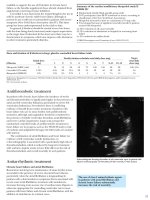
Echocardiographic findings include
hypertrophy of the left and right ventricles Consider amyloidosis
often with a “speckled” visual appearance in patients with left
within the thickened walls (Figure 7.7). ECG, ventricular hypertrophy by
however, shows a low QRS voltage. Systolic echocardiogram, but low
voltage by electrocardiogram.
function is usually preserved until late in the
disease, but not hyperdynamic as it may be
with hypertensive or hypertrophic cardiomyopathy. When a biopsy confirms the diagnosis, immunochemical analysis can reveal the type of
amyloid fibril and implied clinical features (Figures 7.8 and 7.9).
FIGURE 7.7 Echo
features of amyloidosis.
Echocardiogram in 4-chamber
apical view shows left
ventricular hypertrophy with
linear “speckling” of septum
(arrow), right ventricular
free wall hypertrophy, and
left atrial enlargement. Left
ventricular ejection fraction
as
.
Restrictive Cardiomyopathy Due to Amyloidosis • 147
FIGURE 7.8
Endomyocardial biopsy
in amyloidosis. Congo
Red stain showing
characteristic “apple
green” birefringence
under a polarizing
microscope that
often appears with
yellow components.
ascular an interstitial
depositions are also
common.
FIGURE 7.9 Electron
micrograph of
endomyocardial biopsy
in amyloidosis. This
specimen shows cottonlike fibrillar amyloid
material (arrows)
between myocytes from
a patient with familial
ATTR amyloid.
Diagnosis of AL amyloid is
supported by findings of associated immunoglobulin on serum
or urine protein electrophoresis
with immunofixation or noncardiac organ amyloid involvement.
It may be confirmed by endomyocardial biopsy showing interstitial
myocardial deposits of amyloid
protein. The poor prognosis of AL
amyloid is associated with a low
survival when awaiting transplant
(Figure 7.10).21 In AL amyloidosis,
following cardiac transplantation,
the amyloid deposits will recur in
the transplanted heart unless the
patient subsequently undergoes a
bone marrow transplant.
AMYLOIDOSIS CLASSIFICATION
AND CLINICAL FEATURES
Light chain (AL):
Plasma cell dyscrasia related to and occasionally associated with multiple myeloma. Heart disease occurs in one-third to
half of AL patients; heart failure tends to
progress rapidly and has a poor prognosis
Familial (ATTR):
Autosomal dominant transmission;
amyloid derived from a mixture of mutant
and wild-type transthyretin
Senile systemic amyloid:
Almost exclusively found in elderly men;
slowly progressive symptoms
Inflammatory (AA):
Heart disease rare and, if present, rarely
clinically signi cant
148 • Chapter 7: Assessment of Stage C Patients with HF-pEF
FIGURE 7.10 Kaplan-Meier survival curves for patients awaiting heart transplant.
Survival was lower for patients awaiting transplant with AL amyloidosis than for non-amyloid
patients on the waiting list (P < 0.001).21 Source: Adapted with permission from Gray Gilstrap
et al., J Heart Lung Transplant. 2 4 2 4
.
MRI with late gadolinium enhancement may provide an index for the
extent of amyloid protein in the myocardial interstitial space.22 With the
detection of such abnormalities, MRI can serve as a guide for treatment
in this condition.
Additional Causes of Restrictive Cardiomyopathy
Other causes of restrictive cardiomyopathy are numerous but uncommon
(Table 7.2). It may require direct measurement of an elevated pulmonary
capillary wedge pressure to make a diagnosis of restrictive cardiomyopathy. Unfortunately, specific treatment is unavailable for many restrictive
cardiomyopathies. Although wall thickness is usually increased, it may
also be normal (see Chapter 4). It is also important to exclude the potentially treatable diagnosis of constrictive pericarditis.
Other Important Causes of Heart Failure Syndrome • 149
TABLE 7.2 Classification of types of restrictive cardiomyopathy according to cause.
Symbol ++ = relatively common.23 Source: Reprinted with permission Kushwaha et al., N Engl
J Med.
7
4 2 7 27 . opyright
7 assachusetts e ical ociety. ll rights
reserved.
MYOCARDIAL
ENDOMYOCARDIAL
INFILTRATIVE
Endomyocardial fibrosis
Amyloidosis++
Sarcoidosis
Gaucher’s disease
Hurler’s disease
Fatty infiltration
NONINFILTRATIVE
Hypertrophic cardiomyopathy++
Idiopathic cardiomyopathy
Familial cardiomyopathy
Scleroderma
Pseudoxanthoma elasticum
Diabetic cardiomyopathy++
Idiopathic fibrosis
Hypereosinophilic syndrome
Carcinoid heart disease
Metastatic cancer
Radiation++
Toxic effects of adriamycin
Drugs causing fibrous endocarditis:
• serotonin
• methysergide
• ergotamine
• mercurial agents
• busulfan
STORAGE DISEASES
Fabry disease
Glycogen storage disease
Hemochromatosis++
Other Important Causes
of Heart Failure Syndrome
An echocardiogram can suggest three potentially treatable diagnoses
other than HF-rEF or HF-pEF: valvular heart disease, pericardial disease,
or cor pulmonale (Figure 7.11). These conditions require treatment distinct from the usual measures applied to left ventricular dysfunction. Any
of these conditions may be a sole diagnosis or a new precipitant for deterioration in a patient with previously compensated heart failure.
150 • Chapter 7: Assessment of Stage C Patients with HF-pEF
Are there causes of heart
failure other than left
ventricular dysfunction?
YES
Valvular Disease
Pericardial
Disease
Cor Pulmonale
FIGURE 7.11 Potentially treatable causes of heart failure other than left ventricular
dysfunction.
Valvular Heart Disease
Valvular heart disease represents an important treatable cause of heart
failure. Valvular heart disease may be acquired or congenital. In the adult,
the major types of valvular disease associated with heart failure are due
to mechanical deformities of the aortic or mitral valve. The adult with
congenital heart disease may also exhibit pressure or volume overload of
either ventricle. When valvular disease is responsible for clinical heart
failure, consider surgical or percutaneous correction.
Valvular Disease
+
New Heart Failure
Repair
or
Replacement
AORTIC STENOSIS
The presence of aortic stenosis may be subtle in patients with heart failure. The typical systolic ejection murmur may be difficult to auscultate
due to low cardiac output. In patients in cardiogenic shock, echocardiography may be the only way to identify aortic stenosis and even then aortic
valve gradients are more difficult to interpret due to low cardiac output.
Valvular Heart Disease • 151
Echocardiographic Findings
By echocardiogram, the aortic valve appears calcified and restricted in
motion. The peak pressure gradient across the valve can be estimated by
the Bernoulli equation as ∆P = 4 VAO2. The valve area can be assessed by the
continuity equation, as AreaAO = (VLVOT / VAO) × AreaLVOT (abbreviations
below).
Abbreviations for calculating aortic valve area
— peak velocity across valve by Doppler
left ventricular outflo tract
oppler velocity at
Area
cross sectional area of
by 2 echocar iography
A resting aortic peak velocity value of greater than 4.0 m/s, mean
pressure gradient greater than or equal to 30 mm Hg, or valve area less
than 1.0 cm2 usually indicates hemodynamically significant aortic stenosis. Aortic valve replacement should be considered for patients with
symptoms.24 Treadmill testing may unmask symptoms when the clinical
significance of aortic stenosis is uncertain, and hypotension (defined as a
fall in systolic blood pressure of ≥ 20 mm Hg from baseline) can imply a
poor prognosis without valve replacement.25
When echocardiographic findings are equivocal or when coronary
anatomy needs to be determined, cardiac catheterization can be used to
directly measure the pressure gradient across the valve and determine
cardiac output by thermodilution or Fick methods. A valve area can then
be calculated from these direct measurements. A simplified formula estimate of aortic valve area (cm2) is cardiac output (liters/minute) divided by
the square root of the transvalvular peak pressure gradient (mm Hg).26
A patient with heart failure and aortic stenosis may have a significant
stenosis with only a moderate pressure gradient. If aortic valve area is
reduced, valve replacement may still be beneficial despite a low ejection
fraction, especially if no other etiologies for heart failure are present.27
Graded dobutamine infusion during echocardiogram evaluation can be
used to help determine the significance of aortic stenosis versus myocardial dysfunction in low output states. Findings of an increase in valve
gradient during dobutamine infusion and persistent low valve area suggest an expected clinical improvement with valve replacement.
Transcatheter versus Surgical Aortic Valve Replacement
Some patients with aortic stenosis may not be suitable for surgical
valve replacement due to high risk secondary to advanced age, left ventricular dysfunction, or other coexisting conditions.28 An alternate, less
152 • Chapter 7: Assessment of Stage C Patients with HF-pEF
invasive, procedure for such patients is transcatheter aortic-valve replacement (TAVR), which functionally implants a stent-mounted bovine
pericardial valve delivered via catheter (Figure 7.12).28,29
FIGURE 7.12 Transcatheter aortic-valve replacement. Catheter placement of a balloon
expandable bovine pericardial valve.29 Source: Adapted with permission from Smith et al.,
N Engl J Med. 2
4 2 2 87 2 8.
In the PARTNER trial, patients deemed inoperable with severe aortic
stenosis were randomly assigned to undergo transfemoral TAVR versus
standard therapy, including balloon aortic valvuloplasty without valve
replacement (Figure 7.13).28 After a one-year follow up, the rate of death
from any cause in the TAVR group was 30.7%, compared to a 50.7% death
rate in patients who received standard therapy.
Valvular Heart Disease • 153
Death from Any Cause (%)
100
80
Medical therapy
60
40
TAVR
20
p < 0.001
0
0
6
12
18
24
Months
FIGURE 7.13 Mortality rates after TAVR procedure. In patients (n = 179) with comorbidities
that contrain icate surgical aortic valve replacement,
proce ure re uce mortality
compared to medical therapy for patients with severe aortic stenosis. (Hazard ratio 0.55)28
Source: Adapted with permission from Leon et al., N Engl J Med. 2
7
7 7.
In patients with severe aortic stenosis at high risk for surgery who
received either TAVR or surgical aortic valve replacement, all cause
1-year mortality was 24.2% in TAVR patients and 26.8% for those who
received surgery (P = NS). 29 Within 30 days, strokes were more common
with TAVR, however, at 1 year this difference was no longer statistically
significant.
AORTIC REGURGITATION
Left ventricular volume overload due to aortic regurgitation can be acute
or chronic. 30 Two important treatable causes of acute aortic regurgitation
with a nondilated left ventricle are bacterial endocarditis of the aortic
valve or aortic root dissection. Chronic aortic regurgitation with a dilated
left ventricle may be due to aortic root dilatation or a congenitally
deformed bicuspid aortic valve. Congenital conditions that affect connective tissue (e.g., Marfan’s, Loey-Dietz syndrome), rheumatic heart disease,
and rheumatoid arthritis are also associated with aortic regurgitation.
Chronic severe aortic regurgitation can lead to dramatic physical exam
findings including wide pulse pressure, visibly bounding pulse, double (or
bisferiens) pulse, and marked cardiomegaly. When heart failure is present, aortic valve surgery should be considered. Prognosis for aortic valve
replacement is related to the degree of left ventricle chamber enlargement. In the absence of symptoms, progressive or marked left ventricular
enlargement should also suggest a need for aortic valve replacement since
an end-systolic dimension greater than 5.5 cm by echocardiography is
associated with a poorer survival after surgery.30 Ascending aorta replacement may be required in patients with an excessively dilated ascending
154 • Chapter 7: Assessment of Stage C Patients with HF-pEF
aorta. TAVR is currently not an option for patients with aortic regurgitation without aortic stenosis.
MITRAL REGURGITATION
Acute severe mitral regurgitation typically presents as pulmonary edema
with a normal-sized left ventricle and hyperdynamic left ventricular systolic function. Important causes of this condition include destructive
bacterial mitral valve endocarditis, papillary muscle rupture associated
with myocardial infarction or blunt chest trauma, or chordae tendineae
rupture associated with redundant myxomatous mitral valve leaflets
(Figure 7.14).
A
B
FIGURE 7.14 Mitral regurgitation. Panel A: Transesophageal echocardiogram showing
a flail mitral leaflet from a ruptured chordae tendineae. Panel B: Color Doppler showing
associated mitral regurgitation.
When chronic mitral regurgitation leads to heart failure, it is usually
associated with eccentric (dilated) left ventricle chamber enlargement.
Valvular Heart Disease • 155
Ultimately myocardial dysfunction progresses due to excessive wall stress
(pressure × radius/wall thickness) (see Chapter 4). In some cases, it can be
a challenge to determine if mitral regurgitation has a primary valve cause
or is secondary to ventricular enlargement and mitral annular dilation.
Primary chronic mitral regurgitation can be the result of mitral valve
prolapse, mitral annular calcification, or rheumatic heart disease. When
mitral regurgitation is the cause for heart failure, either transthoracic or
transesophageal echocardiography may identify mitral valve deformities
suitable for mitral valve repair. If repair is not possible and the valve is
replaced, preservation of the posterior valve apparatus can blunt subsequent ventricular enlargement. Left ventricular ejection fraction may
initially decrease after correction of mitral regurgitation because of elimination of ventricular systolic ejection retrograde into the lower pressure
left atrium.
Percutaneous options to repair primary or secondary mitral regurgitation are emerging. One example is the catheter placement of a clip that
approximates the edges of the mitral leaflets. When compared to surgical
repair at 12 months, the percutaneous approach was less effective at
reducing mitral regurgitation, but associated with fewer adverse events
and similar clinical outcomes.31 Percutaneous mitral repair techniques
are likely to increase in the future.
MITRAL STENOSIS
Typically mitral stenosis develops decades after acute rheumatic fever. In
the elderly, it can also occur with calcification of the mitral annulus without previous rheumatic fever.32 Patients typically present with symptoms
of shortness of breath due to pulmonary congestion. Because the left
ventricle is “protected” by the narrowed mitral valve, catheter-based balloon valvuloplasty or surgical valve repair or replacement is usually
associated with an excellent recovery of circulatory function.
Right ventricular failure manifested by symptoms of fatigue and a
low cardiac output can predominate in patients with long-standing mitral
stenosis who develop severe pulmonary hypertension. The increase in
resistance to flow through the pulmonary arterial circulation is known as
the “second stenosis” in patients with mitral stenosis. 33 High pulmonary
vascular resistance associated with right ventricular failure can make
operative risk higher and impair functional recovery in these patients.
Once mitral stenosis is relieved, reversal of increased pulmonary vascular
resistance usually occurs over a period of days to weeks. 34
Percutaneous Mitral Valvuloplasty
Percutaneous mitral valvuloplasty using a balloon catheter to dilate
a stenotic mitral valve can be considered an alternative to open valve
repair or replacement in appropriate patients. On average, the mean valve
156 • Chapter 7: Assessment of Stage C Patients with HF-pEF
area doubles (from 1.0 to 2.0 cm2), with a 50% to 60% reduction in transmitral gradient. 35 Complications include cardiac perforation, pericardial
tamponade, severe mitral regurgitation, or cerebral vascular accident.
Patients who have severely calcified valves often require open heart surgery to achieve a good result. 36 In patients with mitral stenosis but pliable
valves, percutaneous mitral valvuloplasty may be preferable to surgical
commissurotomy since it achieves similar results without the liabilities of
thoracotomy and cardiopulmonary bypass. 37
TRANSESOPHAGEAL ECHOCARDIOGRAM FOR VALVE DISEASE
Performance of transesophageal echocardiogram (TEE) is not mandatory
in the diagnosis of valvular diseases and heart failure. Nevertheless, a
transesophageal echocardiogram can be useful when questions remain
after transthoracic echocardiography. A flail mitral valve leaflet, either
due to chordal rupture, papillary muscle tear, or endocarditis, often indicates a need for mitral valve surgery (Figure 7.14). TEE is also useful for
better definition of possible valvular vegetations associated with endocarditis. TEE improves the assessment of prosthetic tissue or mechanical
valves, especially in the mitral position, because echogenic struts limit
visualization with transthoracic echocardiography. Generally, the mitral
valve is better assessed than the aortic valve given the close anatomic
location of the left atrium to the esophagus.
Congenital Heart Disease
Diverse congenital heart lesions can occur in an adult: left-right shunts;
right-left shunts (cyanotic heart disease); stenosis or hypoplasia of heart
valves or ventricles; or great vessel abnormalities. If previously diagnosed
during childhood, the abnormality may have been observed, palliated, or
corrected. Echocardiography and transesophageal echocardiography can
initially define the anatomic and functional significance of a suspected
congenital lesion. Atrial or ventricular arrhythmias may occur associated
with any significant congenital heart lesion even years after successful
surgical correction. Collaboration with a cardiologist experienced in the
management of congenital heart lesions can help in the management of
these patients.38
Pericardial Disease • 157
Pericardial Disease
Pericardial tamponade, pericardial constriction, and mixed effuso-constrictive disease can all be associated with heart failure findings.
Rapid accumulation of fluid within the pericardium can cause acute
pericardial tamponade and circulatory collapse with as little as 100 mL of
fluid. Examples include postoperative bleeding after open-heart surgery,
catheter related coronary vessel or ventricular perforation, or ventricular
rupture post-myocardial infarction. Tamponade is initially assessed by
clinical findings and echocardiographic features (List 7.4).
Effuso-constrictive disease is a mixed diagnosis that is usually confirmed when a significant pericardial effusion is drained and a residual
elevation of ventricular filling pressures remains consistent with pericardial constriction. Neoplastic involvement of the pericardium commonly
results in this finding and may be initially associated with a liter or more
of effusion.
LIST 7.4 Features of Pericardial Disease
Types of pericardial disease
• Pericardial tamponade
• Pericardial constriction
• Effuso-constrictive disease
Clinical findings of tamponade
• Pulsus paradoxus (> 10 mm Hg fall of blood pressure with inspiration)
• Hypotension
• Neck vein distention
• Electrical alternans on ECG
• Increase in size of cardiac silhouette on chest x-ray
Basic echocardiographic findings of tamponade
• Large pericardial effusion (anterior and posterior to heart)
•
collapse ith inspiration
• Atrial collapse with inspiration
• Exaggerated changes in respiratory pattern of transvalvular velocities
CONSTRICTIVE VS. RESTRICTIVE CARDIOMYOPATHY
When a patient has heart failure and normal left ventricular size and
systolic function, distinguishing HF-pEF due to restrictive cardiomyopathy from constrictive pericarditis can be important (Figure 7.15), as
pericardial stripping can lead to marked clinical improvement in the case
of constrictive pericarditis.39 Echocardiography supplemented by CT or
158 • Chapter 7: Assessment of Stage C Patients with HF-pEF
cardiac MRI can help to make a diagnosis of constrictive pericarditis if a
thickened pericardium is identified.40
CP
RC
Thick or calcified pericardium
by CXR, Echo, CT, or MRI
+
-
Respiratory variation in Doppler
Exaggerated tricuspid inflow signal
+
-
LV and RV pressures track
throughout diastole
+
-
-
+
Abnormal myocardial
biopsy
FIGURE 7.15 Laboratory findings to help distinguish constrictive pericarditis (CP) from
restrictive cardiomyopathy (RC).
A previous history of acute pericarditis, granulomatous disease (e.g.,
tuberculosis, histoplasmosis), and autoimmune diseases (e.g., rheumatoid
arthritis), favors constrictive pericarditis. Conversely, a systemic illness,
such as amyloidosis or history of chest radiation therapy, favors restrictive
cardiomyopathy.
Physical exam and Doppler findings help distinguish the two conditions. A constricted pericardium is analogous to a rigid boot surrounding
the heart that isolates the heart from respiratory changes in intrathoracic
pressure and increases reciprocal changes in right and left ventricle inflow.
With constriction, during inspiration (as venous thoracic flow increases),
> 25% increases in tricuspid and decreases in mitral early diastolic transvalvular flows are seen by Doppler echocardiography.39 Conversely, only
small changes in inflow velocities with respiration are seen in restrictive
cardiomyopathy.41 In constrictive pericarditis, in contrast to restrictive
cardiomyopathy, a paradoxical increase in central venous pressure occurs
with respiration (positive Kussmaul’s sign). Left and right heart cardiac
catheterization demonstrates tracking of right and left ventricular diastolic pressure tracings prior to an “a” wave with constriction. Abnormal
histology by endomyocardial biopsy favors restriction.
Occasionally, pericardial constriction can coexist with restrictive
cardiomyopathy, following a previous episode of myopericarditis or
Cor Pulmonale • 159
radiation therapy for neoplasia.42 Final validation of constrictive pericarditis is based on finding hemodynamic improvement following pericardial
stripping. 39,43
Cor Pulmonale
Right heart failure (without left heart failure) due to a primary pulmonary
reason with pulmonary hypertension, known as cor pulmonale, may
occur for reasons such as acute or chronic pulmonary emboli, chronic
obstructive lung disease, or obesity-related hypoventilation syndromes,
including severe sleep apnea.44 The diagnosis of primary pulmonary
hypertension should be considered in the presence of a high pulmonary
vascular resistance of unknown cause after thorough evaluation to
exclude secondary causes of pulmonary hypertension, especially left
heart failure or pulmonary emboli. Trials have shown benefit from treatment with oral and intravenous pulmonary vasodilators for patients with
documented high pulmonary artery pressures and vascular resistance.45
Cor pulmonale can present with clinical findings from low cardiac
output (fatigue or hypotension) and elevated right heart filling pressure
(edema, ascites, and jugular venous distension). The echocardiogram in
cor pulmonale reveals a large right ventricle, a small left ventricle, and a
flattened interventricular septum. Together, this can give an appearance
of a “D” sign on a short axis cross-section of the left ventricle as shown
below (Figure 7.16).
FIGURE 7.16 Echocardiographic left ventricle “D” sign from right ventricular pressure
overload. his image came from a 4 year ol male ith history of surgically correcte
complex congenital heart disease with Eisenmenger’s physiology (high pulmonary vascular
resistance) and right heart failure. Pulmonary artery systolic pressure estimate was 85 mm Hg.