Ebook Harper’s illustrated biochemistry (26th edition): Part 12
Bạn đang xem bản rút gọn của tài liệu. Xem và tải ngay bản đầy đủ của tài liệu tại đây (8.56 MB, 408 trang )
ch33.qxd 3/16/04 10:59 AM Page 286
SECTION IV
Structure, Function, & Replication
of Informational Macromolecules
33
Nucleotides
Victor W. Rodwell, PhD
BIOMEDICAL IMPORTANCE
H
Nucleotides—the monomer units or building blocks of
nucleic acids—serve multiple additional functions. They
form a part of many coenzymes and serve as donors of
phosphoryl groups (eg, ATP or GTP), of sugars (eg,
UDP- or GDP-sugars), or of lipid (eg, CDP-acylglycerol). Regulatory nucleotides include the second messengers cAMP and cGMP, the control by ADP of oxidative phosphorylation, and allosteric regulation of
enzyme activity by ATP, AMP, and CTP. Synthetic
purine and pyrimidine analogs that contain halogens,
thiols, or additional nitrogen are employed for chemotherapy of cancer and AIDS and as suppressors of the
immune response during organ transplantation.
C
H
6
1
C
N
2
H
C
5
4
7
N
3
8
CH
N
C
4
3
N9
H
C
HC
2
5
CH
N
N
CH
6
1
Purine
Pyrimidine
Figure 33–1. Purine and pyrimidine. The atoms are
numbered according to the international system.
Nucleosides & Nucleotides
Nucleosides are derivatives of purines and pyrimidines
that have a sugar linked to a ring nitrogen. Numerals
with a prime (eg, 2′ or 3′) distinguish atoms of the
sugar from those of the heterocyclic base. The sugar in
ribonucleosides is D-ribose, and in deoxyribonucleosides it is 2-deoxy-D-ribose. The sugar is linked to the
heterocyclic base via a -N-glycosidic bond, almost always to N-1 of a pyrimidine or to N-9 of a purine (Figure 33–3).
PURINES, PYRIMIDINES, NUCLEOSIDES,
& NUCLEOTIDES
Purines and pyrimidines are nitrogen-containing heterocycles, cyclic compounds whose rings contain both
carbon and other elements (hetero atoms). Note that
the smaller pyrimidine has the longer name and the
larger purine the shorter name and that their six-atom
rings are numbered in opposite directions (Figure
33–1). The planar character of purines and pyrimidines
facilitates their close association, or “stacking,” which
stabilizes double-stranded DNA (Chapter 36). The oxo
and amino groups of purines and pyrimidines exhibit
keto-enol and amine-imine tautomerism (Figure 33–2),
but physiologic conditions strongly favor the amino
and oxo forms.
NH2
NH
O
OH
Figure 33–2. Tautomerism of the oxo and amino
functional groups of purines and pyrimidines.
286
ch33.qxd 3/16/04 10:59 AM Page 287
NUCLEOTIDES
NH2
NH2
N
N
9
HO
HO
O
OH
OH
OH
HN
9
1
N
H2 N
HO
O
OH
OH
N
O
OH
OH
Guanosine
Cytidine
1
O
N
N
HO
O
Adenosine
N
HN
O
N
N
287
O
O
N
/
OH
Uridine
Figure 33–3. Ribonucleosides, drawn as the syn conformers.
Mononucleotides are nucleosides with a phosphoryl
group esterified to a hydroxyl group of the sugar. The
3′- and 5′-nucleotides are nucleosides with a phosphoryl group on the 3′- or 5′-hydroxyl group of the sugar,
respectively. Since most nucleotides are 5′-, the prefix
“5′-” is usually omitted when naming them. UMP and
dAMP thus represent nucleotides with a phosphoryl
group on C-5 of the pentose. Additional phosphoryl
groups linked by acid anhydride bonds to the phosphoryl group of a mononucleotide form nucleoside
diphosphates and triphosphates (Figure 33–4).
Steric hindrance by the base restricts rotation about
the β-N-glycosidic bond of nucleosides and nuNH2
N
N
Adenine
N
N
cleotides. Both therefore exist as syn or anti conformers
(Figure 33–5). While both conformers occur in nature,
anti conformers predominate. Table 33–1 lists the
major purines and pyrimidines and their nucleoside
and nucleotide derivatives. Single-letter abbreviations
are used to identify adenine (A), guanine (G), cytosine
(C), thymine (T), and uracil (U), whether free or present in nucleosides or nucleotides. The prefix “d”
(deoxy) indicates that the sugar is 2′-deoxy-D-ribose
(eg, dGTP) (Figure 33–6).
Nucleic Acids Also Contain
Additional Bases
Small quantities of additional purines and pyrimidines
occur in DNA and RNAs. Examples include 5-methylcytosine of bacterial and human DNA, 5-hydroxymethylcytosine of bacterial and viral nucleic acids, and
mono- and di-N-methylated adenine and guanine of
CH2
O
O
HO
P
O
–
O
O
P
O–
O
O
P
–
NH2
O
NH2
Ribose
N
HO
N
N
N
N
N
OH
O
Adenosine 5′-monophosphate (AMP)
N
HO
HO
O
N
O
Adenosine 5′-diphosphate (ADP)
Anti
Syn
OH
Adenosine 5′-triphosphate (ATP)
Figure 33–4. ATP, its diphosphate, and its
monophosphate.
OH
OH
OH
Figure 33–5. The syn and anti conformers of adenosine differ with respect to orientation about the N-glycosidic bond.
ch33.qxd 3/16/04 10:59 AM Page 288
288
CHAPTER 33
/
Table 33–1. Bases, nucleosides, and nucleotides.
Base
Formula
Base
X=H
Nucleoside
X = Ribose or
Deoxyribose
Nucleotide, Where
X = Ribose Phosphate
NH2
N
N
N
N
Adenine Adenosine
A
A
Adenosine monophosphate
AMP
Guanine Guanosine
G
G
Guanosine monophosphate
GMP
Cytosine Cytidine
C
C
Cytidine monophosphate
CMP
Uracil
U
Uridine monophosphate
UMP
X
O
H
N
N
H2N
N
N
X
NH2
N
O
N
X
O
H
N
O
N
Uridine
U
X
O
H
O
CH3
N
Thymine Thymidine
T
T
N
Thymidine monophosphate
TMP
dX
NH2
NH2
N
N
O
O
N
N
N
O
O
O
O
O
–
OH
OH
AMP
O
O
O
O
N
O
–
OH
H
dAMP
Figure 33–6. AMP, dAMP, UMP, and TMP.
O
O
O
P
O–
CH3
HN
O
P
O–
O
HN
N
N
P
–
O
N
N
O
P
O–
–
OH
OH
UMP
O
O–
OH
H
TMP
ch33.qxd 3/16/04 11:00 AM Page 289
NUCLEOTIDES
NH2
CH3
N
O
NH2
5-Methylcytosine
O
CH3
O
N
N
N
H2 N
N
H
Dimethylaminoadenine
7
N
7-Methylguanine
mammalian messenger RNAs (Figure 33–7). These
atypical bases function in oligonucleotide recognition
and in regulating the half-lives of RNAs. Free nucleotides include hypoxanthine, xanthine, and uric acid
(see Figure 34–8), intermediates in the catabolism of
adenine and guanine. Methylated heterocyclic bases of
plants include the xanthine derivatives caffeine of coffee, theophylline of tea, and theobromine of cocoa (Figure 33–8).
Posttranslational modification of preformed polynucleotides can generate additional bases such as
pseudouridine, in which D-ribose is linked to C-5 of
uracil by a carbon-to-carbon bond rather than by a
β-N-glycosidic bond. The nucleotide pseudouridylic
acid Ψ arises by rearrangement of UMP of a preformed
tRNA. Similarly, methylation by S-adenosylmethionine
of a UMP of preformed tRNA forms TMP (thymidine
monophosphate), which contains ribose rather than deoxyribose.
O
O
CH3
N
N
N
CH2
O
–
O
P
O
O
OH
OH
Figure 33–9. cAMP, 3′,5′-cyclic AMP, and cGMP.
N
Figure 33–7. Four uncommon naturally occurring
pyrimidines and purines.
H3 C
O
O
O
CH3
N
HN
P
N
N
O
O
N
H2 N
CH2
5-Hydroxymethylcytosine
–
H3 C
N
HN
N
N
N
H
289
O
N
N
O
N
H
NH2
CH2OH
N
/
Nucleotides Serve Diverse
Physiologic Functions
Nucleotides participate in reactions that fulfill physiologic functions as diverse as protein synthesis, nucleic
acid synthesis, regulatory cascades, and signal transduction pathways.
Nucleoside Triphosphates Have High
Group Transfer Potential
Acid anhydrides, unlike phosphate esters, have high
group transfer potential. ∆0′ for the hydrolysis of each
of the terminal phosphates of nucleoside triphosphates
is about −7 kcal/mol (−30 kJ/mol). The high group
transfer potential of purine and pyrimidine nucleoside
triphosphates permits them to function as group transfer reagents. Cleavage of an acid anhydride bond typically is coupled with a highly endergonic process such
as covalent bond synthesis—eg, polymerization of nucleoside triphosphates to form a nucleic acid.
In addition to their roles as precursors of nucleic
acids, ATP, GTP, UTP, CTP, and their derivatives
each serve unique physiologic functions discussed in
other chapters. Selected examples include the role of
ATP as the principal biologic transducer of free energy;
the second messenger cAMP (Figure 33–9); adenosine
3′-phosphate-5′-phosphosulfate (Figure 33–10), the
sulfate donor for sulfated proteoglycans (Chapter 48)
and for sulfate conjugates of drugs; and the methyl
group donor S-adenosylmethionine (Figure 33–11).
N
CH3
P
Figure 33–8. Caffeine, a trimethylxanthine. The dimethylxanthines theobromine and theophylline are
similar but lack the methyl group at N-1 and at N-7, respectively.
Adenine
Ribose
P
O
SO32–
Figure 33–10. Adenosine 3′-phosphate-5′-phosphosulfate.
ch33.qxd 3/16/04 11:00 AM Page 290
290
/
CHAPTER 33
NH2
N
N
N
N
COO–
CH
NH3
CH2
Table 33–2. Many coenzymes and related
compounds are derivatives of adenosine
monophosphate.
NH2
CH3 CH2
CH2
S
+
Adenine
R
HO
O
P
OH
N
N
O
+
O
–
O
Methionine
N
N
O
Adenosine
CH2
n
O
OR'
R'' O
Figure 33–11. S-Adenosylmethionine.
D-Ribose
Coenzyme
R
R
R؆
GTP serves as an allosteric regulator and as an energy
source for protein synthesis, and cGMP (Figure 33–9)
serves as a second messenger in response to nitric oxide
(NO) during relaxation of smooth muscle (Chapter
48). UDP-sugar derivatives participate in sugar epimerizations and in biosynthesis of glycogen, glucosyl disaccharides, and the oligosaccharides of glycoproteins and
proteoglycans (Chapters 47 and 48). UDP-glucuronic
acid forms the urinary glucuronide conjugates of bilirubin (Chapter 32) and of drugs such as aspirin. CTP
participates in biosynthesis of phosphoglycerides,
sphingomyelin, and other substituted sphingosines
(Chapter 24). Finally, many coenzymes incorporate nucleotides as well as structures similar to purine and
pyrimidine nucleotides (see Table 33–2).
Active methionine
Amino acid adenylates
Active sulfate
3′,5′-Cyclic AMP
NAD*
NADP*
FAD
CoASH
Nucleotides Are Polyfunctional Acids
SYNTHETIC NUCLEOTIDE ANALOGS
ARE USED IN CHEMOTHERAPY
Nucleosides or free purine or pyrimidine bases are uncharged at physiologic pH. By contrast, the primary
phosphoryl groups (pK about 1.0) and secondary phosphoryl groups (pK about 6.2) of nucleotides ensure that
they bear a negative charge at physiologic pH. Nucleotides can, however, act as proton donors or acceptors at pH values two or more units above or below
neutrality.
Nucleotides Absorb Ultraviolet Light
The conjugated double bonds of purine and pyrimidine
derivatives absorb ultraviolet light. The mutagenic effect of ultraviolet light results from its absorption by
nucleotides in DNA with accompanying chemical
changes. While spectra are pH-dependent, at pH 7.0 all
the common nucleotides absorb light at a wavelength
close to 260 nm. The concentration of nucleotides and
Methionine*
Amino acid
SO32−
†
†
†
†
H
H
H
H
H PO32−
H PO32−
H
H
PO32− H
H
H
H PO32−
n
0
1
1
1
2
2
2
2
*Replaces phosphoryl group.
†
R is a B vitamin derivative.
nucleic acids thus often is expressed in terms of “absorbance at 260 nm.”
Synthetic analogs of purines, pyrimidines, nucleosides,
and nucleotides altered in either the heterocyclic ring or
the sugar moiety have numerous applications in clinical
medicine. Their toxic effects reflect either inhibition of
enzymes essential for nucleic acid synthesis or their incorporation into nucleic acids with resulting disruption
of base-pairing. Oncologists employ 5-fluoro- or 5iodouracil, 3-deoxyuridine, 6-thioguanine and 6-mercaptopurine, 5- or 6-azauridine, 5- or 6-azacytidine,
and 8-azaguanine (Figure 33–12), which are incorporated into DNA prior to cell division. The purine analog allopurinol, used in treatment of hyperuricemia and
gout, inhibits purine biosynthesis and xanthine oxidase
activity. Cytarabine is used in chemotherapy of cancer.
Finally, azathioprine, which is catabolized to 6-mercaptopurine, is employed during organ transplantation to
suppress immunologic rejection.
ch33.qxd 3/16/04 11:00 AM Page 291
NUCLEOTIDES
I
HN
5
6
O
O
N
HO
O
O
F
HN
HO
H
5-Iodo-2′-deoxyuridine
6
8
N
H2N
N
H
HO
5-Fluorouracil
SH
N
6
N
H
6-Mercaptopurine
H2N
N
N
8-Azaguanine
OH
N
N1
6
5
2
N
N
H
OH
6-Azauridine
SH
N
N
HN
5
O
2′
N
N
HO
O
O
N
291
O
O
HN
/
N
H
N
H
N
6-Thioguanine
N
4
3
Alloburinol
Figure 33–12. Selected synthetic pyrimidine and purine analogs.
Nonhydrolyzable Nucleoside
Triphosphate Analogs Serve as
Research Tools
(absent from DNA) functions as a nucleophile during
hydrolysis of the 3′,5′-phosphodiester bond.
Synthetic nonhydrolyzable analogs of nucleoside
triphosphates (Figure 33–13) allow investigators to distinguish the effects of nucleotides due to phosphoryl
transfer from effects mediated by occupancy of allosteric nucleotide-binding sites on regulated enzymes.
Polynucleotides Are Directional
Macromolecules
POLYNUCLEOTIDES
The 5′-phosphoryl group of a mononucleotide can esterify a second OH group, forming a phosphodiester. Most commonly, this second OH group is the
3′-OH of the pentose of a second nucleotide. This
forms a dinucleotide in which the pentose moieties are
linked by a 3′ → 5′ phosphodiester bond to form the
“backbone” of RNA and DNA.
While formation of a dinucleotide may be represented as the elimination of water between two
monomers, the reaction in fact strongly favors phosphodiester hydrolysis. Phosphodiesterases rapidly catalyze the hydrolysis of phosphodiester bonds whose
spontaneous hydrolysis is an extremely slow process.
Consequently, DNA persists for considerable periods
and has been detected even in fossils. RNAs are far less
stable than DNA since the 2′-hydroxyl group of RNA
Phosphodiester bonds link the 3′- and 5′-carbons of adjacent monomers. Each end of a nucleotide polymer
thus is distinct. We therefore refer to the “5′- end” or
the “3′- end” of polynucleotides, the 5′- end being the
one with a free or phosphorylated 5′-hydroxyl.
Polynucleotides Have Primary Structure
The base sequence or primary structure of a polynucleotide can be represented as shown below. The phosphodiester bond is represented by P or p, bases by a single letter, and pentoses by a vertical line.
A
P
T
P
C
P
A
P
OH
ch33.qxd 3/16/04 11:00 AM Page 292
292
/
CHAPTER 33
O
Pu/Py
R
O
P
O
O
O–
P
SUMMARY
O
O
O–
P
O–
O–
Parent nucleoside triphosphate
O
Pu/Py
R
O
O
P
O
O–
O
P
CH2
O–
P
O–
O–
β,γ-Methylene derivative
O
Pu/Py
R
O
P
O
O
O
–
O
O
H
N
P
–
O–
P
O
–
β,γ-Imino derivative
Figure 33–13. Synthetic derivatives of nucleoside
triphosphates incapable of undergoing hydrolytic release of the terminal phosphoryl group. (Pu/Py, a
purine or pyrimidine base; R, ribose or deoxyribose.)
Shown are the parent (hydrolyzable) nucleoside
triphosphate (top) and the unhydrolyzable β-methylene (center) and γ-imino derivatives (bottom).
Where all the phosphodiester bonds are 5′ → 3′, a
more compact notation is possible:
pGpGpApTpCpA
This representation indicates that the 5′-hydroxyl—
but not the 3′-hydroxyl—is phosphorylated.
The most compact representation shows only the
base sequence with the 5′- end on the left and the 3′end on the right. The phosphoryl groups are assumed
but not shown:
GGATCA
• Under physiologic conditions, the amino and oxo
tautomers of purines, pyrimidines, and their derivatives predominate.
• Nucleic acids contain, in addition to A, G, C, T, and
U, traces of 5-methylcytosine, 5-hydroxymethylcytosine, pseudouridine (Ψ), or N-methylated bases.
• Most nucleosides contain D-ribose or 2-deoxy-Dribose linked to N-1 of a pyrimidine or to N-9 of a
purine by a β-glycosidic bond whose syn conformers
predominate.
• A primed numeral locates the position of the phosphate on the sugars of mononucleotides (eg, 3′GMP, 5′-dCMP). Additional phosphoryl groups
linked to the first by acid anhydride bonds form nucleoside diphosphates and triphosphates.
• Nucleoside triphosphates have high group transfer
potential and participate in covalent bond syntheses.
The cyclic phosphodiesters cAMP and cGMP function as intracellular second messengers.
• Mononucleotides linked by 3′ → 5′-phosphodiester
bonds form polynucleotides, directional macromolecules with distinct 3′- and 5′- ends. For pTpGpTp or
TGCATCA, the 5′- end is at the left, and all phosphodiester bonds are 3′ → 5′.
• Synthetic analogs of purine and pyrimidine bases and
their derivatives serve as anticancer drugs either by
inhibiting an enzyme of nucleotide biosynthesis or
by being incorporated into DNA or RNA.
REFERENCES
Adams RLP, Knowler JT, Leader DP: The Biochemistry of the Nucleic Acids, 11th ed. Chapman & Hall, 1992.
Blackburn GM, Gait MJ: Nucleic Acids in Chemistry & Biology. IRL
Press, 1990.
Bugg CE, Carson WM, Montgomery JA: Drugs by design. Sci Am
1992;269(6):92.
ch34.qxd 2/13/2003 4:04 PM Page 293
Metabolism of Purine &
Pyrimidine Nucleotides
34
Victor W. Rodwell, PhD
BIOMEDICAL IMPORTANCE
(synthesis de novo), (2) phosphoribosylation of purines,
and (3) phosphorylation of purine nucleosides.
The biosynthesis of purines and pyrimidines is stringently regulated and coordinated by feedback mechanisms that ensure their production in quantities and at
times appropriate to varying physiologic demand. Genetic diseases of purine metabolism include gout,
Lesch-Nyhan syndrome, adenosine deaminase deficiency, and purine nucleoside phosphorylase deficiency.
By contrast, apart from the orotic acidurias, there are
few clinically significant disorders of pyrimidine catabolism.
INOSINE MONOPHOSPHATE (IMP)
IS SYNTHESIZED FROM AMPHIBOLIC
INTERMEDIATES
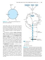
Figure 34–2 illustrates the intermediates and reactions
for conversion of α-D-ribose 5-phosphate to inosine
monophosphate (IMP). Separate branches then lead to
AMP and GMP (Figure 34–3). Subsequent phosphoryl
transfer from ATP converts AMP and GMP to ADP
and GDP. Conversion of GDP to GTP involves a second phosphoryl transfer from ATP, whereas conversion
of ADP to ATP is achieved primarily by oxidative
phosphorylation (see Chapter 12).
PURINES & PYRIMIDINES ARE
DIETARILY NONESSENTIAL
Human tissues can synthesize purines and pyrimidines
from amphibolic intermediates. Ingested nucleic acids
and nucleotides, which therefore are dietarily nonessential, are degraded in the intestinal tract to mononucleotides, which may be absorbed or converted to
purine and pyrimidine bases. The purine bases are then
oxidized to uric acid, which may be absorbed and excreted in the urine. While little or no dietary purine or
pyrimidine is incorporated into tissue nucleic acids, injected compounds are incorporated. The incorporation
of injected [3H]thymidine into newly synthesized DNA
thus is used to measure the rate of DNA synthesis.
Multifunctional Catalysts Participate in
Purine Nucleotide Biosynthesis
In prokaryotes, each reaction of Figure 34–2 is catalyzed by a different polypeptide. By contrast, in eukaryotes, the enzymes are polypeptides with multiple
catalytic activities whose adjacent catalytic sites facilitate channeling of intermediates between sites. Three
distinct multifunctional enzymes catalyze reactions 3,
4, and 6, reactions 7 and 8, and reactions 10 and 11 of
Figure 34–2.
BIOSYNTHESIS OF PURINE NUCLEOTIDES
Purine and pyrimidine nucleotides are synthesized in
vivo at rates consistent with physiologic need. Intracellular mechanisms sense and regulate the pool sizes of
nucleotide triphosphates (NTPs), which rise during
growth or tissue regeneration when cells are rapidly dividing. Early investigations of nucleotide biosynthesis
employed birds, and later ones used Escherichia coli.
Isotopic precursors fed to pigeons established the source
of each atom of a purine base (Figure 34–1) and initiated study of the intermediates of purine biosynthesis.
Three processes contribute to purine nucleotide
biosynthesis. These are, in order of decreasing importance: (1) synthesis from amphibolic intermediates
Antifolate Drugs or Glutamine Analogs
Block Purine Nucleotide Biosynthesis
The carbons added in reactions 4 and 5 of Figure 34–2
are contributed by derivatives of tetrahydrofolate.
Purine deficiency states, which are rare in humans, generally reflect a deficiency of folic acid. Compounds that
inhibit formation of tetrahydrofolates and therefore
block purine synthesis have been used in cancer
chemotherapy. Inhibitory compounds and the reactions
they inhibit include azaserine (reaction 5, Figure 34–2),
diazanorleucine (reaction 2), 6-mercaptopurine (reactions 13 and 14), and mycophenolic acid (reaction 14).
293
ch34.qxd 2/13/2003 4:04 PM Page 294
294
CHAPTER 34
/
Respiratory CO 2
and therefore utilize exogenous purines to form nucleotides.
Glycine
Aspartate
C
6
N1
5
C
7
8
C
10
N -Formyltetrahydrofolate
2
4
3
N
C
AMP & GMP Feedback-Regulate PRPP
Glutamyl Amidotransferase
N
C
9
N
H
N 5,N10 -Methenyltetrahydrofolate
Amide nitrogen of glutamine
Figure 34–1. Sources of the nitrogen and carbon
atoms of the purine ring. Atoms 4, 5, and 7 (shaded) derive from glycine.
“SALVAGE REACTIONS” CONVERT
PURINES & THEIR NUCLEOSIDES TO
MONONUCLEOTIDES
Conversion of purines, their ribonucleosides, and their
deoxyribonucleosides to mononucleotides involves socalled “salvage reactions” that require far less energy
than de novo synthesis. The more important mechanism involves phosphoribosylation by PRPP (structure
II, Figure 34–2) of a free purine (Pu) to form a purine
5′-mononucleotide (Pu-RP).
Pu + PR − PP → PRP + PPi
Two phosphoribosyl transferases then convert adenine
to AMP and hypoxanthine and guanine to IMP or
GMP (Figure 34–4). A second salvage mechanism involves phosphoryl transfer from ATP to a purine ribonucleoside (PuR):
PuR + ATP → PuR − P + ADP
Adenosine kinase catalyzes phosphorylation of adenosine and deoxyadenosine to AMP and dAMP, and deoxycytidine kinase phosphorylates deoxycytidine and
2′-deoxyguanosine to dCMP and dGMP.
Liver, the major site of purine nucleotide biosynthesis, provides purines and purine nucleosides for salvage
and utilization by tissues incapable of their biosynthesis. For example, human brain has a low level of PRPP
amidotransferase (reaction 2, Figure 34–2) and hence
depends in part on exogenous purines. Erythrocytes
and polymorphonuclear leukocytes cannot synthesize
5-phosphoribosylamine (structure III, Figure 34–2)
Since biosynthesis of IMP consumes glycine, glutamine, tetrahydrofolate derivatives, aspartate, and ATP,
it is advantageous to regulate purine biosynthesis. The
major determinant of the rate of de novo purine nucleotide biosynthesis is the concentration of PRPP,
whose pool size depends on its rates of synthesis, utilization, and degradation. The rate of PRPP synthesis
depends on the availability of ribose 5-phosphate and
on the activity of PRPP synthase, an enzyme sensitive
to feedback inhibition by AMP, ADP, GMP, and
GDP.
AMP & GMP Feedback-Regulate
Their Formation From IMP
Two mechanisms regulate conversion of IMP to GMP
and AMP. AMP and GMP feedback-inhibit adenylosuccinate synthase and IMP dehydrogenase (reactions
12 and 14, Figure 34–3), respectively. Furthermore,
conversion of IMP to adenylosuccinate en route to
AMP requires GTP, and conversion of xanthinylate
(XMP) to GMP requires ATP. This cross-regulation
between the pathways of IMP metabolism thus serves
to decrease synthesis of one purine nucleotide when
there is a deficiency of the other nucleotide. AMP and
GMP also inhibit hypoxanthine-guanine phosphoribosyltransferase, which converts hypoxanthine and guanine to IMP and GMP (Figure 34–4), and GMP feedback-inhibits PRPP glutamyl amidotransferase (reaction
2, Figure 34–2).
REDUCTION OF RIBONUCLEOSIDE
DIPHOSPHATES FORMS
DEOXYRIBONUCLEOSIDE
DIPHOSPHATES
Reduction of the 2′-hydroxyl of purine and pyrimidine
ribonucleotides, catalyzed by the ribonucleotide reductase complex (Figure 34–5), forms deoxyribonucleoside diphosphates (dNDPs). The enzyme complex
is active only when cells are actively synthesizing DNA.
Reduction requires thioredoxin, thioredoxin reductase,
and NADPH. The immediate reductant, reduced
thioredoxin, is produced by NADPH:thioredoxin reductase (Figure 34–5). Reduction of ribonucleoside
diphosphates (NDPs) to deoxyribonucleoside diphosphates (dNDPs) is subject to complex regulatory controls that achieve balanced production of deoxyribonucleotides for synthesis of DNA (Figure 34–6).
P
O
5
H
O
CH 2
H
H
H
OH
OH OH
ATP
1
P
O
AMP
Mg 2+
– OOC
5
H
O
2
1
3
H
4
CH 2
H
H
O
4
C
C
OH OH
O
C
6
3
5
PRPP
(II)
N1
H
H2 N
P
N
N
O
CH
R-5- P
O
P
Glutamine
H 2O
2
N
P
O
Glutamate
PPi
+
CH
H
O
CH 2
H
H
O
9
O–
P
+
3
NH 3
7
Glycine
C4
H2C 5
+
NH 3
H
O
Mg 2+
ATP ADP + Pi
CO2
O
C
N
O
H
O
H
C4
H2C 5
H
H2C
C
C
N
N
7
N 5,N10MethenylH 4 folate H 4 folate
4
ATP, Mg 2 +
H2O Ring closure
6
VII SYNTHETASE
C4
H2C5
O
H
N
O
7 8 CH
9
NH
R-5- P
9
Gln
ATP
Mg 2 +
Glu
O
7 8 CH
N
H
Formylglycinamide
ribosyl-5-phosphate
(V)
5
C4
R-5- P
NH
Formylglycinamidine
ribosyl-5-phosphate
(VI)
HN
3
H2C5
VI SYNTHETASE
FORMYLTRANSFERASE
NH 3+
NH
H
R-5- P
CH
R-5- P
N
N
Aminoimidazole
ribosyl-5-phosphate
(VII)
C
HC
OH OH
N
7
VII CARBOXYLASE
HN
CH
OH OH
5
N
CH
Glycinamide
ribosyl-5-phosphate
(IV)
O
C
6
4
R-5- P
11
HC
H2N
5-Phospho-β-D-ribosylamine
(III)
–O
3
H2 N
Ring closure
H 2O
Aminoimidazole
carboxylate ribosyl-5-phosphate
(VIII)
NH3
PRPP GLUTAMYL
AMIDOTRANSFERASE
– OOC
HC
CH2
– OOC
Aspartate
H 2O
8
C
IX SYNTHETASE
O
C
N
IMP CYCLOHYDROLASE
Inosine monophosphate (IMP)
(XII)
ch34.qxd 2/13/2003 4:04 PM Page 295
HC
H 2C
– OOC
N 10 -FormylH 4 folate
H 4 folate
10
FORMYLTRANSFERASE
R-5- P
Formimidoimidazole carboxamide
ribosyl-5-phosphate
(XI)
C
C
H N
H
H2N
Aminoimidazole succinyl
carboxamide ribosyl-5-phosphate
(IX)
CH
R-5- P
N
N
PRPP
SYNTHASE
C
C
α-D-Ribose 5-phosphate
(I)
C
O
COO –
CH
HC
– OOC
9
Fumarate
ADENYLOSUCCINASE
H2 N
H2N
Aminoimidazole carboxamide
ribosyl-5-phosphate
(X)
2–
–
Figure 34–2. Purine biosynthesis from ribose 5-phosphate and ATP. See text for explanations. (᭺
P , PO3 or PO2 .)
ch34.qxd 2/13/2003 4:04 PM Page 296
296
/
CHAPTER 34
–
OOC
O
N
–
H
–
OOC
C C COO
H2
+
H 2O
NH 3
12
HN
N
GTP, Mg 2
N
R-5- P
Inosine monophosphate
(IMP)
NAD
+
–
OOC
H
C
H
C
13
COO
–
NH 2
N
N
+
ADENYLOSUCCINATE
SYNTHASE
N
N
ADENYLOSUCCINASE
R-5- P
Adenylosuccinate
(AMPS)
N
N
R-5- P
Adenosine monophosphate
(AMP)
H 2O
14
NADH
+ H+
IMP DEHYDROGENASE
O
Glutamine
N
HN
O
H
–
C C COO
H2
NH
N
N
N
H
N
O
Glutamate
N
15
ATP
HN
H2N
N
N
TRANSAMIDINASE
R-5- P
Xanthosine monophosphate
(XMP)
R-5- P
Guanosine monophosphate
(GMP)
Figure 34–3. Conversion of IMP to AMP and GMP.
BIOSYNTHESIS OF PYRIMIDINE
NUCLEOTIDES
THE DEOXYRIBONUCLEOSIDES OF
URACIL & CYTOSINE ARE SALVAGED
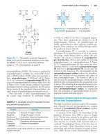
Figure 34–7 summarizes the roles of the intermediates
and enzymes of pyrimidine nucleotide biosynthesis.
The catalyst for the initial reaction is cytosolic carbamoyl
phosphate synthase II, a different enzyme from the mitochondrial carbamoyl phosphate synthase I of urea synthesis (Figure 29–9). Compartmentation thus provides
two independent pools of carbamoyl phosphate. PRPP,
an early participant in purine nucleotide synthesis (Figure 34–2), is a much later participant in pyrimidine
biosynthesis.
While mammalian cells reutilize few free pyrimidines,
“salvage reactions” convert the ribonucleosides uridine
and cytidine and the deoxyribonucleosides thymidine
and deoxycytidine to their respective nucleotides. ATPdependent phosphoryltransferases (kinases) catalyze the
phosphorylation of the nucleoside diphosphates 2′-deoxycytidine, 2′-deoxyguanosine, and 2′-deoxyadenosine
to their corresponding nucleoside triphosphates. In addition, orotate phosphoribosyltransferase (reaction 5,
Figure 34–7), an enzyme of pyrimidine nucleotide synthesis, salvages orotic acid by converting it to orotidine
monophosphate (OMP).
Multifunctional Proteins
Catalyze the Early Reactions
of Pyrimidine Biosynthesis
Five of the first six enzyme activities of pyrimidine
biosynthesis reside on multifunctional polypeptides.
One such polypeptide catalyzes the first three reactions
of Figure 34–2 and ensures efficient channeling of carbamoyl phosphate to pyrimidine biosynthesis. A second
bifunctional enzyme catalyzes reactions 5 and 6.
Methotrexate Blocks Reduction
of Dihydrofolate
Reaction 12 of Figure 34–7 is the only reaction of pyrimidine nucleotide biosynthesis that requires a tetrahydrofolate derivative. The methylene group of N 5,N 10-methylene-tetrahydrofolate is reduced to the methyl group that
is transferred, and tetrahydrofolate is oxidized to dihydro-
ch34.qxd 2/13/2003 4:04 PM Page 297
METABOLISM OF PURINE & PYRIMIDINE NUCLEOTIDES
NH 2
PRPP
NH 2
PP i
N
N
N
H
N
P
Adenine
N
N
H2C
O
Ribonucleoside
diphosphate
O
ADENINE
PHOSPHORIBOSYL
TRANSFERASE
H
2′-Deoxyribonucleoside
diphosphate
Reduced
thioredoxin
H
H
297
RIBONUCLEOTIDE
REDUCTASE
N
N
/
Oxidized
thioredoxin
THIOREDOXIN
REDUCTASE
H
OH OH
NADP+
AMP
O
PRPP
N
HN
O
PP i
N
HN
N
H
Hypoxanthine
N
N
N
P
O
H2C
O
H
H2N
O
N
N
Guanine
H
OH OH
IMP
O
HN
H
H
HYPOXANTHINE-GUANINE
PHOSPHORIBOSYLTRANSFERASE
N
HN
N
H
H2N
PRPP
N
N
NADPH + H+
Figure 34–5. Reduction of ribonucleoside diphosphates to 2′-deoxyribonucleoside diphosphates.
a nucleotide in which the ribosyl phosphate is attached
to N-1 of the pyrimidine ring. The anticancer drug
5-fluorouracil (Figure 33–12) is also phosphoribosylated by orotate phosphoribosyl transferase.
REGULATION OF PYRIMIDINE
NUCLEOTIDE BIOSYNTHESIS
Gene Expression & Enzyme Activity
Both Are Regulated
The activities of the first and second enzymes of pyrimidine nucleotide biosynthesis are controlled by allosteric
PP i
P
O
H2C
H
2′dCDP
CDP
O
–
H
H
–
+
–
H
ATP
OH OH
GMP
Figure 34–4. Phosphoribosylation of adenine, hypoxanthine, and guanine to form AMP, IMP, and GMP,
respectively.
folate. For further pyrimidine synthesis to occur, dihydrofolate must be reduced back to tetrahydrofolate, a reaction catalyzed by dihydrofolate reductase. Dividing cells,
which must generate TMP and dihydrofolate, thus are especially sensitive to inhibitors of dihydrofolate reductase
such as the anticancer drug methotrexate.
+
Orotate phosphoribosyltransferase (reaction 5, Figure
34–7) converts the drug allopurinol (Figure 33–12) to
2′dUDP
UDP
–
–
2′dTTP
–
+
GDP
2′dGDP
2′dGTP
2′dADP
2′dATP
–
+
ADP
Certain Pyrimidine Analogs Are
Substrates for Enzymes of Pyrimidine
Nucleotide Biosynthesis
2′dCTP
Figure 34–6. Regulation of the reduction of purine
and pyrimidine ribonucleotides to their respective
2′-deoxyribonucleotides. Solid lines represent chemical
– ) or positive (᭺
+)
flow. Broken lines show negative (᭺
feedback regulation.
ch34.qxd 2/13/2003 4:04 PM Page 298
298
CHAPTER 34
/
CO 2 + Glutamine + ATP
CARBAMOYL
PHOSPHATE
SYNTHASE II
1
O
–O C 4
+H N
3 3
O
C
O
5 CH 2
+
2
6
1
P + H3 N
Carbamoyl
phosphate
(CAP)
C
O
–O C
4
H2 N 3
ASPARTATE
TRANSCARBAMOYLASE
H
COO
–
2
O
DIHYDROOROTASE
5 CH 2
6
CH
3
1
–
N
COO
H
Carbamoyl
aspartic acid
(CAA)
C
2
C
O
O
Pi
Aspartic
acid
CH 2
HN
C
H2O
N
H
Dihydroorotic
acid (DHOA)
NAD +
DIHYDROOROTATE
DEHYDROGENASE
+
4
NADH + H
O
HN 3
O
CO 2
4
6
5
PP i
O
N
OROTIDYLIC ACID
DECARBOXYLASE
R-5- P
O
COO –
N
R-5- P
UMP
OROTATE
PHOSPHORIBOSYLTRANSFERASE
OMP
ATP
7
NADPH + H+
ADP
NADP+
10
dUDP (deoxyuridine diphosphate)
H2O
UDP
ATP
8
RIBONUCLEOTIDE
REDUCTASE
11
ADP
Pi
dUMP
UTP
ATP
N 5,N10 -Methylene H4 folate
Glutamine
THYMIDYLATE
SYNTHASE
CTP
SYNTHASE
9
H2 folate
NH 2
N
O
12
O
CH 3
HN
N
R-5- P - P - P
CTP
O
N
dR-5- P
TMP
Figure 34–7. The biosynthetic pathway for pyrimidine nucleotides.
O
PRPP
5
HN
2 1 6
CH
COO –
HN
O
N
H
COO –
Orotic acid
(OA)
ch34.qxd 2/13/2003 4:04 PM Page 299
METABOLISM OF PURINE & PYRIMIDINE NUCLEOTIDES
regulation. Carbamoyl phosphate synthase II (reaction
1, Figure 34–7) is inhibited by UTP and purine nucleotides but activated by PRPP. Aspartate transcarbamoylase (reaction 2, Figure 34–7) is inhibited by
CTP but activated by ATP. In addition, the first three
and the last two enzymes of the pathway are regulated
by coordinate repression and derepression.
N
N
N
N
HO H 2C
O
H
HUMANS CATABOLIZE PURINES
TO URIC ACID
Humans convert adenosine and guanosine to uric acid
(Figure 34–8). Adenosine is first converted to inosine
by adenosine deaminase. In mammals other than
higher primates, uricase converts uric acid to the watersoluble product allantoin. However, since humans lack
uricase, the end product of purine catabolism in humans is uric acid.
H
H
H
OH OH
Adenosine
H2O
NH 4+
O
O
N
HN
H2N
N
N
HO H 2C
N
HN
N
N
HO H 2C
O
H
H
O
H
H
H
OH OH
Inosine
H
H
H
OH OH
Guanosine
Pi
Pi
Ribose 1-phosphate
O
O
GOUT IS A METABOLIC DISORDER
OF PURINE CATABOLISM
Various genetic defects in PRPP synthetase (reaction 1,
Figure 34–2) present clinically as gout. Each defect—
eg, an elevated Vmax, increased affinity for ribose 5phosphate, or resistance to feedback inhibition—results
in overproduction and overexcretion of purine catabolites. When serum urate levels exceed the solubility
limit, sodium urate crystalizes in soft tissues and joints
and causes an inflammatory reaction, gouty arthritis.
However, most cases of gout reflect abnormalities in
renal handling of uric acid.
N
HN
N
HN
H 2N
NH
N
Hypoxanthine
NH
N
Guanine
H2O + O2
HN3
O
H2O2
N
HN
O
NH
NH
Xanthine
H2O + O2
H2O2
O
Figure 34–8. Formation of uric acid from purine nucleosides
by way of the purine bases hypoxanthine, xanthine, and guanine. Purine deoxyribonucleosides are degraded by the same
catabolic pathway and enzymes, all of which exist in the mucosa
of the mammalian gastrointestinal tract.
299
NH2
Purine & Pyrimidine Nucleotide
Biosynthesis Are Coordinately Regulated
Purine and pyrimidine biosynthesis parallel one another mole for mole, suggesting coordinated control of
their biosynthesis. Several sites of cross-regulation characterize purine and pyrimidine nucleotide biosynthesis.
The PRPP synthase reaction (reaction 1, Figure 34–2),
which forms a precursor essential for both processes, is
feedback-inhibited by both purine and pyrimidine nucleotides.
/
HN1
O
HN
7
9
3
NH
NH
Uric acid
O
ch34.qxd 2/13/2003 4:04 PM Page 300
300
/
CHAPTER 34
OTHER DISORDERS OF
PURINE CATABOLISM
While purine deficiency states are rare in human subjects, there are numerous genetic disorders of purine catabolism. Hyperuricemias may be differentiated based
on whether patients excrete normal or excessive quantities of total urates. Some hyperuricemias reflect specific
enzyme defects. Others are secondary to diseases such
as cancer or psoriasis that enhance tissue turnover.
Lesch-Nyhan Syndrome
Lesch-Nyhan syndrome, an overproduction hyperuricemia characterized by frequent episodes of uric acid
lithiasis and a bizarre syndrome of self-mutilation, reflects a defect in hypoxanthine-guanine phosphoribosyl transferase, an enzyme of purine salvage (Figure
34–4). The accompanying rise in intracellular PRPP results in purine overproduction. Mutations that decrease
or abolish hypoxanthine-guanine phosphoribosyltransferase activity include deletions, frameshift mutations,
base substitutions, and aberrant mRNA splicing.
Von Gierke’s Disease
Purine overproduction and hyperuricemia in von
Gierke’s disease (glucose-6-phosphatase deficiency)
occurs secondary to enhanced generation of the PRPP
precursor ribose 5-phosphate. An associated lactic acidosis elevates the renal threshold for urate, elevating
total body urates.
Hypouricemia
Hypouricemia and increased excretion of hypoxanthine
and xanthine are associated with xanthine oxidase deficiency due to a genetic defect or to severe liver damage. Patients with a severe enzyme deficiency may exhibit xanthinuria and xanthine lithiasis.
Adenosine Deaminase & Purine
Nucleoside Phosphorylase Deficiency
Adenosine deaminase deficiency is associated with an
immunodeficiency disease in which both thymusderived lymphocytes (T cells) and bone marrow-derived lymphocytes (B cells) are sparse and dysfunctional. Purine nucleoside phosphorylase deficiency is
associated with a severe deficiency of T cells but apparently normal B cell function. Immune dysfunctions appear to result from accumulation of dGTP and dATP,
which inhibit ribonucleotide reductase and thereby deplete cells of DNA precursors.
CATABOLISM OF PYRIMIDINES
PRODUCES WATER-SOLUBLE
METABOLITES
Unlike the end products of purine catabolism, those
of pyrimidine catabolism are highly water-soluble:
CO2, NH3, β-alanine, and β-aminoisobutyrate (Figure
34–9). Excretion of β-aminoisobutyrate increases in
leukemia and severe x-ray radiation exposure due to increased destruction of DNA. However, many persons
of Chinese or Japanese ancestry routinely excrete
β-aminoisobutyrate. Humans probably transaminate
β-aminoisobutyrate to methylmalonate semialdehyde,
which then forms succinyl-CoA (Figure 19–2).
Pseudouridine Is Excreted Unchanged
Since no human enzyme catalyzes hydrolysis or phosphorolysis of pseudouridine, this unusual nucleoside is
excreted unchanged in the urine of normal subjects.
OVERPRODUCTION OF PYRIMIDINE
CATABOLITES IS ONLY RARELY
ASSOCIATED WITH CLINICALLY
SIGNIFICANT ABNORMALITIES
Since the end products of pyrimidine catabolism are
highly water-soluble, pyrimidine overproduction results
in few clinical signs or symptoms. In hyperuricemia associated with severe overproduction of PRPP, there is
overproduction of pyrimidine nucleotides and increased excretion of β-alanine. Since N 5,N 10-methylene-tetrahydrofolate is required for thymidylate synthesis, disorders of folate and vitamin B12 metabolism
result in deficiencies of TMP.
Orotic Acidurias
The orotic aciduria that accompanies Reye’s syndrome
probably is a consequence of the inability of severely
damaged mitochondria to utilize carbamoyl phosphate,
which then becomes available for cytosolic overproduction of orotic acid. Type I orotic aciduria reflects a deficiency of both orotate phosphoribosyltransferase and
orotidylate decarboxylase (reactions 5 and 6, Figure
34–7); the rarer type II orotic aciduria is due to a deficiency only of orotidylate decarboxylase (reaction 6,
Figure 34–7).
Deficiency of a Urea Cycle Enzyme Results
in Excretion of Pyrimidine Precursors
Increased excretion of orotic acid, uracil, and uridine
accompanies a deficiency in liver mitochondrial ornithine transcarbamoylase (reaction 2, Figure 29–9).
ch34.qxd 2/13/2003 4:04 PM Page 301
METABOLISM OF PURINE & PYRIMIDINE NUCLEOTIDES
N
O
N
H
Cytosine
Drugs May Precipitate Orotic Aciduria
1/2 O
2
NH 3
O
O
HN
CH3
HN
O
N
H
Uracil
NADPH + H
NADP
N
H
Thymine
+
O
O
H
H
H
H
CH3
H
H
H
HN
O
N
H
Dihydrothymine
N
H
Dihydrouracil
H2O
H2N
H2O
COO −
CH2
C
CH2
N
H
β-Ureidopropionate
(N -carbamoyl-β -alanine)
O
COO −
CH3
H2N
C
H
C
CH2
N
O
H
β-Ureidoisobutyrate
(N -carbamoyl-β -aminoisobutyrate)
CO2 + NH3
H3N +
CH2
CH2
β-Alanine
COO−
H 3N +
Allopurinol (Figure 33–12), an alternative substrate for
orotate phosphoribosyltransferase (reaction 5, Figure
34–7), competes with orotic acid. The resulting nucleotide product also inhibits orotidylate decarboxylase
(reaction 6, Figure 34–7), resulting in orotic aciduria
and orotidinuria. 6-Azauridine, following conversion
to 6-azauridylate, also competitively inhibits orotidylate
decarboxylase (reaction 6, Figure 34–7), enhancing excretion of orotic acid and orotidine.
SUMMARY
+
O
HN
301
Excess carbamoyl phosphate exits to the cytosol, where
it stimulates pyrimidine nucleotide biosynthesis. The
resulting mild orotic aciduria is increased by highnitrogen foods.
NH2
O
/
CH2
CH
COO−
CH3
β -Aminoisobutyrate
Figure 34–9. Catabolism of pyrimidines.
• Ingested nucleic acids are degraded to purines and
pyrimidines. New purines and pyrimidines are
formed from amphibolic intermediates and thus are
dietarily nonessential.
• Several reactions of IMP biosynthesis require folate
derivatives and glutamine. Consequently, antifolate
drugs and glutamine analogs inhibit purine biosynthesis.
• Oxidation and amination of IMP forms AMP and
GMP, and subsequent phosphoryl transfer from
ATP forms ADP and GDP. Further phosphoryl
transfer from ATP to GDP forms GTP. ADP is converted to ATP by oxidative phosphorylation. Reduction of NDPs forms dNDPs.
• Hepatic purine nucleotide biosynthesis is stringently
regulated by the pool size of PRPP and by feedback
inhibition of PRPP-glutamyl amidotransferase by
AMP and GMP.
• Coordinated regulation of purine and pyrimidine
nucleotide biosynthesis ensures their presence in proportions appropriate for nucleic acid biosynthesis
and other metabolic needs.
• Humans catabolize purines to uric acid (pKa 5.8),
present as the relatively insoluble acid at acidic pH or
as its more soluble sodium urate salt at a pH near
neutrality. Urate crystals are diagnostic of gout.
Other disorders of purine catabolism include LeschNyhan syndrome, von Gierke’s disease, and hypouricemias.
• Since pyrimidine catabolites are water-soluble, their
overproduction does not result in clinical abnormalities. Excretion of pyrimidine precursors can, however, result from a deficiency of ornithine transcarbamoylase because excess carbamoyl phosphate is
available for pyrimidine biosynthesis.
ch34.qxd 2/13/2003 4:04 PM Page 302
302
/
CHAPTER 34
REFERENCES
Benkovic SJ: The transformylase enzymes in de novo purine
biosynthesis. Trends Biochem Sci 1994;9:320.
Brooks EM et al: Molecular description of three macro-deletions
and an Alu-Alu recombination-mediated duplication in the
HPRT gene in four patients with Lesch-Nyhan disease.
Mutat Res 2001;476:43.
Curto R, Voit EO, Cascante M: Analysis of abnormalities in purine
metabolism leading to gout and to neurological dysfunctions
in man. Biochem J 1998;329:477.
Harris MD, Siegel LB, Alloway JA: Gout and hyperuricemia. Am
Family Physician 1999;59:925.
Lipkowitz MS et al: Functional reconstitution, membrane targeting, genomic structure, and chromosomal localization of a
human urate transporter. J Clin Invest 2001;107:1103.
Martinez J et al: Human genetic disorders, a phylogenetic perspective. J Mol Biol 2001;308:587.
Puig JG et al: Gout: new questions for an ancient disease. Adv Exp
Med Biol 1998;431:1.
Scriver CR et al (editors): The Metabolic and Molecular Bases of Inherited Disease, 8th ed. McGraw-Hill, 2001.
Tvrdik T et al: Molecular characterization of two deletion events
involving Alu-sequences, one novel base substitution and two
tentative hotspot mutations in the hypoxanthine phosphoribosyltransferase gene in five patients with Lesch-Nyhansyndrome. Hum Genet 1998;103:311.
Zalkin H, Dixon JE: De novo purine nucleotide synthesis. Prog
Nucleic Acid Res Mol Biol 1992;42:259.
ch35.qxd 2/13/2003 4:12 PM Page 303
Nucleic Acid Structure & Function
35
Daryl K. Granner, MD
The informational content of DNA (the genetic code)
resides in the sequence in which these monomers—
purine and pyrimidine deoxyribonucleotides—are ordered. The polymer as depicted possesses a polarity;
one end has a 5′-hydroxyl or phosphate terminal while
the other has a 3′-phosphate or hydroxyl terminal. The
importance of this polarity will become evident. Since
the genetic information resides in the order of the
monomeric units within the polymers, there must exist
a mechanism of reproducing or replicating this specific
information with a high degree of fidelity. That requirement, together with x-ray diffraction data from
the DNA molecule and the observation of Chargaff
that in DNA molecules the concentration of deoxyadenosine (A) nucleotides equals that of thymidine
(T) nucleotides (A = T), while the concentration of deoxyguanosine (G) nucleotides equals that of deoxycytidine (C) nucleotides (G = C), led Watson, Crick, and
Wilkins to propose in the early 1950s a model of a double-stranded DNA molecule. The model they proposed
is depicted in Figure 35–2. The two strands of this
double-stranded helix are held in register by hydrogen
bonds between the purine and pyrimidine bases of the
respective linear molecules. The pairings between the
purine and pyrimidine nucleotides on the opposite
strands are very specific and are dependent upon hydrogen bonding of A with T and G with C (Figure 35–3).
This common form of DNA is said to be righthanded because as one looks down the double helix the
base residues form a spiral in a clockwise direction. In
the double-stranded molecule, restrictions imposed by
the rotation about the phosphodiester bond, the favored anti configuration of the glycosidic bond (Figure
33–8), and the predominant tautomers (see Figure
33–3) of the four bases (A, G, T, and C) allow A to pair
only with T and G only with C, as depicted in Figure
35–3. This base-pairing restriction explains the earlier
observation that in a double-stranded DNA molecule
the content of A equals that of T and the content of G
equals that of C. The two strands of the double-helical
molecule, each of which possesses a polarity, are antiparallel; ie, one strand runs in the 5′ to 3′ direction
and the other in the 3′ to 5′ direction. This is analogous
to two parallel streets, each running one way but carrying traffic in opposite directions. In the doublestranded DNA molecules, the genetic information re-
BIOMEDICAL IMPORTANCE
The discovery that genetic information is coded along
the length of a polymeric molecule composed of only
four types of monomeric units was one of the major scientific achievements of the twentieth century. This
polymeric molecule, DNA, is the chemical basis of
heredity and is organized into genes, the fundamental
units of genetic information. The basic information
pathway—ie, DNA directs the synthesis of RNA,
which in turn directs protein synthesis—has been elucidated. Genes do not function autonomously; their
replication and function are controlled by various gene
products, often in collaboration with components of
various signal transduction pathways. Knowledge of the
structure and function of nucleic acids is essential in
understanding genetics and many aspects of pathophysiology as well as the genetic basis of disease.
DNA CONTAINS THE
GENETIC INFORMATION
The demonstration that DNA contained the genetic information was first made in 1944 in a series of experiments by Avery, MacLeod, and McCarty. They showed
that the genetic determination of the character (type) of
the capsule of a specific pneumococcus could be transmitted to another of a different capsular type by introducing purified DNA from the former coccus into the
latter. These authors referred to the agent (later shown
to be DNA) accomplishing the change as “transforming
factor.” Subsequently, this type of genetic manipulation
has become commonplace. Similar experiments have
recently been performed utilizing yeast, cultured mammalian cells, and insect and mammalian embryos as recipients and cloned DNA as the donor of genetic information.
DNA Contains Four Deoxynucleotides
The chemical nature of the monomeric deoxynucleotide units of DNA—deoxyadenylate, deoxyguanylate,
deoxycytidylate, and thymidylate—is described in
Chapter 33. These monomeric units of DNA are held
in polymeric form by 3′,5′-phosphodiester bridges constituting a single strand, as depicted in Figure 35–1.
303
ch35.qxd 2/13/2003 4:12 PM Page 304
304
CHAPTER 35
/
O
N
NH
G
5′
CH2
O
N
N
NH2
NH2
N
O
C
P
H
H
H
CH2
H
N
O
O
H3C
O
NH
O
H
T
P
H
H
H
O
N
CH2
H
O
NH2
N
O
N
O
H
A
P
H
H
H
CH2
H
O
O
N
N
O
H
P
H
H
H
H
O
3′
H
P
O
Figure 35–1. A segment of one strand of a DNA molecule in which the purine and pyrimidine bases guanine
(G), cytosine (C), thymine (T), and adenine (A) are held together by a phosphodiester backbone between 2′-deoxyribosyl moieties attached to the nucleobases by an N-glycosidic bond. Note that the backbone has a polarity
(ie, a direction). Convention dictates that a single-stranded DNA sequence is written in the 5′ to 3′ direction (ie,
pGpCpTpA, where G, C, T, and A represent the four bases and p represents the interconnecting phosphates).
sides in the sequence of nucleotides on one strand, the
template strand. This is the strand of DNA that is
copied during nucleic acid synthesis. It is sometimes referred to as the noncoding strand. The opposite strand
is considered the coding strand because it matches the
RNA transcript that encodes the protein.
The two strands, in which opposing bases are held
together by hydrogen bonds, wind around a central axis
in the form of a double helix. Double-stranded DNA
exists in at least six forms (A–E and Z). The B form is
usually found under physiologic conditions (low salt,
high degree of hydration). A single turn of B-DNA
about the axis of the molecule contains ten base pairs.
The distance spanned by one turn of B-DNA is 3.4
nm. The width (helical diameter) of the double helix in
B-DNA is 2 nm.
As depicted in Figure 35–3, three hydrogen bonds
hold the deoxyguanosine nucleotide to the deoxycyti-
dine nucleotide, whereas the other pair, the A–T pair, is
held together by two hydrogen bonds. Thus, the G–C
bonds are much more resistant to denaturation, or
“melting,” than A–T-rich regions.
The Denaturation (Melting) of DNA
Is Used to Analyze Its Structure
The double-stranded structure of DNA can be separated into two component strands (melted) in solution
by increasing the temperature or decreasing the salt
concentration. Not only do the two stacks of bases pull
apart but the bases themselves unstack while still connected in the polymer by the phosphodiester backbone.
Concomitant with this denaturation of the DNA molecule is an increase in the optical absorbance of the
purine and pyrimidine bases—a phenomenon referred
to as hyperchromicity of denaturation. Because of the
ch35.qxd 2/13/2003 4:12 PM Page 305
NUCLEIC ACID STRUCTURE & FUNCTION
/
305
CH3
O
H
N
H
N
N
H
N
N
O
Thymidine
Minor groove
N
S
P
S
P
A
T
S
T
S
C
Adenosine
o
P
A
34 A
S
P
S
G
P
H
P
S
G
C
N
S
N
H
Major groove
N
O
N
H
Cytosine H
N
N
O
N
N
N
H Guanosine
o
20 A
Figure 35–2. A diagrammatic representation of the
Watson and Crick model of the double-helical structure
of the B form of DNA. The horizontal arrow indicates
the width of the double helix (20 Å), and the vertical
arrow indicates the distance spanned by one complete
turn of the double helix (34 Å). One turn of B-DNA includes ten base pairs (bp), so the rise is 3.4 Å per bp.
The central axis of the double helix is indicated by the
vertical rod. The short arrows designate the polarity of
the antiparallel strands. The major and minor grooves
are depicted. (A, adenine; C, cytosine; G, guanine;
T, thymine; P, phosphate; S, sugar [deoxyribose].)
stacking of the bases and the hydrogen bonding between the stacks, the double-stranded DNA molecule
exhibits properties of a rigid rod and in solution is a viscous material that loses its viscosity upon denaturation.
The strands of a given molecule of DNA separate
over a temperature range. The midpoint is called the
melting temperature, or Tm. The Tm is influenced by
the base composition of the DNA and by the salt concentration of the solution. DNA rich in G–C pairs,
which have three hydrogen bonds, melts at a higher temperature than that rich in A–T pairs, which have two hydrogen bonds. A tenfold increase of monovalent cation
concentration increases the Tm by 16.6 °C. Formamide,
which is commonly used in recombinant DNA experiments, destabilizes hydrogen bonding between bases,
thereby lowering the Tm. This allows the strands of DNA
Figure 35–3. Base pairing between deoxyadenosine
and thymidine involves the formation of two hydrogen
bonds. Three such bonds form between deoxycytidine
and deoxyguanosine. The broken lines represent hydrogen bonds.
or DNA-RNA hybrids to be separated at much lower
temperatures and minimizes the phosphodiester bond
breakage that occurs at high temperatures.
Renaturation of DNA Requires
Base Pair Matching
Separated strands of DNA will renature or reassociate
when appropriate physiologic temperature and salt conditions are achieved. The rate of reassociation depends
upon the concentration of the complementary strands.
Reassociation of the two complementary DNA strands
of a chromosome after DNA replication is a physiologic
example of renaturation (see below). At a given temperature and salt concentration, a particular nucleic acid
strand will associate tightly only with a complementary
strand. Hybrid molecules will also form under appropriate conditions. For example, DNA will form a hybrid with a complementary DNA (cDNA) or with a
cognate messenger RNA (mRNA; see below). When
combined with gel electrophoresis techniques that separate hybrid molecules by size and radioactive labeling to
provide a detectable signal, the resulting analytic techniques are called Southern (DNA/cDNA) and Northern blotting (DNA/RNA), respectively. These proce-
ch35.qxd 2/13/2003 4:12 PM Page 306
306
/
CHAPTER 35
dures allow for very specific identification of hybrids
from mixtures of DNA or RNA (see Chapter 40).
There Are Grooves in the DNA Molecule
Careful examination of the model depicted in Figure
35–2 reveals a major groove and a minor groove winding along the molecule parallel to the phosphodiester
backbones. In these grooves, proteins can interact specifically with exposed atoms of the nucleotides (usually by
H bonding) and thereby recognize and bind to specific
nucleotide sequences without disrupting the base pairing of the double-helical DNA molecule. As discussed in
Chapters 37 and 39, regulatory proteins control the expression of specific genes via such interactions.
DNA Exists in Relaxed
& Supercoiled Forms
In some organisms such as bacteria, bacteriophages, and
many DNA-containing animal viruses, the ends of the
DNA molecules are joined to create a closed circle with
no covalently free ends. This of course does not destroy
the polarity of the molecules, but it eliminates all free 3′
and 5′ hydroxyl and phosphoryl groups. Closed circles
exist in relaxed or supercoiled forms. Supercoils are introduced when a closed circle is twisted around its own axis
or when a linear piece of duplex DNA, whose ends are
fixed, is twisted. This energy-requiring process puts the
molecule under stress, and the greater the number of supercoils, the greater the stress or torsion (test this by
twisting a rubber band). Negative supercoils are formed
when the molecule is twisted in the direction opposite
from the clockwise turns of the right-handed double
helix found in B-DNA. Such DNA is said to be underwound. The energy required to achieve this state is, in a
sense, stored in the supercoils. The transition to another
form that requires energy is thereby facilitated by the underwinding. One such transition is strand separation,
which is a prerequisite for DNA replication and transcription. Supercoiled DNA is therefore a preferred form
in biologic systems. Enzymes that catalyze topologic
changes of DNA are called topoisomerases. Topoisomerases can relax or insert supercoils. The best-characterized example is bacterial gyrase, which induces negative
supercoiling in DNA using ATP as energy source. Homologs of this enzyme exist in all organisms and are important targets for cancer chemotherapy.
DNA PROVIDES A TEMPLATE FOR
REPLICATION & TRANSCRIPTION
The genetic information stored in the nucleotide sequence of DNA serves two purposes. It is the source of
information for the synthesis of all protein molecules of
the cell and organism, and it provides the information
inherited by daughter cells or offspring. Both of these
functions require that the DNA molecule serve as a
template—in the first case for the transcription of the
information into RNA and in the second case for the
replication of the information into daughter DNA molecules.
The complementarity of the Watson and Crick double-stranded model of DNA strongly suggests that
replication of the DNA molecule occurs in a semiconservative manner. Thus, when each strand of the double-stranded parental DNA molecule separates from its
complement during replication, each serves as a template on which a new complementary strand is synthesized (Figure 35–4). The two newly formed doublestranded daughter DNA molecules, each containing
one strand (but complementary rather than identical)
from the parent double-stranded DNA molecule, are
then sorted between the two daughter cells (Figure
35–5). Each daughter cell contains DNA molecules
with information identical to that which the parent
possessed; yet in each daughter cell the DNA molecule
of the parent cell has been only semiconserved.
THE CHEMICAL NATURE OF RNA DIFFERS
FROM THAT OF DNA
Ribonucleic acid (RNA) is a polymer of purine and
pyrimidine ribonucleotides linked together by 3′,5′phosphodiester bridges analogous to those in DNA
(Figure 35–6). Although sharing many features with
DNA, RNA possesses several specific differences:
(1) In RNA, the sugar moiety to which the phosphates and purine and pyrimidine bases are attached is
ribose rather than the 2′-deoxyribose of DNA.
(2) The pyrimidine components of RNA differ from
those of DNA. Although RNA contains the ribonucleotides of adenine, guanine, and cytosine, it does not
possess thymine except in the rare case mentioned
below. Instead of thymine, RNA contains the ribonucleotide of uracil.
(3) RNA exists as a single strand, whereas DNA exists as a double-stranded helical molecule. However,
given the proper complementary base sequence with
opposite polarity, the single strand of RNA—as
demonstrated in Figure 35–7—is capable of folding
back on itself like a hairpin and thus acquiring doublestranded characteristics.
(4) Since the RNA molecule is a single strand complementary to only one of the two strands of a gene, its
guanine content does not necessarily equal its cytosine
content, nor does its adenine content necessarily equal
its uracil content.
5475ch35.qxd_ccI 2/27/03 3:33 PM Page 307
NUCLEIC ACID STRUCTURE & FUNCTION
G
OLD
5′
G
307
C
OLD
3′
C
G
/
C
C
Original
parent molecule
G
A
T
A
A
T
G
C
G
C
A
A
First-generation
daughter molecules
T
T
A
G
C
G
C
3′
T
5′
A
T
G
C
C
C
C
T
A
A
G
A
T
T
Second-generation
daughter molecules
T
A
A
A
G
T
T
A
C
G
C
G
A
3′
OLD
T
T
A
T
A
A
G
5′
NEW
3′
NEW
T
T
T
A
A
5′
Figure 35–5. DNA replication is semiconservative.
During a round of replication, each of the two strands
of DNA is used as a template for synthesis of a new,
complementary strand.
OLD
Figure 35–4. The double-stranded structure of DNA
and the template function of each old strand (dark
shading) on which a new (light shading) complementary strand is synthesized.
(5) RNA can be hydrolyzed by alkali to 2′,3′ cyclic
diesters of the mononucleotides, compounds that cannot be formed from alkali-treated DNA because of the
absence of a 2′-hydroxyl group. The alkali lability of
RNA is useful both diagnostically and analytically.
Information within the single strand of RNA is contained in its sequence (“primary structure”) of purine
and pyrimidine nucleotides within the polymer. The
sequence is complementary to the template strand of
the gene from which it was transcribed. Because of this
complementarity, an RNA molecule can bind specifically via the base-pairing rules to its template DNA
strand; it will not bind (“hybridize”) with the other
(coding) strand of its gene. The sequence of the RNA
molecule (except for U replacing T) is the same as that
of the coding strand of the gene (Figure 35–8).
Nearly All of the Several Species of RNA
Are Involved in Some Aspect of Protein
Synthesis
Those cytoplasmic RNA molecules that serve as templates for protein synthesis (ie, that transfer genetic information from DNA to the protein-synthesizing machinery) are designated messenger RNAs, or mRNAs.
Many other cytoplasmic RNA molecules (ribosomal
RNAs; rRNAs) have structural roles wherein they con-
ch35.qxd 2/13/2003 4:12 PM Page 308
308
CHAPTER 35
/
O
N
NH
G
5′
CH2
O
N
NH2
NH2
N
N
O
C
P
H
H
H
CH2
H
O
N
O
O
NH
O
HO
U
P
H
H
H
O
N
CH2
H
O
NH2
N
O
N
O
HO
A
P
H
H
H
CH2
H
O
O
N
N
O
HO
P
H
H
H
H
O
3′
HO
P
O
Figure 35–6. A segment of a ribonucleic acid (RNA) molecule in which the purine and pyrimidine bases—
guanine (G), cytosine (C), uracil (U), and adenine (A)—are held together by phosphodiester bonds between ribosyl moieties attached to the nucleobases by N-glycosidic bonds. Note that the polymer has a polarity as indicated by the labeled 3′- and 5′-attached phosphates.
tribute to the formation and function of ribosomes (the
organellar machinery for protein synthesis) or serve as
adapter molecules (transfer RNAs; tRNAs) for the
translation of RNA information into specific sequences
of polymerized amino acids.
Some RNA molecules have intrinsic catalytic activity. The activity of these ribozymes often involves the
cleavage of a nucleic acid. An example is the role of
RNA in catalyzing the processing of the primary transcript of a gene into mature messenger RNA.
Much of the RNA synthesized from DNA templates
in eukaryotic cells, including mammalian cells, is degraded within the nucleus, and it never serves as either a
structural or an informational entity within the cellular
cytoplasm.
In all eukaryotic cells there are small nuclear RNA
(snRNA) species that are not directly involved in protein synthesis but play pivotal roles in RNA processing.
These relatively small molecules vary in size from 90 to
about 300 nucleotides (Table 35–1).
The genetic material for some animal and plant
viruses is RNA rather than DNA. Although some RNA
viruses never have their information transcribed into a
DNA molecule, many animal RNA viruses—specifically, the retroviruses (the HIV virus, for example)—are
transcribed by an RNA-dependent DNA polymerase,
the so-called reverse transcriptase, to produce a double-stranded DNA copy of their RNA genome. In
many cases, the resulting double-stranded DNA transcript is integrated into the host genome and subsequently serves as a template for gene expression and
from which new viral RNA genomes can be transcribed.
RNA Is Organized in Several
Unique Structures
In all prokaryotic and eukaryotic organisms, three main
classes of RNA molecules exist: messenger RNA
(mRNA), transfer RNA (tRNA), and ribosomal RNA
ch35.qxd 2/13/2003 4:12 PM Page 309
NUCLEIC ACID STRUCTURE & FUNCTION
5′
G
G
C
U
U
U
G
G
C
C
A
A
C
A
G
C
309
Table 35–1. Some of the species of small stable
RNAs found in mammalian cells.
Loop
C
C
G
A
A
A
U
U
C
G
U
U
U
U
C
G
/
Length
Molecules
Name (nucleotides) per Cell
U1
U2
U3
U4
U5
U6
4.5S
7S
7-2
7-3
Stem
3′
Figure 35–7. Diagrammatic representation of the
secondary structure of a single-stranded RNA molecule
in which a stem loop, or “hairpin,” has been formed and
is dependent upon the intramolecular base pairing.
Note that A forms hydrogen bonds with U in RNA.
(rRNA). Each differs from the others by size, function,
and general stability.
A. MESSENGER RNA (MRNA)
This class is the most heterogeneous in size and stability. All members of the class function as messengers
conveying the information in a gene to the proteinsynthesizing machinery, where each serves as a template
on which a specific sequence of amino acids is polymerized to form a specific protein molecule, the ultimate
gene product (Figure 35–9).
165
188
216
139
118
106
91–95
280
290
300
1 × 10
5 × 10⁵
3 × 10⁵
1 × 10⁵
2 × 10⁵
3 × 10⁵
3 x 10⁵
5 × 10⁵
1 × 10⁵
2 × 10⁵
6
Localization
Nucleoplasm/hnRNA
Nucleoplasm
Nucleolus
Nucleoplasm
Nucleoplasm
Perichromatin granules
Nucleus and cytoplasm
Nucleus and cytoplasm
Nucleus and cytoplasm
Nucleus
Messenger RNAs, particularly in eukaryotes, have
some unique chemical characteristics. The 5′ terminal
of mRNA is “capped” by a 7-methylguanosine triphosphate that is linked to an adjacent 2′-O-methyl ribonucleoside at its 5′-hydroxyl through the three phosphates
(Figure 35–10). The mRNA molecules frequently contain internal 6-methyladenylates and other 2′-O-ribose
methylated nucleotides. The cap is involved in the
recognition of mRNA by the translating machinery,
and it probably helps stabilize the mRNA by preventing
the attack of 5′-exonucleases. The protein-synthesizing
machinery begins translating the mRNA into proteins
beginning downstream of the 5′ or capped terminal.
The other end of most mRNA molecules, the 3′-hydroxyl terminal, has an attached polymer of adenylate
residues 20–250 nucleotides in length. The specific
function of the poly(A) “tail” at the 3′-hydroxyl terminal of mRNAs is not fully understood, but it seems that
it maintains the intracellular stability of the specific
mRNA by preventing the attack of 3′-exonucleases.
Some mRNAs, including those for some histones, do
not contain poly(A). The poly(A) tail, because it will
form a base pair with oligodeoxythymidine polymers
attached to a solid substrate like cellulose, can be used
to separate mRNA from other species of RNA, including mRNA molecules that lack this tail.
DNA strands:
Coding
Template
RNA
transcript
5′ —T G G A A T T G T G A G C G G A T A A C A A T T T C A C A C A G G A A A C A G C T A T G A C C A T G — 3′
3′ —A C C T T A A C A C T C G C C T A T T G T T A A A G T G T G T C C T T T G T C G A T A C T G G T A C — 5′
5′
p A U UGUG A GCGG A U A A C A A U U U C A C A C A GG A A A C A GC U A UG A C C A UG
3′
Figure 35–8. The relationship between the sequences of an RNA transcript and its gene, in which the coding and template strands are shown with their polarities. The RNA transcript with a 5′ to 3′ polarity is complementary to the template strand with its 3′ to 5′ polarity. Note that the sequence in the RNA transcript and its
polarity is the same as that in the coding strand, except that the U of the transcript replaces the T of the gene.
ch35.qxd 2/13/2003 4:12 PM Page 310
310
/
CHAPTER 35
DNA
5′
3′
3′
5′
mRNA
5′
3′
Protein synthesis on mRNA template
5′
3′
Ribosome
In mammalian cells, including cells of humans, the
mRNA molecules present in the cytoplasm are not the
RNA products immediately synthesized from the DNA
template but must be formed by processing from a precursor molecule before entering the cytoplasm. Thus,
in mammalian nuclei, the immediate products of gene
transcription constitute a fourth class of RNA molecules. These nuclear RNA molecules are very heterogeneous in size and are quite large. The heterogeneous
nuclear RNA (hnRNA) molecules may have a molecular weight in excess of 107, whereas the molecular
weight of mRNA molecules is generally less than 2 ×
106. As discussed in Chapter 37, hnRNA molecules are
processed to generate the mRNA molecules which then
enter the cytoplasm to serve as templates for protein
synthesis.
B. TRANSFER RNA (TRNA)
tRNA molecules vary in length from 74 to 95 nucleotides. They also are generated by nuclear processing
of a precursor molecule (Chapter 37). The tRNA molecules serve as adapters for the translation of the information in the sequence of nucleotides of the mRNA
into specific amino acids. There are at least 20 species
of tRNA molecules in every cell, at least one (and often
several) corresponding to each of the 20 amino acids required for protein synthesis. Although each specific
tRNA differs from the others in its sequence of nucleotides, the tRNA molecules as a class have many features in common. The primary structure—ie, the nucleotide sequence—of all tRNA molecules allows
extensive folding and intrastrand complementarity to
generate a secondary structure that appears like a
cloverleaf (Figure 35–11).
All tRNA molecules contain four main arms. The
acceptor arm terminates in the nucleotides CpCpAOH.
These three nucleotides are added posttranscriptionally. The tRNA-appropriate amino acid is attached to
the 3′-OH group of the A moiety of the acceptor arm.
Completed
protein
molecule
Figure 35–9. The expression of genetic information in DNA into the form of an mRNA
transcript. This is subsequently translated by
ribosomes into a specific protein molecule.
The D, T⌿C, and extra arms help define a specific
tRNA.
Although tRNAs are quite stable in prokaryotes, they
are somewhat less stable in eukaryotes. The opposite is
true for mRNAs, which are quite unstable in prokaryotes but generally stable in eukaryotic organisms.
C. RIBOSOMAL RNA (RRNA)
A ribosome is a cytoplasmic nucleoprotein structure
that acts as the machinery for the synthesis of proteins
from the mRNA templates. On the ribosomes, the
mRNA and tRNA molecules interact to translate into a
specific protein molecule information transcribed from
the gene. In active protein synthesis, many ribosomes
are associated with an mRNA molecule in an assembly
called the polysome.
The components of the mammalian ribosome,
which has a molecular weight of about 4.2 × 10⁶ and a
sedimentation velocity of 80S (Svedberg units), are
shown in Table 35–2. The mammalian ribosome contains two major nucleoprotein subunits—a larger one
with a molecular weight of 2.8 × 10⁶ (60S) and a
smaller subunit with a molecular weight of 1.4 × 10⁶
(40S). The 60S subunit contains a 5S ribosomal RNA
(rRNA), a 5.8S rRNA, and a 28S rRNA; there are also
probably more than 50 specific polypeptides. The 40S
subunit is smaller and contains a single 18S rRNA and
approximately 30 distinct polypeptide chains. All of the
ribosomal RNA molecules except the 5S rRNA are
processed from a single 45S precursor RNA molecule in
the nucleolus (Chapter 37). 5S rRNA is independently
transcribed. The highly methylated ribosomal RNA
molecules are packaged in the nucleolus with the specific ribosomal proteins. In the cytoplasm, the ribosomes remain quite stable and capable of many translation cycles. The functions of the ribosomal RNA
molecules in the ribosomal particle are not fully understood, but they are necessary for ribosomal assembly
and seem to play key roles in the binding of mRNA to