Ebook Handbook of neurological sports medicine: Part 2
Bạn đang xem bản rút gọn của tài liệu. Xem và tải ngay bản đầy đủ của tài liệu tại đây (14.93 MB, 194 trang )
chapter
11
The Emerging Role
of Subconcussion
C
linical care of the athlete with concussion
has traditionally centered on the recognition of signs and symptoms associated
with a concussive event. As discussed previously, grading scales have been largely replaced
by the recognition and characterization of concussion symptoms and their duration for determination of severity. Additionally, appropriate
management centers on a symptom-free waiting period of physical and cognitive rest to allow
the athlete to, usually, subsequently return to
play. However, emerging research now suggests
that head impacts may commonly occur during
contact or collision sports in which symptoms
may not develop and there are no outward or
visible signs of neurological dysfunction—a
phenomenon termed subconcussion. While
these impacts are often not recognized or identified as a concussion at the clinical level, their
importance cannot be overstated. The concept
of minimal or “subconcussive” injuries thus
requires examination and consideration regarding the role they may play in accruing sufficient
anatomical or physiological damage or both.
Emerging evidence is drawn from laboratory
data in animal models of mild traumatic brain
injury, biophysics data, advanced neuroimaging studies, and forensic analyses of brains of
former athletes who did not have a diagnosis
of concussion during their playing career. Thus,
subconcussion is a previously underrecognized
phenomenon that needs to be further explored
and also contemporaneously appreciated for its
ability to cause important current and future
detrimental neurological effects, such that the
effects of these injuries are potentially expressed
later in life.[3]
A Working Definition
Subconcussion is a cranial impact that does not
result in known or diagnosed concussion on
clinical grounds. It may also occur with rapid
acceleration-deceleration to the body or torso,
particularly when the brain is free to move within
the cranium, creating a “slosh” phenomenon.
Subconcussion has its greatest effect through
repetitive occurrences whereby cumulative exposure becomes deleterious. It should be stressed
that not all head impacts should be considered
potentially harmful. The athlete’s risk of experiencing longstanding effects of repetitive subconcussive blows is likely measured as a cumulative
dose over a lifetime, and could include factors
such as age at exposure, type and magnitude of
exposure, recovery periods, differential rates of
recovery, genotype, and individual vulnerability.
The role of protective equipment and variability
in equipment also are factors that may come into
play, but their contribution is unknown.
•
209
•
210
• • •
Handbook of Neurological Sports Medicine
Laboratory Evidence of
Subconcussive Effects
As discussed earlier in the book, traumatic brain
injury (TBI) is traditionally thought of as involving both primary and secondary injury phases.
[18]
In addition to primary and secondary injury,
a tertiary phase of TBI may now be thought of
as involving ongoing abnormalities in glucose
utilization and cellular metabolism, as well as
membrane fluidity, synaptic function, and structural integrity.[4, 26, 34-36, 51, 52, 56, 60] This phase of
TBI potentially could become chronic and also
compounded if the individual is subjected to
repetitive minor head impacts.
Little attention was paid to repetitive mild
head injury before the year 2000, with only a
few repetitive injury studies having been published.[27, 43, 64] Since that time there has been an
increased interest in laboratory research focused
on repetitive mild TBI.[1, 7, 11, 13, 19, 21, 28, 31, 33, 54, 58, 62, 65]
Most of these studies were performed in rodents;
a few were performed in pig models of TBI. In
one study, DeFord and colleagues showed that as
compared to a single episode of mild TBI, repeat
injury was associated with impairments of complex spatial learning and cognitive impairment.[19]
Interestingly, this was despite no overt cell death
in the cortex or hippocampus or blood–brain
barrier compromise.
Researchers have demonstrated that repetitive
mild TBI (mTBI) causes changes in cortical and
hippocampal cytoskeletal proteins and increases
the brain’s vulnerability to subsequent head
injury compared to single TBI.[27, 31] Some studies have reported evidence of central nervous
system injury despite no overt behavioral deficits,
consistent with subconcussive injury. One study
used microtubule-associated protein-2 (MAP-2)
staining techniques to demonstrate that local
and remote injury was significantly greater if
it occurred in a shorter time window following
the initial injury in mice that exhibited minimal
behavioral response following experimental head
injury.[33]
Some researchers have demonstrated evidence
of deleterious effects following a single subconcussive experimental head injury. Some have
modified the Marmarou weight drop method
concussion model to diminish impact forces to
effect a non–response-altering reaction, thus
simulating less than concussive injury.[2, 40-42] In
these mice, staining for amyloid precursor protein (APP) has shown that these subconcussive
impacts reliably produce tearing of axons and the
formation of axonal retraction bulbs in the brain
stem–level descending motor pathways. These
animals exhibited no alteration of consciousness or responsiveness, but significant numbers
of APP-positive axons were found compared
to observations in control animals. In another
rodent vertical impact mTBI model, Lado and
Persinger found that there was minimal change
in the animals’ behavioral response following
injury, yet at sacrifice the animals showed dark,
swollen neuronal soma.[30]
Lifshitz and Lisembee, in a rodent fluid percussion brain injury model, found at 28 days that
thalamic ventral basal neurons exhibited atrophic
changes without neuronal death.[32] It has been
noted that persistence in a chronic atrophic state
after ipsilateral hippocampal injury deprives the
deafferented basal cholinergic neurons of trophic support, a finding consistent with detailed
autopsy studies on chronic traumatic encephalopathy (CTE) athletes.[45-49] Creed and coauthors
showed that, compared to sham-injured mice,
concussive brain-injured mice had abnormal
spatial acquisition and working memory as
measured by Morris water maze over the first 3
days (p < 0.001) but not later than the fourth day
postinjury.[12] At 1 and 3 days postinjury, intraaxonal accumulation of APP in the corpus callosum and cingulum was associated with neurofilament dephosphorylation, abnormal transport
of Fluoro-Gold and synaptophysin, and deficits
in axonal conductance, which continued until
14 days when axonal degeneration was apparent. What this showed was that although there
may be recovery from acute cognitive deficits,
even subconcussive brain trauma leads to axonal
degeneration and abnormal axonal function.[12]
Shultz and colleagues investigated the effects
of a mild lateral fluid percussion injury (0.500.99 atmosphere (atm) on rat behavior and
neuropathological changes in an attempt to
better understand subconcussive brain injury.
[59]
In their study, male Long-Evans rats received
either a single mild lateral fluid percussion injury
or a sham injury, followed by either a short (24
hours) or long (4 weeks) recovery period. No
The Emerging Role of Subconcussion
significant group differences were found on
behavioral and axonal injury measures; however,
rats given one subconcussive mild fluid percussion injury displayed a significant increase in
microglial activation and reactive astrogliosis at
4 days postinjury.[59] These findings are thought
to be consistent with observations in humans
experiencing a subconcussive impact.[8, 59]
As noted in these studies, such animal models
of mTBI have resulted in a significant number
of damaged corticospinal tract axons, created
permeability in the blood–brain barrier, caused
remote effects away from the cortical impact
site, and altered neuronal soma. All of these
alterations can occur in the absence of behavioral
changes. Thus, there is laboratory evidence that
subconcussive-level impacts can lead to anatomical and physiological alterations and that these
occur particularly if the blows are repetitive.
Clinical Evidence
of Subconcussion
Much of the current clinical work in subconcussion was born out of advanced neuroimaging
studies. Recent biophysics and autopsy studies
have also been suggestive of the phenomenon
of subconcussion. Here we review these clinical
data.
• • •
Biophysics Data
Concussion and subconcussion can occur in
any sport; however, American football has a
high incidence of concussion, largely due to the
style of play, the high rate of impacts, and the
expanse of participation.[25] The mandatory use
of helmets in American football has allowed for
the systematic analysis of injury biomechanics
and real-time measurements of forces, velocities,
accelerations, and frequencies of head impacts via
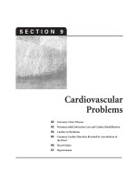
implanted telemetry devices (figure 11.1). Our
understanding of the issue of subconcussion is
clouded by the marked variability between the
thresholds for clinically diagnosed concussion in
terms of linear acceleration, rotational acceleration, and location and number of impacts.[6, 9, 10,
14, 23, 24, 39, 50, 55, 57, 61]
Broglio and colleagues studied 95 high school
football players across four seasons using a helmet
telemetry system to record total number of head
impacts and the associated acceleration forces.[10]
The number of impacts varied with the athletes’
playing position and starting status. The average
player sustained 652 impacts during a 14-week
season. Linemen had the greatest number of
impacts per season (868); the group with the next
highest number of impacts consisted of tight ends,
running backs, and linebackers (619), followed
by quarterbacks (467), receivers, cornerbacks,
1200
1100
Number of hits
1000
Mean g-forces
900
800
700
600
500
400
300
150
50
200
100
6
7
8
211
9 10 11 12 13 14 15 16 17 18 19 20 21 22 23 24 25 26 27 28 29 30 31 32
Youth
High school
College
Pro
Figure 11.1 Subconcussion curve of head impacts and forces over age change and playing level.
E5835/Petraglia/fig 11.1/467639/JG/R1
212
• • •
Handbook of Neurological Sports Medicine
and safeties (372). The seasonal linear acceleration burden averaged 16,746.1 g, while the
rotational acceleration burden was 1,090,697.7
rad/s2. These findings indicate that high school
football players sustain a high number of head
impacts each season, with associated cumulative
impact burdens that are equally impressive.[10]
Talavage and colleagues, using similar technology, found comparable numbers and rates of hit
accumulations.[61]
Eckner and coauthors explored the characteristics of 20 concussion-invoking impacts in
19 high school football players, analyzing the
total number of head impacts, the severity profile values, and cumulative linear and rotational
acceleration values during the same game or
practice session as well as the 30-minute and
1-week periods preceding these impacts.[20] Concussions occurred over a wide range of impact
magnitudes. Interestingly, cumulative impact
burden before a concussion was not different
from nonconcussive impacts of greater magnitudes in the same athletes. Therefore, the authors
concluded that an athlete’s concussion threshold
may be a dynamic feature over time and that
there is a lack of cumulative effects of nonconcussive impacts on concussion threshold. Thus,
the types of impacts that occur in players who
sustain a concussion may be no different from
those that occur in asymptomatic players, further
pointing to the role and potential importance of
subconcussive impacts.
Crisco and colleagues have investigated
impact characteristics in collegiate football players.[14-16] The authors found that player position
and impact location were the largest factors
accounting for differences in head impacts. The
total number of head impacts was a median of
420 and a maximum of 2,492. Studies have
shown variance in the total number of head
impacts in collegiate players, from 950 head
impacts per season[22, 23] to 1,353 per season.[57]
Schnebel and colleagues used accelerometers
embedded in the crown of the helmets in both
high school and collegiate football players.[57]
They found the expected number of high-speed,
open-field collisions occurring in skill position
athletes with forces in the range of 90 to 120
g and a duration of about 15 ms. One of the
most intriguing and unexpected findings of this
study was that linemen experienced impacts
of 20 to 30 g on nearly every play. Due to the
football tradition of linemen starting every play
in the three-point stance and lunging forward
to immediately encounter the opposing player,
head contact occurs on a constant and ubiquitous basis.
Youth football players constitute about 70%
of all American football players and a total of 3.5
million participants. A recent study monitored
seven youth football participants, aged 7 and
8 years, during a football season and noted an
average of 107 impacts per player for the season.
[17]
Linear accelerations ranged from 10 to 100
g, and rotational accelerations ranged from 52
to 7,694 rad/s2. This study was the first to document that very high velocity impacts are possible
at the youth level of football play. Thus, while
youth football players may have fewer helmet
impacts and lower-force hits than their older
counterparts, high-magnitude impacts may occur
nonetheless, and their long-term implications in
an exposure paradigm are uncertain.[18]
Neuropsychological Evaluation
In a recent study, Gysland and colleagues sought
to investigate the relationship between subconcussive impacts and concussion history on clinical
measures of neurological function.[24] Forty-six
collegiate football players completed five clinical
measures of neurological function commonly
employed in the evaluation of concussion before
and after a single season. These tests included
the Automated Neuropsychological Assessment
Metrics, Sensory Organization Test, Standardized Assessment of Concussion, Balance Error
Scoring System, and Graded Symptom Checklist;
impact data were recorded with the Head Impact
Telemetry System (HITS). Even though players
averaged 1,177.3 ± 772.9 head impacts over the
course of a season, the authors found that they
did not demonstrate any clinically meaningful
changes from preseason to postseason on the
measures of neurological function employed.[24]
Similar findings were reported in another study
of college football players.[39] There may be a dose
response with regard to impacts that must be
considered over the course of a player’s career.
Additionally, it is possible that the measures of
neurological function employed were not sensitive enough to detect subclinical neurological
The Emerging Role of Subconcussion
dysfunction in athletes sustaining many repetitive subconcussive impacts.
Other research, though, now suggests that
these nonconcussive impacts may not be benign.
Killam and coauthors found that nonconcussed
collegiate athletes in contact sports actually
scored lower than control subjects in two
memory domains and had lower total scores on
the Repeatable Battery for the Assessment of
Neuropsychological Status (RBANS). [29] Their
data suggest that participation in contact sports
may produce subclinical cognitive impairments in
the absence of a diagnosable concussion, presumably resulting from the cumulative consequences
of multiple mild head injuries. This investigation
showed, and other studies have continued to
demonstrate, that measures of peak acceleration
may not be sufficient to predict cognitive deficit,
and that greater impact forces do not necessarily
correlate with a greater likelihood of neurological
impairment.
McAllister and colleagues studied 214 collegiate Division I football and ice hockey players,
analyzing their accelerometer data and neuropsychological outcomes compared to those for
a control group of noncontact sport athletes.
They found that the athletes in contact sports
had worse performance on tests for new learning, and postseason cognitive testing correlated
with greater head impact exposure. This was
despite the fact that none of the subjects had a
documented sport concussion during the period
of study.[37] Other studies, though, have failed to
detect differences between preseason baseline,
midseason, and postseason assessments in players
who did not sustain concussions.[39] Thus, there
may be specific neuropsychological metrics that
are better suited to or more sensitive for detecting the effects of repetitive subconcussion forces.
It may also be that the symptoms or sequelae of
repetitive subconcussion could require a greater
length of time to develop than a single season.
Neuroradiological Findings
The role of advanced neuroimaging in concussion
has been a progressive one. The use of these new
techniques is especially relevant in the case of
subconcussion because even in cases of concussion, conventional computed tomography and
magnetic resonance imaging (MRI) sequences
• • •
213
are unable to detect macroscopic structural
abnormalities.[53] To test the hypothesis that
subconcussive blows cause an accumulation of
neurophysiological changes, it is necessary to
measure changes in neurological function over
time.
Talavage and colleagues studied a group of
high school football players by performing MRI,
functional MRI (fMRI), and neurocognitive
assessments at three distinct times: (1) before the
start of contact practices, (2) during the season,
and (3) 2 to 5 months after the season concluded.
[61]
In addition to these assessments, the HIT
system was used to record head collisions during
all contact practices and games. The authors
demonstrated quantifiable neurophysiological
changes, in both fMRI and ImPACT testing, in
the absence of outwardly observable symptoms
of concussion. This finding of neuropsychological
disturbance in the absence of classical symptoms
of concussion is consistent with prior observations in seven former National Football League
(NFL) offensive linemen and a wide receiver
as reported by Omalu and colleagues.[46, 47, 49] A
follow-up study by Breedlove and colleagues
demonstrated that the fMRI changes in many
regions of the brain were statistically correlated
to the number and (spatial) distribution of hits
received subsequent to the beginning of contact
practices.[9] This study went on to suggest that the
clinical diagnosis of neurological system deficits
may be dependent on which systems have been
compromised, and that the entire (recent) history
of blows to the head plays a causal role in overall
neurological changes.
A new study using diffusion tensor imaging
(DTI) highlights the emerging clinical evidence
for subconcussive brain injury.[6] Bazarian and
colleagues investigated the ability to detect subject-specific changes in brain white matter (WM)
before and after sport-related concussion. This
prospective cohort study was performed in nine
high school athletes engaged in hockey or football
and six controls. Subjects underwent DTI preand postseason within a 3-month interval. Only
one athlete was diagnosed with a concussion
(scanned within 72 hours), and eight suffered
between 26 and 399 subconcussive head blows.
[6]
While analysis detected significantly changed
WM in a single concussed athlete as expected,
the most striking findings were in those athletes
214
• • •
Handbook of Neurological Sports Medicine
who did not sustain a concussion. Asymptomatic
athletes with multiple subconcussive head blows
had abnormalities in a percentage of their WM
that was over three times higher than in controls.
The significance of these WM changes and their
relationship to head impact forces are currently
unknown.
Necropsy Tissue Analysis
It is now appreciated that the syndrome of CTE,
initially described by Omalu and colleagues in
2005,[47] occurs not only in football players but
also in boxers, wrestlers, hockey players, and
even military personnel.[38, 44, 45] It is believed to be
a lesser form of injury than dementia pugilistica
(DP), initially described by Martland in 1928. In
a series of eight former professional football players, autopsy analysis using detailed and specialized staining techniques for the presence of tau
protein was performed (table 11.1). In all cases,
similar neurobehavioral, neuropsychiatric, and
neuropathological abnormalities were found,
consistent with CTE. Interestingly, none of these
athletes had a history of concussion noted as
a part of the medical and athletic history. It is
unknown whether the methodology at the time
was insufficient to detect the presence of a concussion or whether underreporting occurred due
to player ignorance, motivation, or sport cultural
issues. Seven of the athletes were football linemen, a position associated with constant, mandatory, and often gratuitous head-to-head impacts.
Autopsy data from McKee and coauthors[5, 38]
demonstrate that a subset of athletes in contact
sports, particularly former football players, do
not have a prominent history of known or identified concussions but nonetheless have typical
tauopathy seen in autopsy examination.[38, 45-49]
Taken together, these necropsy tissue findings
point to subconcussion as a pathophysiological
mechanism for unsuspected brain injury in those
exposed to contact and collision sports.
Concluding Thoughts
In recent years there have been major advances in
our understanding of the incidence of mTBI and
the biomechanical forces and cellular responses.
The amount of laboratory research, both animalbased experiments and investigations of the cellular responses underlying concussion, as well
as clinical studies to determine the effects of
concussion, has exponentially increased.[63] In
fact, it is now often stated that the information
from mTBI research produced during the past
decade supersedes the volume and knowledge of
all previous information. An emerging concept
is the phenomenon of subconcussive impacts, as
new evidence highlights their ubiquity in sports,
as well as their potential to contribute to the
development of subacute and chronic sequelae.
As noted previously, Talavage and colleagues
discovered a new category of injured athletes:
those who had no readily observable symptoms
but who instead exhibited functional impairment as measured by neuropsychological testing
Table 11.1 Autopsy Analysis of Former NFL players
Case
1
2
3
4
5
6
7
8
Age
50 years
45 years
45 years
35 years
45 years
39 years
50 years
26 years
Duration of professional career
17 years
7 years
10 years
10 years
12 years
7 years
10 years
5 years
Symptoms
Dep, FB, FM, SA
Dep, FB, FM, SA
Dep, FB, FM
Dep, FB, FM
Dep, drugs, FM
Dep, drugs, FM
Dep, drugs, FM
Dep, personality changes
Cause of death
Cardiac
Suicide
OD
Suicide
OD
OD
OD
Fall from vehicle
Dep, depression; FB, failed business; FM, failed marriage; NFL, National Football League; OD, overdose; SA, substance abuse.
Adapted, by permission, from J.E. Bailes et al., 2013, “Role of subconcussion in repetitive mild traumatic brain injury,” Journal of neurosurgery 119(5):
1235-45.
The Emerging Role of Subconcussion
and fMRI studies.[61] This group of individuals,
who demonstrated abnormal neurological performance despite a lack of symptoms typically
associated with a clinically diagnosed concussion,
may shed light on the issue of subconcussive
impacts and their relationship to chronic neurological syndromes. The research reviewed in
this chapter suggests that the sequence of blows
experienced by a player can mediate the severity of the observed symptoms that lead to the
clinical diagnosis of concussion, or the absence
thereof (e.g., in the case of functionally observed
impairment).
Biophysics data gathered through football
helmet accelerometer studies have shown that
youth, high school, and college players may experience a wide range of head impacts, from 100 to
over 1,000 during the course of a season (table
11.2). Compared to location and magnitude
of forces, it may likely be that the cumulative
number of head impacts best correlates with the
potential for concussion occurrence or chronic
effects. It is uncertain whether head impacts
have a threshold for magnitude or number (or
both) that could result in a cumulative risk for
detrimental effects on brain structures or physiological function.[18]
• • •
215
Our understanding of subconcussion is still
early and evolving but will likely in the future
determine the ultimate risk for those who are
exposed to repetitive mTBI in athletic endeavors.
For now, there is a lack of evidence to permit
a recommendation regarding the number of
subconcussive impacts that should be allowed
prior to ending an athlete’s season or career.
As our knowledge about this emerging concept continues to evolve, refined and advanced
adjunct measures of assessment may someday be
able to help guide such decisions with the aim
of decreasing the incidence of delayed chronic
neurological deficits associated with repetitive
subconcussion. Strategies should be developed to
minimize exposure to recurring cranial impacts
during practice sessions, as Pop Warner Football
has recently done at the youth level. Another
possibility is to change styles of play. Just one
example would be to have linemen in football
start in a squatting “two-point” position or stance,
rather than in a down stance, to remove them
from head contact on every play. It is clear that
further research is needed, but for the time being,
limiting the overall head impact burden as best
as possible is the most prudent recommendation
for today’s athlete.
Table 11.2 Comparison of Head Impacts in Football by Level of Competition
Citations
Daniel et al. 2012
Breedlove et al. 2012
Broglio et al. 2011
Eckner et al. 2011
Schnebel et al. 2007
Talavage et al. 2010
Crisco et al. 2010
Crisco et al. 2011
Guskiewicz et al. 2007
Gysland et al. 2012
Rowson et al. 2012
Schnebel et al. 2007
Extrapolation
Level of competition
Youth
High school
Average head
Age range
impacts per season
5-14 years 107
14-18 years 625§
Range of head
impacts per season
n/a
5-2,235
Collegiate
18-22 years 1,125§
125-2,492
Professional
>22 years
n/a
>1200*
n/a, not available. Note: The number of impacts accrued each season varies by position.
*Estimate based on practice patterns and style of play
§
Head impacts averaged from mean data available from accelerometer studies at each level of competition.
Adapted from: J.E. Bailes et al., 2013, “Role of subconcussion in repetitive mild traumatic brain injury,” Journal of Neurosurgery 119(5): 1235-1245.
216
• • •
Handbook of Neurological Sports Medicine
References
mance in mice. Acad Emerg Med 2004;11(8):809819.
1. Allen GV, Gerami D, Esser MJ. Conditioning
effects of repetitive mild neurotrauma on motor
function in an animal model of focal brain injury.
Neuroscience 2000;99(1):93-105.
14. Crisco JJ, Fiore R, Beckwith JG, Chu JJ, Brolinson
PG, Duma S, et al. Frequency and location of head
impact exposures in individual collegiate football
players. J Athl Train 2010;45(6):549-559.
2. Bailes JE, Mills JD. Docosahexaenoic acid reduces
traumatic axonal injury in a rodent head injury
model. J Neurotrauma 2010;27(9):1617-1624.
15. Crisco JJ, Wilcox BJ, Beckwith JG, Chu JJ,
Duhaime AC, Rowson S, et al. Head impact
exposure in collegiate football players. J Biomech
2011;44(15):2673-2678.
3. Bailes JE, Petraglia AL, Omalu BI, Nauman E,
Talavage T. Role of subconcussion in repetitive
mild traumatic brain injury. J Neurosurg 2013;
119(5):1235-45.
4. Barkhoudarian G, Hovda DA, Giza CC. The
molecular pathophysiology of concussive brain
injury. Clin Sports Med 2011;30(1):33-48, vii-iii.
5. Baugh CM, Stamm JM, Riley DO, Gavett BE,
Shenton ME, Lin A, et al. Chronic traumatic
encephalopathy: neurodegeneration following
repetitive concussive and subconcussive brain
trauma. Brain Imaging Behav. 2012, 6(2):244254.
6. Bazarian JJ, Zhu T, Blyth B, Borrino A, Zhong J.
Subject-specific changes in brain white matter
on diffusion tensor imaging after sports-related
concussion. Magn Reson Imaging 2012;30(2):171180.
7. Bennett RE, Mac Donald CL, Brody DL. Diffusion
tensor imaging detects axonal injury in a mouse
model of repetitive closed-skull traumatic brain
injury. Neurosci Lett 2012;513(2):160-165.
8. Blaylock RL, Maroon J. Immunoexcitotoxicity
as a central mechanism in chronic traumatic
encephalopathy—a unifying hypothesis. Surg
Neurol Int 2011;2:107.
9. Breedlove EL, Robinson M, Talavage TM, Morigaki
KE, Yoruk U, O'Keefe K, et al. Biomechanical correlates of symptomatic and asymptomatic neurophysiological impairment in high school football.
J Biomech 2012;45(7):1265-1272.
10. Broglio SP, Eckner JT, Martini D, Sosnoff JJ,
Kutcher JS, Randolph C. Cumulative head impact
burden in high school football. J Neurotrauma
2011;28(10):2069-2078.
11. Conte V, Uryu K, Fujimoto S, Yao Y, Rokach J,
Longhi L, et al. Vitamin E reduces amyloidosis
and improves cognitive function in Tg2576 mice
following repetitive concussive brain injury. J
Neurochem 2004;90(3):758-764.
12. Creed JA, DiLeonardi AM, Fox DP, Tessler AR,
Raghupathi R. Concussive brain trauma in the
mouse results in acute cognitive deficits and
sustained impairment of axonal function. J Neurotrauma 2011;28(4):547-563.
13. Creeley CE, Wozniak DF, Bayly PV, Olney JW,
Lewis LM. Multiple episodes of mild traumatic
brain injury result in impaired cognitive perfor-
16. Crisco JJ, Wilcox BJ, Machan JT, McAllister TW,
Duhaime AC, Duma SM, et al. Magnitude of head
impact exposures in individual collegiate football
players. J Appl Biomech 2011; 28(2):174-183.
17. Daniel RW, Rowson S, Duma SM. Head impact
exposure in youth football. Ann Biomed Eng
2012;40(4):976-981.
18. Dashnaw ML, Petraglia AL, Bailes JE. An overview
of the basic science of concussion and subconcussion: where we are and where we are going.
Neurosurg Focus 2012;33(6):E5.
19. DeFord SM, Wilson MS, Rice AC, Clausen T,
Rice LK, Barabnova A, et al. Repeated mild brain
injuries result in cognitive impairment in B6C3F1
mice. J Neurotrauma 2002;19(4):427-438.
20. Eckner JT, Sabin M, Kutcher JS, Broglio SP.
No evidence for a cumulative impact effect on
concussion injury threshold. J Neurotrauma
2011;28(10):2079-2090.
21. Friess SH, Ichord RN, Ralston J, Ryall K, Helfaer
MA, Smith C, et al. Repeated traumatic brain
injury affects composite cognitive function in
piglets. J Neurotrauma 2009;26(7):1111-1121.
22. Guskiewicz KM, Mihalik JP. Biomechanics of sport
concussion: quest for the elusive injury threshold.
Exerc Sport Sci Rev 2011;39(1):4-11.
23. Guskiewicz KM, Mihalik JP, Shankar V, Marshall
SW, Crowell DH, Oliaro SM, et al. Measurement
of head impacts in collegiate football players:
relationship between head impact biomechanics and acute clinical outcome after concussion.
Neurosurgery 2007;61(6):1244-1252; discussion
1252-1253.
24. Gysland SM, Mihalik JP, Register-Mihalik JK,
Trulock SC, Shields EW, Guskiewicz KM. The
relationship between subconcussive impacts and
concussion history on clinical measures of neurologic function in collegiate football players. Ann
Biomed Eng 2012;40(1):14-22.
25. Hootman JM, Dick R, Agel J. Epidemiology of
collegiate injuries for 15 sports: summary and
recommendations for injury prevention initiatives. J Athl Train 2007;42:311-319.
26. Johnson GV, Greenwood JA, Costello AC, Troncoso JC. The regulatory role of calmodulin in the
proteolysis of individual neurofilament proteins
by calpain. Neurochem Res 1991;16(8):869-873.
The Emerging Role of Subconcussion
27. Kanayama G, Takeda M, Niigawa H, Ikura Y,
Tamii H, Taniguchi N, et al. The effects of repetitive mild brain injury on cytoskeletal protein
and behavior. Methods Find Exp Clin Pharmacol
1996;18(2):105-115.
28. Kane MJ, Angoa-Perez M, Briggs DI, Viano DC,
Kreipke CW, Kuhn DM. A mouse model of human
repetitive mild traumatic brain injury. J Neurosci
Methods 2012;203(1):41-49.
29. Killam C, Cautin RL, Santucci AC. Assessing the
enduring residual neuropsychological effects
of head trauma in college athletes who participate in contact sports. Arch Clin Neuropsychol
2005;20(5):599-611.
30. Lado WE, Persinger MA. Mechanical impacts to
the skulls of rats produce specific deficits in maze
performance and weight loss: evidence for apoptosis of cortical neurons and implications for clinical
neuropsychology. Percept Mot Skills 2003;97(3 Pt
2):1115-1127.
31. Laurer HL, Bareyre FM, Lee VM, Trojanowski
JQ, Longhi L, Hoover R, et al. Mild head injury
increasing the brain's vulnerability to a second
concussive impact. J Neurosurg 2001;95(5):859870.
32. Lifshitz J, Lisembee AM. Neurodegeneration in the
somatosensory cortex after experimental diffuse
brain injury. Brain Struct Funct 2012;217(1):4961.
33. Longhi L, Saatman KE, Fujimoto S, Raghupathi
R, Meaney DF, Davis J, et al. Temporal window of
vulnerability to repetitive experimental concussive brain injury. Neurosurgery 2005;56(2):364374; discussion 364-374.
34. Mata M, Staple J, Fink DJ. Changes in intraaxonal calcium distribution following nerve crush.
J Neurobiol 1986;17(5):449-467.
35. Maxwell WL, McCreath BJ, Graham DI, Gennarelli TA. Cytochemical evidence for redistribution of membrane pump calcium-ATPase and
ecto-Ca-ATPase activity, and calcium influx in
myelinated nerve fibres of the optic nerve after
stretch injury. J Neurocytol 1995;24(12):925-942.
36. Maxwell WL, Povlishock JT, Graham DL. A mechanistic analysis of nondisruptive axonal injury: a
review. J Neurotrauma 1997;14(7):419-440.
37. McAllister TW, Flashman LA, Maerlender A,
Greenwald RM, Beckwith JG, Tosteson TD, et al.
Cognitive effects of one season of head impacts
in a cohort of collegiate contact sport athletes.
Neurology 2012;78(22):1777-1784.
38. McKee AC, Cantu RC, Nowinski CJ, HedleyWhyte ET, Gavett BE, Budson AE, et al. Chronic
traumatic encephalopathy in athletes: progressive
tauopathy after repetitive head injury. J Neuropathol Exp Neurol 2009;68(7):709-735.
39. Miller JR, Adamson GJ, Pink MM, Sweet JC. Comparison of preseason, midseason, and postseason
• • •
217
neurocognitive scores in uninjured collegiate football players. Am J Sports Med 2007;35(8):12841288.
40. Mills JD, Bailes JE, Sedney CL, Hutchins H, Sears
B. Omega-3 fatty acid supplementation and reduction of traumatic axonal injury in a rodent head
injury model. J Neurosurg 2011;114(1):77-84.
41. Mills JD, Bailes JE, Turner RC, Dodson SC, Sakai
J, Maroon JC. Anabolic steroids and head injury.
Neurosurgery 2012;70(1):205-209; discussion
209-210.
42. Mills JD, Hadley K, Bailes JE. Dietary supplementation with the omega-3 fatty acid docosahexaenoic acid in traumatic brain injury. Neurosurgery
2011;68(2):474-481; discussion 481.
43. Olsson Y, Rinder L, Lindgren S, Stalhammar
D. Studies on vascular permeability changes in
experimental brain concussion. A comparison
between the effects of single and repeated sudden
mechanical loading of the brain. Acta Neuropathologica 1971;19(3):225-233.
44. Omalu B, Bailes J, Hamilton RL, Kamboh MI,
Hammers J, Case M, et al. Emerging histomorphologic phenotypes of chronic traumatic encephalopathy in American athletes. Neurosurgery
2011;69(1):173-183; discussion 183.
45. Omalu BI, Bailes J, Hammers JL, Fitzsimmons RP.
Chronic traumatic encephalopathy, suicides and
parasuicides in professional American athletes:
the role of the forensic pathologist. Am J Forens
Med Pathol 2010;31(2):130-132.
46. Omalu BI, DeKosky ST, Hamilton RL, Minster RL,
Kamboh MI, Shakir AM, et al. Chronic traumatic
encephalopathy in a national football league
player: part II. Neurosurgery 2006;59(5):10861092; discussion 1092-1093.
47. Omalu BI, DeKosky ST, Minster RL, Kamboh
MI, Hamilton RL, Wecht CH. Chronic traumatic
encephalopathy in a National Football League
player. Neurosurgery 2005;57(1):128-134; discussion 128-134.
48. Omalu BI, Fitzsimmons RP, Hammers J, Bailes
J. Chronic traumatic encephalopathy in a professional American wrestler. J Forens Nurs
2010;6(3):130-136.
49. Omalu BI, Hamilton RL, Kamboh MI, DeKosky ST,
Bailes J. Chronic traumatic encephalopathy (CTE)
in a National Football League player: case report
and emerging medicolegal practice questions. J
Forens Nurs 2010;6(1):40-46.
50. Pellman EJ, Viano DC, Tucker AM, Casson IR,
Waeckerle JF. Concussion in professional football:
reconstruction of game impacts and injuries. Neurosurgery 2003;53:799-812.
51. Pettus EH, Povlishock JT. Characterization of a
distinct set of intra-axonal ultrastructural changes
associated with traumatically induced alteration in
218
• • •
Handbook of Neurological Sports Medicine
axolemmal permeability. Brain Res 1996;722(12):1-11.
52. Povlishock JT, Pettus EH. Traumatically induced
axonal damage: evidence for enduring changes in
axolemmal permeability with associated cytoskeletal change. Acta Neurochir Suppl 1996;66:81-86.
53. Prabhu SP. The role of neuroimaging in
sport-related concussion. Clin Sports Med
2011;30(1):103-114, ix.
54. Raghupathi R, Mehr MF, Helfaer MA, Margulies
SS. Traumatic axonal injury is exacerbated following repetitive closed head injury in the neonatal
pig. J Neurotrauma 2004;21(3):307-316.
55. Rowson S, Duma SM, Beckwith JG, Chu JJ, Greenwald RM, Crisco JJ, et al. Rotational head kinematics in football impacts: an injury risk function
for concussion. Ann Biomed Eng 2012;40(1):1-13.
56. Saatman KE, Abai B, Grosvenor A, Vorwerk CK,
Smith DH, Meaney DF. Traumatic axonal injury
results in biphasic calpain activation and retrograde transport impairment in mice. J Cereb Blood
Flow Metab 2003;23(1):34-42.
57. Schnebel B, Gwin JT, Anderson S, Gatlin R. In vivo
study of head impacts in football: a comparison
of National Collegiate Athletic Association Division I versus high school impacts. Neurosurgery
2007;60(3):490-495; discussion 495-496.
58. Shitaka Y, Tran HT, Bennett RE, Sanchez L, Levy
MA, Dikranian K, et al. Repetitive closed-skull
traumatic brain injury in mice causes persistent
multifocal axonal injury and microglial reactivity.
J Neuropathol Exp Neurol 2011;70(7):551-567.
59. Shultz SR, MacFabe DF, Foley KA, Taylor R, Cain
DP. Sub-concussive brain injury in the LongEvans rat induces acute neuroinflammation in the
absence of behavioral impairments. Behav Brain
Res 2012;229(1):145-152.
60. Spain A, Daumas S, Lifshitz J, Rhodes J, Andrews
PJ, Horsburgh K, et al. Mild fluid percussion injury
in mice produces evolving selective axonal pathology and cognitive deficits relevant to human brain
injury. J Neurotrauma 2010;27(8):1429-1438.
61. Talavage TM, Nauman E, Breedlove EL, Yoruk U,
Dye AE, Morigaki K, et al. Functionally-detected
cognitive impairment in high school football
players without clinically-diagnosed concussion.
J Neurotrauma 2010; 31(4):327-338.
62. Uryu K, Laurer H, McIntosh T, Pratico D, Martinez
D, Leight S, et al. Repetitive mild brain trauma
accelerates Abeta deposition, lipid peroxidation,
and cognitive impairment in a transgenic mouse
model of Alzheimer amyloidosis. J Neurosci
2002;22(2):446-454.
63. Weber JT. Experimental models of repetitive brain
injuries. Prog Brain Res 2007;161:253-261.
64. Weitbrecht WU, Noetzel H. [Autoradiographic
investigations in repeated experimental brain concussion (author's transl)]. Arch Psychiatr Nervenk
1976;223(1):59-68.
65. Yoshiyama Y, Uryu K, Higuchi M, Longhi L,
Hoover R, Fujimoto S, et al. Enhanced neurofibrillary tangle formation, cerebral atrophy, and
cognitive deficits induced by repetitive mild brain
injury in a transgenic tauopathy mouse model. J
Neurotrauma 2005;22(10):1134-1141.
chapter
12
Severe Head Injury
and Second Impact Syndrome
T
he last 30 to 40 years have seen a dramatic
decrease in the incidence of severe head
injury in athletics. Rule changes, better
equipment standards and design, increased
awareness, and improved medical care have
all accounted for fewer injuries. While severe
closed head injuries are relatively rare in organized sporting events, the injuries can have
devastating consequences. Understanding the
fundamentals of severe and catastrophic injuries allows the sports medicine practitioner to
be prepared in the event of these occurrences.
Cerebral Contusions
and Intraparenchymal
Hemorrhage
Hemorrhagic brain contusions and intraparenchymal hemorrhages (also known as traumatic
intracerebral hemorrhage) represent regions of
primary neuronal and vascular injury. Contusions are frequent sequelae of head injury and
most commonly occur following acceleration–
deceleration mechanisms. A contusion represents
a heterogeneous area of brain injury that consists
of hemorrhage, cerebral infarction, and necrosis.
These regions of the brain are usually edematous
with areas of punctuate hemorrhages that can
extend deep into the white matter or even the
subdural and subarachnoid spaces. Contusions
commonly occur in coup or contrecoup fashion.
In coup injuries, the brain is injured directly
under the area of impact. The degree of injury
to the underlying brain depends on the energy
transmitted, the area of contact, and the region
of the brain involved, as well as other factors.
Contrecoup injuries occur on the side opposite
the impact as the brain glides and strikes the
skull surface. This results in a hemorrhagic lesion
diametrically opposed to the impact site. After
impact, the brain may also become contused if
it collides with bony protuberances on the inside
surface of the skull. The frontal and temporal
lobes are particularly susceptible to this type of
injury; however, contusions can be observed in
the midbrain and cerebellum, as well.
Contusions vary in size from small, localized
areas to larger areas of injury (figure 12.1). The
important aspect to remember about these types
of brain injuries is that they can demonstrate
progression over time with respect to size and
number of contusions. This progression typically occurs over the first 24 to 48 hours, with
a proportion of cases demonstrating delayed
hemorrhage occurring in areas that were previously free of blood on imaging. Multiple smaller
areas of contusion can coalesce into a largerappearing lesion, more commonly referred to as
an intraparenchymal hemorrhage. Contusions
can be associated with other intra- or extra-axial
hemorrhages, and skull fractures can be present
quite frequently.
•
219
•
220
• • •
Handbook of Neurological Sports Medicine
Figure 12.1 Representative axial computed tomography images demonstrating bifrontal hemorrhagic contusions.
Figure 12.2 Representative axial CT images demon-
The clinical course of these patients varies
greatly depending on the location of the hemorrhagic lesion, as well as the number and extent of
the hemorrhagic contusions. A patient can present with a neurological exam ranging from essentially normal to focal neurological deficits or even
a coma. Involvement of the frontal and temporal
lobes often results in behavioral or mental status
changes. Some athletes have never suffered initial unconsciousness or focal neurological deficit
but may have a headache or period of confusion
after their head injury. The apparent failure to
rapidly clear their mental status is usually what
leads to the diagnosis. Diagnosis is usually made
with a computed tomography (CT) scan, which is
also frequently used for radiographic surveillance
when following patients through their clinical
course. Management is typically conservative
with close observation, but depends on many
factors including the size of the contusion, location, and the patient’s clinical exam.
A traumatic intracerebral hemorrhage is a
parenchymal lesion. It is very similar in radiographic appearance and pathophysiology to a contusion. It represents a localized collection of blood
within the brain and is recognized as a confluent
area of homogenous hemorrhage, which is what
distinguishes it from a contusion (figure 12.2).
As with cerebral contusions, diagnosis is readily
established by CT scan. Patients usually present
with focal neurological deficits but may progress to
further neurological deterioration. Intraparenchymal hemorrhages are among the most common
causes of lethal sport-related brain injuries.
Some patients present with a delayed intracerebral hemorrhage. This entity is typically seen
in older patient populations but should be kept
in mind during evaluation of any patient who
has sustained a significant head impact and has
delayed symptoms. The reported incidence varies
with the resolution of the CT scanner, timing of
the scan, and definition.[103] In those patients with
a Glasgow Coma Scale (GCS) less than or equal
to 8, the reported incidence is approximately
10%.[27, 32, 42, 48, 77, 96] The hemorrhage forms in the
hours to days after the initial trauma, although
most occur within 72 hours after the trauma.[26,
42]
The athlete is generally at risk because these
hematomas are seen more commonly when rotational head trauma has occurred. Factors believed
to contribute to delayed traumatic intracerebral
hemorrhage include local or systemic coagulopathy, hemorrhage into an already contused region
of the brain or an area of necrotic brain softening,
vascular injury, or coalescence of extravasated
microhematomas.[26] The outcome reported in
the literature has generally been poor for these
patients.
strating an intraparenchymal hemorrhage in the right
frontal, parietal, and temporal lobes.
Traumatic Subarachnoid
Hemorrhage
Another acute neurological injury observed in
athletics is traumatic subarachnoid hemorrhage
(SAH). As its name implies, traumatic SAH is
bleeding into the fluid-filled space around the
Severe Head Injury and Second Impact Syndrome
Figure 12.3 Representative axial CT image demonstrating traumatic subarachnoid hemorrhage along the right
sylvian fissure.
brain called the subarachnoid space (figure 12.3).
A large percentage of serious traumatic brain
injuries involve some component of this type
of bleeding. While the hemorrhage can cause
meningeal irritation, the condition is usually not
life threatening, and no immediate treatment is
required for a good outcome. Larger amounts
of SAH may lead to vasospasm, although this
is more typically observed with spontaneous
aneurysmal SAH. Communicating hydrocephalus can occur in a delayed fashion as a result of
SAH and may clinically present with late clinical
deterioration.
Subdural Hematoma
Subdural hematomas (SDHs) are the most
common form of serious and lethal brain injuries
in athletics. A SDH is a collection of blood that
occurs beneath the dura (which is the membrane
overlying the brain). Subdural hematomas in
younger athletes do not behave in the same
manner as those usually seen in the elderly
population. The younger athlete does not possess a large potential subdural space as elderly
people do. As a result, mass effect, increases in
intracranial pressure, and clinical deterioration
occur much more rapidly. These hematomas can
occur both acutely and chronically.
Acute SDHs usually present within 48 to 72
hours after a head injury. According to reports
from the National Center for Catastrophic Sports
Injury Research, an acute SDH is the most
• • •
221
common cause of death due to head injury in
sport.[73] With their research on American football
players, Boden and colleagues demonstrated that
38% of athletes receiving such an injury were
playing while still symptomatic from a prior head
injury that season.[6] Acute SDHs can occur at
any location in the brain and generally occur by
two main mechanisms. These hemorrhages can
result from a tearing of surface or bridging veins
secondary to rotational acceleration-deceleration
during violent head motion. With this etiology,
primary brain damage may be less severe. The
other common cause is a parenchymal laceration
leading to a surrounding subdural accumulation
of blood. In this case there is usually severe primary brain injury. Frequently, the athlete with a
SDH has a small blood collection with underlying brain contusion and hemispheric swelling.
In either case, significant associated underlying
contusions or edema can further compound
brain injury.
Chronic SDHs occur in a later time frame
with more variable clinical manifestations. A
chronic SDH is defined as a hematoma present
at 3 weeks or more after a traumatic injury. The
initial hemorrhage that occurs into the subdural
space may be a small amount that fails to generate any significant brain compression and thus
may not be identified early on. The bleeding or
oozing of blood may continue, and by 4 to 7 days,
a chronic SDH begins to involve the infiltration
of fibroblasts to organize an outer membrane
around the clot.[74] Subsequently, an inner membrane can form and turn the hematoma into an
encapsulated osmotic membrane that interacts
with the production and absorption of cerebrospinal fluid (CSF), creating an active dynamic
process within the membrane layers.
Subdural hematomas can result in a wide
variety of sequelae, ranging from mild symptoms such as headaches to focal neurological
deficits and even death. Athletes may become
unconscious or experience focal neurological
deficits (or both) immediately, or symptoms may
develop more insidiously over time. Typically
athletes with any sizable acute SDH have a significant neurological deficit. Chronic SDHs have
more protean clinical manifestations and may
become symptomatic in a more insidious manner.
Although not common in athletes, a chronic
SDH must always be in the differential diagnosis,
222
• • •
Handbook of Neurological Sports Medicine
especially in those presenting with a remote
history of head impact. Emergent CT diagnosis
is mandatory for the expeditious and successful
treatment of these patients. Acute SDHs appear as
a crescent-shaped mass of increased attenuation
(hyperdense), usually overlying the convexity
of the brain, adjacent to the inner table of the
skull[39, 40] (figure 12.4). However, acute SDHs can
also be interhemispheric, along the tentorium, or
in the posterior fossa. Chronic SDHs have a similar appearance, although they appear hypodense
(approaching the appearance of CSF) on the CT
scan (figure 12.5). Subdural hematomas in general differ from epidural hematomas in that they
are more diffuse, less uniform in appearance, and
usually concave over the surface of the brain.
Figure 12.4 Representative axial CT image demonstrating a left frontoparietal, hyperdense, concave collection
that is consistent with an acute subdural hematoma.
Also note the midline shift to the right.
Figure 12.5 Representative axial CT image demonstrating a right frontoparietal, hypodense, concave collection that is consistent with a chronic subdural hematoma. Also note the midline shift to the left.
Patients with a suspected SDH should be
immediately transported to a facility with neurosurgical services, where an emergent CT can
be obtained and appropriate treatment carried
out. Rapid surgical evacuation of the hematoma
should be considered for symptomatic acute
SDHs that are greater than about 1 cm at the
thickest point (or greater than 5 mm in pediatric
patients).[39, 40] In patients with an underlying
brain contusion, surgical decompression and
evacuation of the hematoma may not improve
the symptoms due to the primary parenchymal
injury.[3]
Skull Fractures
Head injury resulting in the fracture of the skull is
a common occurrence in sports, especially those
in which helmets are not regularly employed.
Additionally, any recreational or sporting activity in which inadvertent head impacts occur
can predispose to skull fracture. Baseball, for
example, is a sport in which an athlete on the
field is unhelmeted and if hit in the head by
a line drive could sustain a skull fracture. Not
uncommonly, spectators are also at risk if struck
in the head with a ball or puck. Diagnosis can
be made with either plain skull radiographs or a
CT scan; the latter can identify any underlying
associated injuries.
Fractures can be linear or comminuted, and
they can also be depressed or nondepressed.
Linear skull fractures are common and can
involve the frontal, parietal, temporal, or occipital
bones (figure 12.6). They usually are the result of
a direct blow to the skull. Linear skull fractures
are not typically depressed, although they can be.
They may occur with a concomitant overlying
scalp laceration, in which case they are considered a compound fracture. More often than not,
there is no misalignment of the bone edges, and
the fractures are not generally considered serious.
They are more important as markers of potential underlying cerebral injury given the large
magnitude of blunt force necessary to create the
fracture. Injury to blood vessels in close proximity
can also occur. Most linear, nondepressed skull
fractures do not require specific treatment other
than conservative observation for any neurological dysfunction or deterioration. These fractures
can heal within several months to years and,
in the absence of any other issues, often do not
Severe Head Injury and Second Impact Syndrome
prevent the athlete from resuming participation,
even in contact sports.
Fractures can also be comminuted and
depressed. Depressed, comminuted skull fractures, like linear fractures, can occur to any of
the surface bones of the skull (figure 12.7). They
usually occur when a relatively small object
makes impact with the skull, resulting in the
depression of the underlying bone. Impacts with
large objects (stationary or moving) can also
result in these complex fractures. Bone fragments
can separate and be driven deep, potentially
lacerating the underlying dura or even invading the brain surface itself. Many patients with
depressed skull fractures do not have significant
brain injury; however, hematomas, CSF leak, or
infection may occur. In contrast to linear skull
fractures, comminuted or depressed skull frac-
• • •
223
tures often require treatment based on the location, contamination, potential regarding cosmetic
appearance, and degree of skull depression.
Epidural Hematoma
Skull fractures that cross the bony grooves harboring blood vessels may cause bony fragments
to lacerate the meningeal vessels, resulting in an
epidural hematoma (EDH). Epidural hematomas
are not an uncommon occurrence secondary to
traumatic brain injury in athletes, especially in
sports in which the participants are not helmeted
such as baseball or golf, although they occur less
commonly than acute SDH. The overall incidence
of EDH is 1% of head trauma admissions, which
is approximately 50% of the incidence of acute
SDH.[39, 40]
An EDH is a collection of blood that occurs
between the dura and the skull (figure 12.8).
Blood accumulates between the skull and outside
the dura, with the dura dissecting until the point
of dural attachment to the overlying cranium.
The bleeding is frequently arterial and fails to
tamponade quickly because of the high arterial
pressure. Approximately 85% of EDHs are due to
arterial bleeding; the middle meningeal artery is
the most common source of middle fossa EDHs.
[39, 40]
The remainder of cases are mainly due to
bleeding from the middle meningeal vein or dural
sinuses. It is important to note that fractured bone
Figure 12.6 Representative axial CT image demonstrating a right-sided linear skull fracture (arrows).
Figure 12.8 Representative axial CT images demon-
Figure 12.7 Representative axial CT image demonstrating a depressed occipital skull fracture (arrows).
Courtesy of University of Rochester Medical Center.
strating (on the left) a large right frontotemporal, hyperdense, biconvex collection that is consistent with an
acute epidural hematoma. The darker (hypodense) areas within the hematoma represent hyperacute hemorrhage. Also note the significant midline shift to the left.
On the right is a left frontal epidural hematoma with a
typical “lentiform” appearance.
224
• • •
Handbook of Neurological Sports Medicine
edges or bleeding from a diploic space can also
result in significant epidural hemorrhage, especially in the pediatric population. A skull fracture
is present in approximately 75% of patients with
an EDH.[53, 76] The majority (70%) of cases occur
laterally over the hemisphere with their epicenter
at the pterion.[39, 40] Epidural hematomas can also
occur in the frontal, occipital, and posterior fossa.
The classic clinical picture of a patient with
an EDH involves a brief posttraumatic loss of
consciousness (LOC), secondary to the force of
impact. This is usually followed by arousal to an
essentially normal level of consciousness. This
is often referred to as a “lucid interval,” which
can last a variable period of time. A short time
thereafter, the athlete may experience a sudden,
excruciating headache followed by a progressive
neurological deterioration. The patient will progress to obtundation, contralateral hemiparesis,
and ipsilateral (same side as clot) pupillary dilation. If this remains untreated, the patient can
go on to exhibit decerebrate posturing-rigidity,
hypertension, respiratory distress, and death
(secondary to brain herniation).[39, 40] While classically clinically characterized this way, a true
“lucid interval” and “textbook” presentation
occurs in less than 10% to 27% of patients with
an EDH,[3, 39, 40] although it has been characterized in some studies as occurring in as many as
47% of patients.[1] The clinical manifestations of
EDH depend on the type of head injury, forces
imparted, and time course of the hematoma
formation.
Any patient or athlete who has sustained a
significant head impact such that a significant
LOC or neurological deficit is present should
undergo a more immediate and thorough medical evaluation including a CT scan. Epidural
hematomas generally (85% of the time) have
a classic appearance of a hyperdense, biconvex
(lenticular) shape adjacent to the skull on head
CT scans[39] (figure 12.8). Mass effect is also frequently associated with EDH. Management can
vary from observation to surgical evacuation of
the EDH and depends on the presence of symptoms, size of the EDH, and age of the patient. It
is essential to recognize this injury early on in
order to commence appropriate management. If
it is treated early, complete neurological recovery
can typically be expected, as EDHs are usually not
associated with other underlying brain injuries.
Diffuse Axonal Injury
Diffuse axonal injury (DAI) is a less localized but
more severe type of acute neurological injury
that can occur in sport. It is a type of injury seen
most commonly in victims of motor vehicle accidents due to significant acceleration–deceleration
forces but is occasionally seen in severe athleticrelated head trauma as well. Diffuse axonal injury
occurs in half of patients with severe TBI and
is responsible for one-third of all head injuryrelated deaths.[34] It is the most common cause
of persistent vegetative state and significant disability following traumatic brain injury.
Diffuse axonal injury is the result of shearing
of multiple axons secondary to rotational forces
(acceleration) on the brain. Parts of the brain
such as the cortex (gray matter) and white matter
have various densities and different physical
properties that accelerate at different speeds upon
impact, resulting in shearing. There is usually
a lack of a mass lesion with severe DAI. Additionally, the rotational acceleration of the head
results in a swirling motion of the brain around
pedicles of blood vessels. A consequence of such
an injury is punctuate hemorrhages from small
vessel tears, in addition to the diffuse tearing of
white matter fiber tracts (figure 12.9). Management varies based on the clinical manifestations
and the severity of the pathophysiology, which
can occur along a spectrum from mild to severe.
Figure 12.9 Diffuse axonal injury in a comatose patient
2 days after a motor vehicle accident. Computed tomography scan displays minimal shear and often can appear
quite normal (left). The corresponding T2-weighted
MRI image (right) reveals extensive bilateral foci of microhemorrhage.
Reprinted from Seminars in Pediatric Surgery, 19(4), S.E. Morrow and
M. Pearson, “Management strategies for severe closed head injuries
in children,” pgs. 279-285, copyright 2010, with permission from
Elsevier.
Severe Head Injury and Second Impact Syndrome
Arterial Dissection
and Stroke
225
golf, volleyball, and softball.[4, 24, 31, 36, 37, 43, 51, 56, 62,
70, 72, 75, 78, 80, 84-86, 88, 90, 92, 94, 100]
Injuries to the head that cause sudden flexion
or extremely rapid rotation of the neck can tear
the intima of the carotid or vertebral arteries.
Injuries to these vessels must not be overlooked
as potential acute neurological injuries. Such
tears or dissections can extend near the skull
base, resulting in vessel occlusion and possible
cerebral ischemia or infarction. Stroke is the most
significant complication of craniocervical arterial
dissection. Dissection occurs more commonly in
the extracranial carotid and vertebral arteries as
compared to the intracranial portions of these
vessels. Cervical internal carotid artery dissections occur typically 2 cm distal to the bifurcation
and may extend distally for a variable distance
(figure 12.10). Extracranial vertebral artery dissection commonly involves the V3 segment at
the C1-C2 levels, where it is most susceptible to
mechanical trauma (figure 12.11).
Athletes with craniocervical arterial dissections can present with nonspecific complaints
and in all settings. Maintaining a high index of
suspicion for carotid or vertebral artery dissection is critical whenever a patient presents with
Athletic trainers, team physicians, pediatricians,
or emergency room physicians are the first providers to see athletes with sport-related stroke.
Thus, it is particularly important that these professionals be aware of the possibility of ischemic
stroke occurring after any form of head or neck
athletic injury. Any athlete with recent head or
neck trauma who presents with acute stroke-like
symptoms should be immediately evaluated for
possible acute ischemic stroke.[29, 82]
Craniocervical arterial dissection is a condition
in which the layers of blood vessel separate from
each other, either spontaneously or secondary
to trauma. Most often this separation occurs
between the intima and media, and it is often
associated with a tear in the luminal lining of
the intima. Craniocervical arterial dissection and
stroke has been reported in a wide spectrum of
athletic activities; among these are soccer, boxing,
wakeboarding, mixed martial arts, scuba diving,
treadmill running, triathlon, springboard diving,
taekwondo, rugby, winter activities, baseball,
Carotid
dissection
Cross section
Normal carotid
artery
Lining of artery
compressed
due to blood
dissecting up
from a tear
Longitudinal section
Blood clot
Common
carotid artery
• • •
Torn artery
wall
Normal
blood
flow
Figure 12.10 Illustration of a carotid dissection.
E5835/Petraglia/fig 12.10/467674/JG/R1
Restricted
blood
flow
226
• • •
Handbook of Neurological Sports Medicine
Figure 12.11 Vertebral artery dissection.
Illustration Copyright © 2014 Nucleus Medical Media, All rights reserved. www.nucleusinc.com.
E5835/Petragila/fig12.11/467675/alw/r1-pulled
unusual focal neurological complaints, particularly if the cranial nerves are involved and if the
patient has a suspicious mechanism of trauma.
A history of cervical hyperextension, flexion, or
rotation should alert the physician to the possibility of dissection. That being said, a direct impact
on the neck can also result in a dissection or
arterial injury. One example is that of a lacrosse
player who was struck in the back of the neck
with a ball and sustained a vertebral artery dissection with diffuse subarachnoid hemorrhage
and subsequently progressed to death (figure
12.12). Injury to the craniocervical vessels can
occur in sports typically felt to be benign, such
as golf. “Golfer’s stroke” has been described by
Maroon and colleagues,[62] as well as others,[24, 46,
93]
and can result in injury or dissection to either
the vertebral or carotid arteries with the minor
repetitive neck motion used in golf.
The diagnosis of an arterial dissection or a
significant hemodynamic process or injury may
require multiple imaging modalities. While
catheter angiography is still considered the gold
standard, recent evidence in the literature suggests that magnetic resonance imaging (MRI),
computed tomography angiogram (CTa), or both
provide highly sensitive and specific diagnostic
information.[23] Thus, catheter angiography may
be needed only in cases in which noninvasive
imaging is negative or inconclusive.[98] It should
be noted that vertebral artery dissections represent a greater diagnostic challenge than carotid
dissections for both MRI (e.g., flow artifacts and
periarterial venous enhancement simulating a
mural hematoma) and CTa (bone artifact, particularly at the skull base).[82]
Expeditious identification and management
are essential for good outcome. There are no clear
recommendations to guide return to participation. Most agree that patients can be encouraged
to participate in noncontact and low-contact
sports. Some have suggested waiting at least 6
Severe Head Injury and Second Impact Syndrome
• • •
227
Figure 12.12 Representative axial CT images in an athlete who was struck in the back of the neck with a ball and who
presented to the hospital emergently for rapid neurological deterioration. The patient was found to have significant
subarachnoid and intraventricular hemorrhage throughout the basal cisterns and ventricular system, secondary to a
traumatic vertebral artery dissection.
months before resumption of contact sports,
while others have reported that they would never
recommend participation in high-contact sports
after arterial dissection and acute ischemic stroke.
[5]
There seems to be more hesitation about letting
a patient with an arterial dissection return to athletics in comparison to a patient with idiopathic
stroke.[5] Though most studies find that the recurrence risk for arterial dissection is approximately
1% per year,[23] the recurrence risk in athletes
(particularly children) with sport-related stroke
may be higher, with rates of up to 30% having
been cited.[5, 55, 82]
Other less common mechanisms of injury to
the craniocervical arteries have been described.
Bow hunter’s stroke is a symptomatic vertebrabasilar insufficiency caused by stenosis or occlusion of the vertebral artery with physiologic head
rotation.[87] In 1978, Sorensen coined the term
bow hunter’s stroke to refer to the sudden onset
of right-sided hemiparesis, contralateral hemisensory changes, and a dilated right pupil in a
39-year-old man after he turned his head during
archery practice.[87] Since that time, additional
series have described patients with bow hunter’s
stroke who presented with either a completed
stroke or transient ischemic attack (TIA) referable to changes in head position.[35, 38, 44, 57, 64, 83,
101]
It is important to note that although the term
stroke is used throughout the literature to refer
to the condition, the condition encompasses a
wide spectrum of rotational hemodynamic insufficiency ranging from a TIA to an acute ischemic
stroke.[54] Various other pathologic conditions
have been reported as causes of bow hunter's
stroke, including far lateral cervical disc herniation reported by Vates and coauthors[97] and
C1-C2 facet hypertrophy reported by Chough
and colleagues.[25] Bow hunter’s stroke most
commonly occurs at the junction of C1 and C2
and less commonly as the vertebral artery enters
the C6 transverse foramen.[69] The predominance
of this site for occlusion is accounted for by the
immobilization of the vertebral artery at the
transverse foramina of C1 and C2 and along the
sulcus arteriosus to where it inserts into the dura.
[54]
Management has varied from conservative
treatment with anticoagulation to surgical fixation and fusion at C1-C2. An alternative treatment is surgical decompression of the vertebral
artery at the site of compression. The long-term
outcome of patients with this disease is not clearly
understood given the overall rarity of the condition; however, any intervention that alleviates
the compression and restores blood flow should
significantly improve outcomes.[54]
Fatalities
While participation in sport is usually regarded
as healthy and safe, athletes are nevertheless
subject to an unpredictable risk of sudden death
during participation.[58, 95] Although most data
regarding these tragic events have had to do with
cardiovascular causes,[59-61] neurological injury
can also lead to fatalities.[21, 22]
An excellent recent study by Thomas and
colleagues sought to define the clinical profile,
epidemiology, and frequency of trauma-related
deaths in young U.S. athletes by analyzing the
30-year U.S. National Registry of Sudden Death
in Young Athletes (1980-2009) using systematic
identification and tracking strategies.[95] Of the
228
• • •
Handbook of Neurological Sports Medicine
1,827 deaths of athletes aged 21 years or younger,
261 (14%) were caused by trauma-related injuries, usually involving the head or neck.[95] This
was noted in 22 different sports. The majority
(90%) of deaths occurred in male athletes. The
highest number of events in a single year was
16, with an average of 9 athletic trauma-related
deaths per year throughout 30 years.[95] The mortality rate in this retrospective study was 0.11 per
100,000 participations. Sports in which traumarelated deaths occurred included track and field
(predominantly pole vaulting), baseball, soccer,
horseback riding, skiing, gymnastics, softball, basketball, cheerleading, hockey, wrestling, cycling,
lacrosse, triathlon or cross country running,
rugby, surfing, weightlifting, American football,
and boxing. The largest number of deaths was
in American football, accounting for 148 fatalities (57%), including 17 deaths in which there
were documented concussions shortly before a
fatal head injury (“second impact syndrome”).[95]
Trauma-related deaths, in this review, occurred
either in competitive events (62%) or during
practice (38%).[95] The majority of the traumarelated deaths were due to injury of the head,
neck, or both (89% of trauma-related deaths),
with other deaths resulting from abdominal,
thoracic, or multiple organ damage. These data
corroborate findings from other similar studies.
[6-14, 21, 22]
Boxing is another sport that has received substantial attention in the media for sport-related
fatalities in recent years. Unfortunately, there is
a paucity of validated epidemiological data on
which to accurately base boxing fatality rates.
[66-68]
Instead, many of the reported fatality data
have been obtained from a combination of media
sources, industry reports, and individual case
reports. From what can be ascertained based on
the available reviews of boxing deaths, it appears
that the rate of boxing fatalities has declined over
the last few decades and that this can in part be
attributed to rule changes, as well as medical
advances that have improved both the diagnosis and treatment of the acutely injured fighter.
The majority of boxing fatalities result from
SDHs, and typically these are associated with an
immediate LOC during the fight. While death
as a result of participation in boxing and other
combat sports has received plenty of media attention, the fatality rate in boxing actually compares
favorably to that in many other sports receiving
less attention with respect to participant safety,
such as horse racing, sky diving, hang gliding,
and mountaineering.
Miele and Bailes analyzed the number and
types of punches landed in a typical professional
match, in bouts considered to be competitive
and in those that ended in fatalities, to determine whether or not this would be a practical
method of differentiating between these groups.
[66]
Several statistically significant differences
were discovered between matches that resulted
in fatalities and the control group; these included
the number of punches landed per round, the
number of power punches landed per round,
and the number of power punches thrown per
round by losing boxers.[66] When the fatal bouts
were compared with the most competitive bouts,
though, these differences were no longer evident.
Thus, based on their findings, the authors concluded that a computerized method of counting
landed blows at ringside could provide sufficient
data to stop matches that might result in fatalities; however, such a process would become less
effective as matches became more competitive.[66]
Other Posttraumatic
Sequelae
Posttraumatic seizures are thought to occur in
approximately 5% of all patients with cranial
cerebral trauma and approximately 15% of those
with severe head injuries, although these may be
underestimates. Certain factors in TBI predispose
the athlete to developing posttraumatic epilepsy.
Patients with lesions such as a contusion or
hematoma (particularly in the temporal lobe),
those with a depressed skull fracture impinging
on the cortical surface, and those experiencing
delayed seizures (later than 1 week following
trauma) are believed to have a higher incidence
of posttraumatic epilepsy.
In the setting of TBI, seizures can occur at a
variety of time points. “Impact seizures” occur
immediately at the time of trauma and are
believed to occur secondary to altered electromechanical conductance due to the impact.
“Immediate seizures” occur within the first 24
hours after trauma, while “early seizures” occur
within the first week after TBI. “Later seizures”
occur at a time remote from the initial injury
Severe Head Injury and Second Impact Syndrome
and are most typically considered to be congruent with posttraumatic epilepsy. While the use
of prophylactic anticonvulsants (such as phenytoin) in patients believed to be at greater risk
for developing posttraumatic seizures has helped
reduce the incidence of seizures within the first
week, there is no effect on the development of
seizures in a delayed fashion. The management
of patients with posttraumatic epilepsy follows
the guidelines for the treatment of patients with
epilepsy of nontraumatic origin.
Another sequela of TBI includes posttraumatic
hydrocephalus. Hydrocephalus is the enlargement of the ventricular system (figure 12.13).
This typically occurs only with more severe
forms of TBI. The incidence of posttraumatic
ventriculomegaly has been reported to range
from 30% to 86%.[49] The enlargement can represent ventriculomegaly alone or symptomatic
hydrocephalus.[41] Particularly following severe
TBI, it is not uncommon to see loss of cerebral
tissue. Imaging with CT or MRI demonstrates
areas of porencephaly, venous–arterial infarction,
or atrophy.[41] The loss of tissue allows for passive
dilation of the ventricular system as the ventricles
expand to fill a void. Diagnosis is typically made
clinically with the aid of imaging. Sometimes
the presentation is as subtle as a leveling off of
neurological improvement in the rehabilitation
of TBI. Some of these patients can improve with
procedures such as a ventriculoperitoneal shunt,
which is commonly used to divert cerebrospinal
fluid away from the point of obstruction.
Another rare complication of traumatic brain
injury is cerebral venous sinus thrombosis
(CVST). Cerebral venous sinus thrombosis is rare,
Figure 12.13 Representative axial CT image demonstrating significantly enlarged lateral ventricles.
• • •
229
with an incidence of around three or four cases
per million people.[30] Overall, in adults, 75% of
CVST cases occur in females.[89] Clinically, CVST
can present with a variety of symptoms. More
commonly patients present with a headache that
develops over several days; much less commonly,
with an acute decrease in consciousness. In some
cases, CVST can result in death. While most cases
of CVST are idiopathic or associated with thrombophilia, pregnancy, or chronic inflammatory
conditions, a rare cause that is often overlooked
is traumatic closed head injury. There have been
several reports of CVST associated with TBI.[2, 15,
45, 52, 63, 102]
Cases are sometimes associated with an
overlying skull fracture, and many may even be
small and go unrecognized.[102] The actual occurrence in traumatic injuries may be higher than
realized if overlooked in the acute TBI setting.
One retrospective review study of 195 patients
with acute traumatic head injuries identified
15.8% of patients as having an occlusive CVST
on imaging.[28] Imaging studies should be carried
out to enable prompt and exacting management.
Treatment with thrombolytics or other potential
interventional procedures may be required to
prevent serious neurological consequences or
even death.
Second Impact Syndrome
The term second impact syndrome (SIS) was
coined in 1984 by Saunders and Harbaugh[79];
however, the phenomenon was actually first
described in 1973 by renowned neurosurgeon
Richard C. Schneider.[20, 81] Second impact syndrome is defined as a fatal, malignant, and uncontrollable increase in intracranial pressure caused
by diffuse cerebral edema that occurs after a head
impact has been sustained before complete recovery from a previous head trauma. The syndrome
occurs when an athlete experiences a head injury,
possibly a concussion or even worse, and then
sustains a second injury before the symptoms
associated with the first injury have cleared.[17-20]
The second blow may be remarkably minor and
involve only a blow to the chest or torso that
may impart forces to the brain.[20] The affected
athlete does not necessarily lose consciousness
and in most cases even remains on the playing
field or walks off under his own power. Then
the stunned but conscious athlete, in a matter of
230
• • •
Handbook of Neurological Sports Medicine
subfalcine herniation, or herniation of the cerebellar tonsils through the foramen magnum
with resultant brain stem compression (figure
12.14). The deterioration is extremely rapid and
often faster than that seen with EDHs. While
MRI can more precisely delineate and characterize the injury, CT scanning is the initial imaging
modality of choice because it can rapidly identify
a potential lesion or brain shift that may require
neurosurgical intervention.[99] On imaging, the
brain appears edematous with a hemispheric
asymmetry; and there is often a small SDH that
can be associated with the injury (figure 12.15).
The notable finding is that the shift is out of proportion to the amount of SDH present. The basal
cisterns may be effaced due to temporal lobe or
diencephalic herniation.[20] If the patient survives
the initial episode of intracranial hypertension,
multifocal bilateral nonhemorrhagic infarction
ensues. This highlights the importance of early
recognition because there is usually little primary
brain injury, and serious or fatal neurological
outcomes are due to secondary brain injury from
raised intracranial pressure and resultant brain
herniation. Prompt treatment with intubation,
hyperventilation, osmotic agents, and surgical
intervention (if ultimately needed) has helped
to reduce the mortality associated with SIS.[16, 19]
seconds to minutes, may precipitously collapse
to the ground, with rapidly dilating pupils, loss
of eye movement, and evidence of respiratory
failure.[17-20]
Significant controversy exists about the validity of this condition, and its precise frequency
in sport is unknown; however, numerous cases
have been reported in the literature, and most
involve adolescent males or young adults.[18, 20,
33, 50, 65, 71, 99]
The precise incidence per 100,000
participants is not known because the population at risk has not been clearly defined. Second
impact syndrome is associated with a high
mortality rate (approaching 50%) and a nearly
100% morbidity rate.[19] It is an important acute
neurological injury to keep in mind when one is
making return-to-play decisions about an athlete
who has suffered a TBI, as clearly prevention is
of utmost importance.
The pathophysiological mechanism is thought
to involve a dysfunction or loss of autoregulation
of the cerebral vasculature, leading to vascular
engorgement and diffuse cerebral edema.[20, 47, 91]
This hyperemic brain swelling markedly increases
intracranial pressure and can lead to brain herniation syndromes. The increased pressure can
cause subsequent inferomedial herniation of
the temporal lobes (transtentorial herniation),
Falx cerebri
Mass
Lateral
ventricle
Subfalcine
herniation
1
Tentorium
cerebelli
2
Central
herniation
3
4
Uncal
transtentorial
herniation
Tonsillar
herniation
Figure 12.14 A mass lesion (like a large hematoma) or significant cerebral edema can cause the brain to herniate in
many different ways, as illustrated, with resultant brain compression.
Reprinted, by permission, from B. Blumenfeld, 2002, Neuroanatomy through clinical cases (Sunderland, MA: Sinauer Associates, Inc.).
E5835/Petraglia/fig 12.14/467681/JG/R2
Severe Head Injury and Second Impact Syndrome
• • •
231
Figure 12.15 Typical imaging findings of dysautoregulation/second impact syndrome (DSIS). (a, b) Admission noncontrast axial CT images and (c) artist's rendition demonstrate a small heterogeneous left frontal subdural hematoma
(SDH; white arrows) that causes complete effacement of the basal cisterns and brain stem distortion. Note the subtle
linear increased density in the region of the circle of Willis (black arrow), consistent with “pseudosubarachnoid hemorrhage,” resulting from the marked elevation in intracranial pressure. Although preservation of the gray–white matter differentiation is seen, there is asymmetric enlargement of the left hemisphere, consistent with hyperemic cerebral
swelling (dysautoregulation). Note that (a) is smaller than (b) even though the left hemisphere is mildly compressed by
the overlying SDH. The extent of mass effect and midline shift is disproportional to the volume of the SDH (compare
with figures 12.3 and 12.4). This 3-day-postoperative Fluid attenuated inversion recovery (FLAIR) magnetic resonance image (d) and artist's rendition (e) demonstrate bilateral multifocal ischemic lesions involving several vascular
territories, including the left posterior cerebral artery, thalamus, insular cortex, basal ganglia, and orbitofrontal cortex.
Reprinted, by permission, from R. Cantu and A.D. Gean, 2010, “Second-impact syndrome and a small subdural hematoma: An uncommon catastrophic result of repetitive head injury with a characteristic imaging appearance,” Journal of Neurotrauma 27(9): 1557-1564. The publisher for this
copyrighted material is Mary Ann Liebert, Inc. publishers.
Concluding Thoughts
While the incidence of severe head injury has
decreased, it is important for caregivers at athletic contests to be cognizant of the spectrum of
injuries that occur in response to brain trauma.
Two important goals in evaluating the potentially head-injured athlete include recognizing
that a head injury may have occurred and that
athletes requiring transport to a medical facility for further workup and treatment must be
accurately identified. While mild traumatic brain
injury or concussion occurs more frequently and
has received a lot of attention lately, other injuries include intracranial hemorrhage, subdural
hematomas, epidural hematomas, skull fractures,
neurovascular injury, and diffuse axonal injury
and can even result in death. Although extremely
rare, second impact syndrome is deadly, and
concern regarding its occurrence has shaped the
conservative, modern-day management of mild
traumatic brain injury.
References
1. Araujo JL, Aguiar Udo P, Todeschini AB, Saade N,
Veiga JC. Epidemiological analysis of 210 cases of
surgically treated traumatic extradural hematoma.
Rev Col Bras Cir 2012;39(4):268-271.
2. Barbati G, Dalla Monta G, Coletta R, Blasetti AG.
Post-traumatic superior sagittal sinus thrombosis.
Case report and analysis of the international literature. Minerva Anestesiol 2003;69(12):919-925.
3. Begso JJ, Lehman RC. Field evaluation and management of head and neck injuries. Clin Sports
Med 1987;6:1.
4. Benedict WJ, Prabhu V, Viola M, Biller J. Carotid
artery pseudoaneurysm resulting from an injury
to the neck by a fouled baseball. J Neurol Sci
2007;256(1-2):94-99.
5. Bernard TJ, deVeber GA, Benke TA. Athletic participation after acute ischemic childhood stroke: a
survey of pediatric stroke experts. J Child Neurol
2007;22(8):1050-1053.
6. Boden B, Tacchetti R, Cantu R, Knowles S, Mueller F. Catastrophic head injuries in high school
and college football players. Am J Sports Med
2000;35:1-7.
7. Boden BP. Direct catastrophic injury in sports. J
Am Acad Orthop Surg 2005;13(7):445-454.
8. Boden BP, Boden MG, Peter RG, Mueller FO,
Johnson JE. Catastrophic injuries in pole vaulters:
a prospective 9-year follow-up study. Am J Sports
Med 2012;40(7):1488-1494.
9. Boden BP, Lin W, Young M, Mueller FO. Catastrophic injuries in wrestlers. Am J Sports Med
2002;30(6):791-795.
10. Boden BP, Pasquina P, Johnson J, Mueller FO.
Catastrophic injuries in pole-vaulters. Am J Sports
Med 2001;29(1):50-54.
11. Boden BP, Tacchetti R, Mueller FO. Catastrophic cheerleading injuries. Am J Sports Med
2003;31(6):881-888.
12. Boden BP, Tacchetti R, Mueller FO. Catastrophic
injuries in high school and college baseball players. Am J Sports Med 2004;32(5):1189-1196.
232
• • •
Handbook of Neurological Sports Medicine
13. Boden BP, Tacchetti RL, Cantu RC, Knowles SB,
Mueller FO. Catastrophic cervical spine injuries
in high school and college football players. Am J
Sports Med 2006;34(8):1223-1232.
14. Boden BP, Tacchetti RL, Cantu RC, Knowles SB,
Mueller FO. Catastrophic head injuries in high
school and college football players. Am J Sports
Med 2007;35(7):1075-1081.
15. Brors D, Schafers M, Schick B, Dazert S, Draf W,
Kahle G. Sigmoid and transverse sinus thrombosis
after closed head injury presenting with unilateral
hearing loss. Neuroradiology 2001;43(2):144-146.
16. Bruce DA, Alavi A, Bilaniuk L, Dolinskas C, Obrist
W, Uzzell B. Diffuse cerebral swelling following
head injuries in children: the syndrome of “malignant brain edema.” J Neurosurg 1981;54(2):170178.
17. Cantu RC. Second impact syndrome a risk in any
contact sport. Phys Sportsmed 1995;23:27-34.
18. Cantu RC. Second impact syndrome: immediate
management. Phys Sportsmed 1992;20:55-58.
19. Cantu RC. Second-impact syndrome. Clin Sports
Med 1998;17(1):37-44.
20. Cantu RC, Gean AD. Second-impact syndrome
and a small subdural hematoma: an uncommon
catastrophic result of repetitive head injury with a
characteristic imaging appearance. J Neurotrauma
2010;27(9):1557-1564.
21. Cantu RC, Mueller FO. Brain injury-related fatalities in American football, 1945-1999. Neurosurgery 2003;52(4):846-852; discussion 852-843.
22. Cantu RC, Mueller FO. Catastrophic football injuries: 1977-1998. Neurosurgery 2000;47(3):673675; discussion 675-677.
23. Caplan LR. Dissections of brain-supplying arteries.
Nat Clin Pract Neurol 2008;4(1):34-42.
24. Choi KD, Oh SJ, Yang TI, Lee TH. Golfer's stroke
from internal carotid artery dissection. Arch
Neurol 2008;65(8):1122-1123.
25. Chough CK, Cheng BC, Welch WC, Park CK.
Bow hunter's stroke caused by a severe facet
hypertrophy of C1-2. J Korean Neurosurg Soc
2010;47(2):134-136.
26. Cooper PR. Delayed traumatic intracerebral hemorrhage. Neurosurg Clin N Am 1992;3(3):659665.
27. Cooper PR, Maravilla K, Moody S, Clark WK.
Serial computerized tomographic scanning and
the prognosis of severe head injury. Neurosurgery
1979;5(5):566-569.
28. Delgado Almandoz JE, Kelly HR, Schaefer PW,
Lev MH, Gonzalez RG, Romero JM. Prevalence of
traumatic dural venous sinus thrombosis in highrisk acute blunt head trauma patients evaluated
with multidetector CT venography. Radiology
2010;255(2):570-577.
29. Dharmasaroja P, Dharmasaroja P. Sports-related
internal carotid artery dissection: pathogenesis and therapeutic point of view. Neurologist
2008;14(5):307-311.
30. Dobbs TD, Barber ZE, Squier WL, Green AL.
Cerebral venous sinus thrombosis complicating traumatic head injury. J Clin Neurosci
2012;19(7):1058-1059.
31. Echaniz-Laguna A, Fleury MC, Petrow P, Arnould
G, Beaujeux R, Warter JM. Internal carotid artery
dissection caused by a kick during French boxing.
Presse Med 2001;30(14):683.
32. Erol FS, Kaplan M, Topsakal C, Ozveren MF,
Tiftikci MT. Coexistence of rapidly resolving
acute subdural hematoma and delayed traumatic
intracerebral hemorrhage. Pediatr Neurosurg
2004;40(5):238-240.
33. Fekete JF. Severe brain injury and death following
minor hockey accidents: the effectiveness of the
"safety helmets" of amateur hockey players. Can
Med Assoc J 1968;99(25):1234-1239.
34. Fick DS. Management of concussion in collision
sports. Guidelines for the sidelines. Postgrad Med
1995;97(2):53-56, 59-60.
35. Fox MW, Piepgras DG, Bartleson JD. Anterolateral decompression of the atlantoaxial vertebral
artery for symptomatic positional occlusion of
the vertebral artery. Case report. J Neurosurg
1995;83(4):737-740.
36. Fridley J, Mackey J, Hampton C, Duckworth E,
Bershad E. Internal carotid artery dissection and
stroke associated with wakeboarding. J Clin Neurosci 2011;18(9):1258-1260.
37. Furtner M, Werner P, Felber S, Schmidauer C.
Bilateral carotid artery dissection caused by springboard diving. Clin J Sport Med 2006;16(1):76-78.
38. George B, Laurian C. Impairment of vertebral
artery flow caused by extrinsic lesions. Neurosurgery 1989;24(2):206-214.
39. Greenberg MS, Arredondo N. Handbook of Neurosurgery. 6th ed. Lakeland, FL/New York: Greenberg Graphics/Thieme Medical; 2006.
40. Greenberg MS. Handbook of Neurosurgery. 7th
ed. Tampa, FL: Greenberg Graphics; 2010.
41. Gudeman SK, Kishore PR, Becker DP, Lipper MH,
Girevendulis AK, Jeffries BF, et al. Computed
tomography in the evaluation of incidence and
significance of post-traumatic hydrocephalus.
Radiology 1981;141(2):397-402.
42. Gudeman SK, Kishore PR, Miller JD, Girevendulis AK, Lipper MH, Becker DP. The genesis and
significance of delayed traumatic intracerebral
hematoma. Neurosurgery 1979;5(3):309-313.
43. Hafner F, Gary T, Harald F, Pilger E, Groell R, Brodmann M. Dissection of the internal carotid artery
after SCUBA-diving: a case report and review of
the literature. Neurologist 2011;17(2):79-82.
Severe Head Injury and Second Impact Syndrome
44. Hanakita J, Miyake H, Nagayasu S, Nishi S,
Suzuki T. Angiographic examination and surgical
treatment of bow hunter's stroke. Neurosurgery
1988;23(2):228-232.
45. Hesselbrock R, Sawaya R, Tomsick T, Wadhwa
S. Superior sagittal sinus thrombosis after closed
head injury. Neurosurgery 1985;16(6):825-828.
46. Hong JM, Kim TJ, Lee JS, Lee JS. Neurological picture. Repetitive internal carotid artery
compression of the hyoid: a new mechanism of
golfer's stroke? J Neurology Neurosurg Psychiatry
2011;82(2):233-234.
47. Junger EC, Newell DW, Grant GA, Avellino AM,
Ghatan S, Douville CM, et al. Cerebral autoregulation following minor head injury. J Neurosurg
1997;86(3):425-432.
48. Kaplan M, Ozveren MF, Topsakal C, Erol FS,
Akdemir I. Asymptomatic interval in delayed
traumatic intracerebral hemorrhage: report of two
cases. Clin Neurol Neurosurg 2003;105(3):153155.
49. Katz RT, Brander V, Sahgal V. Updates on the diagnosis and management of posttraumatic hydrocephalus. Am J Phys Med Rehabil 1989;68(2):9196.
50. Kelly JP, Nichols JS, Filley CM, Lillehei KO,
Rubinstein D, Kleinschmidt-DeMasters BK. Concussion in sports. Guidelines for the prevention of
catastrophic outcome. JAMA 1991;266(20):28672869.
51. Kocyigit A, Cinar C, Kitis O, Calli C, Oran I. Isolated PICA dissection: an unusual complication
of scuba diving: case report and review of the
literature. Clin Neuroradiol 2010;20(3):171-173.
52. Kuether TA, O'Neill O, Nesbit GM, Barnwell
SL. Endovascular treatment of traumatic dural
sinus thrombosis: case report. Neurosurgery
1998;42(5):1163-1166; discussion 1166-1167.
53. Kvarnes TL, Trumpy JH. Extradural haematoma.
Report of 132 cases. Acta Neurochir 1978;41(13):223-231.
54. Lemole GM Jr, Henn JS, Spetzler RF, Zabramski
JM. Bow hunter's stroke. BNI Q2001;17:4-10..
55. Lynch JK, Pavlakis S, Deveber G. Treatment and
prevention of cerebrovascular disorders in children. Curr Treat Options Neurol 2005;7(6):469480.
56. Macdonald DJ, McKillop EC. Carotid artery dissection after treadmill running. Br J Sports Med
2006;40(4):e10; discussion e10.
57. Mapstone T, Spetzler RF. Vertebrobasilar insufficiency secondary to vertebral artery occlusion
froma fibrous band. Case report. J Neurosurg
1982;56(4):581-583.
58. Maron BJ. Sudden death in young athletes. New
Engl J Med 2003;349(11):1064-1075.
• • •
233
59. Maron BJ, Doerer JJ, Haas TS, Tierney DM, Mueller FO. Sudden deaths in young competitive athletes: analysis of 1866 deaths in the United States,
1980-2006. Circulation 2009;119(8):1085-1092.
60. Maron BJ, Haas TS, Doerer JJ, Thompson PD,
Hodges JS. Comparison of U.S. and Italian experiences with sudden cardiac deaths in young
competitive athletes and implications for preparticipation screening strategies. Am J Cardiol
2009;104(2):276-280.
61. Maron BJ, Shirani J, Poliac LC, Mathenge R,
Roberts WC, Mueller FO. Sudden death in young
competitive athletes. Clinical, demographic, and
pathological profiles. JAMA 1996;276(3):199-204.
62. Maroon JC, Gardner P, Abla AA, El-Kadi H,
Bost J. "Golfer's stroke": golf-induced stroke
from vertebral artery dissection. Surg Neurol
2007;67(2):163-168; discussion 168.
63. Matsushige T, Nakaoka M, Kiya K, Takeda T,
Kurisu K. Cerebral sinovenous thrombosis after
closed head injury. J Trauma 2009;66(6):15991604.
64. Matsuyama T, Morimoto T, Sakaki T. Comparison
of C1-2 posterior fusion and decompression of the
vertebral artery in the treatment of bow hunter's
stroke. J Neurosurg 1997;86(4):619-623.
65. McQuillen JB, McQuillen EN, Morrow P. Trauma,
sport, and malignant cerebral edema. Am J Forens
Med Pathol 1988;9(1):12-15.
66. Miele VJ, Bailes JE. Objectifying when to halt a
boxing match: a video analysis of fatalities. Neurosurgery 2007;60(2):307-315; discussion 315-316.
67. Miele VJ, Bailes JE, Cantu RC, Rabb CH. Subdural
hematomas in boxing: the spectrum of consequences. Neurosurg Focus 2006;21(4):E10.
68. Miele VJ, Bailes JE, Voelker JL. Boxing and the
neurosurgeon. Clin Neurosurg 2002;49:396-406.
69. Miele VJ, France JC, Rosen CL. Subaxial positional
vertebral artery occlusion corrected by decompression and fusion. Spine 2008;33(11):E366-370.
70. Miyata M, Yamasaki S, Hirayama A, Tamaki N.
Traumatic middle cerebral artery occlusion. No
Shinkei Geka 1994;22(3):253-257.
71. Mori T, Katayama Y, Kawamata T. Acute hemispheric swelling associated with thin subdural
hematomas: pathophysiology of repetitive head
injury in sports. Acta Neurochir Suppl 2006;96:4043.
72. Motohashi O, Kameyama M, Kon H, Fujimura
M, Onuma T. A case of vertebral artery occlusion
following heading play in soccer. No Shinkei Geka
2003;31(4):431-434.
73. Mueller FO, Cantu RC. National Center for
Catastrophic Sports Injury Research 27th annual
report. Fall 1982 to spring 2009. Chapel Hill, NC
National Center for Catastrophic Sport Injury
Research: 2009.