Ebook Core concepts in the disorders of fluid, electrolytes and acid base balance: Part 2
Bạn đang xem bản rút gọn của tài liệu. Xem và tải ngay bản đầy đủ của tài liệu tại đây (6.24 MB, 204 trang )
6
Diuretic Therapy
Arohan R. Subramanya and David H. Ellison
Introduction
Excluding regional factors or lymphatic obstruction, edema is the clinical consequence of extracellular fluid (ECF) volume expansion. Edema
occurs when dietary sodium intake exceeds renal
Na excretion and is seen in a variety of disorders
including heart failure, cirrhosis, and nephrotic
syndrome. In each of these conditions, the total
body sodium and water content is elevated; therefore, aside from treating the underlying disease,
reducing sodium intake via modifications in diet
is the first intervention in the approach to treating
edema. Water restriction is usually not necessary
when the underlying disease is mild and is usually only recommended when hyponatremia
supervenes [1]. When these interventions are
inadequate or not possible, diuretics are used to
enhance renal sodium and water excretion.
Although diuretics are powerful drugs that are
capable of rapidly improving life-threatening
conditions such as acute pulmonary edema, they
A.R. Subramanya, M.D.
Department of Medicine, Renal-Electrolyte Division,
University of Pittsburgh School of Medicine,
S832 Scaife Hall, 3550 Terrace St,
Pittsburgh, PA 15261, USA
D.H. Ellison, M.D. ( )
Division of Nephrology and Hypertension,
Department of Medicine, Oregon Health
and Science University, 3181 SW Sam Jackson
Park Rd, Portland, OR 97239, USA
e-mail:
are obviously not perfect. Each class bears its
own host of clinical side effects and chronic
diuretic exposure often induces long-term adaptive changes in the kidney that ultimately lead to
diuretic resistance. Fortunately, the current
diverse armamentarium of pharmacologic agents
permits the rational management of these conditions, allowing the clinician to tailor therapy to
the specific needs of his or her patients.
The purpose of this chapter is to review the
classes of diuretic agents and their mechanisms
of action and to discuss their role in treating
edema. Both generalized approaches and treatment of specific edematous states are discussed.
Finally, we address the issue of diuretic resistance
and treatment options for this complex problem.
Diuretic Classes
“Diuretic” is derived from the Greek word diouretikos, which means “to promote urine.”
Traditionally, the term has been reserved for
agents that reduce ECF volume by enhancing urinary solute excretion [2]. The advent of new
drugs that promote solute-free urinary water
excretion, however, has necessitated a novel
scheme of diuretic classification. Most of the
diuretics that are used in clinical practice are
natriuretics; i.e., they increase urine volume by
inhibiting specific sodium transport pathways at
defined anatomic sites along the nephron. Osmotic
diuretics, in contrast, do not have a precise
molecular target, and primarily force diuresis by
D.B. Mount et al. (eds.), Core Concepts in the Disorders of Fluid, Electrolytes and Acid-Base Balance,
DOI 10.1007/978-1-4614-3770-3_6, © Springer Science+Business Media New York 2013
171
172
A.R. Subramanya and D.H. Ellison
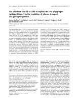
Fig. 6.1 Sites of natriuretic action along the nephron.
Carbonic anhydrase inhibitors such as acetazolamide suppress sodium reabsorption in the proximal tubule. The
loop diuretics (e.g., furosemide, torsemide, bumetanide)
inhibit sodium chloride reabsorption in the thick ascending limb of the loop of Henle. Distal convoluted tubule
natriuretics such as thiazides and thiazide-like diuretics
inhibit NaCl reabsorption in the early and late distal convoluted tubule. Collecting duct natriuretics inhibit electrogenic sodium transport in the cortical collecting duct and
the late distal tubule. Consequently the sites of action of
DCT and collecting duct natriuretics overlap slightly
altering the osmotic pressure of the glomerular
filtrate. Aquaretics constitute a new class of
agents that increase the excretion of solute-free
water by inhibiting vasopressin-mediated renal
water reabsorption.
excretion. As noted in Fig. 6.1, these nephron
segments are responsible for reabsorbing different fractions of the filtered sodium load, and each
segment plays its own important role in controlling ECF volume homeostasis. In general, more
proximal segments of the nephron reabsorb the
bulk of sodium from the glomerular filtrate, while
more distal segments “fine-tune” the urinary
sodium content by reabsorbing smaller fractions
of the total sodium load in a tightly regulated
fashion. The molecular targets and anatomic sites
of action of specific agents define many of their
clinical properties, including their therapeutic
uses, side effects, and chronic effects on nephron
adaptation. Commonly used natriuretics and key
pharmacologic aspects of their clinical use are
summarized in Table 6.1.
Natriuretics
Natriuretics are by far the most frequently used
class of diuretics and are among the most commonly prescribed drugs (source: IMS Health).
These agents promote a solute and water diuresis
by inhibiting the movement of sodium from the
tubular lumen to the blood. Four general subclasses of natriuretics primarily act on different
sites of the nephron to facilitate sodium and water
3–5
5–12
30
50–400
50–100
5–20
50–250
12.5–200
12.5–200
0.5–10
1.25–5
Several days
Several days
2h
2–4 h
2h
2h
1h
1–2 h
0.5–2 h
0.5–1 h
0.5–1 h
0.5 h
2h
125–375
20–320
5–200
0.5–10
25–400
Onset
of action
Oral dose
rangeb (mg)
65
~65
15–25
30–70
50–80
65
65
~95
50
80–100
80–100
100
100
Oral
bioavailability (%)
2–3 days
2–3 days
24 h
7–9 h
6–12 h
24–72 h
~ 24 h
£36 h
6–8 h
6h
4–6 h
12 h
8–12 h
Duration
of action
1.6
5
6–9
~4.5
2.5–14
35–55
20
~14
0.5–1
2–4
1–1.5
1
6–9
Elimination
half-life (t1/2) in
normal adults (h)
Yes (~100 %)
Yes (~100 %)
No
Yes
No
Yes
Yes (~10 %)
Yes (~100 %)
Minimal
Yes (80 %)
Yes
Yes (30 %)
No
Hepatic
metabolism?
b
Each value indicates the approximate maximal fractional excretion of sodium following acute administration of a maximally effective dose of natriuretic
The maximum safe dose of diuretic is rarely indicated or advantageous, and may be associated with excessive side effects
a
Diuretic class
Carbonic anhydrase inhibitors
Acetazolamide
Loop natriuretics
Furosemide
Torsemide
Bumetanide
Ethacrynic acid
Distal convoluted tubule
natriuretics
Hydrochlorothiazide
Chlorthalidone
Metolazone
Indapamide
Collecting duct natriuretics
Spironolactone
Eplerenone
Amiloride
Triamterene
Maximum change
in urinary fractional
excretion of
sodiuma (%)
5–6
Table 6.1 Commonly used natriuretics
Renal, fecal
Renal, fecal
Renal
Renal, fecal
Renal (100 %)
Renal, fecal
Renal, fecal
Renal, fecal
Renal, fecal
Renal
Renal
Renal, fecal
Renal (100 %)
Route of
excretion
6
Diuretic Therapy
173
174
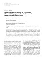
Fig. 6.2 Mechanism of action of carbonic anhydrase
(CA) inhibitors. Diagram of a proximal tubule cell illustrating expression of CA IV in the luminal brush border
and CA II in the cytoplasm. HCO3 from the glomerular
filtrate combines with protons extruded by the sodium
hydrogen exchanger (NHE3) to form carbonic acid
(H2CO3). CA IV breaks down H2CO3 to water and carbon
dioxide, which freely diffuse across cell membranes. CA
II then catalyzes the formation of intracellular bicarbonate
(HCO3) from cytoplasmic CO2 and OH. HCO3 is then
transported into the interstitium by a basolateral sodium
bicarbonate cotransporter (NBC1). CA inhibitors block
the bicarbonate reabsorptive process by inhibiting luminal
CO2 formation and cytoplasmic HCO3 generation by
inhibiting CA IV and CA II. This ultimately suppresses
vectorial sodium reabsorption across the proximal tubule
apical and basolateral membranes (see text)
Proximal Tubule Diuretics (Carbonic
Anhydrase Inhibitors)
Natriuretics that primarily act in the proximal
tubule suppress renal sodium reabsorption
through the inhibition of carbonic anhydrase
(CA). Two isoforms of this enzyme are primarily
responsible for reclaiming greater than 80 % of
the filtered sodium bicarbonate load in the early
proximal tubule (Fig. 6.2) [3]. During this process, protons secreted by proximal tubule cells
into the tubular lumen combine with filtered
bicarbonate to form carbon dioxide and water.
This reaction is catalyzed by type IV CA
expressed at the luminal surface of the proximal
tubule [4]. CO2 is lipid soluble and rapidly diffuses across the apical membrane of the proximal
tubule. Once inside the proximal tubule cell, CO2
combines with OH− in the presence of type II CA
to form HCO3−. Cytoplasmic bicarbonate ions are
then moved across the basolateral membrane of
A.R. Subramanya and D.H. Ellison
the proximal tubule cell in a sodium-dependent
manner via a Na+-HCO3− cotransporter [5]. Thus,
the net effect of this process is to reclaim bicarbonate and sodium from the glomerular filtrate
while maintaining cellular isotonicity.
Although different carbonic anhydrase inhibitors exhibit different isoform specificities [6],
these drugs have been shown to effectively suppress the activity of both type II and type IV CA.
The inhibition of either or both of these enzymes
results in reduced HCO3− reabsorption and a relative increase in luminal nonchloride anions [2].
This change in the anionic composition of the
proximal tubule luminal fluid prevents the apical
reabsorption of sodium cations, ultimately
increasing distal Na+ delivery [7].
In spite of the fact that CA inhibitors are capable of inhibiting proximal tubule Na+ exit by
40–60 %, the natriuretic effect of these drugs is
mild [8]. At most, proximal tubule natriuretics
only enhance net sodium excretion by 3–5 % [9],
except when combined with other agents (see
below). This is largely due to enhanced sodium
reabsorption by more distal nephron segments
[10]. Since chloride is reabsorbed with sodium in
both the thick ascending limb (TAL) of the loop
of Henle, and the distal convoluted tubule, urinary chloride excretion is low in patients treated
with CA inhibitors [11].
The principal effect of CA inhibition on the
urinary electrolyte composition is to increase its
bicarbonate and potassium content. As one might
expect, CA inhibition increases urinary bicarbonate excretion by 25–30 %, elevating the urine pH,
mimicking proximal renal tubular acidosis [7].
This is a direct consequence of the fact that downstream of the proximal tubule, bicarbonate is a
poorly reabsorbable anion [7]. In concert with the
increase in bicarbonaturia, acetazolamide
increases potassium excretion [8]. Current evidence suggests that the kaliuretic effect is indirect, and largely derived from increased potassium
secretion in the distal nephron due to a change in
the lumen-negative voltage and flow induced by
enhanced distal bicarbonate delivery [12].
Acetazolamide is the most commonly prescribed
CA inhibitor in the United States. Used as monotherapy, it is a mild diuretic due to its aforementioned
6
Diuretic Therapy
weak effect on natriuresis, and adaptive processes
downstream of the proximal tubule quickly give rise
to diuretic resistance. Acetazolamide, however, can
be very useful in combination with natriuretics that
block more distal NaCl transport pathways (see
Sect. 12, below).
Aside from its use as a diuretic, acetazolmide
has several other clinical uses. The bicarbonaturia associated with acetazolamide therapy is useful in the prevention of uric acid and cysteine
nephrolithiasis [13]. Raising the pH of the tubular lumen via CA inhibition is a tactic commonly
employed in the treatment of salicylate toxicity
[14]. Due to the fact that aqueous humor formation in the eye is dependent on CA-mediated
bicarbonate production, CA inhibitors [including
dorzolamide and brinzolamide (topical) and
acetazolamide and methazolamide (oral)] are
commonly used to treat chronic open-angle glaucoma [6]. The increased respiratory drive associated with acetazolamide-induced bicarbonaturia
makes it useful as a prophylactic for high-altitude
mountain sickness and pulmonary edema [15].
Generally, acetazolamide and other CA inhibitors are well tolerated. All CA inhibitors are sulfonilamide derivatives, and should be avoided in
patients with severe sulfa allergies. Serum potassium and bicarbonate levels need to be monitored
due to the associated hypokalemia and metabolic
acidosis that often accompany therapy. In contrast
to its therapeutic utility in uric acid and cysteine
stone formers, CA inhibition increases the risk of
nephrolithiasis in patients with hypercalciuria
due to the elevation in urine pH and increased calcium excretion [16]. CNS and other neurologic
symptoms, such as drowsiness, fatigue, and paresthesias, are other known side effects.
Loop Diuretics
Commonly used loop diuretics in the United
States include furosemide, bumetanide, torsemide,
and ethacrynic acid (Table 6.1). The primary
molecular target of these agents is the Na-K-2Cl
cotransporter (NKCC2), which reabsorbs sodium,
potassium, and chloride ions in the TAL of the
loop of Henle [17]. Since this nephron segment is
impermeable to water, NKCC2 plays a crucial
role in generating the hypertonic medullary
175
interstitium that is essential for efficient urinary
concentration [18]. Twenty-five percent of the
filtered NaCl load is reabsorbed by this cotransporter [17]; thus, inhibition of its transport activity leads to a marked increase in sodium chloride
excretion. Indeed, the loop natriuretics constitute
the most potent class of diuretics used in current
clinical practice [2].
Loop diuretics bind to a site on NKCC2
exposed at the apical surface of the epithelium
lining the lumen of the TAL [19]. Loop diuretic
binding to the cotransporter interferes with the
apical translocation of ions passing through the
TAL; this increases the luminal NaCl and K content. The increase in luminal NaCl and K content
correlates with a reduction in the medullary concentration gradient [18]. Consequently, the selective water-reabsorptive response to vasopressin
during loop diuretic-mediated ECF volume contraction is diminished, ensuring that urine volume
increases and urine osmolality approaches that of
plasma.
In addition to increasing Na and Cl excretion
via NKCC2 inhibition in the TAL, loop diuretics
are powerful stimulators of renin release. This
effect is a direct consequence of loop diureticinduced changes in tubular fluid load sensing by
the macula densa, a specialized group of epithelial cells anatomically positioned at the end of the
TAL. Macula densa cells recognize alterations in
fluid delivery by sensing changes in NaCl influx
through NKCC2 cotransporters expressed at the
tubular lumen [20]. A decrease in NKCC2mediated NaCl entry activates local signaling
cascades to trigger renin release from granular
cells in the juxtaglomerular apparatus (JGA) [21].
Since the stimulus for renin release hinges on a
decrease in NKCC2-mediated NaCl influx, direct
inhibition of NKCC2 by loop diuretics dramatically augments the process [22]. The exaggeration in renin release seen with high-dose loop
diuretic therapy may be harmful in some treatment scenarios. In two studies, 1–1.5 mg/kg intravenous boluses of furosemide given to patients
with chronic heart failure (HF) caused a transient
decline in hemodynamic parameters, resulting in
a worsening of HF symptoms over the first hour
of treatment [23, 24]. This finding was attributed
176
to over-activation of the renin-angiotensin and/or
sympathetic nervous systems [25]. Others have
postulated that chronic loop diuretic-induced
renin release may contribute to loop diuretic
resistance [26]. Moreover, chronic deleterious
over-activation of the intrarenal renin-angiotensin
system by long-term diuretic use is a theoretical
risk that could contribute to the development of
chronic kidney disease [27]. Currently, efforts are
being taken to develop agents that may block
paracrine signaling from the macula densa to the
renin-producing cells of the JGA. Such an inhibitor would in all likelihood attenuates the tendency
of loop diuretics to overstimulate the renin-angiotensin system.
NKCC2-mediated NaCl cotransport in the
macula densa is also an essential step in a critical
renal homeostatic process, tubuloglomerular
feedback (TGF). TGF is a negative feedback
mechanism in which the glomerular filtration rate
(GFR) is tightly controlled in response to changes
in tubular fluid delivery to the macula densa.
Luminal sodium chloride is sensed by the macula
densa by way of its cotransport via NKCC2. The
increase in intracellular NaCl then triggers a local
signaling cascade involving adenosine [21]. This
induces preglomerular vasoconstriction [28],
decreasing the GFR and filtration fraction. Loop
diuretics impede TGF by interfering with the
NKCC2 sensing step; this makes the JGA much
less effective at matching GFR with tubular fluid
delivery to the TAL [29]. Thus, through the
blockade of TGF, loop diuretics tend to maintain
the GFR at a higher level than would occur if the
TGF were not blocked.
In addition to their profound natriuretic and
kaliuretic effects, loop diuretics enhance the urinary excretion of calcium and magnesium. Na-K2Cl cotransport in the TAL generates a
lumen-positive transepithelial voltage, largely
owing to the recycling of intracellular potassium
cations back into the tubular lumen via low- and
high-conductance potassium channels [18]. This
voltage gradient favors the paracellular reabsorption of calcium and magnesium. NKCC2 inhibition by loop diuretics dissipates the transepithelial
voltage by disrupting the driving force for K+
recycling; therefore, calcium and magnesium
A.R. Subramanya and D.H. Ellison
reabsorption decreases. Because of their
hypercalciuric effects, loop diuretics are sometimes used to treat hypercalcemia in the volumereplete patient, although they are now generally
reserved for prevention and treatment of hypervolemia in this setting [30].
Furosemide, bumetanide, and torsemide are
absorbed from the gut within 30 min to 2 h following oral administration (Table 6.1). Delayed
absorption may occur in the edematous patient
due to bowel wall edema [31]; this problem is
bypassed with intravenous therapy. Since the oral
bioavailability of furosemide is as low as 50 %,
when converting a patient from an intravenous to
oral formulation, the dose is often doubled; the
same does not hold for bumetanide and torsemide
because the bioavailability is higher. Of these
commonly used loop diuretics, furosemide is the
only one which is cleared primarily by renal processes; in contrast, bumetanide and torsemide are
largely metabolized in the liver. Consequently,
the half-life of furosemide is increased in renal
failure, whereas this is not the case for bumetanide
or torsemide [32].
Owing to their efficacy, loop diuretics are
among the most frequently prescribed drugs in
the world. They are commonly used to treat most
edematous conditions, including HF, renal failure, cirrhosis, and nephrotic syndrome. The treatment of these conditions is discussed in detail
below (see Sect. 7, below).
Although the loop diuretics (particularly furosemide, bumetanide, and torsemide) are well tolerated, several adverse effects are associated with
their clinical use. Due to their kaliuretic effects,
hypokalemia is a common consequence of therapy, and serum potassium levels must be monitored regularly. Periodic replacement of
magnesium and calcium may be required due to
the enhanced urinary excretion of these divalent
cations. As a consequence of increased sodiumdependent proton secretion and aldosterone activity, metabolic alkalosis is often observed in the
setting of aggressive loop diuretic therapy [33].
Ototoxicity is the most common non-renal
toxic effect observed with loop diuretic treatment, and is likely due to cross-reactivity against
the secretory Na-K-2Cl isoform NKCC1, which
6
Diuretic Therapy
is expressed in the lateral wall of the cochlear
duct [34]. The hearing loss associated with loop
diuretics is dependent on the peak level of drug in
the bloodstream [35]. Consequently, this adverse
effect is more commonly seen with intravenous
therapy. Due to its renal clearance, intravenous
furosemide must be administered with care to
avoid ototoxicity in the patient with renal
insufficiency. It has been recommended that furosemide infusion be no more rapid than 4 mg/min
[36]. Ototoxicity may be more common with
ethacrynic acid than the other loop diuretics.
Although hearing loss is often reversible, permanent damage has been reported [36].
Like many other diuretics, furosemide,
bumetanide, and torsemide are sulfonamide
derivatives and should not be used in patients
with severe sulfa allergies. Ethacrynic acid, on
the other hand, is the only loop diuretic available
in the United States that does not contain sulfa
moieties, and is an effective alternative for the
edematous sulfa allergic patient. The former
manufacturer sold production rights for ethacrynic
acid to another company; thus ethacrynic acid
remains available as both an oral and intravenous
preparation.
Distal Convoluted Tubule Diuretics
Thiazides, including chorothiazide and hydrochlorothiazide, and thiazide-like diuretics such as
metolazone and chlorthalidone primarily act in
the distal convoluted tubule (DCT). The major
effect of these drugs is to suppress sodium chloride reabsorption in the DCT [37]. The molecular
target of the DCT diuretics is the thiazide-sensitive Na-Cl cotransporter (NCC), which is responsible for reabsorbing approximately 5 % of the
filtered NaCl load [37]. Given its anatomic position in the distal nephron, NCC plays an important role in “fine-tuning” the final concentration
of NaCl in the urine. Consequently, in the setting
of normal GFR, NCC-mediated NaCl reabsorption is one of the key renal mechanisms involved
in the regulation of ECF volume [38].
Thiazides and thiazide-like diuretics are
organic anions that bind to a luminally exposed
site on NCC cotransporters expressed at the apical surface of DCT cells [39]. Thiazide binding
177
interferes with the ability of NCC to translocate
sodium and chloride ions from the DCT lumen.
The increased natriuresis afforded by the DCT
diuretics contracts ECF volume and reduces
blood pressure, making them effective antihypertensive agents [40].
Structurally similar to CA inhibitors, thiazides
also have modest inhibitory effects on proximal
sodium transport. This proximal effect probably
contributes little to the final urinary NaCl content
[41]. It does, however, contribute to the changes
in renal hemodynamics seen with thiazides.
During acute administration, thiazides activate
TGF, causing pre-glomerular vasoconstriction
and a reduction in the glomerular filtration rate
[42]. The ability of thiazides to inhibit CA likely
plays some role in this process, since the decreased
proximal Na reabsorption seen with CA inhibition increases sodium delivery to the loop of
Henle and macula densa. The effect of thiazides
to stimulate TGF is likely less of an issue during
chronic administration, since the sustained reduction in ECF volume diminishes the delivery of
solutes to the macula densa [43]. As one might
expect, chronic thiazide treatment also enhances
renin release due to decreased macula densa
sodium chloride delivery [43].
When administered chronically, DCT diuretics
decrease urinary calcium excretion, making them
highly effective agents in the treatment of calcium
nephrolithiasis [44]. Several mechanisms have
been proposed to explain the hypocalciuric effect
of thiazides. Recently, work in knockout mice
lacking TRPV5, the major portal for calcium
entry in the distal nephron, still exhibits thiazideinduced hypocalciuria due to enhanced calcium
reabsorption [45]. This observation is likely a
consequence of ECF volume contraction and
enhanced proximal sodium-dependent calcium
transport. Thus, the mechanism by which DCT
diuretics exert their hypocalciuric effect is at least
in part related to enhanced proximal calcium reabsorption. More recent studies, however, confirm
an important effect of thiazide diuretics to reduce
urinary calcium excretion, independent of changes
in sodium balance [46]. In contrast to their proreabsorptive effects on calcium, chronic DCT
diuretics increase urinary magnesium excretion
178
[47]. This may be due to the indirect effect of thiazides to suppress the expression of magnesium
channels in the DCT, owing to structural effects
[45]. Alternatively, thiazides might suppress magnesium reabsorption through the effects of the
drug on the distal nephron transepithelial voltage
[48].
DCT diuretics increase urinary potassium
excretion [12]; this effect is largely due to the
effects of thiazides and thiazide-like drugs on
potassium secretion in the distal nephron. Chronic
thiazide administration increases aldosterone
concentrations, which facilitates distal potassium
secretion via aldosterone-sensitive K channels in
the late DCT and cortical collecting duct [12]. In
addition, thiazides increase luminal sodium and
chloride ionic content in the DCT; this tends to
increase flow to downstream nephron segments
and augment flow-dependent K secretion [49].
The hypomagnesemia seen with thiazide administration also likely contributes to the tendency
for hypokalemia [50].
DCT diuretics are absorbed rather rapidly,
reaching peak concentrations within 90 min to
4 h after ingestion [51]. The half-lives of DCT
diuretics vary widely (Table 6.1). Of the agents
commonly used in the United States, hydrochlorothiazide has a short half-life, while chlorthalidone and metolazone are longer-acting [51]. The
extended half-life of chlorthalidone has been the
subject of speculation that it may be a more potent
diuretic and antihypertensive than hydrochlorothiazide [52]. A recent trial comparing the blood
pressure lowering effects of these two drugs suggests that chlorthalidone might be a more effective antihypertensive agent, although the question
of dose equivalency was difficult to resolve in
this study [53].
The DCT diuretics have many clinical uses. In
patients with normal GFR, thiazides are effective
blood pressure-lowering agents commonly used
to treat essential hypertension [54]. The guidelines of the Seventh Report of the Joint National
Committee of Prevention, Detection, Evaluation,
and Treatment of High Blood Pressure (JNC-7)
recommend that thiazides should be first-line
agents in the treatment of essential hypertension
[55]. DCT diuretics are also commonly used as
A.R. Subramanya and D.H. Ellison
monotherapy to treat edematous disorders such
as HF, but they are usually considered less potent
than loop diuretics in achieving a substantial
diuresis HF [26]. Thiazides and thiazide-like
diuretics are, however, very effective in the treatment of edematous patients who have become
resistant to loop diuretics (see Sect. 7, below).
Owing to their hypocalciuric effects, the DCT
diuretics are the treatment of choice for patients
with idiopathic hypercalciuria and nephrolithiasis [44]. In nephrogenic diabetes insipidus, thiazides exert a paradoxical antidiuretic effect, and
this has been used as an effective treatment of the
disorder. Although the mechanism for the antidiuretic effect of thiazides remains unclear, these
drugs appear to increase collecting duct water
channel expression, increasing free water reabsorption [56, 57]. Other potential mechanisms
include thiazide-induced TGF activation (as
described above), which would reduce GFR and
distal water delivery [42].
As with the other classes of diuretics, thiazides and thiazide-like diuretic agents are generally well tolerated, but several potential adverse
effects deserve mention. Hyponatremia can be
observed with all classes of diuretics, but is particularly common with DCT diuretic therapy
[58]. In fact, hyponatremia can become severe
enough in the setting of DCT diuretic therapy to
become life threatening. There are at least three
mechanisms which contribute to the hyponatremia that can accompany DCT diuretic therapy.
First, the inhibition of solute reabsorption in the
distal convoluted tubule impairs free water excretion (see above). Second, thiazides increase proximal Na reabsorption and inhibit TGF (see
above); these effects impair solute and water
delivery to the distal nephron, reducing free water
clearance. Finally, thiazide treatment stimulates
thirst centers in the brain, increasing water consumption [59]. Risk factors for thiazide-induced
hyponatremia include female gender, low total
body mass, and advanced age [58].
DCT diuretics induce disturbances related to
glucose and lipid metabolism. DCT diuretics
cause a dose-dependent increase in glucose intolerance [60, 61]. This observation was initially
made in the 1950s, and was thought to be a
6
Diuretic Therapy
complication only seen in patients treated with
high doses of diuretics. More recent studies,
however, have revealed that glucose intolerance
may be seen even with lower doses of DCT
diuretics. In ALLHAT, the largest blood pressure
lowering randomized controlled trial conducted
to date, the incidence of new-onset diabetes was
significantly higher in the chlorthalidone-treated
group compared to groups treated with amlodipine or lisinopril (11.9 % vs. 9.8 % or 8.1 %,
respectively) [40]. The mechanism by which
DCT diuretics cause glucose intolerance is not
entirely clear, but may be related to the degree of
diuretic-induced hypokalemia, which may alter
insulin secretion by pancreatic beta cells and glucose uptake by muscle [62]. This was recently
supported by a quantitative review of 59 clinical
trials of thiazide diuretics in which blood glucose
and potassium levels were reported; the results of
this study suggested a dose-dependent inverse
relationship between blood glucose and serum
potassium levels in patients treated with thiazides
[63]. Thus, the risk of new-onset diabetes associated with DCT diuretic therapy may be ameliorated if potassium levels are monitored closely
and maintained within the normal range. The
DCT diuretics also increase the levels of total
cholesterol, low-density lipoprotein, and triglycerides, and reduce HDL. Although the mechanisms underlying the effects of these drugs on the
lipid profile remain unclear, they are probably
linked to those that lead to impaired glucose tolerance. Like the effects of DCT diuretics on blood
glucose, their hyperlipidemic effects are dose
dependent. In ALLHAT, the mean total cholesterol concentrations were higher in the group randomized to chlorthalidone, and averaged 2–3 mg/
dl higher than the other treatment arms [40].
Cortical Collecting Tubule Natriuretics
Three pharmacologically distinct groups of drugs
act to inhibit sodium reabsorption in the cortical
collecting tubule: mineralocorticoid receptor antagonists (spirolactones), pteridines (triamterene),
and pyrazine-carbonyl-guanidines (amiloride).
These agents have a tendency to minimize potassium secretion rather than promote it, as is
commonly seen with diuretics which act on other
179
segments of the nephron. For this reason, the
cortical collecting tubule natriuretics are collectively known as “potassium-sparing diuretics.”
The site of action of potassium-sparing diuretics is the aldosterone-sensitive distal nephron
(ASDN), which by current definitions includes
the late distal convoluted tubule, connecting
tubule, and cortical collecting duct [38]. This is
the final site of sodium reabsorption in the kidney, and is responsible for reclaiming approximately 3 % of the filtered NaCl load. Ultimately,
the effect of the potassium-sparing diuretics is to
inhibit sodium transport by the aldosterone-sensitive epithelial sodium channel (ENaC). ENaC
channels selectively reabsorb sodium ions, and
their synthesis and expression at the apical surface of cells of the ASDN are tightly controlled
by the mineralocorticoid hormone aldosterone
[64]. The potassium-sparing effect of these
diuretics is largely due to their ability to inhibit
ENaC (Fig. 6.3); blocking the reabsorption of
sodium cations in the collecting tubule decreases
the lumen negativity of the segment, which
diminishes the driving force for potassium and
hydrogen ion secretion [65].
The spirolactones inhibit aldosterone action
by binding to intracellular mineralocorticoid
receptors in the ASDN. This causes the retention
of mineralocorticoid receptors in the cytoplasm
and prevents their nuclear translocation, rendering them unable to promote the transcription of
aldosterone-induced gene products [66]. Because
of their effects on gene transcription, the spirolactones have a delayed onset of action, and may
not reach their peak natriuretic effects until several days after starting the drug [51].
Spironolactone has at least a tenfold higher binding affinity to the mineralocorticoid receptor than
its newer cousin eplerenone, but has a greater
tendency to activate the cytochrome P450 system
[67]. Although the half-life of spironolactone is
short, it has long-acting metabolites that greatly
prolong its functional half-life. Although
amiloride and triamterene are structurally different, both of these compounds bind directly to
ENaC and inhibit its activity [68, 69]. At higher
doses, amiloride inhibits multiple ion transport
pathways, most notably the sodium hydrogen
180
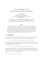
Fig. 6.3 Mechanisms of action of collecting duct
natriuretics. Diagram of a connecting or cortical collecting duct cell illustrating major pathways for sodium entry
and potassium secretion. In the collecting duct, sodium
reabsorption via the epithelial sodium channel (ENaC) is
electrogenic, and generates a lumen-negative voltage of
−30 mV. This voltage provides the driving force for potassium secretion via the renal outer medullary potassium
channel (ROMK). All collecting duct natriuretics ultimately suppress ENaC-mediated Na reabsorption. Their
“potassium-sparing” effect derives from the reduced
potassium secretion seen with the dissipation of the voltage gradient. Amiloride and triamterene block luminal
Na+ entry by binding to the channel, while the aldosterone
antagonists such as spironolactone interfere with cell signaling processes that stimulate ENaC by blocking aldosterone binding to the mineralocorticoid receptor (MR)
exchangers; this effect however is not as relevant
with respect to the low doses of the drug that are
used in clinical practice. All of the potassiumsparing diuretics are weak natriuretics, increase
sodium excretion in normal subjects by no more
than 1–2 % [2]. In clinical practice, triamterene
has weaker diuretic potency than either amiloride
or spironolactone.
The mineralocorticoid receptor antagonists
are effective natriuretics that reduce blood pressure in patients with hyperaldosteronism [70].
This is true for patients with primary aldosterone
excess from either adrenal adenomas or bilateral
adrenal hyperplasia, or secondary hyperaldosteronism from HF, cirrhosis, or nephritic syndrome.
Conversely, spironolactone and eplerenone are
ineffective in inducing a natriuresis in patients
with a nonfunctional adrenal gland. With regard
to the secondary hyperaldosteronemic disorders,
spironolactone and eplerenone are particularly
A.R. Subramanya and D.H. Ellison
effective when used with loop diuretics and ACE
inhibitors to treat HF [71, 72]. RALES and
EPHESUS were two large randomized placebocontrolled trials in which patients with advanced
HF were treated with spironolactone and eplerenone, respectively. In both trials, aldosterone
antagonist therapy reduced the risk of all-cause
mortality in patients with chronic HF and left
ventricular dysfunction following acute myocardial infarction. Although a non-renal effect may
confer the mortality-reducing benefits seen in
these studies, a current debate exists in the literature as to whether the benefit of these agents is
related to the prevention of hypokalemia, a
known risk factor for sudden cardiac death
hypokalemia [73].
In addition, owing to its inhibitory effect on
aldosterone activity, spironolactone has been
shown to be a more effective diuretic than furosemide in the treatment of cirrhotic ascites [74]
(see Sect. 7, below).
Amiloride and triamterene are commonly used
in combination with loop or thiazide diuretics to
reduce potassium loss and the risk of hypokalemia.
Amiloride has been used to treat primary hyperaldosteronism [75] or other potassium wasting
states such as Liddle’s, Bartter’s, or Gitelman’s
syndrome [76, 77]; the weak potency of triamterene renders it incapable of treating these disorders. Amiloride has also been used to treat
lithium-induced nephrogenic diabetes insipidus.
The beneficial effect of amiloride in this disorder stems from its ability to block the intracellular entry of lithium ions through the ENaC
pore [78].
The major adverse effect encountered with the
use of spironolactone or eplerenone is hyperkalemia [79]. Patients that are particularly at risk
for hyperkalemia include those with decreased
GFR and those that are on active potassium supplementation. Consequently, prior to starting
therapy with a mineralocorticoid receptor antagonist, all potassium supplements must be stopped
and serum potassium levels should be monitored.
Spironolactone exerts other endocrine effects due
to its cross-reactivity with androgen and progesterone receptors [71, 80]. Gynecomastia is a
common side effect in males; in RALES, the
6
Diuretic Therapy
incidence was 10 % [71]. Other common symptoms in males include breast tenderness, decreased
libido, and impotence. Females may experience
breast tenderness, hirsutism, or irregular menses.
Eplerenone, in contrast, appears to have greater
specificity for the mineralocorticoid receptor. In
EPHESUS, the incidence of impotence and gynecomastia in men taking eplerenone was not different from placebo [72].
Due to their potassium sparing effects,
amiloride and triamterene can cause hyperkalemia, and should be avoided in patients with
low GFR or those who are taking potassium supplements. Triamterene can promote the formation of renal stones by acting as a nidus for the
precipitation of uric acid or calcium oxalate [81].
Consequently, this drug is contraindicated in
stone formers. In addition, triamterene has been
reported to be associated with acute kidney injury,
particularly when used in combination with indomethacin [82, 83].
Osmotic Diuretics
Osmotic diuretics are substances that are freely
filtered at the glomerulus but are poorly reabsorbed. The ability of these drugs to provoke a
diuresis is dependent on their ability to generate
an osmotic gradient within the tubular lumen.
Thus, the osmotic diuretics do not exert their
diuretic effects through a specific molecular target. Mannitol is the osmotic diuretic used most
commonly in clinical practice. Mannitol infusion
increases the urinary excretion of water, sodium,
calcium, magnesium, and phosphorus [84, 85].
Once mannitol is freely filtered at the glomerulus, its presence in the proximal tubule lumen offsets the osmotic gradient that is usually generated
by the net reabsorption of sodium through specific
transport mechanisms. This minimizes proximal
water reabsorption. As the glomerular filtrate
travels down the nephron, non-reabsorbable mannitol ions replace sodium as the predominant element contributing to the urine osmolality. The
relative displacement of sodium ions decreases
the driving force for sodium reabsorption in multiple nephron segments including the thin loop of
181
Henle and collecting duct; this results in a net
increase in the fractional excretion of Na [84,
85]. In addition, cortical and medullary renal
blood flow is increased [11, 86]. Cortical increases
in renal blood flow contribute to an increase in
the urine flow rate, resulting in a net diuresis. The
increase in medullary renal blood flow “washes
out” the papillary sodium and urea content; this
impairs the urinary concentrating mechanism,
perpetuating mannitol’s diuretic effect [87].
Since mannitol cannot be absorbed from the GI
tract [88], it is administered intravenously. It has a
plasma half-life of approximately 2 h and is almost
entirely cleared by the kidneys [89]. Its osmotic
properties are beneficial as an acute treatment to
lower increases in intraocular or intracranial pressure [90]. Mannitol is also used as prophylaxis
against the dialysis disequilibrium syndrome, a
disorder that typically occurs in patients with
severe azotemia who are initiating hemodialysis
[91, 92]. In this syndrome, patients develop postdialytic acute central nervous system symptoms,
such as nausea, blurred vision, confusion, headaches, and seizures. The rapid removal of solutes
such as urea results in the development of an
osmotic gradient that favors the movement of
water into brain cells [92]. This fluid shift leads to
cerebral edema and neurological dysfunction. To
reduce the magnitude of the osmotic gradient,
mannitol is sometimes infused during the dialysis
treatment to boost the plasma osmolality.
Many of the adverse effects of mannitol treatment are related to problems that can develop if it
is poorly cleared from the circulation, or if too
much of it is administered too quickly. Mannitol
infusion increases the pulmonary capillary wedge
pressure and can cause pulmonary edema in
patients with impaired left ventricular function.
Overzealous use acutely leads to dilution of the
plasma bicarbonate and sodium concentrations,
causing metabolic acidosis and hyponatremia
[93]. In addition, acute high-dose mannitol infusion promotes the extracellular movement of
potassium and causes hyperkalemia [94].
Prolonged mannitol treatment depletes total body
potassium, leading to hypokalemia. Excessive
losses of sodium and water cause volume depletion, and since electrolyte-free water is excreted
A.R. Subramanya and D.H. Ellison
182
Table 6.2 V2 Receptor antagonists currently under development or in clinical use
Compound
Conivaptan (YM-087)
Receptor
V1a + V2
Route
Intravenous
Tolvaptan (OPC 41–061)
V2
Oral
Lixivaptan (VPA-985)
V2
Oral
Satavaptan (SR-121463)
V2
Oral
in excess relative to sodium, hypernatremia develops [95]. In extreme cases of mannitol intoxication, the drug can be rapidly removed from the
circulation with hemodialysis mannitol [96].
Aquaretics (Vasopressin Receptor
Antagonists)
The vasopressin receptor antagonists promote the
excretion of solute-free water, and thus are known
as “aquaretics.” One of these agents is currently
available for intravenous administration. Oral
analogues are currently the subject of clinical trials, designed to determine their efficacy and clinical use in various disease states. As discussed in
detail below, they hold promise for serving as
effective diuretics to treat edematous conditions
accompanied by hyponatremia owing to the
excessive release of arginine vasopressin (AVP,
antidiuretic hormone).
The vasopressin receptor antagonists are competitive inhibitors of AVP action in water-reabsorptive segments of the nephron. The actions of
AVP are carried out by two receptors, V1 and V2.
V1 receptors are divided into two major subtypes.
V1a receptors are expressed in multiple tissues,
including vascular smooth muscle, platelets, and
myocardium. V1b receptors are predominantly
found in cells of the anterior pituitary gland. V2
receptors are predominantly localized to principal cells of the distal nephron, including the connecting tubule and cortical and medullary
collecting duct [97]. During states of high osmolality or extreme ECF volume contraction, AVP
is released from its storage centers in the posterior pituitary. Once the hormone binds to V2
Current status
FDA approved for treatment of euvolemic and
hypervolemic hyponatremia
FDA approved for treatment of euvolemic and
hypervolemic hyponatremia
Phase 3 clinical trials in hyponatremic patients
with heart failure
Phase 3 clinical trials in hyponatremic patients
with ascites
receptors located at the basolateral membrane of
distal nephron principal cells, it triggers a cyclic
AMP-dependent signaling cascade that leads to
an increase in the expression of aquaporin-2
water channels at the luminal surface. Ultimately,
this enhances water reabsorption and normalizes
the serum osmolality and extracellular fluid volume. Although some of the vasopressin receptor
antagonists show cross-reactivity towards V1
receptors, all of the members of the class competitively inhibit AVP binding to V2 receptors;
binding of the drug to these receptors thus
decreases water reabsorption, enhancing free
water excretion and urinary flow [97].
V2 receptor antagonists that are currently
being clinically used or are under development
for commercial use are listed in Table 6.2. To
date, the Food and Drug Administration has only
approved one member of the class, conivaptan,
for clinical use in the United States. Conivaptan
(YM-087) is a vasopressin receptor antagonist
that exhibits activity towards both V1a and V2
receptors [98]. Clinical trials illustrate its diuretic
potency. In one study performed in healthy volunteers, oral conivaptan increased urinary flow
by sevenfold and reduced urinary osmolality
from 600 mOsm/kg to less than 100 mOsm/kg
within 2 h of administration [99]. In accordance
with studies performed in laboratory animals, the
peak effect of conivaptan was seen 2 h after giving the dose, and persisted for at least 6 h. Despite
the oral efficacy seen in this study, there are concerns about its interactions with other drugs
metabolized by the CYP3A4 pathway, so the
agent has been developed for clinical use as an
intravenous preparation only [97]. The primary
FDA-approved indication for conivaptan use was
6
Diuretic Therapy
for the treatment of euvolemic hyponatremia in
hospitalized patients, but this has recently been
hypervolemic hyponatremia, as well. The initial
approval was based on a double-blinded placebocontrolled study of 56 patients with euvolemic
hyponatremia. In this trial, a loading dose, followed by a 4-day continuous infusion at 20 or
40 mg/day increased the serum Na concentration
at least 4 mEq/l from the baseline concentration at
the start of the study [97]. Conivaptan was also
shown to correct hyponatremia in decompensated
CHF during 4 days of intravenous infusion [100].
Tolvaptan (OPC-41061) is an oral once-daily
vasopressin receptor antagonist that exhibits
higher selectivity for V2 receptors. This agent
has received much attention due to several recent
clinical studies evaluating its utility in hyponatremic and edematous disorders, particularly
CHF. Although the drug is currently not FDA
approved for the treatment of these conditions, it
may become clinically available in the near
future. In ACTIV in CHF [101], patients with
New York Heart Association class III or IV HF
were treated with between 30 and 90 mg of
tolvaptan or placebo and were reassessed at 24 h
or 7 weeks post treatment. In this study, the
tolvaptan-treated patients exhibited decreased
edema and body weight and a higher serum
sodium compared to the placebo arm. No changes
in serum potassium levels, heart rate, or blood
pressure were noted. Although these findings
illustrate the diuretic potency of tolvaptan, no
significant difference in rate of rehospitalization
was appreciated. These data were very recently
echoed in two papers describing the short- and
long-term results from the Efficacy of Vasopressin
Antagonism in Heart Failure Outcome Study
with Tolvaptan (EVEREST) [102, 103].
EVEREST was a large-scale randomized placebo-controlled trial that evaluated the effect of a
30 mg daily dose of tolvaptan on the outcomes of
more than 4,000 patients with decompensated
CHF. The aggregate results implicate tolvaptan
as a safe and effective treatment that is capable of
reducing some of the adverse symptoms of decompensated HF, when added to current pharmacologic standard of care. The observed
improvement in CHF symptoms was related to
183
the diuretic effect of tolvaptan, since the beneficial
change in symptom score was driven by a reduction in body weight. It is important to note however that despite adequate power, there was no
measurable beneficial effect of tolvaptan on the
long-term composite primary end point of allcause mortality or rehospitalization for HF.
The Studies of Ascending Levels of Tolvaptan
(SALT-1 and SALT-2) were identical randomized
double-blinded placebo-controlled trials conducted in parallel in Europe and the United States
[104]. Both of these trials enrolled more than 200
hyponatremic patients with normal or expanded
ECF volume, and randomized them to treatment
with 15 mg tolvaptan per day or placebo. Based
on the serum sodium levels, the dose could be
increased to 30 or 60 mg/day. The results of these
studies demonstrated that tolvaptan significantly
increased the serum sodium concentration relative to patients receiving placebo. No significant
changes in renal function, blood pressure, or
heart rate were noted, and the serum Na concentration fell back to baseline within 7 days of stopping the drug. Consistent with other studies of
vasopressin receptor antagonists, the observations from SALT-1 and SALT-2 suggest that
tolvaptan may be an effective acute treatment for
hyponatremia.
As mentioned above, tolvaptan is currently
not FDA approved for the treatment of hyponatremia or CHF. It did, however, recently receive a
“Fast-Track” designation from the FDA for the
treatment of autosomal dominant polycystic kidney disease (ADPKD). The Fast-Track designation was granted on the basis of a current lack of
effective treatments for ADPKD, and empiric
evidence suggesting that vasopressin receptor
antagonists may reduce cystic fluid accumulation, expansion, and rupture [105, 106]. Phase III
clinical trials are currently being conducted to
determine the role of tolvaptan in reducing
ADPKD symptoms and disease progression.
The side effect profile of the vasopressin receptor antagonists continues to evolve. Major side
effects that have been reported during treatment
include dry mouth and thirst. As one might expect,
hypernatremia has been observed; consequently,
serum sodium levels should be closely monitored
184
during treatment. Infusion site reactions are
common during conivaptan therapy, occurring in
greater than 63 % of subjects treated at a dose
higher than 20 mg/day, according to the package
insert. Conivaptan and tolvaptan are both primarily metabolized by the cytochrome P450 isoenzyme CYP3A4, and their concentrations may be
increased by CYP3A4 inhibitors, including ketoconazole, indinavir, and clarithromycin [98].
Concomitant use of these agents is contraindicated. In addition, conivaptan and tolvaptan inhibit
CYP3A4 activity, so their use with drugs that are
metabolized by the isoenzyme (including some
HMG CoA reductase inhibitors) should be avoided
if possible. Conivaptan reduces the rate of digoxin
clearance, and clinicians should be aware of the
possibility that blood digoxin levels may rise.
Tolvaptan, in contrast, does not significantly affect
the serum digoxin concentration [97].
Clinical Use of Diuretics
General Concepts
Determinants of Maximal Diuresis
The change in urinary flow seen during the
administration of a diuretic depends on many factors, including its mechanism of action, dose,
kinetics of entry into the bloodstream, and delivery to its site of action.
In many cases, the site of action of a diuretic
determines its potency. For example, loop diuretics are more potent than the DCT diuretics such
as hydrochlorothiazide. This observation is
largely related to the fact that loop diuretics
inhibit a transport pathway responsible for reabsorbing up to 30 % of the filtered sodium load,
while DCT diuretics inhibit a pathway responsible for reabsorbing only 5–10 %. Similarly, mineralocorticoid antagonists have a mild natriuretic
effect due to the fact that they suppress a pathway
responsible for reabsorbing only 3 % of the
filtered Na load. There are, of course, exceptions
to this rule. The carbonic anhydrase inhibitors,
which reduce proximal tubule reabsorption, are
only weakly natriuretic due to adaptive changes
in the loop of Henle and DCT [10].
A.R. Subramanya and D.H. Ellison
Diuretic efficacy is highly dependent on the
kinetics of drug entry into the bloodstream. The
dynamics of drug absorption may be perturbed in
certain clinical situations, and this might result in
a diminished effect. This is exemplified by the
pharmacokinetics of furosemide. In normal individuals, the rate of furosemide absorption from
the GI tract is not rapid, and a reservoir of drug
can persist long after the diuretic is administered
[51]. This reservoir provides a consistent source
of diuretic that, when dosed appropriately, maintains the blood furosemide concentrations above
the natriuretic threshold. In certain edematous
states, however, impaired absorption from the gut
may slow furosemide absorption to a point where
it dips below the diuretic threshold, rendering it
ineffective [31]. To compensate for this, switching to different loop diuretics with higher bioavailabilities, such as torsemide or bumetanide,
might facilitate a brisker diuresis [107]. Another
even more effective approach would be to switch
to an intravenous loop diuretic preparation, which
is of course 100 % bioavailable.
The effectiveness of a diuretic is also dependent on its rate of delivery to its site of action. In
the case of furosemide, its rate of delivery to
NKCC2 binding sites in the tubular lumen can be
inferred by measuring the rate of urinary furosemide excretion. If the rate of urinary furosemide excretion is low, few binding sites are
inhibited, leading to poor diuretic effectiveness.
Conversely, a high rate of furosemide delivery
will lead to diuretic inefficiency, since any furosemide molecules in excess of the total number
of binding sites will be wasted as they move past
their sites of action and into the collecting system. Brater established that diuretics such as
furosemide have an excretion rate of maximal
efficiency, i.e., a rate of diuretic delivery that is
associated with a maximal natriuretic response
[108]. This concept helps to explain why an orally
administered dose of furosemide can be more
effective than an equivalent single intravenous
dose in individuals with normal GI absorption
(Fig. 6.4). When a dose of furosemide is given as
an intravenous bolus, the rate of diuretic excretion is very high early on in the time course, substantially greater than the rate of maximal
6
Diuretic Therapy
Fig. 6.4 Time course of urinary furosemide excretion
following intravenous (solid line) and oral (dashed line)
dosing. The curves are shown in relation to the furosemide
excretion rate with maximal efficiency (thick solid line).
Following a bolus intravenous dose, a large area of deviation from the max efficiency rate (light gray shading) is
observed. This is greater than the area of deviation seen
with the equivalent oral dose (dark gray shading), illustrating that the overall efficiency of oral dosing is greater
than bolus intravenous dosing. Note: These findings are
only relevant in individuals with normal gastrointestinal
absorption kinetics. Adapted from ref. [108]
efficiency. This rate tapers down over time, but
the curve quickly dips below the maximal
efficiency rate. In contrast, oral administration of
the same dose of diuretic reaches the bloodstream
more gradually due to “absorption-limited” kinetics. Thus, a constant reservoir of furosemide is
present in the GI tract, and when optimized, the
rate of absorption into the bloodstream (and
hence, the rate of furosemide delivery to its site
of action in the urinary space) keeps the circulating level above the natriuretic threshold and close
to the rate of maximal diuretic efficiency for a
longer period. When the kidney becomes less
responsive to a diuretic, however, the same situation may not hold true. For example, a patient
with edema from heart failure may demonstrate
an increased natriuretic threshold (above the horizontal line, in Fig. 6.4). In this case, an intravenous dose may be effective, when an oral dose is
185
not, because the oral dose does not lead to serum
levels above the natriuretic threshold.
With the exception of the mineralocorticoid
receptor antagonists and aquaretics, all diuretics
must access the tubule lumen to mediate their
effects. Thus, they must be delivered to their site
of action by either glomerular filtration or tubular
secretion. Since most diuretics are tightly bound
to albumin, they primarily access the urinary
space via secretion, particularly in the proximal
tubule. Loop and DCT diuretics and CA inhibitors are negatively charged, and they are transported into the tubular lumen via the proximal
organic anion secretory pathway. Two basolateral
organic anion transporters (OAT-1 and OAT-3)
transport thiazides, loop diuretics, and CA inhibitors into the proximal tubule epithelial cell [109].
In order to facilitate this process, OAT-1 and
OAT-3 exchange anions with intracellular
a(alpha)-ketoglutarate. The pathways for the apical secretion of organic anions are less well
defined, but likely involve voltage-driven mechanisms and/or urate countertransport [110].
Amiloride and triamterene are organic cations
that are transported to the proximal tubule lumen
via the organic cation transport pathway.
Basolateral entry is mediated by OCTs, organic
cation transporters that facilitate the diffusion of
cations in either direction [111]. Apical efflux of
organic cations is carried out by an organic cation/proton exchange mechanism.
These transport processes are relatively
nonspecific, and a single transporter type can
facilitate the movement of a variety of similarly
charged molecules into the tubular lumen.
Accordingly, any exogenous or endogenous substance that competes with a diuretic for one of
these transport processes can potentially limit the
efficient arrival of that diuretic to its site of action.
For instance, cimetidine, an organic cation, has
been shown to inhibit the tubular secretion of
creatinine [112]. Several substances, including
nonsteroidal anti-inflammatory drugs, probenecid,
penicillins, and uremic anions, all compete with
loop and thiazide diuretics for tubular secretion
probenecid [113, 114]. In certain disease states,
competition between different drugs or endogenous substances for transport to the tubular lumen
186
A.R. Subramanya and D.H. Ellison
Fig. 6.5 Dose response for loop diuretics. The fractional
sodium excretion (FENa) is plotted versus the logarithm
of the serum diuretic concentration. In patients with
chronic kidney disease (CKD) the curve is shifted to the
right, but the maximal natriuresis is unchanged. In patients
with edema, such as heart failure, the curve is shifted
down and to the right
may lead to diuretic resistance. The prototypical
example of such a condition is chronic kidney
disease (CKD), in which diuretic delivery to the
urine is impaired [32]. In CKD, impaired drug
delivery shifts the diuretic dose response curve to
the right, and a higher dose is required to achieve
a diuretic effect (Fig. 6.5). This potentially could
unmask the competitive effects of two different
pharmacologic agents on an organic ion transport
process, since a slight decrease in the rate of
transport of the diuretic to the urinary space could
make the tubular diuretic concentration fall
beneath its threshold of effectiveness.
systemic adaptations take place in response to
diuretic therapy. Early on in treatment, diuretic
adaptation helps to protect the body from ECF
volume depletion and maintain volume homeostasis in the presence of daily diuretic dosing.
Eventually, however, these adaptive changes can
counteract the ability of the diuretic to reduce
edema, and thus become a major cause of diuretic
resistance.
Diuretic adaptations can be generally classified
into immediate, short-term, and chronic changes
[2]. Immediate adaptations refer to the instantaneous changes in sodium transport that the kidney undergoes during the period of diuretic-induced
natriuresis. An example of an immediate diuretic
adaptation would be the increased sodium reabsoprtion seen in the loop of Henle during active
carbonic anhydrase inhibition by acetazolamide
(see above). As discussed, this effect is a major
Diuretic Adaptation and Resistance
Typically, the brisk increase in urinary solute and
water excretion following each dose of diuretic
wanes during the first week of treatment. This
phenomenon occurs because certain renal and
6
Diuretic Therapy
factor that limits the natriuretic effectiveness of
carbonic anhydrase inhibitors. Coadministration
of a loop diuretic with a carbonic anhydrase
inhibitor can limit sodium reabsorption in the
TAL and counteract the immediate diuretic adaptations seen during CA inhibition.
Short-term adaptation refers to the tendency
of sodium reabsorptive processes in the kidney
to rebound once the drug concentration of a
diuretic falls beneath the natriuretic threshold.
This phenomenon is often referred to as “postdiuretic NaCl retention,” and has been attributed
to three factors. First, short-term changes occur
in response to an acute decrease in ECF volume.
These effects are both renal and systemic and
involve the activation of the renin-angiotensinaldosterone axis and sympathetic nervous system, changes in GFR, and suppression of atrial
natriuretic peptide secretion (reviewed in ref.
[115]). The net effect of these responses is to
enhance renal NaCl retention in an effort to
increase ECF volume. Second, the decline of a
diuretic drug concentration to a level beneath
the natriuretic threshold induces rebound effects
at its direct site of action. For example, in the
case of loop diuretics, the number of NKCC2
cotransporters expressed at the apical surface of
the TAL increases in response to a reduction in
intracellular chloride concentration [116]. While
a loop diuretic is present in the lumen of the
TAL at its appropriate therapeutic concentration, this cellular response is ineffective at
increasing sodium reabsorption, since any
NKCC2 cotransporter reaching the luminal surface of the TAL epithelium will be inhibited by
the drug. Once the dose of loop diuretic drops
beneath its therapeutic threshold, however, the
inhibitory effect is unmasked and Na-K-2Cl
cotransport will be increased to a higher rate
than baseline. Third, post-diuretic NaCl retention occurs as a consequence of changes in
sodium chloride reabsorption at nephron segments downstream of the diuretic’s molecular
site of action. In the case of loop diuretic therapy, the number of thiazide-sensitive cotransporters in the DCT increases as early as 60 min
following the drug administration [117]. This
effect is likely a consequence of changes in the
187
luminal sodium chloride concentration, which
activates molecular mechanisms that stimulate
NCC synthesis and delivery to the DCT apical
surface.
Chronic adaptations are those mechanisms
that cause the “braking phenomenon,” which
refers to the tendency of daily dosed diuretics to
lose their effectiveness over time as NaCl balance returns to neutral. The braking phenomenon
is likely due to a combination of factors. These
factors include those that contribute to postdiuretic NaCl retention, such as the chronic intermittent stimulation of the sympathetic nervous
and renin-angiotensin-aldosterone systems from
ECF volume contraction. But other, more longterm changes also take place. One of the most
significant of these is the capacity of chronic
diuretic therapy to induce structural changes in
the epithelium lining the nephron. Specifically,
chronic diuretic therapy can lead to both hypertrophy and hyperplasia of sodium chloride-reabsorbing cells [118, 119]. These effects act together
to enhance the sodium chloride reabsorbing
capacity of the nephron, which ultimately leads
to the braking phenomenon. For instance, in the
case of chronic loop diuretic infusion, as little as
7 days of continuous treatment with furosemide
increases the number and size of distal convoluted cells in the kidney [118, 119]. Accordingly,
this also increases the total number of active thiazide-sensitive NaCl cotransporters in the DCT
[118, 120, 121]. These changes result in enhanced
DCT sodium chloride reabsorption, which undermines the therapeutic effectiveness of loop diuretics and contributes to diuretic resistance. Since
chronic loop diuretic therapy increases the fraction of thiazide-sensitive NaCl reabsorption,
combining a low-dose thiazide with a loop
diuretic can be a highly effective approach to
counteracting resistance (see below).
Approach to the Treatment
of Generalized Edema
Edema is a direct consequence of an increase in
capillary hydrostatic pressure or permeability, an
increase in interstitial oncotic pressure, or a
188
Table 6.3 Causes of generalized edema
Increased capillary hydrostatic pressure
Heart failure
Cirrhosis
Nephrotic syndrome
Drug-induced
Minoxidil
Diazoxide
Calcium channel blockers (e.g., Nifedipine)
Estrogens
Corticosteroids
Premenstrual edema
Reduced capillary oncotic pressure
Cirrhosis
Malnutrition
Nephrotic syndrome
Protein-losing enteropathy
Increased interstitial oncotic pressure
Hypothyroidism
Increased microvascular permeability
Allergic reactions, anaphylaxis, angioedema
Sepsis
Bone marrow transplant [122]
Drug-induced
Calcium channel blockers
Interleukin-2 therapy
reduction in capillary oncotic pressure. Under
any of these circumstances, a shift in vessel
hemodynamics occurs, and fluid moves from the
intravascular space to the interstitium. As with
any clinical sign or symptom, the first step
towards treatment is to identify the underlying
clinical disorder. The differential diagnosis of
generalized edema is broad and can be classified
by the four major factors that dictate capillary
hemodynamics (Table 6.3) [122].
Renal salt and water retention is crucial in the
pathogenesis of generalized edema. In the case of
acute kidney injury or CKD with reduced GFR,
the retention of salt and water results primarily
from renal parenchymal damage, which reduces
the number of functional nephrons that are capable of excreting electrolytes and water. In all
other disorders that result in generalized edema,
the renal NaCl and water retention is a secondary,
compensatory phenomenon. In these clinical situations, fluid movement from the intravascular
A.R. Subramanya and D.H. Ellison
space to the interstitium results in a reduction in
capillary hydrostatic pressure and “effective”
arterial blood volume (EABV). The reduction in
EABV is sensed by the homeostatic mechanisms
involved in the preservation of tissue perfusion
and stimulates the reabsorption of sodium and
water by the kidney. These mechanisms include
neurohormonal responses to low intravascular
blood volume that culminate in the release of catecholamines, renin, and vasopressin [123].
The treatment of generalized edema consists
of four key interventions: optimizing treatment
of the underlying disorder, dietary sodium and
fluid restriction, measures to mobilize fluid from
edematous tissues, and diuretic drug therapy.
Treating the Edema-Causing Disorder
Initial attempts at treating edema should be
directed at identifying its underlying cause and
optimizing disease management. In the case of
HF, this might involve assessing cardiac function
with diagnostic studies such as echocardiography, ruling out dietary indiscretion, medication
noncompliance, or an ischemic insult to the myocardium which might have compromised cardiac
output, or optimizing the medication regimen
with inotropes or improved afterload reduction.
The treatment of HF and other common edematous states is discussed in greater detail below.
Dietary Sodium and Fluid Restriction
As mentioned above, every patient with generalized edema suffers from excessive renal sodium
and water retention, either from primary renal
dysfunction or secondary compensatory homeostatic mechanisms directed towards preserving
EABV. Consequently, patients with edema are
sensitive to fluctuations in dietary sodium and
water intake, and an acute ingestion of sodium
above baseline can dramatically worsen an edematous state. Sodium restriction is an essential
component in the management of ECF volume
expansion. Typically, dietary sodium is restricted
to 2 g (88 mEq) per day and should be sufficient
for maintaining neutral sodium balance as long
as measures are being taken to increase sodium
excretion (i.e., diuretic therapy). Generally, in
edematous states, negative fluid balance cannot
6
Diuretic Therapy
be achieved solely by restricting dietary sodium,
since the kidneys of patients with these disorders
are unable to increase Na excretion above the
level of Na intake. Thus, sodium restriction does
not reverse the severity of edema, but rather only
prevents the edema from worsening.
Common edematous disorders, such as HF,
nephrotic syndrome, and cirrhosis, are in part
caused by a defect in water excretion [124]. In
each of these conditions, water can be retained in
excess of sodium, leading to hypervolemic
hyponatremia. Although fluid restriction is not
indicated for edema in the absence of hyponatremia [1], fluid restriction should be recommended when hyponatremia supervenes. The
typical inpatient recommendation of fluid restriction to 2 l/24 h is often not stringent enough, and
restricting fluid intake to 1 or 1.5 l/24 h may be
necessary to achieve the desired results.
Realistically, fluid restriction is extremely difficult
to accomplish in outpatients given multiple factors, the most important of which is the excess
thirst caused by excess vasopressin release.
Mobilization of Edema
Once edema fluid transudes into the interstitium
of peripheral tissues, it can be difficult to recruit
back into the intravascular space. This is at least
in part related to the pooling of fluid into gravitydependent areas. By altering the effect of gravity,
edema fluid can be moved from pooled compartments in the interstitium, thus leading to increased
venous return. Bed rest can be highly effective in
mobilizing edema fluid from peripheral tissues,
although this may raise the risk for venous thrombosis . Alternatively, patients with leg edema can
achieve a similar effect if they elevate their lower
extremities above the level of the heart four times
per day.
Compression stockings are also extremely
helpful at minimizing dependent edema [125].
Knee or thigh-high compression stocking may be
used to mobilize edema fluid; thigh-high stockings are generally less well tolerated due to
patient discomfort, but are more effective at minimizing fluid accumulation in the legs than kneehigh stockings. Patients should be measured by
an expert to make sure that the appropriate level
189
of compression is being attained. Often, moderate
levels of compression (i.e., 30 mmHg) are needed
to achieve satisfactory results. Depending on the
clinical situation, higher levels of compression
(50 mmHg) may be necessary.
Diuretic Therapy
As mentioned above, diuretics are associated
with a host of side effects, potentially deleterious
neurohormonal changes, and chronic renal adaptations that ultimately lead to resistance.
Consequently, treatment of the underlying disease and dietary sodium restriction should be
tried before initiating diuretic therapy.
In general, the goal of diuretic therapy in
patients with ECF volume overload is to facilitate an efficient negative NaCl balance without
compromising EABV. In order to accomplish this
goal, volume removal needs to occur at a rate that
allows for adequate vessel refilling from the
interstitial space. This rate varies depending on
the clinical situation. For instance, in the generalized edema seen in HF, fluid readily moves from
the interstitium to the intravascular compartment.
Therefore, 2 l of edema fluid can be removed per
day without major concerns of intravascular volume depletion from inadequate refilling. In contrast, the rate of refilling in patients with cirrhosis
and ascites can be slower, especially when peripheral edema is absent, and a negative fluid balance
on the order of up to only 750 ml/day can be
safely achieved without depleting intravascular
volume [126]. Thus, in all outpatient and most
inpatient situations where diuretic therapy is
required, gentle but consistent fluid removal is
the rule of thumb. Life-threatening pulmonary
edema is the one major exception to this rule. In
this situation, diuretics should be used more
aggressively to facilitate the efficient and rapid
removal of edema fluid.
Many patients will initially present to their
physician with pedal edema or leg swelling. In
the absence of severe cases or skin breakdown,
peripheral edema should be viewed largely as a
cosmetic issue and, by itself, is not an absolute
indication for diuretic treatment. The key factor
that should drive a clinician’s decision to start
therapy for peripheral edema is the underlying
190
cause of the condition. If, for instance, the patient
has developed pedal edema in the setting of
known left ventricular dysfunction, it would be
reasonable to consider starting diuretic therapy in
order to avoid the development of pulmonary
vascular congestion. Conversely, if the patient
has peripheral edema related to the menstrual
cycle, or drug-induced edema from agents such
as calcium channel blockers, diuretic therapy is
not an ideal early intervention.
In the outpatient setting, the goal of therapy
should be to find the minimum dose of diuretic
that consistently ensures a natriuretic response.
Due to their effectiveness in ensuring a brisk
diuresis, loop diuretics are often the initial treatment of choice for patients who present with
significant generalized edema. Patients with normal GFR who are naïve to the effects of a loop
diuretic can develop a natriuresis with as little as
10 mg of furosemide per day. In contrast, those
with a reduced number of functioning nephrons,
such as in CKD, may require a higher initial dose
to experience an effective natriuresis [32]. In
either case, one way a clinician can monitor for
the effectiveness of a diuretic dose would be to
simply ask the patient whether he/she experiences an increase in urine output within hours
after taking an oral dose of loop diuretic [115]. In
addition, any patient on diuretic therapy should
be measuring his/her weight on a daily basis,
preferably at the same time of the day. If the
patient does not perceive a significant difference
in urine output after each dose of diuretic, and if
the patient’s weight has not significantly changed
within a few days of starting diuretic therapy, it is
unlikely that the prescribed diuretic dose is generating a negative fluid balance. The initial management of edema in the outpatient setting should
be conducted carefully, and both the volume status and blood chemistries (including the electrolytes, blood urea nitrogen, and creatinine) of the
patient should be closely monitored to ensure
that he/she is not developing intravascular volume depletion from overdiuresis or developing
hypokalemia or other electrolyte abnormalities.
Once the physical exam suggests euvolemia, the
initial diuretic dose may need to be titrated to
ensure neutral sodium and water balance,
A.R. Subramanya and D.H. Ellison
although this might not be necessary since the
aforementioned short-term adaptive effects may
allow the kidneys to adjust sodium excretion to
match sodium intake over the initial 1–2 weeks of
treatment [127, 128].
When a patient is hospitalized for edema, it is
often useful to use loop diuretics intravenously to
obviate problems associated with limited bioavailability. When switching from intravenous to oral
doses however, it is generally recommended that
twice the intravenous dose of furosemide be
administered. In clinical settings where a maximum natriuretic response is necessary, continuous
diuretic infusion appears to be more effective than
bolus intermittent diuretic dosing. In a prospective randomized crossover trial that studied modes
of diuretic administration in patients with HF,
continuous furosemide infusion preceded by a
loading dose produced a greater diuresis and
natriuresis than a 24-h dose equivalent of furosemide given in boluses intermittently [129]. No
significant differences in side effects were noted
between the two groups. Similar findings were
reported from a study of patients with CKD, in
which bumetanide was administered either by
bolus or infusion [130]. In this case, side effects
were also reduced by the continuous infusion.
The effectiveness of continuous loop diuretic
infusion likely results from the fact that a constant
supply of diuretic is being maintained in the
bloodstream. This serves to clamp urinary furosemide levels at a concentration above the diuretic
threshold, close to the concentration of maximal
diuretic efficiency (Fig. 6.4). Moreover, continuous therapy has the benefit of minimizing the
adaptive effect of post-diuretic NaCl retention,
and therefore generally can facilitate negative
fluid balance much more effectively than if an
identical dose of intravenous diuretic was given
intermittently over the same period of time [131].
A possible alternative to intravenous treatment
is the use of diuretics that are better absorbed.
Both torsemide and bumetanide are much more
bioavailable than furosemide and their absorption
is more consistent [132]. Bumetanide is very short
acting, however, whereas torsemide’s action is
longer (Table 6.1). Unblinded data suggest that
the use of torsemide to treat HF may be associated
6
Diuretic Therapy
with a reduced rate of exacerbations [107]. In a
blinded trial of patients with CKD, torsemide and
furosemide were equally effective at reducing
blood pressure [133]. Thus, despite pharmacokinetic differences, a clear demonstration of the
superiority of specific loop diuretics awaits randomized trials.
Although loop diuretics are commonly prescribed as the initial therapy to treat generalized
edema, other diuretic classes have specific uses
in certain edema-causing disorders. These special
clinical situations are discussed in the subsequent
section (see below).
Diuretic Treatment of Specific
Generalized Edematous States
Heart Failure
Heart failure with systolic dysfunction is characterized by impaired myocardial contractility,
which leads to the sensing of low EABV by normally functioning homeostatic mechanisms. One
of the most important contributors to the
pathophysiology of HF is the kidney, which
responds to elevations in catecholamines, vasopressin, and aldosterone by enhancing sodium
and water retention. This increases blood volume,
capillary hydrostatic pressure, and pulmonary
vascular congestion in the face of poor left ventricular outflow. Initially, these effects increase
venous return, helping to preserve cardiac output.
Ultimately, however, the changes in intravascular
pressure translate into the transudation of fluid
into the interstitium, and edema.
Conventional management of systolic HF
includes both measures shown to prolong life,
such as angiotensin converting enzyme inhibitors
(or alternatively angiotensin receptor blockers, or
hydralazine + long-acting nitrates), beta adrenergic blocking drugs, and aldosterone blocking
drugs (see below). Symptomatic interventions
include digoxin and inotropes, such as dobutamine and milrinone. Diuretics are required
whenever symptomatic HF is present and,
although these drugs have not tested in controlled
trials, observational studies and practical considerations suggest that they may reduce mortality
191
and are clearly an essential aspect of treatment
[134–136]. Mild HF may be treated with a single
DCT diuretic such as hydrochlorothiazide or
chlorthalidone. As HF symptoms progress, loop
diuretics become the treatment of choice, owing
to their effectiveness at increasing sodium and
water excretion to remove edema fluid and maintain intravascular volume at acceptable levels.
Loop diuretics are an essential component of
the treatment regimen for severe HF, owing to
their effectiveness at controlling HF symptoms.
However, they are not completely benign drugs.
A recent study pointed out a dose-dependent
association of loop diuretic use and all-cause
mortality in patients with HF [137]. Although
this effect might simply reflect a correlation
between loop diuretic dose and more severe salt
and water retention owing to worse disease state,
other factors might play a role. This is supported
by observations that the incidence of arrhythmic
deaths is higher in patients using loop diuretics—
an association which is likely related to the
increased risk of diuretic-induced hypokalemia
[138]. Moreover, as mentioned above, ample evidence suggests that loop diuretic therapy is associated with activation of the renin angiotensin
aldosterone system, which aggravates salt and
water retention and amplifies the fluid retentive
state [26]. Finally, evidence suggests that loop
diuretics exert their beneficial effect at the
expense of reducing cardiac output [139], and
care must be taken to dose diuretic appropriately
and ensure that intravascular volume does not
become so low as to compromise organ tissue
perfusion. Indeed, it seems likely that, in the case
of loop diuretic therapy, the very treatments that
are employed to improve HF symptoms and quality of life accelerate disease progression as the
HF becomes more severe.
For the reasons described above, chronic loop
diuretic therapy in HF is commonly associated
with diuretic resistance. Short-term adaptive
effects can be minimized with twice-daily diuretic
dosing, but over time chronic adaptations, including DCT cell hypertrophy and hyperplasia, overcome the ability of loop diuretics to facilitate
adequate sodium and water excretion. Once a
total furosemide dose of 240 mg orally per day is
192
insufficient to maintain volume status, a DCT
diuretic should usually be added to the regimen.
Adding a DCT diuretic such as metolazone or
chlorthalidone effectively counteracts the
enhanced sodium chloride reabsorption caused
by long-term DCT adaptation to chronic loop
diuretic therapy [140–143]. In the setting of loop
diuretic resistance, even adding a very low dose
of metolazone (e.g., 2.5 mg orally every other
morning) can have a surprisingly robust diuretic
effect. Consequently, when initiating DCT
diuretic therapy to counterbalance loop diuretic
resistance, patients should monitor their weight
carefully, potassium needs to be supplemented,
and K+ levels need to be followed to keep them
from becoming volume depleted and hypokalemic.
Typically, the DCT diuretic takes approximately
30 min to 1 h prior to the oral ingestion of loop
diuretic to ensure that sodium transport pathways
in the DCT are inhibited at the time the loop
diuretic reaches the urinary space.
In addition to loop diuretic therapy, aldosterone receptor antagonists are commonly prescribed in severe (New York Heart Association
Class IV) HF [144]. Low-dose diuretic treatment
with either spironolactone or eplerenone has been
shown to reduce mortality in patients with severe
HF who are already on an ACE inhibitor and loop
diuretic [71, 145]. The beneficial effects of the
aldosterone antagonists are believed to be a consequence of their ability to suppress the neurohormonal activation of the renin-angiotensinaldosterone system, but also may be related to
their ability to attenuate renal potassium secretion and hypokalemia [71].
The utility of aquaretics in the treatment of HF
is currently under investigation. From a
pathophysiological point of view, V2 receptor
antagonism makes good sense, since vasopressin
levels are elevated early on in systolic HF, and
their circulating concentrations correlate with the
severity of disease [146]. The recent results from
the EVEREST trial support the use of tolvaptan
to increase free water excretion and improve
symptoms in HF, although the results from this
well-designed study did not detect a difference in
all-cause mortality compared to placebo [103]
A.R. Subramanya and D.H. Ellison
(also, see above). Thus, the current evidence
suggests that V2 receptor antagonists should not
be incorporated into the pharmacologic standard
of care for severe HF. Nevertheless, they may
serve as a safe and effective therapy to facilitate
relief from the symptoms of volume overload, in
patients with hyponatremia and HF.
Acute Kidney Injury
Acute kidney injury (AKI) is defined as an abrupt
decrease in renal function over 48 h, manifesting
as an increase in the serum creatinine concentration of 0.3 mg/dl or 50 % above baseline, or the
development of oliguria [147]. AKI can be
classified in a number of different ways, but one
of the most important distinctions is whether the
renal failure associated with the insult is oliguric
or nonoliguric. Oliguric renal failure, that is AKI
associated with a total urine output of less than
400 ml/24 h, is associated with a markedly worse
prognosis than the nonoliguric variety [148]. This
is due to the fact that nonoliguric AKI is associated with fewer of the metabolic complications
associated with renal failure and also is less likely
to require dialysis.
Loop diuretic therapy has been proposed to
serve as a potential treatment for AKI. The rationale for this idea was in part supported by studies
suggesting that loop diuretics increase the degree
of oxygenation of the renal medulla [149], possibly
due to the inhibition of active transport in the TAL.
In addition, since volume overload is commonly an
indication for dialysis in patients with AKI, it was
thought that loop diuretics might improve outcomes by minimizing the number of patients that
require acute dialysis. Finally, it was also proposed
that in many cases, loop diuretics could increase
urinary flow and convert oliguric renal failure to
nonoliguria, and thus could reduce the mortality
associated with a low urine output state.
Multiple small randomized controlled trials
used loop diuretic therapy as an intervention to
treat acute renal failure [150, 151]. The results
from these studies were negative; in each case,
although loop diuretics were able to increase the
urine output above the defined oliguric threshold,
they did not improve patient mortality or reduce
6
Diuretic Therapy
the need for dialysis. It is important to note, however, that these trials were small and statistically
underpowered. More recently, Mehta et al. [152]
conducted a large-scale multicenter retrospective
analysis of the outcomes of all patients hospitalized in intensive care units with AKI who were
seen in nephrology consultation over a 6-year
period. In this study, diuretic treatment was associated with an increased risk of death and lack of
recovery of renal function. Although these
findings suggest that high-dose furosemide therapy might be harmful to patients with AKI, it is
important to note that this was an observational
study, and therefore the findings do not invoke
causality. Indeed, a recent meta-analysis of nine
acute renal failure trials encompassing 849
patients was unable to replicate the association
between loop diuretic therapy and higher patient
mortality [153]. Thus, the current state of the literature illustrates the need for large-scale, adequately powered randomized controlled trials to
definitively establish the role of diuretics in AKI.
Current evidence does suggest however that while
loop diuretic therapy may not be harmful to critically ill patients with AKI, it does not lead to
improved outcomes, and should not serve as a
means to delay renal replacement therapy. Early
nephrologist involvement—shortly after the onset
of AKI—can be extremely helpful in determining
the appropriate time course for initiating dialysis.
Cirrhosis
In cirrhosis, the initiating event that leads to volume expansion is the dilation of the splanchnic
vasculature [154]. Coupled with the alterations in
portal pressure and decreased plasma oncotic
pressure from hypoproteinemia, these changes all
contribute to ECF volume expansion. During this
process, volume expansion in cirrhosis is associated with an up-regulation of the renin-angiotensin system, and circulating aldosterone
concentrations increase. In addition, the perceived
low EABV leads to an increase in vasopressin
secretion from the posterior pituitary. Together,
these neurohormonal changes lead to enhanced
renal salt and water retention. Similar to the clinical situation seen in HF, as the disorder reaches its
193
more advanced stages, excessive vasopressin
release can lead to hyponatremia.
Owing to changes in the portal venous pressure, edema fluid tends to accumulate in the peritoneum. As volume overload progresses,
peripheral edema is also observed. Ascites fluid
tends to refill the intravascular space slowly, with
a maximum rate of absorption of approximately
750 ml/day [126, 155]. For most patients with
ascites however, the rate of reabsorption from the
peritoneal space is significantly lower than this
number. Thus, the clinician must devise an
approach that will ultimately lead to net negative
fluid balance while being mindful of the tendency
of patients with this condition to refill their vasculature poorly. Moreover, the clinician must be
especially careful not to diurese a patient with
liver failure too aggressively due to the propensity to develop encephalopathy. Specifically,
overzealous diuresis in cirrhosis can lead to
azotemia, and the secondary hyperaldosteronism
associated with this condition favors hypokalemia
and increased renal ammoniagenesis during
diuretic therapy hepatic encephalopathy [156].
Both the azotemia and increased ammonia levels
can ultimately lead to mental status changes and
overt encephalopathy. In light of these special
considerations, patients with cirrhotic ascites
need to be particularly mindful of their volume
status. Close monitoring of the weight and regular clinic visits to track the GFR and electrolytes
are essential. The importance of sodium restriction should be emphasized to the patient.
A reasonable initial daily negative fluid balance in a cirrhotic patient with ascites should
total approximately 750 ml/24 h. Given the overactivity of the renin-angiotensin system in cirrhotic ascites, aldosterone receptor antagonists
are the first-line diuretic of choice. Spironolactone
is typically prescribed initially at a dose of up
to 100 mg orally per day. If the patient does
not appear responding to aldosterone receptor
antagonist monotherapy, a loop diuretic such as
furosemide may be added, usually starting at
20–40 mg orally per day. The American Society
for the Study of Liver Disease recommends that
the ratio of furosemide to spironolactone be
194
40 mg/100 mg [1]. In the setting of tense ascites
requiring large-volume paracentesis, diuretic
therapy may need to be adjusted to account for
any fluid shifts that might occur following the
bulk removal of peritoneal fluid. Aquaretic therapy may eventually be useful to facilitate a water
diuresis in the cirrhotic patient with edema and
hyponatremia, and studies are currently being
conducted to confirm the safety and efficacy of
this novel treatment modality.
Nephrotic Syndrome
Nephrotic edema poses unique problems to the
clinician. Despite considerable effort, a unifying
hypothesis that explains all of the pathophysiologic features of edema in the nephrotic syndrome has been elusive. Currently, a debate exists
in the literature as to how edema is formed in the
nephrotic syndrome. The traditional “underfill”
hypothesis, which proposes that the underlying
etiology is purely driven by hypoalbuminemia
leading to secondary renal sodium retention, has
been challenged by several pieces of evidence.
An alternative “overflow” model suggests that a
primary defect in renal sodium excretion drives
ECF volume expansion and edema formation
[157]. According to this hypothesis, the distal
sodium reabsorptive machinery becomes less
responsive to systemic mediators of sodium
excretion, such as ANP, thus leading to an
increase in the capillary hydrostatic pressure and
transudation of fluid into the interstitium. More
recently, the interstitial inflammation that often
accompanies massive proteinuria has been proposed to be an additional factor that modulates
sodium reabsorption in the nephrotic syndrome
[158]. Given the heterogeneity of the various
underlying glomerulopathies that give rise to the
nephrotic syndrome, it seems likely that multiple
factors may work together to generate the
sodium-avid phenotype seen in patients with the
disease.
Loop diuretics are the treatment of choice for
edema in the nephrotic syndrome due to the fact
that other diuretic classes are less capable of
facilitating a clinically significant natriuretic
effect. Massive proteinuria, the hallmark of the
nephrotic syndrome, diminishes the effective-
A.R. Subramanya and D.H. Ellison
ness of loop diuretic therapy. When Brater and
colleagues measured the diuretic efficiency of
furosemide in nephrotic rats, sodium reabsorption was decreased relative to urinary furosemide
excretion, compared to non-nephrotic controls
[159]. This finding illustrates that, compared to
their efficacy in some edematous disorders, loop
diuretics are less capable of provoking a natriuresis in the nephrotic syndrome. The authors
suggested that this observation may be due to
the fact that a large fraction of the furosemide
that enters the loop of Henle during diuretic
therapy remains bound to albumin and is therefore unable to inhibit Na-K-2Cl cotransport. Yet
work by the same group later showed that albumin binding to loop diuretics in the tubule lumen
is not a major contributor to diuretic resistance;
agents that reduce diuretic binding to albumin
had no substantial effect on diuretic efficacy in
nephrotic patients [160]. Hypoalbuminemia also
may act to diminish the effectiveness of loop
diuretics. Once loop diuretics are absorbed into
the bloodstream, they become largely bound to
albumin. A low serum albumin level diminishes
the total blood concentration of loop diuretic
and increases its volume of distribution.
Therefore, less diuretic will be conveyed by the
renal circulation to the nephron, and less will be
extruded by basolateral-to-apical proximal
tubule organic anion transport into the tubule
lumen for delivery to the TAL. This scenario
provides the rationale for infusing albumin
together with loop diuretics to patients with substantial hypoalbuminemia, a suggestion that has
received some support in the literature [161–
165]. Yet there is little evidence that such an
approach is useful, if the serum albumin concentration exceeds 2 g/dl [165].
Three important clinical practice points come
to mind when one considers the various
pathophysiologic factors that make nephrotic
edema more resistant to diuretic treatment. First,
since both loop diuretic drug delivery and
efficiency are diminished in the nephrotic syndrome, clinicians may need to use a higher dose
to achieve the desired natriuretic effect. Second,
since the “overfill” mechanism suggests that the
distal nephron is less able to adjust its sodium
6
Diuretic Therapy
195
Table 6.4 Ceiling doses of loop diuretics
Renal insufficiency
GFR 20–50 ml/min
GFR < 20 ml/min
Severe acute renal failurea
Nephrotic syndrome
Cirrhosis
Congestive heart failure
Furosemide
i.v.
p.o.
Bumetanide
i.v.
p.o.
Torsemide
i.v.
p.o.
80–160
200
500
120
40–80
40–80
160
240
NA
240
80–160
80–160
4–8
8–10
12
3
1
1–2
50
8–10
200
20–50
10–20
10–20
50
100
NA
20–50
20
20
4–8
8–10
NA
3
1–2
1–2
Ceiling dose indicates the dose that produces the maximal increase in FENa. Larger doses may increase net daily natriuresis by increasing the duration of natriuresis without increasing the maximal rate. All doses in milligrams. (Based on
Brater DC: Diuretic therapy. N Engl J Med. 1998;339:387–95)
a
This dose is not usually recommended; instead, a continuous infusion may be utilized. NA, not available
reabsorptive capacity to changes in volume,
incorporating combination loop and DCT diuretic
therapy early in treatment may protect the patient
from volume overload. Finally, when hypoalbuminemia is severe, combining loop diuretic treatment with intravenous albumin infusion may help
to improve glomerular hemodynamics [162] and
facilitate loop diuretic delivery to its site of action.
When considering any or all of these approaches,
one should always keep the underlying clinical
disorder in mind. Like many heterogeneous syndromes, measures that did not work well in one
case of nephrotic syndrome may be exceedingly
effective under a different set of circumstances
and glomerular pathologies.
Chronic Kidney Disease
CKD typically increases extracellular fluid volume, even in the absence of overt edema [166].
Although CKD alone typically does not cause
edema, its presence greatly complicates the treatment of edema owing to heart failure, cirrhosis,
or nephrotic syndrome. In addition, the dependence of hypertension on salt intake is enhanced
as CKD progresses [167]. Therefore, salt restriction and diuretics are central features of the treatment of hypertension in patients with CKD.
Diuretics also stimulate K+ and H+ excretion by
increasing distal NaCl and fluid delivery through
the distal nephron. Thus, loop diuretics are useful
in patients with CKD to prevent or treat the
hyperkalemia and acidosis that can often occur,
especially during concomitant use of drugs that
block the renin-angiotensin-aldosterone axis.
Ceiling doses of loop diuretics have been evaluated (Table 6.4). Ceiling doses are those that
produce a maximal increase in fractional Na+
excretion. A further increase in dose may
produce a further modest increase in Na+ loss by
prolonging the duration of the natriuresis, but
repeating the ceiling dose is preferable to avoid
diuretic toxicity. In CKD, ceiling doses are higher
than normal, for a number of reasons. Increasing
the dose of furosemide above the ceiling increases
the plasma level sharply with the possibility of
precipitating ototoxicity [35].
Although many factors in patients with CKD
conspire to limit the response of the kidney to
loop diuretics [168], only a few can be addressed
therapeutically. The first is drug effects that limit
diuretic effectiveness. Several drugs, most commonly the nonsteroidal anti-inflammatory drugs,
compete with loop diuretics for proximal secretion and thereby diminish diuretic efficacy [114,
169]. The second is the effect of CKD on the loop
diuretic dose response curve (Fig. 6.5). As discussed above, CKD is associated with a shift in
the loop diuretic dose response to the right. This
means that a higher dose of diuretic is necessary
to elicit the same increase in fractional sodium
excretion [170]. The practitioner should exercise
caution in such patients, however, because furosemide is metabolized by the kidneys and can
accumulate in renal failure. Therefore, when
needed for prolonged, high-dosage therapy in
CKD, bumetanide or torsemide may be preferred
because they are metabolized by the liver and do
not accumulate.