Ebook Acute nephrology for the critical care physician: Part 2
Bạn đang xem bản rút gọn của tài liệu. Xem và tải ngay bản đầy đủ của tài liệu tại đây (5.65 MB, 140 trang )
Part III
Prevention and Protection
Prevention of AKI and Protection
of the Kidney
11
Michael Joannidis and Lui G. Forni
11.1
Introduction
Acute kidney injury (AKI) poses a significant risk to patients resulting in an increase
in both mortality and morbidity. As discussed in previous chapters, the major
causes of AKI in the ICU include renal hypoperfusion, sepsis and septic shock,
heart failure and direct nephrotoxicity although in most cases the aetiology is
multifactorial with a combination of events leading to AKI. Major risk factors have
been identified which predispose to the development of AKI (Table 11.1). Given the
poor outcomes of patients with AKI it is of the highest priority for physicians
treating critically ill patients. Given that to-date no single pharmaceutical
intervention has proven effective in preventing AKI a more systemic approach
should be considered which includes three major issues:
1. Ensuring adequate renal perfusion
2. Modulation of renal physiology
3. Avoiding further, additional renal insult
M. Joannidis, MD (*)
Division of Intensive Care and Emergency Medicine,
Department of Internal Medicine, Medical University Innsbruck,
Innsbruck A-6020, Austria
e-mail:
L.G. Forni
Department of Intensive Care Medicine, Royal Surrey County Hospital
NHS Foundation Trust, Surrey Perioperative Anaesthesia Critical Care Collaborative
Research Group (SPACeR) and Faculty of Health Care Sciences,
University of Surrey, Guildford, UK
e-mail:
© Springer International Publishing 2015
H.M. Oudemans-van Straaten et al. (eds.), Acute Nephrology for the Critical
Care Physician, DOI 10.1007/978-3-319-17389-4_11
141
142
M. Joannidis and L.G. Forni
Table 11.1 Major risk
factors for AKI
Patient factors
Pre-existing
co-morbidities
Current
susceptibilities
Exposures
Surgery
Drugs
Advanced Age
Female
Black Race
Chronic Kidney Disease (CKD)
Liver Disease
Respiratory Disease
Heart Failure
Diabetes: Especially with proteinuria
Cancer
Volume Depletion
Dehydration
Hypoalbuminemia
Critical Illness
Sepsis
Circulatory Shock
Burns
Cardiac Surgery (especially with CPB)
Trauma
Nephrotoxic Agents
Radiocontrast
Adapted from KDIGO
11.2
Ensuring Adequate Kidney Perfusion
According to large cohort studies hypovolemia, sepsis and heart failure have been
shown to be the most frequent causes of AKI, it follows that as a consequence
reduced renal perfusion is considered a major risk factor as well as a trigger for this
syndrome. However, the practicalities of how to provide optimal renal perfusion are
far from straightforward but are best achieved by a systematic approach with the
main targets being:
(a) Optimizing systemic haemodynamics
(b) Reducing factors compromising renal perfusion and filtration
(c) Selective vasodilation of the renal vascular bed
11.2.1 Optimizing Systemic Hemodynamics
Optimisation of systemic hemodynamics is accomplished through enhanced hemodynamic monitoring. Usual targets include adequate oxygen delivery achieved by
normalizing the stroke index and arterial oxygen saturation. Central venous saturation and lactate clearance may be additionally included for evaluation but the results
must be viewed in context. Detailed recommendations on how to guide hemodynamic management is outside the remit of this chapter but was recently addressed in
the recommendations by the European Society of Intensive Care Medicine [1].
11
Prevention of AKI and Protection of the Kidney
143
11.2.1.1 Vasopressors
Vasopressors are the mainstay of therapy in vasodilatory shock: Noradrenalin is the
preferred choice over adrenaline or dopamine given they are associated with higher
rates of arrhythmias [2, 3]. Vasopressin may be an option in vasoplegic states where
noradrenalin use fails to attain target values and some recent studies suggest a lower
incidence of AKI stage 1 when vasopressin rather than noradrenalin is used [4].
11.2.1.2 Inotropes
Where reduced cardiac output predominates the clinical picture, inotropic agents
including inodilators are a reasonable option. Interestingly, recent data indicates
that the calcium sensitizers levosimendan may be superior with regard to effects on
renal function compared to dobutamine especially in the setting of sepsis [5, 6].
11.2.1.3 Volume Therapy
Both relative and overt hypovolaemia contribute to reduced cardiac filling pressures and potentially lead to reduced renal perfusion and therefore timely, appropriate fluid administration is a preventive measure which should be effective both
through the restoration of the circulating volume and potentially minimising drug
induced nephrotoxicity [7]. Where volume replacement is indicated this should
be performed in a controlled fashion directed by hard end points with hemodynamic monitoring [8] as injudicious use of fluids carries its own inherent risk [9]
(see below).
Volume replacement may employ 5 % glucose (i.e. free water), crystalloids (isotonic, half isotonic), colloids or a combination thereof. Glucose solutions substitute
free water and are mainly used to correct hyperosmolar states. Given free water is
distributed throughout the extracellular volume, glucose solutions provide only
about half of the effects on volume expansion as compared to crystalloids. Isotonic
crystalloids represent the mainstay for correction of extracellular volume depletion.
However, increased chloride load resulting from normal saline may result in a
hyperchloraemic acidosis and potential renal vasoconstriction as well as altered perfusion of other organs such as the gut [10]. Recent investigations suggest increased
risk of AKI and RRT as well as increased mortality associated with use of large
volumes of 0.9 % saline as compared to so called ‘balanced solutions’ which contain significantly lower chloride concentrations [11–13]. However, to-date there are
no published randomised controlled studies comparing saline to balanced solutions
and the effects on renal function and recent evidence suggest that other cofounders
may also play a role in the development of AKI. Whereas crystalloids expand
plasma volume by approximately 25 % of the infused volume, colloid infusion
results in a greater expansion of plasma volume. The degree of expansion is dependent on concentration, mean molecular weight and (for starches) the degree of
molecular substitution. Furthermore, volume effects of colloids are dependent on
the integrity of the vascular barrier which is often compromised in the presence of a
severe SIRS response as well as sepsis. Artificial colloids used clinically include
gelatines, starches and dextrans. Human albumin (HA) is the only naturally occurring colloid with additional pleiotropic properties outside the scope of this chapter.
144
M. Joannidis and L.G. Forni
Hydroxyethyl starches (HES) are highly polymerised non-ionic sugar molecules
characterised by molecular weight, grade of substitution, concentration and C2/C6
ratio. Their volume effect is greater than that of albumin especially when larger sized
polymers are employed. These molecules degrade through hydrolytic cleavage the
products of which undergo renal elimination. However, these degradation products
may be reabsorbed and contribute to osmotic nephrosis and possibly medullary
hypoxia [14–16]. A further problem with HES may be dose dependant tissue deposition and associated pruritus [17–19] which appear to be characteristic for all preparations of HES independent of molecular size and substitution grade. Recent randomized
controlled trials (RCT) have substantiated increased risk for AKI and renal replacement therapy by using starches especially in sepsis [20–22] leading to the recommendation not to use starches in critically ill patients [23, 24]. Gelatines have an average
molecular weight of ca. 30 KD and the observed intravascular volume effect is shorter
than that observed with HA or HES although potential side effects of there use include
the possibility of prion transmission, histamine release and coagulation problems particularly with the use of large volumes [25, 26]. Furthermore, there is a theoretical
risk of osmotic nephrosis with gelatine use although data is scarce and studies fail to
demonstrate any deleterious effects on renal function as determined by changes in
serum creatinine [27–29]. Dextrans are single chain polysaccharides comparable to
albumin in size (40–70 kDa) and with a reasonably high volume effect though again
anaphylaxis, coagulation disorders and indeed AKI may occur at doses higher than
1.5 g/kg/day [30–33]. Osmotic nephrosis has also been reported for dextranes [16].
HA may appear attractive in hypooncotic hypovolaemia but in some countries is
costly [34–36]. A large multicenter RCT comparing 20 % albumin to crystalloid
failed to demonstrate any difference in outcomes including renal function, but
proved that albumin itself was safe [37]. The most recent trial in patients with sepsis
showed improved survival and a better negative fluid balance in patients with septic
shock [38]. Importantly, to-date no negative effect on renal function have been
reported from RCTs using 20 % albumin.
11.2.2 Reducing Factors Compromising Renal Perfusion
According to the currently available data a fluid overload of >10 % has been found
to be associated with increased mortality in critically ill patients [39]. Moreover,
fluid overload has also been demonstrated to be a significant risk factor for
AKI. Volume overload may impair renal function through effects on glomerular
filtration through several mechanisms. General organ oedema increases interstitial
pressure throughout and in organs which are encapsulated, such as the kidneys, the
limited ability to mitigate this change through distension leads to a further rise compromising function. Venous congestion with volume overload reflected by a rise in
central venous pressure has been shown to be associated with a reduced glomerular
filtration rate (GFR) and increased sodium reabsorption in animal studies. Moreover,
recent investigations demonstrate an association between increased central venous
pressures (>12 mmHg) and the rate of AKI in critically ill patients [40]. Thirdly,
11
Prevention of AKI and Protection of the Kidney
145
massive fluid overload is a major risk factor for abdominal hypertension which further impairs renal function through its putative effects on renal perfusion.
Furthermore, volume overload is associated with lung injury requiring increased
ventilation pressures, especially positive endexpiratory pressure (PEEP) which also
increases central venous pressure (CVP) and subsequently intrabdominal pressure.
Treatment of volume overload includes aggressive pursuit of a negative fluid balance with volume restriction and diuretic usage. Volume overload may lead to the
initiation of renal replacement therapy (RRT) if a negative fluid balance cannot be
achieved over the desired period and indeed intractable volume overload is considered an absolute indication for commencing renal replacement therapy [41].
11.2.3 Selective Renal vasodilation
11.2.3.1 Dopamine
Dopamine when used at so-called ‘renal doses’ is still widely used but is ineffective
in improving renal function although an increased diuresis on the first day of use has
been observed [42]. Indeed, dopamine may worsen renal perfusion in patients with
acute kidney injury as determined by change in observed renal resistive indexes
[43]. Despite showing promising results in pilot studies on patients at risk of contrast nephropathy [44, 45] and sepsis-associated acute kidney injury [46, 47], selective dopamine A1 agonists such as fenoldopam have failed to demonstrate significant
renal protection in larger studies of either early presumed acute tubular necrosis [48,
49] or contrast nephropathy [50].
11.2.3.2 Prostaglandins
Prostaglandins have been investigated mainly in the setting of contrast nephropathy. Both prostaglandin E1 (PGE1) and PGI (Iloprost) administered intravenously
resulted in attenuated rise of serum creatinine after the use of contrast media [51,
52]. However, major adverse events include hypotension as well as flushing and
nausea at higher doses thereby limiting their extensive use.
11.2.3.3 Natriuretic Peptide
Natriuretic peptides improve renal blood flow through afferent glomerular dilatation resulting in an increase in both GFR and urinary sodium excretion and, in addition, B-type natriuretic peptides (BNPs) inhibit aldosterone. Atrial natriuretic
peptide (ANP) use in human studies has been controversial attenuating rise in serum
creatinine in ischemic renal failure [53] or in AKI after liver transplantation but it is
ineffective in large RCTs of both non-oliguric [54] and oliguric AKI [55]. A recent
study using low-dose BNP (nesiritide) suggested there was some preservation of
renal function in patients with chronic kidney disease stage 3 undergoing cardiopulmonary bypass surgery [56].
Currently, the most promising preliminary reports in the intensive care setting do
exist for the adenosine antagonist theophylline for either contrast nephropathy
[57–59] as well as some types of nephrotoxic AKI like cisplatin associated renal
146
M. Joannidis and L.G. Forni
dysfunction [60]. A randomized placebo controlled trial in neonates with perinatal
asphyxia showed significant increase in creatinine clearance after a single dose of
theophylline within the first hour of birth [61].
11.3
Modulation of Renal Physiology
11.3.1 Renal Metabolism, Tubular Obstruction
Diuretics, particularly those acting on the loop of Henle, have provided most data
regarding the potential pharmacological manipulation of renal metabolism and inhibition of tubular obstruction. Loop diuretics are known to reduce oxygen consumption within the renal medulla and increased oxygen tension in the renal medulla in
both animals and healthy volunteers has been observed [62]. However, a randomized controlled trial performed in established renal failure could not demonstrate
improvement in outcome. Application of very high doses of furosemide, on the
other hand, increases risk of serious adverse events like hearing loss significantly
and as such cannot be recommended [63].
11.3.2 Oxygen Radical Damage
Several roles have been proposed for reactive oxygen species (ROS) under both
normal and pathological conditions, with the NAD(P)H oxidase system pivotal in
their formation and instrumental in the development of certain pathophysiological
conditions [64, 65]. Under certain circumstances a role for antioxidant supplementation may be proposed with potential candidates including N-acetylcysteine (NAC),
selenium and the antioxidant vitamins (vitamin E (α-tocopherol) and vitamin C
(ascorbic acid)). However, most studies involving antioxidant supplementation suffer from a lack of data regarding optimal dosing as well as timing.
11.3.2.1 N-acetylcysteine
N-acetylcysteine, has been investigated in multiple trials particularly in the setting
of contrast nephropathy. Despite several reports showing prevention of contrast
nephropathy [66, 67] evaluation of this substance by meta–analyses yields controversial results [68]. Furthermore NAC was ineffective in other circumstances where
AKI is common such as major cardiovascular surgery or sepsis [69, 70–72].
Finally studies of IV NAC in both human volunteers as well as patients receiving
contrast media demonstrate a decrease in serum creatinine not reflected by concomitant changes of cystatin C considered the more sensitive marker of early
changes in GFR [73, 74].
11.3.2.2 Mannitol
Mannitol, an osmotic diuretic with potential oxygen radical scavenging properties
was investigated in randomized trials for the prevention of contrast nephropathy but
11
Prevention of AKI and Protection of the Kidney
147
generally was inferior to general measures such as volume expansion [75]. Some
authors favour mannitol for treatment of AKI following crush injuries but controlled
trials are still awaited [76].
11.3.2.3 Selenium
Selenium is an essential component of the selenoenzymes including glutathione peroxidase and thioredoxin reductase. Selenium supplementation reduces oxidative
stress, nuclear factor-B translocation, and cytokine formation as well as attenuating
tissue damage. Angstwurm et al. performed a small RCT in 42 patients and showed
that selenium supplementation decreased the requirement for RRT from 43 to 14 %
[77]. This finding was not reproduced in a consequent prospective RCT in septic
shock although selenium appeared to reduce 28 days mortality [78].
Cocktails of antioxidants have been investigated in several small studies showing
controversial results. In one randomized trial in patients undergoing elective aortic
aneurysm repair use of an antioxidant cocktail resulted in an increased creatinine
clearance on the second postoperative day but the incidence of renal failure was
very low [79].
11.3.2.4 Ascorbic Acid
Ascorbic acid used in preclinical at high-doses can prevent or restore ROS-induced
microcirculatory flow impairment, prevent or restore vascular responsiveness to
vasoconstrictors and potentially preserve the endothelial barrier [80]. When given
PO 2 h pre-contrast in a single centre trial there appeared to be protection against
the development of contrast nephropathy but the rate of AKI in the control group
was high and no patients required renal support [81]. A recent meta-analysis on this
subject found a renal protective effect of ascorbic acid against contrast-induced AKI
[82]. To-date no multicentre randomised control trials have demonstrated any benefit in reducing the rate of AKI by using antioxidant supplementation.
11.3.3 Avoiding Additional Nephrotoxic Damage
The use of nephrotoxic drugs can cause or worsen acute kidney injury, or delay
recovery of renal function. Moreover when renal function declines, failure to appropriately adjust the doses of medications can cause further adverse effects. The
potential for inappropriate drug use in patients with, or at risk of developing, acute
kidney injury is high and this is potentially a preventable cause of AKI. Therefore,
any assessment of a patient at risk or with AKI must include a thorough review of
prescribed medications. Particular agents associated with AKI in the critically ill
include aminoglycosides, amphotericin and the angiotensin-converting enzyme
inhibitors (ACEI) and angiotensin receptor blockers (ARBs) [8].
11.3.3.1 Aminoglycoside
Aminoglycoside antimicrobial agents are highly potent, bactericidal antibiotics
effective against multiple bacterial pathogens particularly when administered with
148
M. Joannidis and L.G. Forni
beta-lactams and other cell-wall active antimicrobial agents. Despite their well
documented side effects including nephrotoxicity, and to a lesser degree ototoxicity
and neuromuscular blockade there use continues to increase due to progressive
antimicrobial resistance to other antimicrobial agents and lack of new alternatives.
However, given the potential risks aminoglycosides should be used for as short a
period of time as possible and care should be taken in those groups most susceptible
to nephrotoxicity. This includes older patients, patients with chronic kidney disease,
sepsis (particularly in the presence of intravascular volume depletion), diabetes
mellitus and concomitant use of other nephrotoxic drugs. Aminoglycoside
demonstrates concentration-dependent bactericidal activity which enables extended
interval dosing which optimizes efficacy and minimizes toxicity. This dosing
strategy, together with meticulous attention to therapeutic drug monitoring when
used for more than a 24 h period may limit the risk of nephrotoxicity.
11.3.3.2 Amphotericin B
Amphotericin B is a polyene antifungal agent which is insoluble in water and has
been the standard of treatment for life threatening systemic mycoses for over
50 years. This is despite its well known and common drug-induced toxicity which
includes thrombophlebitis, electrolyte disturbances, hypoplastic anemia and nephrotoxicity the latter of which is associated with higher mortality rates, increased
LOS, and increased total costs of health care. An alternative approach is to use,
where possible, non-amphotericin B antifungal agents which are better tolerated.
11.3.3.3 Angiotensin-Converting Enzyme Inhibitors
Angiotensin-converting enzyme inhibitors (ACEI) and angiotensin receptor blockers (ARBs) are widely used in the management of hypertension and heart failure
and are often used in patients with CKD particularly in the presence of significant
proteinuria. These agents are potentially nephrotoxic medications given that they
antagonize the normal physiological response to a reduction in renal blood flow.
ACEI and ARBs, cause vasodilation of efferent blood vessels, resulting in AKI in
susceptible patients as the body’s normal compensatory response to a decreased
GFR is impeded. Hence in the critically ill and in those at risk of hypovolaemia they
should be withheld unless there is an impelling clinical reason for continuing therapy. It is important to stress that on the patient’s recovery the reintroduction of these
agents should not be forgotten where continuing therapy is needed.
References
1. Cecconi M, De BD, Antonelli M, Beale R, Bakker J, Hofer C, et al. Consensus on circulatory
shock and hemodynamic monitoring. Task force of the European Society of Intensive Care
Medicine. Intensive Care Med. 2014;40(12):1795–815.
2. De BD, Biston P, Devriendt J, Madl C, Chochrad D, Aldecoa C, et al. Comparison of dopamine and norepinephrine in the treatment of shock. N Engl J Med. 2010;362(9):779–89.
3. Dellinger RP, Levy MM, Rhodes A, Annane D, Gerlach H, Opal SM, et al. Surviving Sepsis
Campaign: international guidelines for management of severe sepsis and septic shock, 2012.
Intensive Care Med. 2013;39(2):165–228.
11
Prevention of AKI and Protection of the Kidney
149
4. Gordon AC, Russell JA, Walley KR, Singer J, Ayers D, Storms MM, et al. The effects
of vasopressin on acute kidney injury in septic shock. Intensive Care Med. 2010;36(1):
83–91.
5. Morelli A, De CS, Teboul JL, Singer M, Rocco M, Conti G, et al. Effects of levosimendan on
systemic and regional hemodynamics in septic myocardial depression. Intensive Care Med.
2005;31(5):638–44.
6. Hasslacher J, Bijuklic K, Bertocchi C, Kountchev J, Bellmann R, Dunzendorfer S, et al.
Levosimendan inhibits release of reactive oxygen species in polymorphonuclear leukocytes
in vitro and in patients with acute heart failure and septic shock: a prospective observational
study. Crit Care. 2011;15(4):R166.
7. Badr KF, Ichikawa I. Prerenal failure: a deleterious shift from renal compensation to
decompensation. N Engl J Med. 1988;319(10):623–9.
8. Kellum JA, Lameire N. Diagnosis, evaluation, and management of acute kidney injury: a
KDIGO summary (Part 1). Crit Care. 2013;17(1):204.
9. Himmelfarb J, Joannidis M, Molitoris B, Schietz M, Okusa MD, Warnock D, et al. Evaluation
and initial management of acute kidney injury. Clin J Am Soc Nephrol. 2008;3(4):962–7.
10. Wilkes NJ, Woolf R, Mutch M, Mallett SV, Peachey T, Stephens R, et al. The effects of
balanced versus saline-based hetastarch and crystalloid solutions on acid–base and electrolyte
status and gastric mucosal perfusion in elderly surgical patients. Anesth Analg. 2001;93(4):
811–6.
11. Yunos NM, Bellomo R, Glassford N, Sutcliffe H, Lam Q, Bailey M. Chloride-liberal vs.
chloride-restrictive intravenous fluid administration and acute kidney injury: an extended
analysis. Intensive Care Med. 2015;41(2):257–64.
12. Yunos NM, Bellomo R, Hegarty C, Story D, Ho L, Bailey M. Association between a chlorideliberal vs chloride-restrictive intravenous fluid administration strategy and kidney injury in
critically ill adults. JAMA. 2012;308(15):1566–72.
13. Shaw AD, Raghunathan K, Peyerl FW, Munson SH, Paluszkiewicz SM, Schermer CR.
Association between intravenous chloride load during resuscitation and in-hospital mortality
among patients with SIRS. Intensive Care Med. 2014;40(12):1897–905.
14. Legendre C, Thervet E, Page B, Percheron A, Noel LH, Kreis H. Hydroxyethylstarch
and osmotic-nephrosis-like lesions in kidney transplantation. Lancet. 1993;342(8865):
248–9.
15. Bernard C, Alain M, Simone C, Xavier M, Jean-Francois M. Hydroxyethylstarch and osmotic
nephrosis-like lesions in kidney transplants. Lancet. 1996;348(9041):1595.
16. Dickenmann M, Oettl T, Mihatsch MJ. Osmotic nephrosis: acute kidney injury with
accumulation of proximal tubular lysosomes due to administration of exogenous solutes. Am
J Kidney Dis. 2008;51(3):491–503.
17. Bork K. Pruritus precipitated by hydroxyethyl starch: a review. Br J Dermatol. 2005;152(1):
3–12.
18. Barron ME, Wilkes MM, Navickis RJ. A systematic review of the comparative safety of
colloids. Arch Surg. 2004;139(5):552–63.
19. Wiedermann CJ, Joannidis M. Accumulation of hydroxyethyl starch in human and animal
tissues: a systematic review. Intensive Care Med. 2014;40(2):160–70.
20. Gattas DJ, Dan A, Myburgh J, Billot L, Lo S, Finfer S. Fluid resuscitation with 6 %
hydroxyethyl starch (130/0.4 and 130/0.42) in acutely ill patients: systematic review of effects
on mortality and treatment with renal replacement therapy. Intensive Care Med. 2013;39(4):
558–68.
21. Myburgh JA, Finfer S, Bellomo R, Billot L, Cass A, Gattas D, et al. Hydroxyethyl starch or
saline for fluid resuscitation in intensive care. N Engl J Med. 2012;367(20):1901–11.
22. Perner A, Haase N, Guttormsen AB, Tenhunen J, Klemenzson G, Aneman A, et al.
Hydroxyethyl starch 130/0.42 versus Ringer’s acetate in severe sepsis. N Engl J Med. 2012;
367(2):124–34.
23. Mutter TC, Ruth CA, Dart AB. Hydroxyethyl starch (HES) versus other fluid therapies: effects
on kidney function. Cochrane Database Syst Rev. 2013;7, CD007594. doi:10.1002/14651858.
CD007594.pub3.:CD007594.
150
M. Joannidis and L.G. Forni
24. Reinhart K, Perner A, Sprung CL, Jaeschke R, Schortgen F, Johan Groeneveld AB, et al.
Consensus statement of the ESICM task force on colloid volume therapy in critically ill
patients. Intensive Care Med. 2012;38(3):368–83.
25. Mardel SN, Saunders FM, Allen H, Menezes G, Edwards CM, Ollerenshaw L, et al. Reduced
quality of clot formation with gelatin-based plasma substitutes. Br J Anaesth. 1998;80(2):
204–7.
26. Tabuchi N, de Haan J, Gallandat Huet RC, Boonstra PW, van Oeveren W. Gelatin use impairs
platelet adhesion during cardiac surgery. Thromb Haemost. 1995;74(6):1447–51.
27. Beyer R, Harmening U, Rittmeyer O, Zielmann S, Mielck F, Kazmaier S, et al. Use of modified
fluid gelatin and hydroxyethyl starch for colloidal volume replacement in major orthopaedic
surgery. Br J Anaesth. 1997;78(1):44–50.
28. Schortgen F, Lacherade JC, Bruneel F, Cattaneo I, Hemery F, Lemaire F, et al. Effects of
hydroxyethylstarch and gelatin on renal function in severe sepsis: a multicentre randomised
study. Lancet. 2001;357(9260):911–6.
29. Thomas-Rueddel DO, Vlasakov V, Reinhart K, Jaeschke R, Rueddel H, Hutagalung R, et al.
Safety of gelatin for volume resuscitation–a systematic review and meta-analysis. Intensive
Care Med. 2012;38(7):1134–42.
30. Kurnik BR, Singer F, Groh WC. Case report: dextran-induced acute anuric renal failure. Am J
Med Sci. 1991;302(1):28–30.
31. Laxenaire MC, Charpentier C, Feldman L. Anaphylactoid reactions to colloid plasma
substitutes: incidence, risk factors, mechanisms. A French multicenter prospective study. Ann
Fr Anesth Reanim. 1994;13(3):301–10.
32. Mailloux L, Swartz CD, Capizzi R, Kim KE, Onesti G, Ramirez O, et al. Acute renal failure
after administration of low-molecular weight dextran. N Engl J Med. 1967;277(21):1113–8.
33. Messmer KF. The use of plasma substitutes with special attention to their side effects. World J
Surg. 1987;11(1):69–74.
34. Horstick G, Lauterbach M, Kempf T, Bhakdi S, Heimann A, Horstick M, et al. Early albumin
infusion improves global and local hemodynamics and reduces inflammatory response in
hemorrhagic shock. Crit Care Med. 2002;30(4):851–5.
35. Inoue M, Okajima K, Itoh K, Ando Y, Watanabe N, Yasaka T, et al. Mechanism of furosemide
resistance in analbuminemic rats and hypoalbuminemic patients. Kidney Int. 1987;32(2):
198–203.
36. Zhang H, Voglis S, Kim CH, Slutsky AS. Effects of albumin and Ringer’s lactate on production of lung cytokines and hydrogen peroxide after resuscitated hemorrhage and endotoxemia
in rats. Crit Care Med. 2003;31(5):1515–22.
37. Finfer S, Bellomo R, Boyce N, French J, Myburgh J, Norton R. A comparison of albumin and
saline for fluid resuscitation in the intensive care unit. N Engl J Med. 2004;350(22):2247–56.
38. Caironi P, Tognoni G, Masson S, Fumagalli R, Pesenti A, Romero M, et al. Albumin replacement in patients with severe sepsis or septic shock. N Engl J Med. 2014;370(15):1412–21.
39. Prowle JR, Echeverri JE, Ligabo EV, Ronco C, Bellomo R. Fluid balance and acute kidney
injury. Nat Rev Nephrol. 2010;6(2):107–15.
40. Legrand M, Dupuis C, Simon C, Gayat E, Mateo J, Lukaszewicz AC, et al. Association
between systemic hemodynamics and septic acute kidney injury in critically ill patients: a
retrospective observational study. Crit Care. 2013;17(6):R278.
41. Rosner MH, Ostermann M, Murugan R, Prowle JR, Ronco C, Kellum JA, et al. Indications and
management of mechanical fluid removal in critical illness. Br J Anaesth. 2014;113(5):
764–71.
42. Friedrich JO, Adhikari N, Herridge MS, Beyene J. Meta-analysis: low-dose dopamine
increases urine output but does not prevent renal dysfunction or death. Ann Intern Med.
2005;142(7):510–24.
43. Lauschke A, Teichgraber UK, Frei U, Eckardt KU. ‘Low-dose’ dopamine worsens renal
perfusion in patients with acute renal failure. Kidney Int. 2006;69(9):1669–74.
44. Stone GW, McCullough PA, Tumlin JA, Lepor NE, Madyoon H, Murray P, et al. Fenoldopam
mesylate for the prevention of contrast-induced nephropathy: a randomized controlled trial.
JAMA. 2003;290(17):2284–91.
11
Prevention of AKI and Protection of the Kidney
151
45. Chu VL, Cheng JW. Fenoldopam in the prevention of contrast media-induced acute renal
failure. Ann Pharmacother. 2001;35(10):1278–82.
46. Brienza N, Malcangi V, Dalfino L, Trerotoli P, Guagliardi C, Bortone D, et al. A comparison
between fenoldopam and low-dose dopamine in early renal dysfunction of critically ill
patients. Crit Care Med. 2006;34(3):707–14.
47. Morelli A, Ricci Z, Bellomo R, Ronco C, Rocco M, Conti G, et al. Prophylactic fenoldopam
for renal protection in sepsis: a randomized, double-blind, placebo-controlled pilot trial. Crit
Care Med. 2005;33(11):2451–6.
48. Tumlin JA, Finkel KW, Murray PT, Samuels J, Cotsonis G, Shaw AD. Fenoldopam mesylate
in early acute tubular necrosis: a randomized, double-blind, placebo-controlled clinical trial.
Am J Kidney Dis. 2005;46(1):26–34.
49. Bove T, Zangrillo A, Guarracino F, Alvaro G, Persi B, Maglioni E, et al. Effect of fenoldopam
on use of renal replacement therapy among patients with acute kidney injury after cardiac
surgery: a randomized clinical trial. JAMA. 2014;312(21):2244–53.
50. Heyman SN, Goldfarb M, Shina A, Karmeli F, Rosen S. N-acetylcysteine ameliorates renal
microcirculation: studies in rats. Kidney Int. 2003;63(2):634–41.
51. Koch JA, Plum J, Grabensee B, Modder U. Prostaglandin E1: a new agent for the prevention
of renal dysfunction in high risk patients caused by radiocontrast media? PGE1 Study Group.
Nephrol Dial Transplant. 2000;15(1):43–9.
52. Spargias K, Adreanides E, Giamouzis G, Karagiannis S, Gouziouta A, Manginas A, et al.
Iloprost for prevention of contrast-mediated nephropathy in high-risk patients undergoing a
coronary procedure. Results of a randomized pilot study. Eur J Clin Pharmacol. 2006;62(8):
589–95.
53. Sward K, Valsson F, Odencrants P, Samuelsson O, Ricksten SE. Recombinant human atrial
natriuretic peptide in ischemic acute renal failure: a randomized placebo-controlled trial. Crit
Care Med. 2004;32(6):1310–5.
54. Allgren RL, Marbury TC, Rahman SN, Weisberg LS, Fenves AZ, Lafayette RA, et al. Anaritide
in acute tubular necrosis. Auriculin Anaritide Acute Renal Failure Study Group. N Engl J Med.
1997;336(12):828–34.
55. Lewis J, Salem MM, Chertow GM, Weisberg LS, McGrew F, Marbury TC, et al. Atrial
natriuretic factor in oliguric acute renal failure. Anaritide Acute Renal Failure Study Group.
Am J Kidney Dis. 2000;36(4):767–74.
56. Chen HH, Redfield MM, Nordstrom LJ, Horton DP, Burnett Jr JC. Subcutaneous administration
of the cardiac hormone BNP in symptomatic human heart failure. J Card Fail. 2004;10(2):115–9.
57. Ix JH, McCulloch CE, Chertow GM. Theophylline for the prevention of radiocontrast
nephropathy: a meta-analysis. Nephrol Dial Transplant. 2004;19(11):2747–53.
58. Bagshaw SM, Ghali WA. Theophylline for prevention of contrast-induced nephropathy: a
systematic review and meta-analysis. Arch Intern Med. 2005;165(10):1087–93.
59. Huber W, Schipek C, Ilgmann K, Page M, Hennig M, Wacker A, et al. Effectiveness of
theophylline prophylaxis of renal impairment after coronary angiography in patients with
chronic renal insufficiency. Am J Cardiol. 2003;91(10):1157–62.
60. Benoehr P, Krueth P, Bokemeyer C, Grenz A, Osswald H, Hartmann JT. Nephroprotection
by theophylline in patients with cisplatin chemotherapy: a randomized, single-blinded,
placebo-controlled trial. J Am Soc Nephrol. 2005;16(2):452–8.
61. Bhat MA, Shah ZA, Makhdoomi MS, Mufti MH. Theophylline for renal function in term
neonates with perinatal asphyxia: a randomized, placebo-controlled trial. J Pediatr.
2006;149(2):180–4.
62. Epstein FH, Prasad P. Effects of furosemide on medullary oxygenation in younger and older
subjects. Kidney Int. 2000;57(5):2080–3.
63. Ho KM, Sheridan DJ. Meta-analysis of frusemide to prevent or treat acute renal failure. BMJ.
2006;333(7565):420.
64. Evans RG, Fitzgerald SM. Nitric oxide and superoxide in the renal medulla: a delicate balancing act. Curr Opin Nephrol Hypertens. 2005;14(1):9–15.
65. Haugen E, Nath KA. The involvement of oxidative stress in the progression of renal injury.
Blood Purif. 1999;17(2–3):58–65.
152
M. Joannidis and L.G. Forni
66. Tepel M, van der GM, Schwarzfeld C, Laufer U, Liermann D, Zidek W. Prevention of
radiographic-contrast-agent-induced reductions in renal function by acetylcysteine. N Engl J
Med. 2000;343(3):180–4.
67. Marenzi G, Assanelli E, Marana I, Lauri G, Campodonico J, Grazi M, et al. N-acetylcysteine
and contrast-induced nephropathy in primary angioplasty. N Engl J Med. 2006;354(26):
2773–82.
68. Bagshaw SM, McAlister FA, Manns BJ, Ghali WA. Acetylcysteine in the prevention of
contrast-induced nephropathy: a case study of the pitfalls in the evolution of evidence. Arch
Intern Med. 2006;166(2):161–6.
69. Hynninen MS, Niemi TT, Poyhia R, Raininko EI, Salmenpera MT, Lepantalo MJ, et al.
N-acetylcysteine for the prevention of kidney injury in abdominal aortic surgery: a randomized, double-blind, placebo-controlled trial. Anesth Analg. 2006;102(6):1638–45.
70. Macedo E, Abdulkader R, Castro I, Sobrinho AC, Yu L, Vieira Jr JM. Lack of protection of
N-acetylcysteine (NAC) in acute renal failure related to elective aortic aneurysm repair-a
randomized controlled trial. Nephrol Dial Transplant. 2006;21(7):1863–9.
71. Komisarof JA, Gilkey GM, Peters DM, Koudelka CW, Meyer MM, Smith SM. N-acetylcysteine
for patients with prolonged hypotension as prophylaxis for acute renal failure (NEPHRON).
Crit Care Med. 2007;35(2):435–41.
72. Haase M, Haase-Fielitz A, Bagshaw SM, Reade MC, Morgera S, Seevenayagam S, et al. Phase
II, randomized, controlled trial of high-dose N-acetylcysteine in high-risk cardiac surgery
patients. Crit Care Med. 2007;35(5):1324–31.
73. Hoffmann U, Fischereder M, Kruger B, Drobnik W, Kramer BK. The value of N-acetylcysteine
in the prevention of radiocontrast agent-induced nephropathy seems questionable. J Am Soc
Nephrol. 2004;15(2):407–10.
74. Poletti PA, Saudan P, Platon A, Mermillod B, Sautter AM, Vermeulen B, et al. I.v.
N-acetylcysteine and emergency CT: use of serum creatinine and cystatin C as markers of
radiocontrast nephrotoxicity. AJR Am J Roentgenol. 2007;189(3):687–92.
75. Solomon R, Werner C, Mann D, D’Elia J, Silva P. Effects of saline, mannitol, and furosemide
to prevent acute decreases in renal function induced by radiocontrast agents. N Engl J Med.
1994;331(21):1416–20.
76. Abassi ZA, Hoffman A, Better OS. Acute renal failure complicating muscle crush injury.
Semin Nephrol. 1998;18(5):558–65.
77. Angstwurm MW, Schottdorf J, Schopohl J, Gaertner R. Selenium replacement in patients with
severe systemic inflammatory response syndrome improves clinical outcome. Crit Care Med.
1999;27(9):1807–13.
78. Angstwurm MW, Engelmann L, Zimmermann T, Lehmann C, Spes CH, Abel P, et al. Selenium
in Intensive Care (SIC): results of a prospective randomized, placebo-controlled, multiplecenter study in patients with severe systemic inflammatory response syndrome, sepsis, and
septic shock. Crit Care Med. 2007;35(1):118–26.
79. Wijnen MH, Vader HL, Van Den Wall Bake AW, Roumen RM. Can renal dysfunction after
infra-renal aortic aneurysm repair be modified by multi-antioxidant supplementation? J
Cardiovasc Surg (Torino). 2002;43(4):483–8.
80. Oudemans-van Straaten HM, Spoelstra-de Man AM, de Waard MC. Vitamin C revisited. Crit
Care. 2014;18(4):460.
81. Spargias K, Alexopoulos E, Kyrzopoulos S, Iokovis P, Greenwood DC, Manginas A, et al.
Ascorbic acid prevents contrast-mediated nephropathy in patients with renal dysfunction
undergoing coronary angiography or intervention. Circulation. 2004;110(18):2837–42.
82. Sadat U, Usman A, Gillard JH, Boyle JR. Does ascorbic acid protect against contrast-induced
acute kidney injury in patients undergoing coronary angiography: a systematic review with
meta-analysis of randomized, controlled trials. J Am Coll Cardiol. 2013;62(23):2167–75.
Part IV
Renal Replacement Therapy
Timing of Renal Replacement Therapy
12
Marlies Ostermann, Ron Wald, Ville Pettilä,
and Sean M. Bagshaw
12.1
Introduction
Acute kidney injury (AKI) is a common complication among critically ill patients
supported in an intensive care unit (ICU) setting [1]. Recent epidemiologic data
indicate the incidence of AKI is increasing and may characterize the ICU course in
up to two-thirds of patients [2–5]. Among those with more severe AKI or those with
complications attributable to AKI, renal replacement therapy (RRT) is commonly
initiated [6, 7].
The decision to initiate RRT is often multi-factorial; however, it clearly results in
an escalation in both the complexity and costs of care [8, 9] Epidemiologic data
would imply that these critically ill patients are at increased risk of substantial morbidity, including non-recovery of kidney function, long-term dialysis dependence
[10], and excess mortality; with case-fatality rates approaching 60 % [4, 6, 7, 11].
M. Ostermann, MD, PhD
Department of Critical Care and Nephrology,
Guy’s and St Thomas Hospital, London SE1 9RT, UK
R. Wald, MD
Division of Nephrology, St. Michael’s Hospital,
30 Bond Street, Toronto, ON M5B 1 W8, Canada
V. Pettilä, MD, PhD
Intensive Care Units, Division of Anaesthesia and Intensive Care Medicine,
Department of Surgery, Helsinki University Central Hospital,
Box 340, Haartmaninkatu 4, 00290 Helsinki, Finland
S.M. Bagshaw, MD, MSc, FRCPC (*)
Division of Critical Care Medicine, Faculty of Medicine and Dentistry,
University of Alberta, 2-124E, Clinical Sciences Building,
8440-112 ST NW, EdmontON AB T6G 2B7, Canada
e-mail:
© Springer International Publishing 2015
H.M. Oudemans-van Straaten et al. (eds.), Acute Nephrology for the Critical
Care Physician, DOI 10.1007/978-3-319-17389-4_12
155
156
M. Ostermann et al.
Table 12.1 Benefits and drawbacks of earlier RRT in critically ill patients with AKI
Benefits
Earlier control of electrolyte/
metabolic derangement
Earlier control of acid–base
derangement
Avoidance and earlier control of
complications of uremia
Earlier management of fluid
status and avoidance of excessive
fluid accumulation and overload
Avoidance of unnecessary
diuretic exposure
Potentially beneficial
immunomodulation
Drawbacks
Iatrogenic episodes of hemodynamic instability that may
impede kidney repair and recovery
Insertion of dialysis catheter and risk of catheter-associated
complication (i.e., bleeding, thrombosis, bloodstream
infection, and pneumothorax)
Uncertain clearance of micronutrients, trace elements and
sub-therapeutic levels of vital medications (i.e.,
antimicrobials, anti-epileptics)
Unnecessary exposure to RRT in those who will
spontaneously recover kidney function with conservative
management
Need for immobilization
Use of health resources and increased health care costs
RRT may be considered as one of the core life sustaining technologies used to
support patients with critical illness, multiple organ dysfunction and AKI [12]. In
general, the main goals of RRT are to: (1) achieve and maintain fluid and electrolyte, acid–base, and uremic solute homeostasis; and (2) facilitate additional supportive measures (i.e., enable the delivery of antimicrobials or other vital
medications, nutritional support, and blood transfusions without limitation or complications as indicated). In addition, RRT in critical illness should also serve: (3) to
prevent additional or worsening non-renal organ dysfunction that may have been
contributed to by AKI; (4) to help avoid further insults to the kidney; and importantly; (5) to facilitate renal recovery; and (6) to improve patient outcome [1].
The optimal time to initiate RRT in critically ill patients with AKI remains
uncertain, which unfortunately results in practice variation for the prescription and
delivery of acute RRT in this population [13]. Life-threatening complications of
AKI such as cardiac toxicity attributable to hyperkalemia, profound acidemia, and
fluid overload precipitating pulmonary edema can be readily corrected with RRT
[12]. In these situations, the need to initiate RRT is unequivocal. However, for
patients who have severe AKI in the absence of overt or impending life-threatening
complications, the optimal time for starting RRT is unknown [14, 15].
Earlier initiation of RRT in critically ill patients with AKI, in the absence of overt
life-threatening complications, will theoretically lead to better electrolyte, acid–base,
and uremic homeostasis, better control of extracellular volume accumulation, and
potentially modulate systemic inflammation (Table 12.1). Similarly, earlier RRT
may prevent the development of life-threatening complications such as hyperkalemia
or pulmonary edema. Accordingly, earlier RRT would appear at face value to confer
a variety of benefits and is supported by data from observational studies [16–18].
On the other hand, there is no robust high quality evidence to support the practice
that earlier initiation of RRT, in the absence of a life-threatening complication of
AKI, impacts important patient centered outcomes such as renal recovery or survival. These perceived benefits of RRT have to naturally be balanced with the potential harm attributable to RRT, including risks associated with iatrogenic episodes of
12
Timing of Renal Replacement Therapy
157
hemodynamic instability, central venous insertion of a dialysis catheter, exposure of
blood to an extracorporeal circuit, need for anticoagulation of the extracorporeal
circuit, uncertain medication clearance (i.e., antimicrobials) and unwanted depletion of micronutrients. In addition, there is a possibility that with a more conservative strategy of supportive management and watchful waiting, and initiation of RRT
only when a life-threatening complication develops, some patients with severe AKI
may indeed recover kidney function spontaneously [19]. As a result, early RRT in
some patients may unnecessarily expose patients to the risks of RRT and result in
less favorable outcomes, unnecessary bedside resources and incremental costs [20].
12.2
Triggers for Starting RRT
When considering whether to initiate RRT, most clinicians make this decision based
on the following clinical, physiologic and laboratory factors and their trajectories:
serum creatinine, and urea including the presence of uremic complications, serum
potassium, acid–base status, urine output, fluid balance, overall course and prognosis of the patient’s illness, and the patient’s preferences for escalation of lifesustaining therapy with RRT [21]. Among these triggers, some are considered
absolute indications to avert potentially life threatening complications and others
are considered more relative (Table 12.2). Recently, the issue of fluid balance,
Table 12.2 Summary of absolute and relative indications for starting RRT in critically ill patients
with AKI
Absolute
indications
Relative
indications
In the absence of contraindications or limitations of organ support, indications for
urgent/emergency RRT include:
Refractory, rapidly rising, or cardiac toxicity associated hyperkalemia
(K > 6.5 mmol/L)
Refractory metabolic acidosis (pH ≤7.2 despite normal or low arterial pCO2)
Refractory pulmonary or non-renal organ edema unresponsive to diuretic
therapy
Symptoms or complications attributable to uremia (i.e., pericarditis,
encephalopathy, and coagulopathy)
Overdose/toxicity from a dialyzable drug/toxin
In the absence of life threatening complications of AKI, important factors that
might influence the decision to start RRT include:
Limited physiological reserve to tolerate the consequences of AKI (i.e.,
pre-morbid advanced CKD)
Advanced non-renal organ dysfunction intolerant to excessive fluid
accumulation (i.e., impaired cardiac function)
Anticipated solute burden (i.e., tumor lysis syndrome; rhabdomyolysis; and
intravascular hemolysis)
Need for large fluid administration (i.e., nutritional support, medications, or
blood products)
Severity of the underlying disease (affecting the likelihood of recovery of
kidney function)
Concomitant accumulation of poisons or toxic drugs which can be removed by
RRT (i.e., salicylates, ethylene glycol, methanol, and metformin)
158
M. Ostermann et al.
accumulation and/or overload has received focused attention as a potential
modifiable factor associated with outcome and has emerged as a determinant for
considering RRT [22–24]. To know whether there is a role for the application of
routine RRT primarily for immunomodulation to remove inflammatory mediators
such as in sepsis is the focus of ongoing investigations [25].
12.3
Literature Review
The optimal timing for RRT remains unclear [26, 27]. Very few randomized clinical
trials and numerous observational studies of variable methodological rigor have
evaluated the issue of timing of RRT initiation in critically ill patients with AKI
[16–18]. These studies vary widely in their criteria for defining “early” and “late”
RRT, often using arbitrary cut-offs for serum creatinine, serum urea or urine output,
fluid balance, time from ICU admission or duration of AKI. This has created
challenges for making clear inferences to inform clinical practice.
In a pilot trial, Bouman et al randomized 106 critically ill predominantly cardiac
surgical patients with oliguric AKI despite fluid resuscitation, inotropic support and
diuretic therapy, to a strategy of early versus late initiation of RRT [28]. The early
group started RRT within 12 h of fulfilling eligibility, defined by oliguria (<30 ml/h
for 6 h and no response to a diuretic challenge or hemodynamic optimization), or a
creatinine clearance <20 ml/min. The late group started RRT when classic indications were fulfilled including a serum urea >40 mmol/L, potassium of >6.5 mmol/L
or evidence of pulmonary edema. In this study, there were no differences in survival, recovery of kidney function or health resource utilization beyond
RRT. However, this trial was not adequately designed to assess these outcomes; was
not viewed as widely generalizable due to an unexpectedly high observed survival
and a large number of patients who had cardiac surgery-associated AKI. Notably,
six patients allocated to the late group did not start RRT (four due to renal recovery;
and two due to death) and of those who started RRT, 50 % had developed fluid overload and pulmonary edema. In a small single-centre trial from India, 208 hospitalized patients with community-acquired AKI were randomized to either (1) early
RRT, characterized by starting RRT after serum urea exceeded 23 mmol/L or serum
creatinine exceeded 618 μmol/L irrespective of other AKI complications, or (2)
standard of care where RRT was only initiated in the setting of medically-refractory
hyperkalemia, acidosis or volume overload or in the setting of uremic symptoms
[29]. In this study, there were no observed differences in mortality or recovery of
kidney function. This trial also has limited generalizability due to the young demographics of enrolled patients (mean age 42 years), the predominant aetiology of
AKI (>50 % tropical infections or obstetric complications), and due to most patients
not being critically ill.
Several single-centre controlled trials in cardiac surgery patients have suggested
that earlier RRT, most often defined as initiation within 8 h of surgery, can reduce
12
Timing of Renal Replacement Therapy
159
morbidity, improve survival and reduce overall post-operative resource use [30–35].
The concluding inference from these small non-randomized trials is that early
initiation of RRT for patients with AKI following cardiac surgery should be triggered by a worsening oliguria rather than actual serum creatinine results.
Several observational studies have also evaluated the optimal timing of RRT for
critically ill patients with AKI, as summarized in recent systematic reviews [16–18].
While these studies have numerous methodological limitations, low quality, and
high risk of bias, the majority have suggested that “earlier” initiation of RRT was
associated with improved outcomes [36–41].
In a secondary analysis of the multinational Beginning and Ending Supportive
Therapy (BEST) for the Kidney cohort study, the timing of initiation of RRT was
evaluated in 1,238 critically ill patients with AKI [42]. Late RRT, defined relative to
time from ICU admission (≥5 days) was associated with higher adjusted-mortality
(OR, 1.95; 95 % CI, 1.30–2.92; p = 0.001). Furthermore, the duration of RRT and
hospitalization, and the rate of RRT dependence at hospital discharge, were greater
when the interval from ICU admission to RRT initiation was prolonged. Other studies have shown similar results [20, 36]. In a multi-centre prospective Canadian
study, the characteristics of critically ill patients with AKI at the time RRT was initiated, were evaluated [43]. At RRT initiation, serum creatinine and urea were 331
(225–446) μmol/L and 22.9 (13.9–32.9) mmol/L, respectively. Oligo-anuria
(<400 mL/24 h) was present in 32.9 %, and 92.2 % had a positive fluid balance.
Notably, only 16.2 % had hyperkalemia (serum potassium ≥5.5 mmol/L) and
33.8 % had metabolic acidosis (serum bicarbonate ≤15 mmol/L) at RRT initiation.
These data highlight that the decision to initiate RRT was often influenced by
numerous patient-specific factors and that the majority (>80 %) had two or more
recognized triggers; however, this study also found that the occurrence of life threatening urgent indications for RRT initiation was relatively infrequent in the ICU. In
a secondary analysis of 239 critically ill patients with severe AKI treated with RRT
in the FINNAKI study, the impact of the presence of classic indications for RRT on
90-day all-cause mortality were evaluated [44]. The primary exposure was the timing of starting RRT relative to evidence of developing one or more “conventional”
indications for RRT which included hyperkalemia, severe acidemia, uremia, oligoanuria and severe fluid overload with pulmonary edema. Timing was classified as
“pre-emptive” if RRT was started in the absence of these criteria; “classic – urgent”
if started within 12 h of developing one of these indications; and “classic – delayed”
when started more than 12 h after developing one of these indications. In multivariable and propensity-adjusted analyses, pre-emptive RRT was associated with lower
90-day mortality compared with RRT after a classic indication developed (30 % vs.
49 %; odds ratio [OR] 2.1; 95 % CI 1.0–4.1). Ninety-day mortality was also markedly lower among patients having “classic – urgent” RRT compared with when RRT
was delayed (39 % vs. 68 %; OR 3.9; 95 % CI 1.5–10.2). Moreover, mortality
among patients with pre-emptive RRT was found lower compared to those with
AKI not treated with RRT in an adjusted propensity-matched analysis.
160
12.4
M. Ostermann et al.
Current Clinical Practice Guideline Recommendations
Since 2012, the Kidney Disease Improving Global Outcomes (KDIGO) consortium
and the National Institute for Health and Care Excellence (NICE) in the United
Kingdom have published official recommendations related to the timing of RRT
[26, 27].
The KDIGO Clinical Practice Guideline (CPG) for AKI acknowledged that both
the ideal indication and the optimal timing for initiation of RRT in patients with
AKI were uncertain, [26] and accordingly, by consensus, KDIGO provided the following recommendations:
(i) Initiate RRT emergently when life-threatening changes in fluid, electrolyte, and
acid–base balance exist (Sect. 5.1.1 – Not Graded).
(ii) Consider the broader clinical context, the presence of conditions that can be
modified with RRT, and trends of laboratory tests—rather than single BUN and
creatinine thresholds alone—when making the decision to start RRT (Sect.
5.1.2 – Not Graded)
The KDIGO CPG clearly recognizes that there is a paucity of a strong evidence
base for these recommendations and suggests that clinicians assess not only the
presence of life-threatening complications when considering RRT, but also the
wider clinical status of the patient, including the underlying trajectory of illness
severity, burden of non-renal organ dysfunction and the expectation of whether
complications attributable to AKI will arise.
Similarly, the NICE CPG for AKI, based on the findings from two randomized
trials and three prospective observational studies, made the following recommendations pertaining to initiation of RRT [27]:
(i) Discuss any potential indications for renal replacement therapy with a
nephrologist, pediatric nephrologist and/or critical care specialist immediately to ensure that the therapy is started as soon as needed.
(ii) Refer adults, children and young people immediately for RRT if any of the following are not responding to medical management:
• Hyperkalemia
• Metabolic acidosis
• Complications of uremia (i.e., pericarditis or encephalopathy)
• Fluid overload
• Pulmonary edema
(iii) Base the decision to start RRT on the condition of the adult, child or young
person as a whole and not on an isolated urea, creatinine or potassium value.
The NICE recommendations also highlight the lack of evidence to support when
to optimally start RRT. Moreover, NICE emphasizes that better tools are needed to
identify those patients with AKI who are less likely to recover renal function with a
conservative strategy alone and need a period of renal support, and patients in whom
12
Timing of Renal Replacement Therapy
161
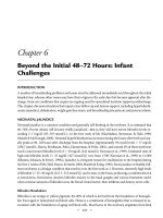
AKI
Presence of life threatening complications of AKI
which cannot be reversed quickly by simple means
Start RRT
(unless not appropriate)
Reverse hypovolemia (unless contraindicated)
Optimize hemodynamic status
Discontinue/avoid nephrotoxic drugs (if possible)
Regular assessment of clinical status,
including metabolic/acid-base profile, illness severity,
fluid balance andinitial response to resuscitation
Persistent or worsening AKI and evidence of ≥1 of
the following:
• Progressive fluid accumulation and/or cumulative
fluid balance >10 % of body weight
• Persistent or worsening acidosis (pH <7.25)
• Persistent or worsening hyperkalemia (K >6
mmol/L)
• Persistent or worsening oliguria (urine < 0.5
mL/kg/h × 6–12 h or <500 mL/24h)
• Persistent or worsening non-renal organ
dysfunction
• Expectation of significant fluid and/or solute
burden
Yes
Consider RRT
(unless not appropriate
or prognosis futile)
No
Fig. 12.1 Proposed algorithm to aid in clinical decision making on when to initiate RRT in
critically ill patients with AKI [21]
RRT can be safely avoided. It is possible that some of the newly discovered
biomarkers for AKI will fulfil this role. Figure 12.1 gives some guidance for clinical
management and decision making at the bedside [21].
12.5
Discontinuation of RRT in the ICU
There is a relative paucity of data about the optimal circumstance and time to wean
and/or discontinue RRT in critically ill patients with AKI [45, 46]. In the BEST
Kidney study, an increase in urine output was the most important determinant of
162
M. Ostermann et al.
recovery of kidney function and likelihood of successful weaning from RRT [45].
Those patients with a spontaneous urine output >400–450 mL/day without diuretics
or >2,300 mL/day with exposure to diuretics had a >80 % probability of sustained
weaning from RRT. In a similar retrospective study of 304 post-operative patients
with severe AKI treated with RRT from the National Taiwan University Surgical,
I. C. U. Acute Renal Failure Study Group, predictors of successful weaning from
RRT were higher (and increasing) urine output on the day following cessation of
RRT along with a shorter cumulative duration of renal support, younger age and
lower non-renal organ dysfunction [46]. Accordingly, apart from increasing spontaneous urine output, there are few reliable clinical signs or tests to predict recovery
sufficient to successfully wean RRT.
12.6
Future Clinical Trials
In addition to the limited high quality evidence on optimal timing of RRT for critically ill patients with AKI, there are a number of ongoing or recently completed
randomized controlled trials (RCTs) addressing this issue. In Canada, the STARRTAKI trial, a multi-centre pilot RCT has recently been completed [47].This trial
enrolled critically ill patients with severe AKI to a strategy of early RRT (within
12 h of eligibility) or standard initiation of RRT (based on persistent AKI and/or
development of more classic indications).The ongoing IDEAL-ICU trial is a multicentre RCT in France with a target enrolment of 824 patients that seeks to randomize critically ill patients with septic shock and severe AKI (defined as a three-fold
rise in serum creatinine and urine output <0.3 mL/kg/h for 12 h) [48]. The early
strategy calls for starting RRT within 12 h of fulfilling AKI criteria whereas in the
late arm RRT commences 48–60 h thereafter. Finally, the AKIKI trial, another
multi-centre RCT in France, proposes to enrol 620 critically ill patients with AKI
randomized to early RRT immediately upon fulfilling Risk-Injury-Failure-Endstage-Loss (RIFLE) category FAILURE or a conservative strategy whereby RRT is
started only after fulfilling RIFLE FAILURE criteria and an additional classical
indication for RRT [49]. The findings from these trials are eagerly awaited and
should help to better inform practice on when to optimally initiate RRT and reduce
unnecessary variation in practice.
12.7
Conclusions: Decision Making on Starting RRT
at the Bedside
The accumulated evidence from clinical studies to date would imply that the optimal timing of starting RRT for critically ill patients with AKI is uncertain and that
the decision should largely be individualized and informed by best practice whenever possible. Evidence from high quality RCTs addressing this issue are anticipated and will hopefully help to inform best clinical practice, reduce unnecessary
variation in how RRT is prescribed, and provide critical data to update clinical
12
Timing of Renal Replacement Therapy
163
practice guidelines. In the absence of life threatening complications of AKI, the
patient’s current and evolving illness severity, burden of non-renal organ dysfunction, fluid balance, and physiological reserve to the consequences of AKI and
response to medical treatment should all be considered and continuously reassessed
when deciding whether RRT should be initiated. These factors should naturally be
weighted in the context of the perceived risks associated with starting RRT along
with the patient’s stated preferences for life-sustaining therapy.
References
1. Bellomo R, Kellum JA, Ronco C. Acute kidney injury. Lancet. 2012;380:756–66.
2. Bagshaw SM, George C, Bellomo R. Changes in the incidence and outcome for early acute
kidney injury in a cohort of Australian intensive care units. Crit Care. 2007;11:R68.
3. Hsu RK, McCulloch CE, Dudley RA, Lo LJ, Hsu CY. Temporal changes in incidence of
dialysis-requiring AKI. J Am Soc Nephrol. 2013;24:37–42.
4. Vaara ST, Pettila V, Reinikainen M, Kaukonen KM. Population-based incidence, mortality and
quality of life in critically ill patients treated with renal replacement therapy: a nationwide
retrospective cohort study in Finnish intensive care units. Crit Care. 2012;16:R13.
5. Hoste EA, Clermont G, Kersten A, et al. RIFLE criteria for acute kidney injury are associated
with hospital mortality in critically ill patients: a cohort analysis. Crit Care. 2006;10:R73.
6. Nisula S, Kaukonen KM, Vaara ST, et al. Incidence, risk factors and 90-day mortality of
patients with acute kidney injury in Finnish intensive care units: the FINNAKI study. Intensive
Care Med. 2013;39:420–8.
7. Uchino S, Kellum JA, Bellomo R, et al. Acute renal failure in critically ill patients: a
multinational, multicenter study. JAMA. 2005;294:813–8.
8. Hamel MB, Phillips RS, Davis RB, et al. Outcomes and cost-effectiveness of initiating dialysis
and continuing aggressive care in seriously ill hospitalized adults. SUPPORT Investigators.
Study to Understand Prognoses and Preferences for Outcomes and Risks of Treatments. Ann
Intern Med. 1997;127:195–202.
9. Korkeila M, Ruokonen E, Takala J. Costs of care, long-term prognosis and quality of life in
patients requiring renal replacement therapy during intensive care. Intensive Care Med.
2000;26:1824–31.
10. Wald R, Quinn RR, Luo J, et al. Chronic dialysis and death among survivors of acute kidney
injury requiring dialysis. JAMA. 2009;302:1179–85.
11. Bagshaw SM, Laupland KB, Doig CJ, et al. Prognosis for long-term survival and renal
recovery in critically ill patients with severe acute renal failure: a population-based study. Crit
Care. 2005;9:R700–9.
12. Joannidis M, Forni LG. Clinical review: timing of renal replacement therapy. Crit Care.
2011;15:223.
13. Ricci Z, Ronco C, D’Amico G, et al. Practice patterns in the management of acute renal failure
in the critically ill patient: an international survey. Nephrol Dial Transplant. 2006;21:690–6.
14. Bagshaw SM, Uchino S, Kellum J, et al. Association between renal replacement therapy in
critically ill patients with severe acute kidney injury and mortality. J Crit Care. 2013;28(6):
1011–8.
15. Clec’h C, Gonzalez F, Lautrette A, et al. Multiple-center evaluation of mortality associated
with acute kidney injury in critically ill patients: a competing risks analysis. Crit Care.
2011;15:R128.
16. Karvellas CJ, Farhat MR, Sajjad I, et al. A comparison of early versus late initiation of renal
replacement therapy in critically ill patients with acute kidney injury: a systematic review and
meta-analysis. Crit Care. 2011;15:R72.
164
M. Ostermann et al.
17. Seabra VF, Balk EM, Liangos O, Sosa MA, Cendoroglo M, Jaber BL. Timing of renal
replacement therapy initiation in acute renal failure: a meta-analysis. Am J Kidney Dis.
2008;52:272–84.
18. Wang X, Jie Yuan W. Timing of initiation of renal replacement therapy in acute kidney injury:
a systematic review and meta-analysis. Ren Fail. 2012;34:396–402.
19. Clark EG, Bagshaw SM. Unnecessary renal replacement therapy for acute kidney injury is
harmful for renal recovery. Semin Dial. 2015;28(1):6–11.
20. Shiao CC, Ko WJ, Wu VC, et al. U-curve association between timing of renal replacement
therapy initiation and in-hospital mortality in postoperative acute kidney injury. PLoS One.
2012;7, e42952.
21. Ostermann M, Dickie H, Barrett NA. Renal replacement therapy in critically ill patients with
acute kidney injury–when to start. Nephrol Dial Transplant. 2012;27(6):2242–8.
22. Bouchard J, Soroko SB, Chertow GM, et al. Fluid accumulation, survival and recovery of
kidney function in critically ill patients with acute kidney injury. Kidney Int. 2009;76:422–7.
23. Prowle JR, Echeverri JE, Ligabo EV, Ronco C, Bellomo R. Fluid balance and acute kidney
injury. Nat Rev Nephrol. 2010;6:107–15.
24. Sutherland SM, Zappitelli M, Alexander SR, et al. Fluid overload and mortality in children
receiving continuous renal replacement therapy: the prospective pediatric continuous renal
replacement therapy registry. Am J Kidney Dis. 2010;55(2):316–25.
25. Rimmele T, Kellum JA. Clinical review: blood purification for sepsis. Crit Care. 2011;15:205.
26. Kidney Disease: Improving Global Outcomes (KDIGO) Acute Kidney Injury Work Group.
KDIGO clinical practice guideline for acute kidney injury. Kidney Int. 2012;2012:1–138.
27. National Institute for Health and Care Excellence Acute Kidney Injury Workgroup. Acute
kidney injury: prevention, detection and management of acute kidney injury up to the point of
renal replacement therapy. Clinical guidelines, CG169. ( />28. Bouman CS, Oudemans-Van Straaten HM, Tijssen JG, Zandstra DF, Kesecioglu J. Effects of
early high-volume continuous venovenous hemofiltration on survival and recovery of renal
function in intensive care patients with acute renal failure: a prospective, randomized trial. Crit
Care Med. 2002;30:2205–11.
29. Jamale TE, Hase NK, Kulkarni M, et al. Earlier-start versus usual-start dialysis in patients with
community-acquired acute kidney injury: a randomized controlled trial. Am J Kidney Dis.
2013;62:1116–21.
30. Demirkilic U, Kuralay E, Yenicesu M, et al. Timing of replacement therapy for acute renal
failure after cardiac surgery. J Card Surg. 2004;19:17–20.
31. Durmaz I, Yagdi T, Calkavur T, et al. Prophylactic dialysis in patients with renal dysfunction
undergoing on-pump coronary artery bypass surgery. Ann Thorac Surg. 2003;75:859–64.
32. Elahi MM, Lim MY, Joseph RN, Dhannapuneni RR, Spyt TJ. Early hemofiltration improves
survival in post-cardiotomy patients with acute renal failure. Eur J Cardiothorac Surg.
2004;26(5):1027–31.
33. Manche A, Casha A, Rychter J, Farrugia E, Debono M. Early dialysis in acute kidney injury
after cardiac surgery. Interact Cardiovasc Thorac Surg. 2008;7:829–32.
34. Sugahara S, Suzuki H. Early start on continuous hemodialysis therapy improves survival rate
in patients with acute renal failure following coronary bypass surgery. Hemodial Int. 2004;8:
320–5.
35. Iyem H, Tavli M, Akcicek F, Buket S. Importance of early dialysis for acute renal failure after
an open-heart surgery. Hemodial Int. 2009;13:55–61.
36. Andrade L, Cleto S, Seguro AC. Door-to-dialysis time and daily hemodialysis in patients with
leptospirosis: impact on mortality. Clin J Am Soc Nephrol. 2007;2:739–44.
37. Gettings LG, Reynolds HN, Scalea T. Outcome in post-traumatic acute renal failure when
continuous renal replacement therapy is applied early vs. late. Intensive Care Med. 1999;25:
805–13.
38. Shiao CC, Wu VC, Li WY, et al. Late initiation of renal replacement therapy is associated
with worse outcomes in acute kidney injury after major abdominal surgery. Crit Care. 2009;13:
R171.
12
Timing of Renal Replacement Therapy
165
39. Wu VC, Ko WJ, Chang HW, et al. Early renal replacement therapy in patients with postoperative acute liver failure associated with acute renal failure: effect on postoperative outcomes. J
Am Coll Surg. 2007;205:266–76.
40. Carl DE, Grossman C, Behnke M, Sessler CN, Gehr TW. Effect of timing of dialysis on
mortality in critically ill, septic patients with acute renal failure. Hemodial Int. 2010;14:11–7.
41. Liu KD, Himmelfarb J, Paganini E, et al. Timing of initiation of dialysis in critically ill patients
with acute kidney injury. Clin J Am Soc Nephrol. 2006;1:915–9.
42. Bagshaw SM, Uchino S, Bellomo R, et al. Timing of renal replacement therapy and clinical
outcomes in critically ill patients with severe acute kidney injury. J Crit Care. 2009;24:
129–40.
43. Bagshaw SM, Wald R, Barton J, et al. Clinical factors associated with initiation of renal
replacement therapy in critically ill patients with acute kidney injury-a prospective multicenter
observational study. J Crit Care. 2012;27:268–75.
44. Vaara ST, Reinikainen M, Wald R, Bagshaw SM, Pettila V, for the FINNAKI Study Group
FS. Timing of RRT based on the presence of conventional indications. Clin J Am Soc Nephrol.
2014;9:1577–85.
45. Uchino S, Bellomo R, Morimatsu H, et al. Discontinuation of continuous renal replacement
therapy: a post hoc analysis of a prospective multicenter observational study. Crit Care Med.
2009;37:2576–82.
46. Wu VC, Ko WJ, Chang HW, et al. Risk factors of early redialysis after weaning from
postoperative acute renal replacement therapy. Intensive Care Med. 2008;34:101–8.
47. Smith OM, Wald R, Adhikari NK, et al. Standard versus accelerated initiation of renal
replacement therapy in acute kidney injury (STARRT-AKI): study protocol for a randomized
controlled trial. Trials. 2013;14:320.
48. Initiation of Dialysis Early Versus Late in Intensive Care Unit. ClinicalTrials.gov, 2013.
Accessed 20 Dec 2014, at />49. Artificial Kidney Initiation in Kidney Injury, a Multicenter Randomised Trial. ClinicalTrials.
gov, 2013. Accessed 20 Dec 2014, at />