Ebook Drug and device selection in heart failure: Part 2
Bạn đang xem bản rút gọn của tài liệu. Xem và tải ngay bản đầy đủ của tài liệu tại đây (16.52 MB, 142 trang )
C H ap t er
Percutaneous Mechanical Support
8
Raphael E Bonita, Kariann Abbate
INTRODUCTION
There are approximately 5.8 million people in the United States living with heart
failure (HF).1 These numbers are expected to increase over the next decade due
to the aging population. Despite the increasing prevalence of HF, the number of
donor hearts available for transplant has remained stagnant. In 2010, there were
2,333 heart transplants performed in the United States.2 A severe donor shortage
has limited the availability of donor hearts resulting in prolonged waits for organs
for patients with advanced HF. Furthermore, more than 200,000 patients with heart
failure are not eligible for transplant due to age or comorbidities.3 The advent of
mechanical circulatory support has decreased mortality and improved the quality
of life for patients awaiting heart transplantation as well as for those ineligible for
transplantation.
INDICATIONS FOR MECHANICAL ASSIST DEVICES
Mechanical assist devices are indicated for patients who are failing optimal
medical therapy. When patients are exhibiting evidence of end-organ dysfunction
despite optimal medical therapy, mechanical assist devices should be considered.
Mechanical assist devices are used for both acute and chronic HF. The National
Institute of health has developed profiles using data from Interagency Registry for
Mechanically Assisted Circulatory Support (INTERMACS) to assist in clarification
of target populations for mechanical assist devices (Table 1). There are three main
indications for mechanical assist devices: (1) bridge to myocardial recovery, (2)
bridge to cardiac transplantation, and (3) destination therapy.
Bridge to Recovery
Mechanical assist devices are commonly used to support patients suffering from
postcardiotomy shock that are unable to be weaned from cardiopulmonary bypass
Ch-8.indd 110
10-09-2013 15:18:56
111
Chapter 8: Percutaneous Mechanical Support
Table 1
INTERMACS profile description
Clinical presentations
Time frame for
intervention
Profile 1: Critical cardiogenic shock
Patients with life-threatening hypotension despite rapidly escalating Definitive inter
inotropic support, critical organ hypoperfusion, often confirmed by vention needed
worsening acidosis and/or lactate levels. “Crash and burn”
within hours
Profile 2: Progressive decline
Patients with declining function despite intravenous inotropic support may
be manifested by worsening renal function, nutritional depletion, inability
to restore volume balance, “sliding on inotropes”. Also describes declining
status in patients unable to tolerate inotropic therapy
Definitive
intervention
needed within a
few days
Profile 3: Stable but inotrope dependent
Patients with stable blood pressure, organ function, nutrition, and
symptoms on continuous intravenous inotropic support (or a temporary
circulatory support device or both), but demonstrated repeated failing to
wean from support due to recurrent symptomatic hypotension or renal
dysfunction “dependent stability”
Definitive
intervention
elective over a
period of weeks
to months
Profile 4: Resting symptoms
Patients can be stabilized close to normal volume status but experiences
daily symptoms of congestion at rest or during ADL. Doses of diuretics
generally fluctuate at very high levels. More intensive management and
surveillance strategies should be considered, which may in some cases
reveal poor compliance that would compromise outcomes with any
therapy. Some patients may shuttle between 4 and 5
Definitive
intervention
elective over a
period of weeks
to months
Profile 5: Exertion intolerance
Comfortable at rest and with ADL but unable to engage in any other
activity, living predominantly within the house. Patients are comfortable
at rest without congestive symptoms, but may have underlying refractory
elevated volume status, often with renal dysfunction. If underlying
nutritional status and organ function are marginal, patient may be more at
risk than INTERMACS 4 and require definitive intervention
Variable urgency
depends upon
maintenance of
nutrition, organ
function and
activity
Profile 6: Exertion limited
Patient without evidence of fluid overload is comfortable at rest and
with activities of daily living and minor activities outside the home but
fatigues after the first few minutes of any meaningful activity. Attribution
to cardiac limitation requires careful measurement of peak oxygen
consumption, in some cases with hemodynamic monitoring to confirm
severity of cardiac impairment. “Walking wounded”
Variable urgency
depends upon
maintenance of
nutrition, organ
function and
activity level
Profile 7: Advanced NYHA III
A placeholder for more precise specification in future, this level includes
patients who are without current or recent episodes of unstable fluid
balance, living comfortably with meaningful activity limited to mild
physical exertion
Transplantation
or circulator
support may
not currently be
indicated
Contd...
Ch-8.indd 111
10-09-2013 15:18:56
112
Drug and Device Selection in Heart Failure
Contd...
Modifiers for profiles
Possible profiles
to modify
Temporary circulatory support can modify only patients in hospital 1,2,3 in hospital
(other devices would be INTERMACS devices) includes IABP, ECMO,
TandemHeart®, Levitronix, BVS 5000 or AB5000, Impella®
Arrhythmia (A)—Can modify any profile. Recurrent ventricular Any profile
tachyarrhythmias that have recently contributed substantially to clinical
compromise. This includes frequent ICD shock or requirement for
external defibrillator, usually more than twice weekly
Frequent Flyer (FF)—Can modify only outpatients, designate a patient 3 if at home, 4,
requiring frequent emergency visits or hospitalizations for diuretics, 5, 6. A frequent
flyer would rarely
ultrafiltration, or temporary intravenous vasoactive therapy
be profile 7
INTERMACS, interagency registry for mechanically assisted circulatory support; ADL, activities of daily
living; NYHA, New York Heart Association; IABP, Intra-aortic balloon pump; ECMO, extracorporeal
membrane oxygenating system; ICD, implantable cardioverter-defibrillator
and hemodynamically unstable patients following acute myocardial infarction (MI)
or acute viral myocarditis. These patients are most often in INTERMACS profile
1. Percutaneous, temporary assist devices, such as extracorporeal membrane
oxygenating system (ECMO), intra-aortic balloon pump (IABP), TandemHeart®,
and Impella® are frequently used in these situations because of their relative
ease of insertion. If a patient does not adequately recover with these temporary
devices, a more permanent ventricular assist device (VAD) may be considered.
Often, a temporary device is used as a “bridge-to-bridge”, stabilizing the patient’s
hemodynamics so that a more definitive VAD can be placed or transplant can be
performed. If both right- and left-heart support is needed, devices such as the
Thoratec CentriMag or Thoratec VAD system can be used.
Occasionally, patients with chronic HF symptoms, usually due to a nonischemic
cardiomyopathy, have a left ventricular assist device (LVAD) placed and recover
enough myocardial function to have the LVAD explanted. In a retrospective study
conducted by Mancini et al., only 5% of patients fell into this category and had the
LVAD successfully explanted. Exercise testing may be a useful modality to identify
those patients in whom the device can be explanted.4
Bridge to Transplant
This is the most common indication for LVAD placement. Due to long waiting times
on the transplant list, especially for patients with blood type O, mechanical assist
devices are often used to prevent end-organ damage and improve quality of life
while patients are waiting for heart transplant. LVAD placement prior to transplant
can improve end-organ function, enhance nutritional status, and allow patients
Ch-8.indd 112
10-09-2013 15:18:56
113
Chapter 8: Percutaneous Mechanical Support
to participate in rehabilitation programs, which can improve post-transplant
outcomes.5 The timing of VAD placement in these patients is sometimes challenging.
For patients awaiting transplant, performing multiple sternotomy procedures may
predispose patients to adverse outcomes, such as sternal wound infection following
their definitive transplant surgery. Conversely, waiting too long for VAD placement
may compromise renal and other end-organ function and allow deconditioning
and cardiac cachexia to occur, resulting in worse outcomes at the time of transplant.
Jarvik 2000 is a continuous flow LVAD approved in the United States for bridge to
transplant. This device can be placed via left thoracotomy, which can spare patients
a sternotomy procedure. HeartMate II is the most commonly used for bridge to
transplant.
Destination Therapy
In 2001, the Randomized Evaluation of Mechanical Assistance for the Treatment
of Congestive Heart Failure (REMATCH) group published a landmark study
demonstrating a mortality benefit of LVAD versus optimal medical therapy in
patients with end-stage heart failure. REMATCH was a multicenter, controlled
trial that randomly assigned 129 patients with advanced heart failure who were
ineligible for transplant to receive HeartMate XVE LVAD versus optimal medical
therapy (including inotropes). Survival analysis showed a reduction of 48% in the
risk of death from any cause in the group that received LVADs as compared with
the medical-therapy group (relative risk, 0.52; 95% confidence interval, 0.34–0.78;
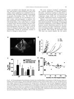
P = 0.001, Fig. 1). The rates of survival at 1 year were 52% in the device group and
25% in the medical-therapy group (P = 0.002), and the rates at 2 years were 23% and
8% (P = 0.09), respectively.6 REMATCH demonstrated a significant improvement in
survival for patients receiving pulsatile flow LVAD compared with medical therapy,
but it also revealed limitations of these devices for long-term support. Survival at
1 year was 52% in the LVAD group and at 2 years was only 25%; 65% of patients
surviving to 2 years required device replacement due to mechanical failure. Driveline
infections were common. Some have questioned whether there was simply a mode
switch of death in patients receiving first generation LVADs for destination therapy.7
Second generation axial flow LVADs have shown improved survival compared to
the pulsatile devices studied in REMATCH. Axial flow devices have overcome many
of the limitations of pulsatile flow devices. Pulsatile flow devices require a large
reservoir to store blood, which limits its utility in small women and children. Axial
flow devices are smaller and can be used in a more diverse group of patients. Axial
flow devices are quieter and more comfortable. Although driveline infections are still
a problem with axial flow devices, they are much less frequent compared to pulsatile
devices. Currently, HeartMate II is the only axial flow device approved for destination
therapy in the United States. This device was approved based on a study conducted
Ch-8.indd 113
10-09-2013 15:18:56
114
Drug and Device Selection in Heart Failure
Figure 1: Kaplan-Meier survival curves in patients receiving left ventricular (LV) assist devices
versus optimal medical therapy.
Source: From Rose EA, Gelijns AC, Moskowitz AJ, et al. Long-term use of a left ventricular assist
device for end-stage heart failure. N Engl J Med. 2001;345:1435-45, with permission.
by the HeartMate II investigators in 2009. It randomized patients with advanced HF
who were ineligible for transplantation, in a 2:1 ratio, to undergo implantation of a
continuous-flow device or pulsatile-flow device. The primary composite end- point
was, at 2 years, survival-free from disabling stroke and reoperation to repair or
replace the device. The primary composite end-point was achieved in more patients
with continuous-flow devices than with pulsatile-flow devices [62 of 134 (46%) vs.
7 of 66 (11%); P < 0.001; hazard ratio, 0.38; 95% confidence interval, 0.27–0.54; P <
0.001], and patients with continuous-flow devices had superior actuarial survival
rates at 2 years (58% vs. 24%, P = 0.008). Adverse events and device replacements
were less frequent in patients with the continuous-flow device.8
DEVICES USED FOR SHORT-TERM MECHANICAL CIRCULATORY
SUPPORT
The choice of mechanical circulatory support is based on stability of the patient, the
amount and type of circulatory support needed, and the expected duration the device
will be used. Mechanical circulatory support devices can be placed percutaneously or
surgically and can be extracorporeal, paracorporeal, or intracorporeal. For patients
in cardiogenic shock, the most effective devices are relatively easy to implant and
have a good safety profile.
Ch-8.indd 114
10-09-2013 15:18:56
115
Chapter 8: Percutaneous Mechanical Support
Percutaneous Devices
The IABP, TandemHeart®, and Impella® are the percutaneous devices that are
currently available in the United States. They are most frequently used as rescue
devices for patients in cardiogenic shock or to provide support for patients
undergoing high-risk percutaneous coronary interventions (PCIs) or surgeries.
The advantage of percutaneous devices is that they can be placed with relative
ease in the cardiac catheterization laboratory. As with all mechanical circulatory
support devices, they carry the risks of bleeding, hemolysis, thrombus formation,
infection, and device failure. Percutaneous devices carry the additional risk of
peripheral vascular complications. This is particularly important, as many of the
patients eligible for these devices are at high risk of peripheral vascular disease. If
a percutaneous device is being considered, imaging of the distal aorta, iliac, and
femoral vessels with angiography, computed tomography, or magnetic resonance
imaging should be considered.9
Intra-aortic Balloon Pump
The IABP consists of a cylindrical polyethylene balloon that sits in the aorta,
approximately 2 cm from the left subclavian artery. It deflates in systole
increasing cardiac output by reducing afterload and decreases myocardial oxygen
consumption. Inflation during diastole increases coronary artery perfusion. Based
on American College of Cardiology and American Heart Association Guidelines,
IABP is a Class IB indication for patients in cardiogenic shock. It is commonly
used following an acute MI and has proven beneficial in patients who suffer
from mechanical complications of acute MI, including mitral regurgitation and
rupture of the ventricular septum.10,11 IABP is also used to provide hemodynamic
support during high-risk PCI or cardiac surgery and is sometimes placed to assist
in weaning patients from cardiopulmonary bypass following cardiac surgery.
Although hemodynamics is improved in patients suffering from cardiogenic shock
after placement of IABP, it is unclear if placement of IABP provides a mortality
benefit. To date, there are no randomized control trials comparing IABP to
standard therapy in patients suffering from cardiogenic shock. In 2009, a metaanalysis was performed evaluating the available evidence of IABP in ST segment
elevation myocardial infarction (STEMI) with or without cardiogenic shock. The
pooled randomized data do not support IABP in patients with high-risk STEMI.
The meta-analysis of cohort studies in the setting of STEMI complicated by
cardiogenic shock supported IABP therapy adjunctive to thrombolysis. In contrast,
the observational data did not support IABP therapy adjunctive to primary PCI.
These findings should be taken with caution, as the authors noted that currently
available observational data concerning IABP therapy in the setting of cardiogenic
shock is hampered by bias and confounding.12
Ch-8.indd 115
10-09-2013 15:18:56
116
Drug and Device Selection in Heart Failure
Contraindications to the use of IABP include severe aortic valve insufficiency,
aortic dissection, severe peripheral vascular disease, and irreversible brain damage.
Inflation and deflation of the intra-aortic balloon is timed with the electrocardiogram
(EKG), rendering IABP ineffective in unstable rhythms.
The complication rate for IABP placement ranges from 8.7% to 29% and
averages 15%.13-15 Most complications are vascular in nature with the most severe
complications being arterial thrombosis and limb loss. Other vascular complications
include compartment syndrome, arterial dissection, hematoma, and retroperitoneal
bleeding. Infectious complications can occur, especially in situations where IABP is
used for a long duration. Risk factors for complications include peripheral vascular
disease, female sex, and diabetes.9
TandemHeart®
TandemHeart® is manufactured by CardiacAssist, Pittsburgh, Pennsylvania. It
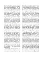
is comprised of three components: (1) a centrifugal continuous flow pump, (2)
a microprocessor-based controller, and (3) a 21-French transseptal cannula
(Fig. 2). The inflow catheter can be placed percutaneously in the left atrium through
a transseptal approach and whose outflow cannula is placed in the femoral artery.
TandemHeart can provide up to 5 L of flow per minute when placed percutaneously.
If placed in the or using direct surgical cannulation technique, TandemHeart® can
provide up to 8 L per minute of flow. The indications for TandemHeart® are similar to
the indications for IABP support: cardiogenic shock due to acute MI, postcardiotomy,
or decompensated heart failure. Because TandemHeart® is capable of providing 5 L
of flow per minute, it may be preferred to IABP in patients with severe cardiogenic
shock. However, TandemHeart® is more technically challenging to place compared
to an IABP. Thiele et al. conducted a randomized control trial in 2005 comparing
IABP to TandemHeart for patients presenting with cardiogenic shock and acute MI.
They found that the hemodynamic and metabolic parameters in cardiogenic shock
were reversed more effectively with TandemHeart® compared to IABP treatment.
However, there were more complications encountered with the TandemHeart®
including severe bleeding and limb ischemia. The study was not powered to detect
a mortality difference. The authors speculate that the complications associated
with VAD therapy may be due to a systemic inflammatory response triggered by the
extracorporeal circulation. This response may play a role in triggering disseminated
intravascular coagulation (DIC). In patients treated with VAD for more than 2 days,
nearly all patients required a blood transfusion as a consequence of DIC. The VAD
took 25 minutes to place, compared to 11.5 minutes for placement of IABP.16
Given the higher complication rate with TandemHeart® compared to IABP,
TandemHeart® is usually considered only after a patient suffering from cardiogenic
shock has failed medical therapy and IABP support. TandemHeart® is used as first
Ch-8.indd 116
10-09-2013 15:18:57
117
Chapter 8: Percutaneous Mechanical Support
Figure 2: Tandem heart.
Source: CardiacAssist, Inc. [online] Available from: . [Accessed
April, 2013].
line support in patients who are predicted to require more than 3 L of additional
cardiac output.
TandemHeart® is contraindicated in patients with predominant right ventricular
(RV) failure because the low left atrial pressure does not permit adequate pumping.
It is also relatively contraindicated in patients with a ventricular septal defect due to
the risk of hypoxemia secondary to right-to-left shunting. Other contraindications
include aortic insufficiency and severe peripheral arterial disease.17
Impella 2.5
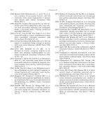
Impella 2.5 (Abiomed Europe GmbH, Aachen, Germany) is a catheter-based,
impeller-driven axial flow pump with a maximal flow rate of 2.5 L/minute from the
left ventricle to the ascending aorta (Fig. 3). It can be implanted percutaneously.
Impella 5.0 is capable of generating 5 L/minute of flow, but must be placed in the OR.
The Placebo-controlled Randomized Study of the Selective A1 Adenosine Receptor
Antagonist Rolofylline for Patients Hospitalized with Acute Decompensated Heart
Failure and Volume Overload to Assess Treatment Effect on Congestion and Recent
Ch-8.indd 117
10-09-2013 15:18:57
118
Drug and Device Selection in Heart Failure
Figure 3: Impella 2.5.
Source: ABIOMED [online]. Available from: www.abiomed.com [Accessed April, 2013].
Function (PROTECT 1) trial was a randomized control trial that demonstrated
improved hemodynamics, safety, and efficacy of Impella 2.5 in use prior to high
risk PCI. The Impella LP 2.5 vs. IABP in Cardiogenic Shock (ISAR-SHOCK) trial
was a randomized control trial that demonstrated that Impella 2.5 improved
hemodynamics, increased cardiac output, and was safe in patients receiving Impella
for acute MI. Seyfarth et al. published a randomized control study in 2008 comparing
Impella LP 2.5 to IABP in patients suffering from cardiogenic shock secondary to
acute MI. The study was not powered to detect mortality. The investigators found
that hemodynamics including cardiac index were statistically significantly improved
in the Impella group compared to the IABP group. There was no increased risk of
major bleeding, distal limb ischemia, arrhythmia, or infection in the Impella group.
Transient hemolysis was noted in the Impella group. USpella is a United States
multicenter registry of Impella 2.5 patients evaluating the safety and feasibility of
left ventricular support with the Impella 2.5 during high-risk PCI and treatment of
acute MI. It examined approximately 181 patients. In situations where Impella 2.5
was used to facilitate high-risk PCI, the registry showed that overall major adverse
event rate was low at 6% and 30-day survival rate was 97%. In patients who received
an Impella for acute MI, Impella improved hemodynamics by increasing cardiac
index from 1.9 to 2.5 l/min/m2 and mean arterial pressure from 62 to 87 mmHg.
It was able to successfully lower wedge pressure from 28 to 20 mmHg and systemic
vascular resistance (SVR). After Impella 2.5 support, overall ejection fraction in AMI
patients improved from 29 to 37%. Impella successfully supported AMI refractory
shock patients with 69% survival to the next therapy or on to recovery. Also, 58% of
AMI shock patients and 89% of AMI patients with no shock were discharged.18
Ch-8.indd 118
10-09-2013 15:18:57
119
Chapter 8: Percutaneous Mechanical Support
Contraindications to Impella 2.5 include prosthetic aortic valves, moderate
to severe aortic insufficiency, heavily calcified aortic valves, documented left
ventricular thrombus, severe peripheral vascular disease, and in patients who are
unable to tolerate anticoagulation.
The most commonly reported complications of Impella 2.5 placement and
support include limb ischemia, vascular injury, and bleeding requiring blood
transfusion. Hemolysis has been reported. Other potential complications include
aortic valve damage, displacement of the distal tip of the device into the aorta,
infection, and sepsis.19
Impella 2.5 is most commonly used in patients with cardiogenic shock
who have failed IABP or in patients with cardiogenic shock or prior to high-risk
PCI who are anticipated to require more hemodynamic support than an IABP can
provide.
Extracorporeal Membrane Oxygenation
Extracorporeal membrane oxygenation is similar to cardiopulmonary support
provided during cardiac surgery but can be delivered for a more prolonged period.
There are two types of ECMO: (1) venovenous (VV) and (2) venoarterial (VA). Both
provide respiratory support, but only VA ECMO can provide hemodynamic support.
Although there are data to support a mortality benefit with the use of VV ECMO for
acute respiratory failure, the literature supporting the use of VA ECMO for patients
in cardiogenic shock is less robust. To date, no randomized control trials have been
conducted to determine the efficacy of this modality in hemodynamically unstable
patients.
Venoarterial extracorporeal membrane oxygenation is comprised of a
cannula that is inserted into the femoral artery and a cannula that is inserted
into the femoral vein. The ECMO circuit is based on a centrifugal pump and a
hollow-fiber membrane oxygenator. All circuit components are heparin surface
coated. During ECMO, blood is extracted from the native vascular system and
circulated outside the body by a mechanical pump. The blood passes through an
oxygenator and heat exchanger, where the hemoglobin becomes fully saturated
with oxygen and carbon dioxide is removed. The blood is then reinfused into
the native vascular system. VA ECMO has the capability of providing as much
augmentation to cardiac output as an LVAD but can be placed quickly and less
invasively. ECMO is often placed percutaneously. It can provide hemodynamic
support to both the right and left hearts. It has become a popular option for
patients in cardiogenic shock who require rapid implementation of hemodynamic
and/or respiratory support. Indications for VA ECMO include support after
cardiac arrest, inability to wean from cardiopulmonary bypass following cardiac
surgery, cardiogenic shock following acute MI, bridge-to-decision or bridge-to-
Ch-8.indd 119
10-09-2013 15:18:57
120
Drug and Device Selection in Heart Failure
transplant in advanced heart failure patients, and cardiogenic shock associated
with myocarditis, poisoning or hypothermia. ECMO has proven successful in
supporting patients with fulminant myocarditis (FM). Chen and Yu described
their experience with ECMO in patients with FM in 2004. From 1995 to 2001, they
used ECMO as first-line mechanical support to treat 15 FM patients with shock,
including five under external cardiopulmonary resuscitation (CPR) and ten
with high-degree atrioventricular block. Their results revealed 93.3% (14/15) in
successful weaning rate and 73.3% (11/15) in discharge survival rate. The average
ECMO support time was 129 ± 50 hours (127 ± 83 hours for the survivors). As
compared with ABIOMED BiVAD use for FM, ECMO group had lower morbidity
rate than VAD group: mechanical related thromboembolism was 6.7% in ECMO
group and 40–27.3% in VAD group; re-exploration for hemostasis was 20% in
ECMO group and 45.5% in VAD group. They pointed out that since FM tends to
recover within 2 weeks, ECMO is an appropriate option for this relatively short
duration. ECMO is easier to wean off than VAD, and ECMO can be converted to
VAD at any time if necessary.20
Since anticoagulation is necessary to prevent blood from clotting in the
ECMO circuit, ECMO is contraindicated in patients who are not candidates for
anticoagulation, such as patients with active bleeding issues, recent surgery, and
recent intracranial injury. ECMO is also relatively contraindicated in patients with
irreversible cardiac failure who are not candidates for transplant or more permanent
VADs. Other factors to consider before implementing ECMO include age, body mass
index, neurological function, and prior functional status.
The most frequent complication of ECMO is bleeding. Patients on ECMO are
predisposed to bleeding due to the need to receive a continuous infusion of heparin
or a similar anticoagulant and platelet dysfunction that is caused by the ECMO circuit.
Thromboembolism due to thrombus formation in the ECMO circuit is another
serious complication. Vascular complications can result from cannula placement,
such as limb ischemia, vessel perforation, and/or vessel dissection. Complications
specific to VA ECMO include pulmonary hemorrhage, pulmonary infarction, aortic
thrombosis, coronary ischemia, and stroke.
Centrifugal Ventricular Assist Device
A centrifugal assist device is an extracorporeal cone-shaped rotor contained within
a plastic or metal housing. Blood flows into the pump at the cone’s apex and exits
at the edge of the base. The spinning of the rotor creates a centrifugal force that
is imparted to the blood, generating a constant, nonpulsatile flow. These devices
are indicated for short-term support including cardiopulmonary bypass surgery,
postcardiotomy shock, and bridge-to-bridge situations. Centrifugal pumps can also
be used to provide RV support after cardiac transplant (Fig. 4). These devices can
Ch-8.indd 120
10-09-2013 15:18:57
121
Chapter 8: Percutaneous Mechanical Support
Figure 4: BPX-80 BIO Pump plus centrifugal blood pump.
Source: [online] Available from: />products-therapies/cardiovascular/cardiopulmonary-products/bpx-80-bio-pump-pluscentrifugal-blood-pump/index.htm [Accessed April 2013].
provide up to 10 L/min of flow. The most commonly used centrifugal device is BioMedicus Perfusion System (Medtronic).
Pulsatile Assist Devices for Short-term Use
Pulsatile flow devices consist of plastic or metal housing with a mechanically
driven volume displacement chamber that fills either passively or by suction
applied during chamber expansion. Blood enters through an inflow valve and fills
the chamber as it expands. The blood is then forced out through an outflow valve
as the chamber contracts. These pumps mimic the cyclic systole and diastole of
the heart and generate pulsatile blood flow. The inflow valve (bioprosthetic or
mechanical) allows unidirectional flow into the device and prevents regurgitation
during mechanical systole and the outflow valve prevents regurgitation during
mechanical relaxation.
Abiomed AB5000 is a pulsatile assist device that can supply one or both sides of
the heart. The AB5000 ventricle is vacuum-assisted technology with clear housing to
allow clinicians a view into the device. Regardless of whether the device is supporting
one or both ventricles, the AB5000 only requires one driver. The AB Portable Driver is
designed to allow patients to leave their hospital rooms and walk within the hospital
and on hospital grounds.
Ch-8.indd 121
10-09-2013 15:18:57
122
Drug and Device Selection in Heart Failure
Figure 5: Thoratec percutaneous ventricular assist device.
Source: Thoratec Corporation [online]. Available from: />sionals/vad-product-information/thoratec-pvad.aspx [Accessed April, 2013].
Right Ventricular Assist Devices and Biventricular Assist Devices
Right ventricular failure presents a unique challenge when considering VADs.
Because the right ventricle is less muscular and thinner than the left ventricle, it is
more difficult to canalize the right ventricle. There is greater risk of complications
when the right ventricle is canalized, such as right ventricular perforation or
displacement of the cannula. Because of the tenuous nature of RVADs and BiVADs,
most patients who require these devices are confined to the hospital. Abiomed
AB5000 (as discussed above) is an example of a device that can provide biventricular
support. Other biventricular VADs include Thoratec percutaneous ventricular
assist device [(PVAD) Fig. 5], Thoratec CentriMag Blood Pump, and the Thoratec
IVAD. The Thoratec IVAD is the only biventricular device that allows patients to be
discharged home. Indications for RVADs or BiVADs are bridge to recovery or bridge
to transplant.
References
1. Lloyd-Jones D, Adams RJ, Brown TM, et al. Heart disease and stroke statistics—2010
update: a report from the American Heart Association. Circulation. 2010;121(7):
e46-215.
2. Transplant: Transplant Year (2009-2010) by Organ US Transplants Performed: January 1,
1988-September 30, 2011 Based on OPTN Data as of December 23, 2011. Available from:
Accessed on December 31, 2011.
3. Ammar KA, Jacobsen SJ, Mahoney DW, et al. Prevalence and prognostic significance
of heart failure stages: application of the American College of Cardiology/American
Heart Association heart failure staging criteria in the community. Circulation.
2007;115:1563-70.
4. Mancini DM, Beniaminovitz A, Levin H, et al. Low incidence of myocardial recovery
after left ventricular assist device implantation in patients with chronic heart failure.
Circulation. 1998;98:2383-9.
5. Ashton RC, Goldstein DJ, Rose EA, et al. Duration of left ventricular assist device support
affects transplant survival. J Heart Lung Transplant. 1996;15:1151-7.
6. Rose EA, Gelijns AC, Moskowitz AJ, et al. Long-term use of a left ventricular assist device
for end-stage heart failure. N Engl J Med. 2001;345:1435-43.
7. Deng MC, Naka Y. Mechanical circulatory support therapy in advanced heart failure.
London: Imperial College Press; 2007.
Ch-8.indd 122
14-09-2013 12:27:45
123
Chapter 8: Percutaneous Mechanical Support
8. Slaughter MS, Rogers JG, Milano CA, et al. Advanced heart failure treated with
continuous-flow left ventricular assist device. N Engl J Med. 2009;361:2241-51.
9. Feldman AM. Heart failure: device management. Oxford: Blackwell Publishing; 2010.
10. Decker AL, Reesink KD, Van Der Veen FH, et al. Intra-aortic balloon pumping in acute mitral
regurgitation reduces aortic impedance and regurgitrant fraction. Shock. 2003;19:334-8.
11. Bouchart F, Bessou JF, Tabley A, et al. Urgent surgical repair of postinfarction ventricular
septal rupture: early and late outcomes. J Cardiac Surg. 1998;12:104-12.
12. Sjaw KD, Engstrom AE, Vis MM, et al. A systematic review and meta-analysis of intraaortic balloon pump therapy in ST-elevation myocardial infarctions: should we change
the guidelines? Eur Heart J. 2009;30(4):459-68.
13. Arafa OE, Pedersen TH, Svennevig JL, et al. Vascular complications of the intra-aortic
balloon pump in patients undergoing open-heart operations: 15-year experience. Ann
Thorac Surg. 1999;67:645-51.
14. Cohen M, Dawson MS, Kopistansky C, et al. Sex and other predictors of intra-aortic
balloon counterpulsation-related complications: prospective study of 1119 consecutive
patients. Am Heart J. 2000;139:282-7.
15. Cook L, Pillar B, McCord G, et al. Intra-aortic balloon pump complications: A five-year
retrospective study of 283 patients. Heart Lung. 1999;28:195-202.
16. Thiele H, Sick P, Boudriot E, et al. Randomized comparison of intra-aortic balloon support
with a percutaneous left ventricular assist device in patients with revascularized acute
myocardial infarctions complicated by cardiogenic shock. Eur Heart J. 2005;26:1276-83.
17. De Suoza CF, de Suoza Brito F, De Lima VC, et al. Percutaneous mechanical assistance
for the failing heart. J Interv Cardiol. 2010;23:195-202.
18. Cath Lab Digest [Internet] Available from: /> 19. McCulloch B. Use of Impella 2.5 in high-risk percutaneous coronary intervention. Crit
Care Nurse. 2011;31:e1-16.
20. Chen YS, Yu HY. Choice of mechanical support for fulminant myocarditis: ECMO vs.
VAD? Eur J Cardiothorac Surg. 2005;27:931-2.
Ch-8.indd 123
10-09-2013 15:18:57
C H a pt e r
Cardiac Resynchronization
Therapy
9
Toshimasa Okabe, Behzad B Pavri
INTRODUCTION
Cardiac resynchronization therapy (CRT), also known as biventricular (BiV) pacing,
has revolutionized the treatment of chronic drug-refractory heart failure (HF). The
American College of Cardiology (ACC)/American Heart Association (AHA)/Heart
Rhythm Society (HRS) 2008 guidelines for device-based therapy provide a Class I
indication for CRT in New York Heart Association (NYHA) Class III or ambulatory
IV HF patients with left ventricular ejection fraction (LVEF) less than or equal to
35% and QRS duration greater than or equal to 120 ms who are already on optimal
recommended medical therapy.1 In this population, CRT is capable of improving
exercise tolerance and NYHA functional class and reducing both mortality and HF
hospitalization. As an adjunct therapy, CRT has proved to be as powerful as other
established HF pharmacotherapy including beta-blockade and renin-angiotensinaldosterone inhibition.2
Benefits of CRT have also been studied in less symptomatic HF (NYHA
Class I and II HF), HF patients with atrial fibrillation (AF), patients with bradycardia
requiring frequent right ventricular (RV) pacing, and HF patients with narrow QRS
complex. Investigational efforts have also been aimed at improving response rates
in patients who do not respond to CRT and methods to optimize the response to
CRT.
This chapter summarizes:
1. Rationale for CRT.
2. Review of major CRT trials.
3. Effects of CRT.
4. Emerging indications and expanding roles of CRT.
5. CRT nonresponders and methods to improve response.
6. Complications of CRT.
7. Future directions.
Ch-9.indd 124
10-09-2013 15:18:28
125
Chapter 9: Cardiac Resynchronization Therapy
RATIONALE FOR CARDIAC RESYNCHRONIZATION THERAPY
Widening of the QRS complex is seen in up to 30% of patients with HF, most commonly
as a left bundle branch block (LBBB) pattern, and is associated with increased
1-year sudden and total mortality rate.3 Clinically, LBBB has been associated with
higher event rates in HF patients, and is also a risk factor for developing future HF in
asymptomatic patients.4,5 LBBB may itself cause a form of dilated cardiomyopathy
related to abnormal electrical propagation and resulting mechanical [interventricular
(V-V) and intraventricular] dyssynchrony.6-8 Animal and human studies have shown
that an abnormal ventricular activation with LBBB is associated with abnormal
systolic septal movement, alterations in regional myocardial perfusion, increased
energy utilization, structural changes, and impaired cardiac performance.
Similar adverse hemodynamic effects are also seen in RV pacing. Similar to the
activation sequence of LBBB, RV pacing results in earlier activation of the RV and the
left ventricular (LV) septum contracts before the lateral wall of the LV.9 The resulting
mechanical dyssynchrony not only reduces systolic function, but also impairs cardiac
energetics as demonstrated in canine asynchronous ventricular pacing models.10,11
Various repercussions of conduction disturbance, including LBBB, right bundle
branch block (RBBB) and nonspecific intraventricular conduction delay (IVCD),
are also seen at cellular levels, including regional alteration in protein expression,
myocyte hypertrophy, apoptosis, and fibrosis. Finally, a canine model of LBBB has
demonstrated that there are a variety of electrophysiologic effects of dyssynchrony,
including reduced conduction velocity, action potential duration, and refractory
periods in late-activated lateral LV segments.12 In this model, distribution of connexin
43 was altered from intercalated disks to lateral myocyte membranes. In addition,
the normal gradient in conduction velocity from epicardium to endocardium was
reversed. These profound mechanical, electrophysiologic, and clinical abnormalities
seen in dyssynchronous ventricles provide the rationale for CRT.
Cardiac resynchronization therapy was first described in 1983 by de Teresa et
al. at the 7th World Symposium on Cardiac Pacing. The authors described four
patients with LBBB who underwent aortic valve replacement and atrial synchronous
“epicardial” LV pacing. The atrioventricular (AV) delay was adjusted to allow
for fusion beat between native conduction through a right bundle branch and
epicardial LV pacing, causing resynchronization of the two ventricles. There was
an impressive 25% increase in LVEF and improvement in dyssynchrony based on
angioscintigraphy.13 The importance of these observations went unappreciated for
almost a decade.
In history of pacing therapy in HF, initial efforts were focused on resynchroni
zation of AV timing. Prolonged AV conduction time (commonly seen in HF patients)
results in atrial systole occurring too early in diastole, leading to an ineffective
contribution of atrial contraction to ventricular filling. By programming a shorter
Ch-9.indd 125
10-09-2013 15:18:28
126
Drug and Device Selection in Heart Failure
AV, AV resynchronization resulted in virtually 100% RV pacing. Initial enthusiasm
for AV resynchronization by way of dual chamber rate adaptive pacemaker (DDDR)
pacing, however, was hampered by an unexpected increase in new or worsening HF
and death in major pacing mode trials.14,15
In 1994, Cazeau et al. reported the first use of CRT in a patient with alcoholinduced dilated cardiomyopathy; LBBB, prolonged PR interval, and NYHA Class IV
symptoms, which were clinically deteriorating despite optimal medical therapy.16
M-mode echocardiography demonstrated significant septal-to-posterior wall
contraction delay. To correct the conduction abnormalities, the authors implanted
a four-chamber pacer (transvenously placed right atrial, left atrial, RV leads,
and epicardially placed LV via thoracotomy). The patient experienced acute
improvements in pulmonary capillary wedge pressure, and cardiac output, and QRS
duration. Six weeks after the implantation, the patient’s functional class improved
from NYHA Class IV to Class II. Shortly after this report, several small case series of
the acute benefits of ventricular resynchronization utilizing epicardial LV leads were
reported.6,17,18
In 1998, Daubert et al. described the first transvenous insertion of an LV lead
into a branch of the coronary sinus and this has become the standard implantation
technique.19 This approach to LV lead placement simplified the implanting
procedure, enabled nonsurgeon operators to implant the device in non-OR setting
with lower operative risks.
These initial small reports provided the foundation for the larger clinical trials
that followed. In 2001, the Food and Drug Administration (FDA) approved the first
BiV pacemaker for treating drug-refractory HF. Subsequently, CRT was incorporated
into defibrillators, thereby increasing the therapeutic potential of these devices.
REVIEW OF MAJOR CARDIAC RESYNCHRONIZATION
THERAPY TRIALS
The short-term clinical response to CRT has been examined in numerous studies.20-27
Consistently, these studies showed improved symptoms and functional capacity in
patients with severe HF symptoms (NYHA Class III or IV), LVEF less than 35% and
widened QRS (Table 1). The Multicenter InSync Randomized Clinical Evaluation
(MIRACLE) study was the first large randomized double-blinded study comparing
optimal medical management and CRT in 453 patients.23 Over a 6-month followup, the study found significant improvement in NYHA functional class, 6-minute
walk distances (6MWDs) and quality-of-life (QoL) scores in patients randomized to
CRT. Furthermore, patients assigned to CRT had significantly greater improvements
in LVEF, increase in measured maximum aerobic/exercise capacity (VO2 max),
decrease in mitral regurgitation (MR), and decrease in left ventricular end-diastolic
dimensions (LVEDDs). These favorable responses were seen within 1 month after
Ch-9.indd 126
10-09-2013 15:18:28
127
Chapter 9: Cardiac Resynchronization Therapy
Table 1
Major cardiac resynchronization therapy trials
Trial, year
published
(reference)
Number of
patients
(CRT/
control)
CONTaK® CD, 245/245
200320
Inclusion criteria
Mean
Statistically
QRS
significant
duration improvements
NYHa LVeF QRS
(ms)/
class
cut-off duration follow-up
(%)
cut-off
duration
(ms)
(months)
II–IV
35
120
158/3–6
LV dimensions and
LVeF
MUSTIC,
200122
58
(crossover)-
III
35
150
176/6
6MWD, QoL
score, VO2 max
MIRaCLe,
200223
228/225
III, IV
35
130
166/6
6MWD, QoL
score, NYHa class,
LVeF
Meta-analysis
of CONTaK
CD, MUSTIC,
MIRaCLe, and
InSync ICD,
200324
809/825
II–IV
35
120–150 158–76/
3–6
Death from HF, HF
hospitalization
COMPaNION, CRT-P 617; III, IV
200425
CRT-D 595/
control 308
35
120
159/12
Death or HF
hospitalization, allcause mortality
CaRe-HF,
200526
409/404
III, IV
35
120
160/29.4
Death or
cardiovascular
hospitalization, allcause mortality
InSync® ICD,
200227
186/176
II–IV
35
130
165/6
QoL score, NYHa
class, 6MWD
CRT, cardiac resynchronization therapy; NYHa class, New York Heart association Functional
Classification; LVeF, left ventricular ejection fraction; LV, left ventricular; ICD, implantable cardioverter
defibrillator; QoL, quality of life; 6MWD, six-minute walk distance; MUSTIC, Multisite Stimulation
in Cardiomyopathies; VO2 max, maximum aerobic/exercise capacity; MIRaCLe, Multicenter InSync
Randomized Clinical evaluation Study; HF, heart failure; COMPaNION, Comparison of Medical
Therapy, Pacing, and Defibrillation in Heart Failure Trial; CRT-P, cardiac resynchronization
therapy pacemaker; CRT-D, cardiac resynchronization therapy defibrillator; CaRe-HF, Cardiac
Resynchronization in Heart Failure Trial.
device implantation in the majority of patients, and were sustained at 6-month and
1-year follow-up.
The Comparison of Medical Therapy, Pacing, and Defibrillation in Heart Failure
(COMPANION)25 and Cardiac Resynchronization in Heart Failure (CARE-HF)26
trials were designed to test mortality benefit of CRT. The largest study to date,
COMPANION, randomized 1,520 patients with NYHA Class III or IV HF, LVEF less
than or equal to 35%, QRS greater than or equal to 120 msec and sinus rhythm to
Ch-9.indd 127
10-09-2013 15:18:29
128
Drug and Device Selection in Heart Failure
cardiac resynchronization therapy pacemaker (CRT-P) (BiV pacing only), cardiac
resynchronization therapy defibrillator (CRT-D) (defibrillator with BiV pacing),
or optimal medical therapy in a 1:2:2 ratio. Over a follow-up period of 12 months,
both CRT-P and CRT-D arms showed a comparable and statistically significant
improvement in the primary end-point of death or hospitalization from any cause
[CRT-P versus medical therapy: hazard ratio (HR) = 0.81, P = 0.014; CRT-D versus
medical therapy: HR = 0.80, P = 0.01]. While CRT-P did not reach a statistically
significant reduction in death (P = 0.059), there was a significant reduction in the risk
of death in the CRT-D arm (P = 0.003) likely due to aborted sudden cardiac deaths.
Cardiac Resynchronization in Heart Failure trial compared CRT-P (BiV pacing
without a defibrillator) and optimal medical management in 813 patients over
a mean follow-up duration of 29.4 months. Eligible patients had to have sinus
rhythm, NYHA Class III or IV HF, LVEF less than or equal to 35%, LVEDD greater
than or equal to 30 mm, and QRS greater than or equal to 120 ms. Additionally,
patients with QRS duration between 120 and 149 ms were required to meet two
of three indices of echocardiographic dyssynchrony. CARE-HF was the first
trial to demonstrate a significant survival benefit with CRT-P compared with
medical management (P < 0.002). Both COMPANION and CARE-HF confirmed
prior findings of significant improvements in clinical symptoms and LV reverse
remodeling.
EFFECTS OF CARDIAC RESYNCHRONIZATION THERAPY
Improvements in cardiac hemodynamics are often seen shortly after the initiation
of BiV pacing. Hemodynamic monitoring during CRT device implantation
demonstrated acute improvements in systolic blood pressure, cardiac output,
peak rate of pressure change in the ventricle (dP/dt) and LVEF, accompanied by
a decline in pulmonary capillary wedge pressure.28-31 Mechanisms for these acute
improvements include changes in loading conditions, reduced MR and enhanced
contractile function. Importantly, these changes occur without an increase in
myocardial oxygen consumption (VO2), suggesting improved cardiac efficiency as
the predominant acute effect of CRT.31
Numerous studies have reported decrease in functional MR.22,23,26,32,33 The
mechanisms for this improvement are multifactorial,33 including improved
ventricular contractile function and increased transmitral gradient, enabling earlier
mitral valve closure,34 restoration of coordinated papillary muscle activation, and
reduction in mitral annular dilatation due to favorable LV reverse remodeling.34-36
A patient with nonischemic cardiomyopathy (NICM), a nondilated LV (LVEDD
<75 mm), and mild-to-moderate MR would have a greater than 90% predicted
probability of favorable response to CRT.37 However, a patient with marked MR prior
to CRT may not show improvement.37,38
Ch-9.indd 128
10-09-2013 15:18:29
129
Chapter 9: Cardiac Resynchronization Therapy
Favorable cardiac remodeling effects of CRT are also seen at the atrial level.
Improved atrial systolic function, atrial compliance, and atrial dimensions have
been observed in patients after CRT.39
Cardiac resynchronization therapy also benefits the maladaptive neurohormonal
responses seen in HF. Studies suggest that sympathetic nerve activity is reduced
after CRT (greater than the reduction seen with optimal medical therapy alone)
and sustained after CRT is turned off, as reflected by improved cardiac 123I-metaiodobenzylguanidine (123I-MIBG) uptake.40,41 The CARE-HF trial also demonstrated
a large reduction in N-terminal brain natriuretic peptide (BNP)26 and several studies
have shown significant improvement in heart rate variability and heart rate profiles
after initiation of CRT. Cardiac resynchronization therapy shifts the neurohormonal
balance away from sympathetic excess that is ubiquitous in HF.42
Recent data have shed light on numerous beneficial effects of BiV pacing beyond
improvement in LV systolic function including improvements in sleep apnea,43
pulmonary hypertension,30 RV function, tricuspid regurgitation,29,44 augmentation
of coronary flow,45 and His-Purkinje conduction system (so-called “electrical
remodeling”).46
EMERGING INDICATIONS AND EXPANDING ROLES OF
CARDIAC RESYNCHRONIZATION THERAPY
Patients with Mild Heart Failure
Based on the mortality and morbidity benefits of CRT in patients with NYHA Class
III or IV HF, CRT was tested in patients with reduced LV function and wide QRS
complexes, but milder (NYHA Class I and II) HF symptoms. Three major randomized
trials and one meta-analysis demonstrated convincing morbidity and mortality
benefits of CRT in patients with mild HF symptoms.47-50
The Resynchronization Reverses Remodeling in Systolic Left Ventricular
Dysfunction (REVERSE) trial showed that CRT was associated with positive LV
remodeling and delayed progression to symptomatic HF at 1 year,47 most notably in
patients with the widest QRS complexes (>150 ms) and in patients with nonischemic
cardiomyopathy.51 The Multicenter Automatic Defibrillator Implantation Trial with
Cardiac Resynchronization Therapy (MADIT-CRT) trial also showed reduced HF
event after 2.4 years with CRT, along with significant improvement in LV volumes and
LVEF (11% increase in the CRT-D group vs. 3% increase without CRT, P < 0.001).52
As in the REVERSE trial, clinical benefit in MADIT-CRT was mainly seen in patients
with a QRS greater than or equal to 150 ms and LBBB morphology.53 Finally, the
Resynchronization-Defibrillation for Ambulatory Heart Failure Trial (RAFT)
investigators showed that after 40 months, the primary end-point of death from any
cause or HF hospitalization was lower in the CRT-D group (HR = 0.75, P = 0.003), as
were the secondary end-points of all-cause mortality, cardiovascular death, and HF
Ch-9.indd 129
10-09-2013 15:18:29
130
Drug and Device Selection in Heart Failure
hospitalization.49 Once again, the subgroup of patients with an intrinsic QRS greater
than or equal to 150 ms and with LBBB morphology derived the greatest benefit.
A recent meta-analysis of randomized controlled CRT trials in adults with HF
and LVEF less than or equal to 40% concluded that in patients with NYHA Class I and
II HF, CRT reduced all-cause mortality [95% confidence interval (CI), 0.72–0.96] and
HF hospitalization (95% CI, 0.57–0.87) without improving functional class or QoL.50
The authors concluded that CRT was beneficial for patients with symptomatic HF,
reduced LVEF and prolonged QRS, “regardless of NYHA class”. A small observational
study concluded that CRT resulted in greater improvements in general health and
social functioning in patients with NYHA Class II HF as compared to patients with
NYHA Class III HF.54
Based on these data, the recently updated guidelines of the European Society of
Cardiology extended recommendations for CRT to include patients with mild HF
and a QRS duration greater than or equal to 150 ms.55 In 2010, the FDA approved use
of a CRT device in patients with mild or asymptomatic HF and LBBB.56
Patients with Atrial Fibrillation
Atrial fibrillation is a common occurrence with HF and the prevalence of AF
increases with the severity of HF from 6% in patients with mild HF to more than 40%
in patients with advanced HF.57,58 However, most of the major clinical trials on CRT
have excluded patients with AF. The use of CRT in AF is a Class IIA recommendation
in the ACC/AHA/HRS 2008 guidelines in patients with NYHA Class III or ambulatory
Class IV, LVEF less than or equal to 35%, and QRS greater than or equal to 120 ms.1
Atrial fibrillation, in addition to eliminating normal AV synchrony, is particularly
problematic when ventricular rates are rapid (faster than the programmed pacing
rate). This prevents delivery of BiV pacing, and blunts the benefits of CRT. Recent
data suggest that the greatest mortality benefit from CRT is observed when the
percentage of true BiV pacing is greater than 98%.59
Cardiac resynchronization therapy recipients with AF may be subdivided into
two groups. The first group includes “AF patients who have bradycardia” (AV block,
either spontaneous or as a result of AV node ablation, and patients who have slow
ventricular rates in AF). With conventional (RV only) pacing, such patients would
become 100% dyssynchronously paced. The second subgroup consists of “AF
patients without bradycardia”, in whom conventional pacing is not indicated, but
who have a wide QRS complex, and therefore could benefit from CRT.
Several small trials have reported on AF patients who had previously undergone
AV nodal ablation or had high RV pacing burden due to standard pacing indications,
and tested the efficacy of CRT in comparison to conventional RV pacing.60-63 In
these studies, CRT appeared to be superior to conventional RV pacing in terms of
improvements in functional class and LV function. These favorable outcomes led to
Ch-9.indd 130
10-09-2013 15:18:29
131
Chapter 9: Cardiac Resynchronization Therapy
a larger randomized single-blind study in patients with symptomatic HF and chronic
AF (>30 days) undergoing AV node ablation. Patients were assigned to receive CRT or
a RV pacing system.64 At 6 months, CRT provided a significant improvement in 6MWD
and LVEF compared to RV pacing, with the greatest benefits in patients with LVEF less
than or equal to 45% or NYHA Class II or III HF; there was no mortality benefit during
a 6-month follow-up. More recently, the assessment of Cardiac Resynchronization
Therapy in Patients with Permanent Atrial Fibrillation (APAF) trial65 enrolled patients
with permanent AF who were undergoing AV node ablation for either (1) rapid
ventricular rates or (2) drug-refractory HF with reduced LVEF. All patients underwent
AV node ablation and implantation of a CRT device, and were randomized to CRT or
RV apical pacing. During a median follow-up of 20 months, the primary composite
end-point of death from HF, HF hospitalization or worsening HF occurred in 11%
in the CRT group and 26% in the RV pacing group (HR = 0.37, P = 0.005); once again,
there was no difference in mortality. The role of CRT in HF patients with relatively
preserved LVEF, HF patients with different types of AF (paroxysmal or persistent) and
demonstration of mortality benefit await future studies.
Far fewer data exist on the benefit of CRT in patients with AF but without a
standard pacing indication. Several studies have reported on the efficacy of CRT
in patients with AF (whether heart rate was controlled pharmacologically or via AV
node ablation) in comparison to patients without AF.66-69 A recent meta-analysis
showed that the presence of AF itself was associated with an increased probability
of nonresponse and greater all-cause mortality among CRT recipients.70 Recent
data from the Multicenter Longitudinal Observational Study (MILOS) group registry
suggest that when rate control is not attained pharmacologically (as assessed by less
than 86% true BiV pacing), ablation of the AV junction (with resultant increase in
BiV pacing) improves all-cause mortality, cardiac mortality, and HF mortality at a
median follow-up of 34 months.69
In conclusion, the benefit of CRT may be reduced in patients with AF, especially in
the setting of rapid ventricular rates. Patients with AF who undergo AV node ablation
and receive 100% BiV pacing appear to derive similar CRT benefit compared with
patients in sinus rhythm, although a prospective randomized study in evaluating
this strategy has not been conducted. In patients with AF who receive CRT, it is
imperative that ventricular rates are optimally controlled to ensure maximal delivery
of BiV pacing. Features available in many contemporary CRT devices, such as “sense
assurance”, “conducted AF response”, “triggered pacing”, and “rate regulation” are
designed to promote BiV pacing but have yet to be tested for clinical benefit. Holter
monitoring studies indicate that device-based pacing counters overestimate the
degree of true BiV pacing, probably because of underlying fusion and pseudofusion
beats; only patients with very high percentage of complete BiV capture, as confirmed
by 12-lead Holter recordings, demonstrated favorable response to CRT.71
Ch-9.indd 131
10-09-2013 15:18:29
132
Drug and Device Selection in Heart Failure
Whether CRT reduces the incidence of AF among HF patients is debatable.
In major CRT trials, such as CARE-HF and COMPANION, patients with AF were
excluded and the incidence of new AF during follow-up did not differ between
CRT-treated and control patients. In a small comparison study of patients with HF
undergoing CRT, the incidence of new-onset AF was lower compared with age- and
gender-matched controls with comparable LVEF.72 The proposed mechanism of
the benefit of CRT in reducing AF occurrence includes left atrial reverse remodeling
and reduction of MR, but only a small minority of patients with either persistent or
permanent AF will show spontaneous conversion to sinus rhythm after CRT.73 At the
present time, the effect of CRT on incidence of either new or recurrent AF remains
inconclusive.
Patients with Heart Failure and Narrow QRS Complex
The duration of the QRS complex on 12-lead electrocardiogram (ECG) has been
used as the identifying marker of LV dyssynchrony, and consequently, only patients
with QRS greater than or equal to 120 ms were enrolled into large clinical trials.
Subsequent studies, however, demonstrated that prolonged QRS duration (electrical
dyssynchrony) does not completely reflect true LV mechanical dyssynchrony.
Significant mechanical LV dyssynchrony may be present in patients with narrow
QRS complex (QRS ≤120 ms).74-78 Although a wide QRS complex is associated with
a high prevalence (~70%) of mechanical dyssynchrony, about a third of HF patients
with a narrow QRS complex also exhibited mechanical dyssynchrony.74 This
discrepancy between electrical (QRS duration) and mechanical (echocardiographic)
dyssynchrony may be due to the fact that the QRS duration primarily reflects total
ventricular activation time, but may not reflect regional inhomogeneities of LV
contraction (i.e., intra-LV dyssynchrony). Rapid RV depolarization may offset
electrical delays in LV, consequently normalizing QRS duration on a surface ECG.79
Thus, QRS duration may be a convenient but inaccurate surrogate for the ventricular
dyssynchrony that CRT is designed to correct. Since approximately two-thirds
of HF patients have a narrow QRS complex,80 a large portion of HF patients with
mechanical LV dyssynchrony will not be offered CRT.
Initially, several small single-center, nonrandomized studies provided
promising results in the efficacy of CRT among patients with a narrow (≤120 ms)
QRS complex and echocardiographically-detected mechanical dyssynchrony81,82
(Table 2). In one study, only those HF patients with echocardiographic
dyssynchrony showed reduction in left ventricular end-systolic volume (LVESV),
left ventricular end-diastolic volume (LVEDV), and improved LVEF. The degree
of LV reverse remodeling was found to be similar between the wide- and narrowQRS groups, provided the extent of mechanical dyssynchrony was comparable,
regardless of the baseline QRS duration, suggesting that mechanical dyssynchrony
Ch-9.indd 132
10-09-2013 15:18:29
Ch-9.indd 133
Study arms
Narrow QRS
(n = 14), wide
QRS
(n = 38)
Narrow QRS
(n = 33), wide
QRS
(n = 33)
Narrow QRS
(n = 51,
only 27 with
mechanical
dyssynchrony),
wide QRS
(n = 51)
Narrow QRS
(n = 98)
Echocardiographic criteria
for dyssynchrony and other
inclusion criteria
Posterolateral LV wall
activation delay greater than
the interval between QRS
onset and transmitral filling,
difference between RV and
LV electromechanical delay
>20 ms, incomplete LBBB,
NYHA III or IV, LVEF ≤0.35
Maximum delay between
peak systolic velocities
among the four walls in LV
≥65 ms by TDI, NYHA III
or IV HF, LVEF ≤0.35
Standard deviation of time
to peak systolic velocity in
12 LV segments (asynchrony
index) >32.6 ms by TDI,
NYHa III or IV, LVeF <0.40
Three trials listed above are
included in the analysis
achilli et al.
200381
Bleeker et al.
200682
Yu et al.
200683
Meta-analysis,
Jeevanantham
et al. 200884
at least 3
3
6
6
Change from
baseline in NYHa
Class, 6MWD, and
LVeF
NYHa Class, QoL,
LVeSV, LVeF,
MR, 6MWD, and
maximal exercise
capacity
NYHa Class, QoL,
6MWD, LVeSV,
LVeDV, and LVeF
NYHa Class, LVeF,
LVeSD, LVeDD,
MR, and 6MWD
Study
Studied outcome
follow-up
duration
(months)
Contd...
Significant improvements in NYHa
class, 6MWD, and LVeF
all clinical and echo parameters
improved. Those with narrow QRS and
mechanical dyssynchrony showed a
greater extent of LV remodeling than
those with narrow QRS but without
dyssynchrony
all clinical and echo parameters
improved and the magnitude of
improvement was comparable in both
groups
Improvement in 6MWD was greater
in wide-QRS group than in narrowQRS group. Otherwise, similar and
significant improvements in clinical and
echo parameters seen in both groups
Findings
Cardiac resynchronization therapy trials in heart failure patients with narrow QRS complex
Trial, year
published,
reference
Table 2
Chapter 9: Cardiac Resynchronization Therapy
133
10-09-2013 15:18:30
Ch-9.indd 134
NYHa class, QoL,
LVeSV, and LVeDV
Primary endpoint:
increased peak
oxygen consumption (VO2) during
cardiopulmonary
exercise, secondary
end-points: NYHa,
QoL, 6MWD, and
HF events
NYHa class and QoL improved. No
difference in LVeSV or LVeDV
No difference in primary end-point
LV, left ventricular; RV, right ventricular; LBBB, left bundle branch block; NYHa class, New York Heart association Functional Classification; LVeF, left
ventricular ejection fraction; LVeSD, left ventricular end-systolic diameter; LVeDD, left ventricular end-diastolic diameter; MR, mitral regurgitation; 6MWD, sixminute walk distance; TDI, tissue Doppler imaging; HF, heart failure; QoL, quality of life; LVeDV, left ventricular end-diastolic volume; LVeSV, left ventricular
end-systolic volume; CRT, cardiac resynchronization therapy; eSTeeM-CRT, evaluation of Screening Techniques in electrically-Normal, MechanicallyDyssynchronous Heart Failure Patients in Cardiac Resynchronization Therapy Study.
Single arm
6 and 12
(CRT, n = 67)
Standard deviation of time
to peak velocity of 12 LV
segments >28.7 ms, NYHa
class III HF, LVEF ≤0.35%,
QRS <120 ms, optimal
medical therapy
eSTeeM-CRT,
Donahue et al.
200887
6
Randomly
assigned to
CRT on
(n = 87) or
CRT off
(n = 85)
An opposing wall delay ≥65
ms on TDI or a mechanical
dyssynchrony in the septal-toposterior wall ≥130 ms on
M-mode, NYHa class III HF,
LVEF ≤0.35%, QRS <130
ms, optimal medical therapy
RethinQ,
Beshai et al.
200785
Contd...
134
Drug and Device Selection in Heart Failure
10-09-2013 15:18:30