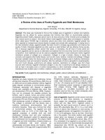
A review of the expression of clinical, pathological and immunohistochemical features in 76 cases of thymoma
Bạn đang xem bản rút gọn của tài liệu. Xem và tải ngay bản đầy đủ của tài liệu tại đây (2.56 MB, 7 trang )
Journal of military pharmaco-medicine no1-2019
A REVIEW OF THE EXPRESSION OF CLINICAL,
PATHOLOGICAL AND IMMUNOHISTOCHEMICAL
FEATURES IN 76 CASES OF THYMOMA
Tran Ngoc Dung1; Dinh Tien Truong1; Nguyen Khac Tuyen1
Nguyen Manh Hung1; Tran Viet Tien1
SUMMARY
Introduction: Thymoma is the most common tumor in mediastinum. In Vietnam, there has not
been any study regarding the pathological classification according to 2015 WHO classification,
and the expression of imumunohistochemical markers of thymoma. Objectives: To determine
these histological types of thymoma and expression of imumunohistochemical markers in
thymoma according to 2015 WHO classification. Subjects and methods: 76 patients
pathologically diagnosed with thymoma, underwent thymectomy at Department of Thoracic
Surgery, 103 Military Hospital, from January 2010 to October 2018. Samples of thymoma were
taken and performed H&E staining and imunohistochemical staining with these markers which
were CD3, CD20, CD45, CD117, CKAE1/AE3, EMA, CK19, Ki67, p53. Results: A total of
76 patients, mean age 47.97 ± 13.43 and mean tumor size 4.09 ± 2.27 cm; patients with stage I
disease were the most frequent (55.3%). 44.7% of patients had the invasive tumor. 59.21% of
patients had accompanying myasthenia gravis, mostly at stage IIA (57.8%). The two most
common types of thymic tumors were determined type B1 (26.3%) and B2 (26.3%). Conclusion:
Generally, thymoma is rarely exposed on children. There was no difference between genders.
The tumor size was quite large. Patients with stage I were the most frequent. The tumor could
invade into the capsule. Type B1 and B2 thymoma were the most common. Myasthenia gravis
could be found in patients with thymoma. Thymoma had a high mitotic count and poor expression
of the mutation in tumor suppressor genes.
* Keywords: Thymoma; Myasthenia gravis; Imumunohistochemical features.
INTRODUCTION
Thymomas are the most common
tumors in the mediastinum, represent
more than 45% in adults with mediastinal
tumor. Patients at the age of 40 - 60 are
more common, there is no sex
predominance [4, 5]. According to
American Cancer Society, the overall
incidence of thymoma in the United State
is about 0,15 per 100.000 person-years [6].
It is widely accepted that thymomas are
malignant because of these invasive
potentials. About 50% of patients with
thymomas are asymptomatic, 30% of
neoplasms manifest clinically by symptoms
of myasthenia gravis (MG) [7]. Determining
pathological subtypes of thymomas yield
advantages in prognosis and treatment. In
Vietnam, studies have been involved in
clinical and radio-characteristics but not in
pathological ones. This study was done
with two goals:
1. 103 Military Hospital
Corresponding author: Tran Ngoc Dung ()
Date received: 20/10/2018
Date accepted: 11/12/2018
166
Journal of military pharmaco-medicine no1-2019
- To evaluate the symptom of
myasthenia gravis, pathological types of
thymomas according to 2015 WHO
classification.
- To determine the expression of
immunohistochemistry (IHC) markers in
thymomas.
SUBJECTS AND METHODS
1. Subjects.
The study included 76 patients who
had tumors in the anterior mediastinum,
underwent thymectomy at Department of
Thoracic Surgery, 103 Military Hospital;
the tumors were subsequently pathologically
diagnosed with thymomas, from January
2010 to October 2018
2. Methods.
This is a retrospective and descriptive
study, including:
- Age, sex, Masaoka’s classification:
Stage of thymoma, Osserman's classification:
severity of clinical myasthenia gravis [8, 9].
- Pathological characteristics of the
tumor: Size, pathological types according
to 2015 WHO classification [10], the
expression of immunohistochemistry
markers CD3, CD20, CD45, CD117,
CKAE1/AE3, EMA, CK19, Ki67, p53.
(Ki67 is positive if there were more than
10% of tumor nuclei reacted)
* Statistical analysis: Using SPSS 22.0
for Windows.
RESULTS AND DISCUSSION
1. Clinical features.
Table 1: Age and gender.
Gender
Age
Male
Total
Female
n
%
20 - 29
5
4
9
11.8
30 - 39
5
5
10
13.2
40 - 49
13
10
23
30.3
50 - 59
6
11
17
22.4
≥ 60
9
8
17
22.4
Total
38
38
76
100
X ± SD
47.24 ± 13.98
48.71 ± 12.99
47.97 ± 13.43
The mean age of the study group was 47.97 ± 13.43. The age ranged from 21 to 77.
The group of 40 - 49 years old made up the highest proportion (30.3%). Patients older
than 40 years old were predominant (75.1%). Male to female ratio was 1/1. There was
no significant difference in mean age of male to female (p = 0.64).
167
Journal of military pharmaco-medicine no1-2019
Nguyen Khac Kiem (2010) conducted
a study on 71 patients showed that the
age ranged from 15 to 75, and patients at
the age of 30 - 50 accounted for the
majority with 56.3% [1]. In a studying on
209 patients with thymoma, Huynh Quang
Khanh reported that the mean age was
44.30 ± 15.15 years old, patients aged
31 - 60 were predominant, there was no
difference between genders.
into perithymic adipose tissue (Masaoka
IIB) and 3 cases (3.9%) invaded to the
lung parenchyma (Masaoka III) (the tumor
invaded to the lung although the size was
under 4 cm). This result was different
from Huynh Quang Khanh’s, most cases
(67.2%) had noninvasive tumors (stage I),
17.2% of cases had capsular invasion
(stage II) and the percentage of tumors
with perithymic tissue was 15.6% [2].
Thus, most of studies revealed that
thymoma occurred in adults. There was
no sex predominant.
Table 3: Severity of clinical
(Osserman's classification).
Table 2: Tumor size.
Size (cm)
%
≤5
60
78.9
5 - 10
14
18.4
> 10
2
2.7
76
100
X ± SD
4.09 ± 2.27
Mean tumor size was 4.09 ± 2.27,
ranged from 0.5 to 12 cm. According to
Khanh's study, the mean tumor size was
8.17 ± 2.44 cm [2]. This difference comes
from the different ways of choosing surgery
methods. Our study involved both open
and endoscopic thymectomy while Khanh’s
study only involved open thymectomy.
* Masaoka’s stage:
+ Noninvasive tumors (Masaoka I):
42 cases (55.3%) which was the most
common.
+ Invasive tumors: 43 cases include:
25 cases (32.9%) with capsular invasion
(Masaoka IIA), 6 cases (7.9%) extended
168
n
%
31
40.8
Class I
18
23.7
Class IIA
26
34.2
Class III
1
1.3
Total
76
100
No MG
n
Total
Clinical features
MG
MG
45 cases (59.21%) had signs of MG.
Class IIA was the most common (57.8%).
Our result was higher than others’ finding.
Pham Huu Lu’s research (2015) showed
that MG was associated with 11.54% of
patients with thymoma [3], this incidence
was 36.5% in the Zhefeng Zhang's study
(2016) [5].
Myasthenia gravis is the most common
presentation among neuroimmune diseases
associated with thymomas. The incidence
of thymoma with MG ranges from 10 - 25%.
The signs vary from time to time, there
have been progressive and regressive
periods alternatively. Signs of MG can
manifest before or after thymectomy.
Journal of military pharmaco-medicine no1-2019
The manifestation of MG is the presence
of fatigable muscle weakness including
ocular, facial, oropharyngeal or extremity
muscle. The prostigmin test or the antiacetylcholine receptor (AChR) antibody
test can be used for diagnosing MG.
Table 4: Pathological classification of thymomas.
Type
n
%
A
15
19.7
AB
18
23.8
B1
20
26.3
B2
20
26.3
B3
3
3.9
76
100
Total
Type B1 and B2 thymoma accounted for 26.3% separately and were the most
common, type B3 (3.9%) was the least common. The incidence of type B3 thymoma
was lower than Zhefeng Zhang’s results (18.4%) [5]. The other types were consistent
with other researchers’ findings [10].
Table 5: Correlation between pathological types and MG.
Clinical features
Pathological type
No MG
MG
Total
%
A
6
9
15
19.7
AB
7
11
18
23.8
B1
8
12
20
26.3
B2
7
13
20
26.3
B3
3
0
3
3.9
Total
31
45
76
100
p
0.519
(χ² = 11.11, df = 12, p = 0.519)
In some previous studies, there was the correlation between pathological types
and MG, the incidence of MG was the highest in type B2. In our research, there was no
statistically significant difference between pathological types and MG (p > 0.05).
According to Zhefeng Zhang (2016), the incidences of correlation MG with types A,
AB, B1, B2, B3 were 2.6%; 13.2%; 13.2%; 52.6% and 18.4%, respectively, type B2
thymoma was the most common (52.6%) [5].
169
Journal of military pharmaco-medicine no1-2019
Table 6: The expression of IHC markers in thymomas.
Type A
Type AB
Type B1
Type B2
Type B3
Markers
(+)
(-)
Σ
(+)
(-)
Σ
(+)
(-)
Σ
(+)
(-)
Σ
(+)
(-)
Σ
CD3
7
1
8
16
1
17
18
0
18
15
2
17
2
0
2
CD20
0
7
7
2
12
14
10
7
17
3
14
17
1
0
1
CD45
2
2
4
8
0
8
7
0
7
6
0
6
1
0
1
CD117
2
4
6
0
1
1
0
4
4
1
4
5
AE1/AE3
14
1
15
17
0
17
14
3
17
19
0
19
3
0
3
EMA
2
3
5
5
3
8
1
3
4
1
4
5
2
0
2
3
1
4
1
1
2
7
0
7
CK19
Ki67
3
0
3
5
0
5
7
0
7
0
5
5
1
0
1
p53
3
4
7
1
0
1
1
1
2
1
3
4
0
0
0
- CD3 was positive in 100% of type B1 and B3 thymomas and most of types A, AB
and B2 thymomas.
- A small number of CD20-positive can be found in type AB and B2, CD20 was
negative in type A. 58.8% of cases of type B1 thymoma reacted positively with CD20,
which approved that type B1 thymoma tumor had the lymphocytic component which
stained with CD20.
- Most of cases showed positive staining for CD45 and CD3, this result demonstrated
the lympho T - rich pattern in thymomas.
- True to the epithelial origination, the neoplastic cells of thymomas were positive for
CK AE1/AE3. This result was consistent with WHO 2015.
- The positive expression of Ki67 in most of cases revealed the high proliferation
activity of tumor cells, although some cases demonstrated weak positive results.
- p53 revealed weak positive in 14 cases.
- There have been difficulties in using IHC in thymomas because thymomas are
neoplasms composed of many kinds of cells with varying numbers.
170
Journal of military pharmaco-medicine no1-2019
a
b
c
d
Figure 1: Pathological types of thymomas: Type A (a); type B1 (b);
type B2 (c) and type B3 (d).
CONCLUSION
- Generally, thymoma rarely exposes
on children. Mean age 47.97 ± 13.43.
There was no difference between genders.
The tumor size was quite large, mean
size 4.09 ± 2.27 cm. Patients with stage I
were the most common (55.3%). 44.74%
of cases had invasive tumors. 59.21% of
patients with thymomas had MG,
57.8% of cases were in class IIA MG.
- Type B1 and B2 thymoma were the
most common, followed by type AB thymoma
(23.8%), B3 thymoma was the least.
- The expression of IHC markers was
different from types depending on tumor
patterns, but there has a high proliferation
activity of tumor cells and poor expression
of the mutation in tumor suppressor genes.
REFERENCES
1. Nguyễn Khắc Kiểm, Hoàng Đình Chân.
Nghiên cứu týp mô bệnh học và kết quả điều
trị u tuyến ức tại Bệnh viện K 2003 - 2008.
Tạp chí Y học Thực hành. 2010, 716 (5),
tr.19-22.
2. Huỳnh Quang Khánh. Nghiên cứu
kết quả điều trị u trung thất nguyên phát bằng
phẫu thuật nội soi lồng ngực. Trường
Đại học Y Dược Thành phố Hồ Chí Minh.
TP. Hồ Chí Minh. 2015.
171
Journal of military pharmaco-medicine no1-2019
3. Phạm Hữu Lư. Nghiên cứu điều trị u
trung thất bằng phẫu thuật nội soi lồng ngực
tại Bệnh viện Việt Đức. Trường Đại học Y Hà
Nội. Hà Nội. 2015.
4. Marchevsky A.M, Wick M.R. Pathology
of the mediastinum. Cambridge University
Press. Cambridge. 2014.
5. Zhefeng Zhang, Youbin Cui, Rui Jia et al.
Myasthenia gravis in patients with thymoma
affects survival rate following extended
thymectomy. Oncology Letters. 2016, 11,
pp.4177-4182.
6. Engels E.A, Pfeiffer R.M. Malignant
thymoma in the United States: Demographic
patterns in incidence and associations with
172
subsequent malignancies. J Cancer. 2003,
105, pp.546-551.
7. Thomas, C.R, Wright, C.D, Loehrer, P.J.
Thymoma: State of the Art. J Clin Oncol.
1999, 17, pp.2280-2289.
8. Masaoka A, Monden Y, Nakahara K,
Tanioka T. Follow-up study of thymomas with
special reference to their clinical stages.
Cancer. 1981, 48 (11), pp.2485-2492.
9. Osserman K.E. Myasthenia gravis.
New York: Grune and Stratton. 1958, p.80.
10. William D.T, Elisabeth B, Allen P et al.
WHO classification of tumours of the lung,
pleura, thymus and heart. 4 Ed, IARC Press.
Lyon. 2015, pp.183-298.