Ebook Exam preparatory manual for undergraduates ophthalmology: Part 2
Bạn đang xem bản rút gọn của tài liệu. Xem và tải ngay bản đầy đủ của tài liệu tại đây (37.65 MB, 170 trang )
CHAPTER
15
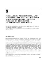
Lids
1. What are the layers of eyelids?
••
••
••
••
••
••
••
Fig. 15.1: From the front to back
Cutaneous layer (skin)
Areolar layer (loose areolar tissue)
Muscular layer (orbicularis oculi)
Sub Muscular areolar tissue
Fibrous layer (tarsal plate)
Layer of smooth muscle (Muller’s muscle)
Conjunctival layer (mucous layer)
−− Grey line separates fibrous layer from muscular layer.
mebooksfree.com
Lids 257
2. Write short notes on blepharitis:
Chronic inflammation of lid margin.
Types
••
••
••
••
Squamous
Ulcerative
Posterior blepharitis or meibomitis
Parasitic blepharitis – due to Demodex folliculorum and Phthiriasis palpebrarum.
Squamous blepharitis
Fig. 15.2: Squamous blepharitis
••
••
••
••
••
Due to seborrheic dermatitis
Usually associated with dandruff of scalp
White coloured scales accumulate among lashes
Eyelashes fall out but are replaced without distortion
On removal of scales, the underlying surface is red.
Ulcerative blepharitis
Fig. 15.3: Ulcerative blepharitis
•• Yellow scales glue the lashes together
•• Hyperemia, telangiectasia and scaling
•• On removing of the crusts, small ulcers are seen around base of the lashes. These ulcers
bleed. Lashes fall and replaced by misdirected ones.
mebooksfree.com
258 Exam Preparatory Manual for Undergraduates Ophthalmology
•• Caused by staphylococcal aureus and epidermidis
•• Secondary changes caused by hypersensitivity to staphylococcus exotoxins are:
−− Mild papillary conjunctivitis
−− Marginal keratitis.
Symptoms
••
••
••
••
••
••
Itching
Redness
Soreness
Lacrimation
Mild photophobia
Lid crusting.
Sequela
••
••
••
••
••
••
Trichiasis
Tylosis (thickening of lid margin)
Madarosis ( loss of eye lashes)
Poliosis (whitening of eye lashes)
Ectropion
Stye
−− Usually secondary to poor ocular hygiene, frequent rubbing of eyes, exposure to
smoke and pollutants and asthenopia.
Differences between squamous and ulcerative blepharitis
Features
Squamous
Ulcerative
Etiology
Seborrhea
Staphylococcus
Deposits on eyelashes
Shiny waxy
Brittle scales/ulcers
Removal of deposits leaves
Hyperaemic zone
Bleeding ulcers
Hair follicles
Not destroyed
Destroyed
Meibomian secretion
Vicarious
Normal
Eyelashes
Normal
Trichiasis, Madarosis and
Poliosis
Lid margin may present
Chalazion
Stye
Conjunctiva
Mild conjunctivitis
Severe conjunctivitis
Dryness
Absent
Present
Corneal complications
•• Punctate epithelial erosions
•• Marginal ulcers
May be seen
Absent
Commonly seen
Present
Treatment
Local
•• Removal of scales, crusts and diseased lashes
•• Cleaning lid margin with 3% NaHCO₃, betadine, or baby shampoo and artificial tears.
mebooksfree.com
Lids 259
Specific: Antibiotic eye ointment, betadine lotion applied for 2–3 weeks.
General: Improvement of general health and personal hygiene.
Dandruff of scalp: Selenium sulphide 2.5% shampoo twice weekly.
Posterior blebharitis: Systemic tetracycline or doxycycline or erythromycin and antibiotic
steroid eye ointment at the lid margin.
•• Parasitic blepharitis – Removal of nits by forceps and antibiotic ointment to lid margin.
•• Delousing of patient and family members.
3. Write short notes on Hordeolum externum (Stye):
Fig. 15.4: Zeis gland
Fig. 15.5: Stye
Acute suppurative inflammation of Zeis or Moll’s glands.
mebooksfree.com
260 Exam Preparatory Manual for Undergraduates Ophthalmology
Etiology
Due to staphylococcal infection associated with boils, acne, common in children and young
adults. Also seen in immuno compromised person.
Symptoms
Acute pain along the lid margin.
Signs
Tender, hard swelling seen near lid margin. An abscess may form pointing near base of
lashes. Pain subsides if it gets ruptured. There are two stages:
•• Stage of cellulitis
•• Stage of abscess.
Treatment
Hot fomentation in early stage:
•• Systemic – broad spectrum antibiotic
•• Analgesics and anti-inflammatory drugs
•• Removal of pus by pulling or epilating the involved lash or incision and drainage
•• In case of recurrent stye, diabetes and refractive errors have to be ruled out.
4. Write short notes on chalazion:
Fig. 15.6: Tarsal or meibomian cyst
Chronic granulomatous inflammation of meibomian gland.
Etiology
Due to chronic irritation caused by an organism of low virulence:
•• Small nontender hard swelling in the lid, slightly away from lid margin
•• Cosmetic problem
•• May cause astigmatism against rule due to pressure on cornea.
Sequela
•• Spontaneous resolution
•• Contents may extrude through conjunctiva to resemble fungating mass
mebooksfree.com
Lids 261
••
••
••
••
Calcification
Meibomian cell carcinoma
May get secondary infection and form Hordeolum internum
Marginal chalazionoccurs in ducts of meibomian gland. The granulation tissue projects
as a reddish grey nodule on the intermarginal strip.
Treatment
•• Incision and curettage: The lid is everted and vertical incision is made with 11 blade
and mucoid material scooped out and the cavity curetted. Vertical incision is made to
avoid damage to adjacent meibomian glands.
•• Intralesional injection of Triamcinolone may help in smaller chalazion.
5. Write short notes on Hordeolum internum:
Acute suppurative inflammation of meibomian gland.
•• Occurs due to secondary infection of chalazion.
•• Symptoms are more than in stye because the gland is larger and is embedded deeply
in dense fibrous tissue.
Treatment: Broad spectrum antibiotics and anti-inflammatory drugs to control pain. Latter
incision and curettage as for chalazion.
6. What is Molluscum contagiosum?
It is a viral infection of the lids, commonly affecting children. It is caused by poxvirus.
Clinical features: They are multiple, pale, waxy, umblicated swellings around the lid
margin.
Complications: Chronic follicular conjunctivitis and superficial keratitis.
Treatment: They should be incised and expressed and the interior touched with 5%
povidone iodine or pure carbolic acid.
7. Write short notes on trichiasis:
Fig. 15.7: Eyelashes are misdirected backwards and rub against cornea
Etiology
•• Stage IV Trachoma
•• Spastic entropion in elderly person.
mebooksfree.com
262 Exam Preparatory Manual for Undergraduates Ophthalmology
•• Tight bandaging
•• Ulcerative blepharitis
•• Scars of lid.
Symptoms
•• Foreign body sensation and photophobia
•• Redness of conjunctiva and lacrimation.
Signs
•• Misdirected cilia touching the cornea
•• Blepharospasm and photophobia if cornea is involved
•• Superficial corneal opacities.
Complications
•• Chronic conjunctivitis
•• Recurrent corneal abrasions
•• Nonhealing corneal ulcer.
Treatment
•• If single or few cilia are involved, epilation or electrolysis at root of hair follicle
•• When many cilia are misdirected, surgical correction similar to cicatrical entropion is
advised.
8. Write short notes on entropion:
Fig. 15.8: Lid margin rolls inwards
Types
•• Senile or involutional: Affects only lower lid.
•• Spastic: Due to spasm of orbicularis oculi of lower lid due to tight bandaging. Occurs
in lower lid of old people.
mebooksfree.com
Lids 263
•• Cicatrical: Due to contraction of palpebral conjunctiva involves both lids. It occurs in
trachoma stage IV, ulcerative blepharitis, Burns, Steven Jhonson syndrome,
diphtheritic membranous conjunctivitis and pemphigus (pemphigoid)
•• Congenital entropion rare: Due to deformity of tarsal plate. It involves lower lid only.
•• Mechanical: Due to lack of support provided by eyeball—seen in phthisis bulbi or
enucleated or eviscerated eyes.
Symptoms and Signs
Same as for Trichiasis and those of disturbances of stability of tear film. They are foreign
body sensation, irritation, abrasion and corneal ulcer.
Three grades
1. Mild: Posterior border of lid inturned
2. Moderate: Intermarginal strip rotates inwards
3. Severe: Entire lid margin rolls inwards.
Senile entropion: Seen in old age. Usually above 60 years.
Etiology
•• Overriding of preseptal part of orbicularis over pretarsal part with atrophy of subcutaneous tissue with ageing.
•• Inturning of lower lid tarsus due to weak lower lid retractors and laxity of medial and
lateral ligaments.
•• Loss of posterior support in enophthalmos due to atrophy of orbital fat.
Treatment
Aim of surgery: To restore vertical and horizontal tautness of lid:
•• Reattach retractors to tarsal plate.
•• Shortening of horizontal width of tarsal plate.
•• Forming a cicatrix between pretarsal and preseptal parts of orbicularis:
−− Short term relief: By sticking an adhesive tape to pull lower lid outwards.
−− Everting sutures: 5–0 vicryl chromic catgut from conjunctiva to skin of lid adjacent
to inferior border of tarsus creates cicatrical barrier that maintains eyelid in everted
position.
−− Modified Wheeler operation: Skin incision—parallel to lower lid. Orbicularis is dissected and 3mm strip of muscle is double breasted and stitched together with an
excision of base down triangle of tarsus. Redundant (excess) skin is excised.
−− Quickert’s procedure: Transverse lid split + everting sutures + horizontal lid shortening.
−− Modified Jones procedure: Inferior lid retractors are plicated or attached to the tarsus.
Spastic entropion
Due to spasm of Orbicularis oculi:
•• Evert the lid by pulling it with adhesive plaster
•• Botulinum toxin injected into pretarsal orbicularis to prevent it from overriding.
mebooksfree.com
264 Exam Preparatory Manual for Undergraduates Ophthalmology
Cicatrical Entropion
Treatment: Mild to moderate cases with thickened tarsus is treated with wedge resection
of tarsus. If tarsus is not thickened, Modified Burrow’s operation or Tarsal fracture is
done. Very extensive scarring may necessitate the replacement of conjunctiva by a mucous
membrane graft and a distorted tarsal plate by cartilage graft.
Congenital Entropion
abnormal tarsal plate is resected. Abnormal skin fold is excised.
9. Write short notes on Ectropion:
Fig. 15.9: Outward turning of eyelid margin
Types
••
••
••
••
••
••
Senile or Involutional
Spastic
Cicatrical
Mechanical
Paralytic
Congenital.
•• Senile ectropion occurs only in lower lid due to horizontal laxity of eyelid, medial
canthal or lateral canthal laxity.
•• Spastic: Due to blepharospasm when lids are well supported by globe. Occurs in
children and young adults.
•• Cicatrical: Due to scarring of skin by chronic conjunctivitis, blepharitis, injuries, burns,
ulcers, etc.
•• Mechanical: Due to dragging of lid tumor with its weight.
•• Paralytic: Paralysis in orbicularis as in facial palsy - affecting lower lid.
•• Congenital: Due to deficient eyelid skin - skin grafting to be done.
Symptoms
Epiphora and constant watering of the eyes.
Signs
•• Conjunctiva becomes dry and thickened.
•• Chronic conjunctivitis due to exposure, exposure keratitis.
mebooksfree.com
Lids 265
Three grades
1. Mild: Only punctum is everted
2. Moderate: Palpebral conjunctiva is visible
3. Severe: Lower fornix is exposed.
Treatment
•• Spastic: Underlying cause of blepharospasm is treated
•• Cicatricial: Free lid margin from scar tissue and restore lid to normal position.
Mild cases
Fig. 15.10: V-Y operation
V shaped incision is made in the skin of lower lid which includes the scar. Skin is excised
and wound is sutured in Y shaped pattern thus correcting ectropion.
Extensive scarring: Excision of scar tissue and application of skin graft. Split skin graft
or full thickness grafts are taken from upper lid, behind the ear, inner side of upper arm or
thigh.
Senile ectropion: Depending on severity the following operations are done.
−− Mild to moderate cases, a horizontal spindle shaped piece of conjunctiva subconjunctival tissue is removed 5 mm from the punctum and margins are sutured.
−− For severe cases in mid portion of lower lid, full thickness shortening of lid is done.
Fig. 15.11: Bryon Smith modification of Kuhnt Szymanowski’s procedure
Full thickness inverted house (pentagon) shaped excision at least 5 mm away from
punctum is done and margins are sutured.
−− Bryon Smith modification of Kuhnt Szymanowski’s procedure: for severe ectropion
which is more marked in the lateral half of lid. A base up pentagonal full thickness
excision from the lateral third of lid is combined with triangular excision of skin
from the area just lateral to lateral canthus to elevate the lid.
−− Medial ectropion is corrected by modified lazy T operation in which medial vertical
pentagon of full thickness lid is excised 4 mm lateral to lower punctum.
Paralytic ectropion: It occurs due to paralysis of facial nerve, in Bell’s palsy, parotid
surgeries, trauma and tumors such as an acoustic neuroma.
mebooksfree.com
266 Exam Preparatory Manual for Undergraduates Ophthalmology
Treatment: Initially lubricants and taping of the lid is done. For a permanent solution,
lateral tarsorraphy is indicated. In this operation the palpebral aperture is shortened by
uniting the lids at lateral canthus. The edges of the upper and lower lids are freshened for
requisite distance and then sutured.
10. Write short notes on symblepharon:
Fig. 15.12: Adhesion of palpebral and bulbar conjunctiva
Etiology
Due to formation of raw surface on two opposing surfaces.
Causing adhesions during healing of:
•• Burns, ulcer, diphtheria, operative scar
•• Ocular pemphigus
•• Steven Johnsons syndrome.
Types
•• Anterior – lid margin is involved
•• Posterior – fornix is involved
•• Total – Both lids get completely adherent to the globe.
Symptoms
••
••
••
••
Cosmetic disfigurement
Difficulty in lid movements
Diplopia due to restricted mobility of eye
Lagophthalmos - Inability to close lids properly.
Signs
•• Bands of fibrous tissue stretching between lid and globe are seen.
Treatment
•• Prophylaxis: Applying eye ointment and moving a glass rod in the fornices several
times a day
mebooksfree.com
Lids 267
•• Bandage contact lens
•• Bands once formed need excision and the raw surfaces to be coverd by conjunctival or
buccal or amniotic membrane graft.
11. Discuss the types, evaluation, and treatment of ptosis:
Fig. 15.13: Dropping of upper lid below its normal position which is 1–2 mm below upper limbus
It is due to weakness of Levator Palpabrae superioris or Muller’s muscle.
Pseudoptosis
••
••
••
••
••
Lack of support - Phthisis bulbi
Contralateral lid retraction- Thyrotoxicosis
Ipsilateral hypotropia
Brow ptosis (blepharochalosis)
Dermatochalosis (excessive eyelid skin).
Classification
•• Congenital
•• Acquired
−− Neurogenic (oculomotor nerve palsy)
−− Myogenic (myopathy or myasthenic)
−− Traumatic
−− Mechanical - lid tumor
−− Aponeurotic - senile, postoperative evaluation:
-- Marginal reflex distance
-- Upper lid excursion - elevator action
-- Vertical fissure height
-- Bell’s phenomenon
-- Associated features
Marginal reflex distance
−− Distance between upper lid margin and light reflex
−− Mild ptosis – 2 mm of droop
mebooksfree.com
268 Exam Preparatory Manual for Undergraduates Ophthalmology
−− Moderate – 3 mm
−− Severe – 4 mm or more.
Upper lid excursion or Levator action
−− Normal – 15 mm or more
−− Good – 12 mm or more
−− Fair – 5–11 mm
−− Poor – 4 mm or less.
Vertical fissure height
−− Distance between upper and lower lid margin. Comparison determines unilateral
ptosis.
Upper lid crease
−−
−−
−−
−−
−−
Distance between lid margin and lid crease in downgaze.
Female – 10 mm (normal)
Male – 8 mm (normal)
Absent in congenital ptosis
High lid crease suggests aponeurotic ptosis.
Bell’s phenomenon
(upward rotation of eye ball on attempted lid closure)
−− If it is poor – risk of post operative corneal exposure.
−− Presence indicate intact superior rectus function.
Ocular movements
−− Weakness of superior rectus
−− Weakness of superior rectus and inferior oblique of one eye – Double Elevator
Palsy.
Marcus Gunn jaw winking phenomenon
−− Changes in lid position on attempted masticatory movements or side to side
movement of jaws
−− Indicate congenital synkinetic phenomenon.
Corneal sensation
Tensilon or Neostigmine test: To exclude myasthenia gravis
10% phenylephrine test: To differentiate Horner’s syndrome.
Schirmer’s test: For corneal dryness
Management
Mild ptosis: Fasanella servat operation
Tarsus-conjunctival excision along with Muller muscle.
Moderate to severe ptosis
•• Frontalis sling operation = if poor levator action
•• Levator Resection = if levator action is good
mebooksfree.com
Lids 269
•• Ever Busch operation – skin approach
•• Blaskowicz operation – conjunctival approach.
12.Write short notes on lagophthalmos:
Fig. 15.14: Lagophthalmos is a Greek word meaning incomplete closure of eyelids
Causes
•• Bell’s palsy
•• Postsurgical–following ptosis correction, eyelid reconstruction, inferior rectus
recession
•• Thyroid ophthalmopathy
•• Lower lid ectropion
•• Proptosis
•• Extremely ill or comatosed patients.
Symptoms
•• Exposure keratopathy - Burning sensation, Increased lacrimation, tear film instability,
Decreased break up time
•• Inferior corneal ulcer.
Differential Diagnosis
•• Neurotrophic keratopathy
•• Dry eye syndrome.
Treatment
•• Tear supplements and lubricating eye ointment
•• Temporary corneal protection – Bandage contact lens.
Surgical
•• Lower lid ectropion - surgery
•• Orbital decompression for thyroid ophthalmopathy
mebooksfree.com
270 Exam Preparatory Manual for Undergraduates Ophthalmology
•• Lateral tarsorrhaphy
•• Treat the cause.
13. Write short notes on lid tumors:
Benign
••
••
••
••
Xanthelasma or xanthoma
Cysts - Moll’s gland
Nevus
Hemangioma
−− Capillary
−− Cavernous
•• Lymphangioma
•• Neurofibromatosis.
Malignant
••
••
••
Squamous cell carcinoma (Epithelioma)
Basal cell carcinoma
Other: Lymphoma, lymphosarcoma, malignant melanoma, metastatic tumor, leukemia.
Treatment: Excision with radiation.
•• Xanthoma
−− Yellowish round plaque. Usually seen in upper lid close to medial canthus.
−− May be symmetrical
−− Elderly, obese, female who have diabetic or hyperlipoproteinemia.
Treatment: Excision or treated with tricholoroacetic acid.
•• Cysts
Retention cyst of Moll’s gland.
Small, clear or whitish cysts - seen among base of cilia in old people.
Treatment: Incision.
•• Nevus: Pigmentation seen on lid margin.
Treatment: Excision
•• Hemangioma
−− Capillary: Red or portwine stain. Dilated capillaries.
−− Cavernous: Bluish in colour. Dilated large venous spaces located deeper, localized
and encapsulated . May increase in size on lowering the head, crying or coughing.
-- Part of Sturge Weber syndrome.
-- May be associated with choroidal or leptomeningeal hemangioma.
Treatment
−− Spontaneous regression
−− Injection of sclerosing agent
−− Intralesional triamcinolone
mebooksfree.com
Lids 271
−− Cryosurgery
−− Surgical excision.
•• Lymphangioma: Rarely involves lid.
•• Neurofibromatosis (Plexiform neuroma)
−− von Recklinghausen disease
−− Phakomatosis involves lids and orbit
−− Lids are swollen
−− Skin: Shown multiple thickened nerves like knots, cords or bag of worms and
−− Glioma of optic nerve.
Malignant Tumors
•• Squamous cell carcinoma:
−− Usually seen at edges of skin in elderly people.
−− Preauricular and submandibular lymph nodes are enlarged.
•• Basal cell carcinoma – (Rodent ulcer):
−− Occur near medial canthus and is more common.
−− Starting as pimple, ulceration or induration.
−− Grown deeper and all around.
−− Locally malignant.
−− Lymph gland not involved.
−− Basal cell carcinoma is radiosensitive.
mebooksfree.com
Lacrimal
Apparatus
CHAPTER
16
1. Describe briefly the anatomy of lacrimal apparatus:
•• Lacrimal gland
•• Lacrimal passage.
Lacrimal Gland - Two parts
1. Orbital lobe - orbital roof
2. Palpebral part - superior fornix.
Lacrimal Passage
•• Lacrimal puncta
− 6 mm from medial canthus
− On posterior border of lid margin.
•• Canaliculi
−− From puncta to lacrimal sac.
•• Lacrimal sac
−− Lies in lacrimal fossa
- formed by lacrimal bone and
- frontal process of maxilla
- 10 mm in length
−− Opens into nasolacrimal duct.
•• Nasolacrimal duct
− 12 mm in length
Fig. 16.1: Lacrimal apparatus
mebooksfree.com
Lacrimal Apparatus 273
− Opens into inferior meatus of nose
− Directed downwards, slightly outwards and backwards.
Lacrimal Apparatus
Dacryocystitis
•• Inflammation of the lacrimal sac.
Classification
•• Congenital
•• Acquired
−− Acute
−− Chronic.
2. Describe the etiology, clinical features, differential diagnosis, and treatment of
congenital dacryocystitis:
•• Dacryocystitis in the newborn.
Etiology
•• Failure in the canalization of nasolacrimal duct (NLD)
•• Lumen blocked by epithelial debris
•• Commonly at inferior end.
Symptoms
•• Epiphora
−− Normally tears are secreted only 3–4 weeks after birth
•• Purulent discharge or conjunctivitis.
Signs
•• Sticky mucopurulent discharge
•• Persistent epiphora
•• Regurgitation on pressure over the sac area.
Differential Diagnosis
•• Ophthalmia neonatorum
•• Congenital glaucoma.
Complications
•• Recurrent conjunctivitis
•• Lacrimal abscess
•• Fistula formation.
Treatment
•• Conservative:
−− Massage over lacrimal sac area and clear the discharge several times by downward
pressing from sac area towards ala of the nose
mebooksfree.com
274 Exam Preparatory Manual for Undergraduates Ophthalmology
−− Increase in hydrostatic pressure helps to open up membranous occlusion, followed
by instillation of antibiotic drops 3–4 times daily.
•• Surgical
−− Probing of nasolacrimal duct through upper punctum after 3 months of age
−− Dilate the punctum and pass probe vertically downwards, then inwards until
lacrimal bone is reached
−− The probe is then rotated towards midline and pushed down the nasolacrimal duct
−− Lacrimal duct syringing
−− Intubation with silicone tube
−− To be kept for 6 months
−− Dacryocystorhinostomy(DCR) after 4 years.
3. Describe the etiology, clinical features, complications, and treatment of acute
dacryocystitis:
Acute suppurative inflammation of the lacrimal sac.
Two Forms:
1. Acute suppurative dacryocystitis
2. Acute suppurative pericystitis.
Fig. 16.2: Acute dacryocystitis
Acute Suppurative Dacryocystitis
•• Acute exacerbation of chronic dacryocystitis.
•• May start spontaneously.
Etiology
•• Pneumococcus, Staphylococcus, and Streptococci are usual pathogens.
Symptoms and signs
••
••
••
••
Marked swelling, redness, and tenderness of the skin over the sac area
Conjunctival congestion
Submaxillary lymph nodes enlargement
Lacrimal fistula – if the abscess bursts repeatedly.
mebooksfree.com
Lacrimal Apparatus 275
Clinical picture
Three stages:
1. Stage of cellulitis
2. Stage of lacrimal abscess
3. Stage of fistula formation
Stage of cellulitis
••
••
••
••
Painful swelling in lacrimal sac region
Epiphora
Fever and malaise
Redness and edema
−− May spread to lids and cheek.
Fig. 16.3: Lacrimal abscess
Stage of lacrimal abscess
•• Occlusion of canaliculi due to edema
−− Sac filled with pus distends
−− Anterior wall ruptures
−− Forms pericystic swelling – lacrimal abscess
−− Points below and outer side of sac.
Stage of fistula
•• Lacrimal abscess discharges
•• Leaving external fistula below medial palpebral ligament
•• May open internally into nose forming internal fistula.
Fig. 16.4: Stage of external lacrimal fistula – left eye
mebooksfree.com
276 Exam Preparatory Manual for Undergraduates Ophthalmology
Complications
•• Orbital cellulitis and thrombophlebitis
•• Cavernous sinus thrombosis
•• Hypopyon ulcer with minor trauma to cornea.
Differential diagnosis
••
••
••
••
••
Inflamed sebaceous cyst
Furuncle
Acute sinusitis
Dental abscess
Acute mucocele of frontal or anterior ethmoidal sinuses.
Treatment
••
••
••
••
Systemic antibiotics
Analgesics and anti-inflammatory drugs
In lacrimal abscess: Vertical incision and drain the pus
In lacrimal fistula: Excision of the fistulous tract and removal of sac.
Acute Suppurative Pericystitis
••
••
••
••
Infection of perilacrimal tissue
Symptoms are similar to acute dacryocystitis
May burst to form fistula
Treatment: Same as for acute dacryocystitis.
4. Describe the etiology, clinical features, complications, and management of chronic
dacryocystitis:
Common suppurative inflammation of the lacrimal sac that usually results from obstruction
of nasolacrimal duct.
Etiology
•• Stasis of tear fluid in the lacrimal sac.
•• It occurs due to inflammation of mucosa of the nasolacrimal duct.
•• Mucosa becomes swollen and congested causing block favoring stasis.
−− Source of infection- from neighboring structures- nose, sinuses, conjunctiva and
pericystic tissue.
Factors responsible for stasis and inflammation:
•• Anatomical factors
−− Narrow bony canal - Male: Female = 1:3
−− Hypertrophied inferior turbinate
−− Deviated nasal septum
−− Polyps in the nose.
•• General infection: Influenza, mumps, chicken pox
•• Lacrimation: Excess secretion – retention and
−− Atony of the sac – chronic inflammation – BLOCK
Foreign bodies in canaliculi or sac.
mebooksfree.com
Lacrimal Apparatus 277
Clinical Features
Depending on the type:
•• Catarrhal - commonest
•• Encysted
•• Suppurative.
Catarrhal form
••
••
••
••
••
••
Swelling in the lacrimal sac region below the level of inner canthus
Swelling: Not freely mobile
Skin over the swelling is normal
No tenderness
Regurgitated material is mucoid
Syringing: Nonpatency with regurgitation of fluid from upper punctum
Encysted mucocele
••
••
••
••
Obstruction at the upper part of NL duct and sac canalicular junction
In a closed sac: Encysted mucocele forms
No regurgitation
Syringing: Fluid comes back from the same punctum.
Fig. 16.5: Lacrimal mucocele
Suppurative form
••
••
••
••
Erythema of the skin over sac
Conjunctival congestion
Epiphora
Syringing: Discharge of pus.
Complications
••
••
••
••
••
Unilateral conjunctivitis
It can be a source of infection
Hypopyon ulcer with minor trauma to cornea
Eczema of lower lid
Ectropion due to repeated wiping of tears.
mebooksfree.com
278 Exam Preparatory Manual for Undergraduates Ophthalmology
Differential Diagnosis
•• Lacrimal sac tumor
−− Blood stained discharge
•• Cold abscess
•• Dermoid cyst
•• Sebaceous cyst
•• Mucocele of the anterior ethmoidal sinus or frontal sinus.
Investigations
•• Nasal examination
−− To exclude deviation of septum, growth and atrophic rhinitis.
•• Radiological examination
−− Dacryocystography
-- Lipiodol, urograffin, or iodized oil outlines the lacrimal excretory passage.
X-ray taken immediately and after 15 minutes to find out the size of the sac
and site of obstruction.
−− Substraction macrodacryocystography
-- Done with canalicular cathetarization.
−− Gamma scanning
-- Radioactive tracer (technetium) instilled into the conjunctival sac. Its excretion
is visualized by gamma camera.
Treatment
•• In recent cases
−− Repeated syringing of nasolacrimal duct with frequent instillation of antibiotic
drops—reduces swelling of inflamed mucosa and restore the patency by clearing
epithelial debris.
•• In recurrent cases
−− Dacryocystectomy (DCT)
−− Dacryocystorhinostomy (DCR).
•• Insertion of tube
−− Lester jones tube or
−− Pawar’s implant - Inserted into the lacrimal sac
−− Facilitates drainage of tears into nasal cavity.
Dacryocystectomy
•• Complete excision of lacrimal sac
•• Advantage: Operation is easy and takes less time
•• Disadvantage: Watering persists.
Dacryocystorhinostomy
•• Conventional
−− From skin approach: Nasal drainage operation
−− Advantage: No postoperative epiphora
mebooksfree.com
Lacrimal Apparatus 279
−− Early steps are as same as excision of sac. Nasal mucosa in middle meatus is
anastamosed with medial wall of sac.
•• Endonasal DCR
−− Approach from the nose
−− No skin incision
−− Success – 80%.
Features
Endoscopic DCR
External DCR
External scar
Absent
Present
Blood loss during surgery
Less
More
Operating time
Less (15–30 min)
More (45–60 min)
Cost
Expensive
Cheap
Success rate
Less (70–80%)
More (90–95%)
Surgical technique
Needs skill in endoscopy
Endoscopy not required
•• Endolaser DCR
−− Diode laser – Approach from canaliculi
−− Takes less time
−− Success – 70%.
Contraindications for DCR
• Atrophic rhinitis
• Rhinosporidiosis
• Bleeding diseases
• Tumors of lacrimal sac
• Tuberculosis of lacrimal bone
• Extremes of age.
5. Write short notes on tear film:
Tears form a thin (7.6 μ to 8 μ) layer over the cornea and conjunctiva which is known as
tear film. It consists of 3 layers.
1. Inner mucin layer (0.2 μ)
−− Produced by the goblet cells. It consists of glcoproteins.
2. Aqueous layer (6.5 to 7.5 μ)
−− Lacrimal gland and accessory glands of Krause and Wolfring – constitute 90% of
tear film.
−− Content – water with dissolved salts, glucose, lysozymes, lactoferrin, immunoglobulin A and lactoferrin.
3. Outer lipid layer (0.1 μ) produced by the Meibomian glands. It consists of cholesterol
esters.
Functions
•• Mucous layer: Converts corneal epithelium to hydrophilic surface.
•• Aqueous layer: Aids oxygenation, removes debris and antimicrobial
−− Barrier to ocular infection
−− Optical clarity.
•• Lipid layer: Reduces evaporation and aids lubrication.
−− Prevents lid margin overflow.
mebooksfree.com
280 Exam Preparatory Manual for Undergraduates Ophthalmology
6. Describe the etiology, investigations, and treatment of dry eye:
Dry eye is a clinical condition of ocular discomfort caused by deficient tear production
and/or excessive tear evaporation.
•• Mucin deficiency
•• Aqueous tear deficiency
•• Lipid deficiency
•• Impaired eye lid function
•• Corneal epitheliopathies.
Punctate epithelial erosion filaments and plaques.
Mucin Deficiency Dry Eye
•• Hypovitaminosis A
•• Severe conjunctival scarring
•• Trachoma, Steven Johnson syndrome, Chemical burns, radiation and ocular phem
phigus.
Aqueous Deficiency Dry Eye
•• Pure keratoconjunctival sicca (KCS) – only lacrimal glands are damaged by mononuclear
cells.
•• Primary Sjögren’s syndrome (Sicca complex), KCS and dry mouth.
•• Secondary Sjögren’s syndrome - sicca complex and conjunctival tissue disease (usually
rheumatoid arthritis).
•• Riley day syndrome (familial dysautonomia)
•• Idiopathic hyposecretion.
Lipid Deficiency Dry Eye
•• Congenital absence of meibomian glands
•• Chronic blepharitis and meibomitis
•• Neurological lesion of non Sjögren KCS.
Impaired Eye Lid Function - Dry Eye
••
••
••
••
••
Bell’s palsy
Dellen
Symblepheron
Lagophthalmos
Ectropion.
Miscellaneous Causes
•• Computer vision syndrome
•• After LASIK surgery
•• After keratoplasty.
mebooksfree.com