Ebook ECMO - Extracorporeal life support in adults: Part 1
Bạn đang xem bản rút gọn của tài liệu. Xem và tải ngay bản đầy đủ của tài liệu tại đây (3.13 MB, 239 trang )
ECMOExtracorporeal Life
Support in Adults
Fabio Sangalli
Nicolò Patroniti
Antonio Pesenti
Editors
123
ECMO-Extracorporeal Life Support in Adults
Fabio Sangalli • Nicolò Patroniti
Antonio Pesenti
Editors
ECMO-Extracorporeal Life
Support in Adults
Editors
Fabio Sangalli
Department of Anaesthesia
and Intensive Care Medicine
San Gerardo Hospital
Monza (MB)
Antonio Pesenti
Health Science Department
Università Milano Bicocca Facoltà
Medicina e Chirurgia
Monza (MB)
Italy
Italy
Nicolò Patroniti
Health Sciences Department,
Urgency and Emergency Department
Milano-Bicocca University
San Gerardo Hospital
Monza (MB)
Italy
ISBN 978-88-470-5426-4
ISBN 978-88-470-5427-1 (eBook)
DOI 10.1007/978-88-470-5427-1
Springer Milan Heidelberg New York Dordrecht London
Library of Congress Control Number: 2014934677
© Springer-Verlag Italia 2014
This work is subject to copyright. All rights are reserved by the Publisher, whether the whole or part of
the material is concerned, specifically the rights of translation, reprinting, reuse of illustrations, recitation,
broadcasting, reproduction on microfilms or in any other physical way, and transmission or information
storage and retrieval, electronic adaptation, computer software, or by similar or dissimilar methodology
now known or hereafter developed. Exempted from this legal reservation are brief excerpts in connection
with reviews or scholarly analysis or material supplied specifically for the purpose of being entered and
executed on a computer system, for exclusive use by the purchaser of the work. Duplication of this
publication or parts thereof is permitted only under the provisions of the Copyright Law of the Publisher's
location, in its current version, and permission for use must always be obtained from Springer.
Permissions for use may be obtained through RightsLink at the Copyright Clearance Center. Violations
are liable to prosecution under the respective Copyright Law.
The use of general descriptive names, registered names, trademarks, service marks, etc. in this publication
does not imply, even in the absence of a specific statement, that such names are exempt from the relevant
protective laws and regulations and therefore free for general use.
While the advice and information in this book are believed to be true and accurate at the date of
publication, neither the authors nor the editors nor the publisher can accept any legal responsibility for
any errors or omissions that may be made. The publisher makes no warranty, express or implied, with
respect to the material contained herein.
Printed on acid-free paper
Springer is part of Springer Science+Business Media (www.springer.com)
Foreword
The best way to temporarily support or substitute vital organs is based on the availability of reliable and effective tools able to vicariate the failing natural organ. This
opportunity was achieved long ago for the kidney and, later on, for the heart and
lung. The technological improvement that miniaturized the apparatus improved the
vascular access, increased the performance of the artificial support, and has allowed
to expand the use of circulatory and respiratory extracorporeal support to several
clinical situations and to different ICUs (cardiac, respiratory, general). The advent
of new fulminant diseases (H1N1 respiratory failure) and the improvement of outof-hospital care for cardiac arrest are two situations that recently have seen extracorporeal support as a possible life-saving application. In order to correctly use the new
technologies, a specific competency and skills should be developed and implemented: as it happens for the achievement of positive results in the ICU setting, the
entire team (perfusionists, nurses, and doctors) has to be trained and should have
specific knowledge of the new technologies. Moreover, in this time where the adequate allocation of resources appears to be very important, it is mandatory that the
indications for the use of expensive and long-lasting techniques should be accurately weighed and shared among professionals.
The aim of this book is to provide readers with the theory and practical issues
that experts in the field of extracorporeal circulatory and respiratory support believe
could help in understanding and improving the practice of this medical device.
Milan, Italy
Roberto Fumagalli
v
Preface
Extracorporeal membrane oxygenation (ECMO) is not a new technique. It has been
used in clinical practice for the last four decades, but the complexity of management
and the relevant complications limited its diffusion to few specialized centers.
In recent years, the development of new materials and the simplification of the
procedure led to a dramatic increase in the centers providing extracorporeal life support (ECLS) and in the number of ECMO runs, for both respiratory and circulatory
indications.
A growing number of publications on all aspects pertaining to ECLS are populating the medical literature.
Despite this expansion in the use of ECLS and in ECLS-related research, the
clinical management of ECMO remains mainly based on local protocols and procedures, and guidelines are lacking on many aspects of this practice. The ELSO
(Extracorporeal Life Support Organization) registry and website, together with their
so-called Red Book, represent the most authoritative resource, and many websites
provide protocols and management guidelines from different ECMO centers. Still,
such indications are mainly locally based or not regularly updated.
For this reason we tried to collate the most relevant aspects pertaining to ECLS,
following two different approaches. Some chapters present an in-depth analysis of
the current evidence and literature on the different indications, while other chapters
face technical aspects with a more practical approach. These latter chapters are
obviously influenced by the practice in the authors’ centers, but we tried to integrate
this with literature and different experiences whenever possible, particularly for the
aspects where centers’ attitudes diverge, such as left ventricle venting, cannulation
techniques, and management of the lung during respiratory support, to name some.
ECLS remains a fast-evolving technique and some aspects still need research
and optimization. Some of these are outlined in the conclusive chapter of the book,
but more are still to be faced. Ample bibliographic references are provided at the
end of every chapter for the interested reader to further explore specific aspects.
ECLS represents a relatively easy technique, but it is not simply a “procedure” to
be learned and performed. ECMO is an excellent tool for organ support, but it
requires sound physiologic and pathophysiologic knowledge and needs to be combined with top-level standard care.
vii
viii
Preface
We are aware that, as a first edition, the readers will find aspects of the book that
might be improved, and we will welcome any suggestion in this regard. We still
hope that the present work will be useful in disseminating ECLS knowledge and
stimulate further study and research.
Fabio Sangalli, Nicolò Patroniti, Antonio Pesenti, Monza (MB), Italy
Contents
Part I
History and Technical Aspects
1
History of Extracorporeal Life Support . . . . . . . . . . . . . . . . . . . . . . .
Fabio Sangalli, Chiara Marzorati, and Nerlep K. Rana
3
2
Developing a New ECMO Program . . . . . . . . . . . . . . . . . . . . . . . . . .
Antonio F. Arcadipane and Giovanna Panarello
11
3
Basic Aspects of Physiology During ECMO Support . . . . . . . . . . . .
Vittorio Scaravilli, Alberto Zanella, Fabio Sangalli,
and Nicolò Patroniti
19
4
Percutaneous Cannulation: Indication, Technique,
and Complications . . . . . . . . . . . . . . . . . . . . . . . . . . . . . . . . . . . . . . . .
Maurizio Migliari, Roberto Marcolin, Leonello Avalli,
and Michela Bombino
37
5
Surgical Cannulation: Indication, Technique, and Complications .
Francesco Formica, Silvia Mariani, and Giovanni Paolini
49
6
Materials: Cannulas, Pumps, Oxygenators . . . . . . . . . . . . . . . . . . . .
Umberto Borrelli and Cristina Costa
65
7
Coagulation, Anticoagulation, and Inflammatory Response . . . . . .
Marco Ranucci
77
Part II
8
9
ECMO for Circulatory Support
Extracorporeal Life Support: Interactions
with Normal Circulation . . . . . . . . . . . . . . . . . . . . . . . . . . . . . . . . . . .
Michele G. Mondino, Filippo Milazzo, Roberto Paino,
and Roberto Fumagalli
ECMO for Ischemic Cardiogenic Shock. . . . . . . . . . . . . . . . . . . . . . .
Francesco Formica, Fabio Sangalli, and Antonio Pesenti
93
105
ix
x
Contents
10
ECMO for Refractory Cardiac Arrest . . . . . . . . . . . . . . . . . . . . . . . .
Leonello Avalli, Margherita Scanziani, Elena Maggioni,
and Fabio Sangalli
117
11
ECMO for Postcardiotomic Shock . . . . . . . . . . . . . . . . . . . . . . . . . . .
Massimo Baiocchi, Fabio Caramelli, and Guido Frascaroli
127
12
ECMO in Myocarditis and Rare Cardiomyopathies . . . . . . . . . . . . .
Barbara Cortinovis, Monica Scanziani,
and Simona Celotti
137
13
ECMO for High-Risk Procedures . . . . . . . . . . . . . . . . . . . . . . . . . . . .
Fabio Ramponi, Paul Forrest, John F. Fraser, Korana Musicki,
and Michael P. Vallely
151
14
ECMO for Severe Accidental Hypothermia . . . . . . . . . . . . . . . . . . . .
Peter Mair and Elfriede Ruttmann
163
15
ECMO in Drug Intoxication . . . . . . . . . . . . . . . . . . . . . . . . . . . . . . . .
Piergiorgio Bruno, Piero Farina, and Massimo Massetti
171
16
Newer Indications for ECMO: Pulmonary Embolism,
Pulmonary Hypertension, Septic Shock and Trauma . . . . . . . . . . . .
Michela Bombino, Sara Redaelli, and Antonio Pesenti
179
17
Left Ventricular Rest and Unloading During VA ECMO . . . . . . . . .
Gianluca Greco, Barbara Cortinovis, and Leonello Avalli
193
18
Weaning from Extracorporeal Circulatory Support . . . . . . . . . . . . .
Anna Coppo, Lucia Galbiati, and Gianluigi Redaelli
207
19
Treatment Options for End-Stage Cardiac Failure . . . . . . . . . . . . . .
Gurmeet Singh
217
Part III
ECMO for Respiratory Support
20
Ventilatory Management of ARDS Before and During ECMO . . . .
Giacomo Bellani, Giacomo Grasselli, and Antonio Pesenti
239
21
Respiratory Monitoring of the ECMO Patient . . . . . . . . . . . . . . . . .
Alberto Zanella, Francesco Mojoli, Luigi Castagna,
and Nicolò Patroniti
249
22
Structure of an ECMO Network for Respiratory Support . . . . . . . .
Maria Grazia Calabrò, Federico Pappalardo,
and Alberto Zangrillo
265
23
ECMO and Thoracic Surgery . . . . . . . . . . . . . . . . . . . . . . . . . . . . . . .
Alia Noorani and Alain Vuylsteke
273
24
ECMO in the Awake/Extubated Patient . . . . . . . . . . . . . . . . . . . . . . .
Giorgio A. Iotti, Francesco Mojoli, and Mirko Belliato
281
Contents
xi
25
ECMO as a Bridge to Lung Transplant . . . . . . . . . . . . . . . . . . . . . . .
Stefania Crotti and Alfredo Lissoni
293
26
Low-Flow ECMO and CO2 Removal . . . . . . . . . . . . . . . . . . . . . . . . .
Vito Fanelli, Andrea Costamagna, Pierpaolo P. Terragni,
and V. Marco Ranieri
303
27
Weaning from VV ECMO . . . . . . . . . . . . . . . . . . . . . . . . . . . . . . . . . .
Giacomo Grasselli, Paolo Mangili, Simone Sosio,
and Nicolò Patroniti
317
Part IV
ECMO for Organ Procurement
28
Heart-Beating and Non-Heart-Beating Donors . . . . . . . . . . . . . . . . .
Marinella Zanierato, Francesco Mojoli, and Antonio Braschi
327
29
Lung Reconditioning . . . . . . . . . . . . . . . . . . . . . . . . . . . . . . . . . . . . . .
Franco Valenza, Jacopo Fumagalli, Valentina Salice,
and Luciano Gattinoni
337
Part V
Monitoring the ECMO Patient
30
Patient Care During ECMO . . . . . . . . . . . . . . . . . . . . . . . . . . . . . . . .
Michela Bombino, Sara Redaelli, and Nicolò Patroniti
345
31
Echocardiography in Venoarterial and Venovenous ECMO . . . . . .
Nicola Bianco, Leonello Avalli, and Fabio Sangalli
361
32
Haemodynamic Monitoring . . . . . . . . . . . . . . . . . . . . . . . . . . . . . . . . .
Fabio Guarracino and Rubia Baldassarri
375
33
Respiratory Monitoring During VA ECMO. . . . . . . . . . . . . . . . . . . .
Daniela Pasero, Pietro Persico, Tommaso Tenaglia,
and Vito Marco Ranieri
383
34
Neurological Monitoring During ECMO . . . . . . . . . . . . . . . . . . . . . .
Paolo Zanatta, Enrico Bosco, Alessandro Forti,
Elvio Polesel, and Carlo Sorbara
389
35
Monitoring the ECMO Patient: The Extracorporeal Circuit . . . . . .
Stefano Isgrò, Francesco Mojoli, and Leonello Avalli
401
Part VI
36
Complications of ECMO
Complications of Extracorporeal Support
and Their Management . . . . . . . . . . . . . . . . . . . . . . . . . . . . . . . . . . . .
Antonio Rubino, Richard Haddon, Fabrizio Corti,
and Fabio Sangalli
415
xii
37
Contents
Troubleshooting Common and Less Common Problems . . . . . . . . .
Lisen Hockings and Alain Vuylsteke
Part VII
425
Transport of the ECMO Patient
38
Air Transport: Fixed-Wing and Helicopter . . . . . . . . . . . . . . . . . . . .
Antonio F. Arcadipane and Gennaro Martucci
445
39
Ground Transport: Ambulance . . . . . . . . . . . . . . . . . . . . . . . . . . . . . .
Stefano Isgrò, Roberto Rona, and Nicolò Patroniti
455
Part VIII
40
Conclusion
Newer Indications and Challenges . . . . . . . . . . . . . . . . . . . . . . . . . . .
Marco Giani, Alberto Zanella, Fabio Sangalli, and Antonio Pesenti
463
Index . . . . . . . . . . . . . . . . . . . . . . . . . . . . . . . . . . . . . . . . . . . . . . . . . . . . . . . .
473
Part I
History and Technical Aspects
1
History of Extracorporeal Life Support
Fabio Sangalli, Chiara Marzorati, and Nerlep K. Rana
ECMO (extracorporeal membrane oxygenation), also called ECLS (extracorporeal
life support), in its actual application is an evolution of the heart–lung machines
used in cardiac surgery. Depending on its configuration – venovenous or venoarterial – it is used to support respiratory function, circulation, or both. This treatment
provides a bridge, either to healing of the natural organs or to long-term devices or
transplantation. In fact, although ECMO has the capability to support cardiorespiratory function temporarily, it is not a cure for the underlying disease. As Warren
Zapol, one of the fathers of respiratory ECMO, pinpointed in an editorial in the New
England Journal of Medicine in 1972, the goal of ECLS is to “buy time” while
sustaining an adequate tissue perfusion [1].
Despite the fact that the origins of ECLS stem from cardiac surgery and the
heart–lung machine, its main applications – at least until recent years – and most
of the related research were carried out in the setting of severe respiratory
failure.
Artificial oxygenation is a theme that has always fascinated scientists since the
beginning of modern medicine.
The first attempt to artificially oxygenate blood in an extracorporeal circulation
was achieved in 1869 by Ludwig and Schmidt, by shaking together defibrinated
blood with air in a balloon [2]. Next step was reached 10 years later, by artificially
perfusing for the first time an isolated kidney using the first simple “bubble oxygenator.” In the same year, Frey and Gruber described the first “two-dimensional,”
F. Sangalli (*) • C. Marzorati
Department of Anaesthesia and Intensive Care Medicine, San Gerardo Hospital,
University of Milano-Bicocca, Via Pergolesi 33, Monza 20900, Italy
e-mail: ;
N.K. Rana
Anaesthesiology and Critical Care Department, Città della Salute e della Scienza,
Ospedale S. Giovanni Battista-Molinette, Corso Bramante, 88, Turin 10126, Italy
e-mail:
F. Sangalli et al. (eds.), ECMO-Extracorporeal Life Support in Adults,
DOI 10.1007/978-88-470-5427-1_1, © Springer-Verlag Italia 2014
3
4
F. Sangalli et al.
direct-contact extracorporeal oxygenator that exposed a thin film of blood to air
in an inclined cylinder that was rotated at a frequency of 30/min by an electric
motor [3–7].
Several bubble- and surface-type oxygenators were developed in the first two
decades of the twentieth century. The main problems that hampered the development of the technique were thrombosis and hemolysis [8]. The turning point was the
discovery of heparin by Jay Maclean, in 1916. This led to overcome most of the
problems due to the contact of blood with air and the resulting prothrombotic activation [3, 4, 7].
The first whole-body extracorporeal perfusion was realized on a dog in 1929 by
Brukhonenko and Tchetchuline in Russia [3, 7, 9–11].
Between 1930 and 1953 three important oxygenators were developed; they
paved the way to apply the technique to men:
• The film oxygenator developed by Gibbon between 1937 and 1953 consisted of
a stationary screen oxygenator [12–15] made up of a series of six to eight wire
mesh screens arranged vertically and in parallel in a plastic container down
which the blood flowed, forming a stable film that was exposed to a flow of oxygen [5, 11]. Kirklin et al. [16–19], at the Mayo Clinic in Rochester, Minnesota,
further developed the Gibbon-type stationary screen oxygenator into the MayoGibbon pump-oxygenator apparatus.
• The rotating disc oxygenator was described in 1948 by Bjork. It was further
modified for clinical use by several scientists and improved with the development of materials.
• The bubble oxygenator was described in 1952 by Clarke, Gollan, and Gupta. They
reported that although small bubbles with their large surface area to volume ratio
favored oxygen uptake, they were less buoyant. This means that smaller bubbles
are less likely to rise spontaneously to the surface and are more likely to remain in
suspension – air embolism is therefore more likely. An optimum balance has therefore to be obtained. This optimum is believed to exist if the bubbles are between
2 mm and 7 mm in diameter. Alternatively, a mixture of small and big bubbles may
be used. This oxygenator was subsequently modified and improved until the
DeWall oxygenator, a “sequential bubble oxygenator,” i.e., its components (bubbler, defoamer, reservoir, and pump), are arranged linearly in series [7, 20].
The first successful extracorporeal cardiopulmonary bypass was performed in
1953, by the surgeon John Gibbon. In 1954, Gibbon described how the heart–lung
machine could be used, in case of emergency, to support respiratory and circulatory
activities. This theoretical intuition clashed with the practical impossibility of
extending the duration of extracorporeal circulation over 6 h. This was mainly due
to the cellular damage caused by the direct exposure of blood to gas. Interposing a
gas exchange membrane between the blood and the gas flow solved most of this
problem, and with this technological innovation the machine became more effective, allowing to perform ECMO for longer periods.
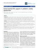
The first successful use of prolonged life support with a heart–lung machine was
conducted by J. Donald Hill in 1971. The patient was 24 years old affected by posttraumatic ARDS, who was supported with ECMO during the acute phase of his
1
History of Extracorporeal Life Support
5
Fig. 1.1 The first successful ECMO patient
pathology, for 3 days. The patient was eventually weaned from ECLS and survived
(Fig. 1.1) [21].
This success was of fundamental importance for the subsequent development
and spread of ECMO. In the same period, ICUs were developing and hemodialysis
was introduced for the treatment of acute renal failure. ARDS remained a fundamental issue for critically ill patients, and the ECMO success was a hope for a
definitive solution to this problem: thanks to that treatment physicians could allow
the functional recovery of the damaged lung. The interest linked to ECLS treatment
was especially about its effectiveness as a respiratory support. This led to the creation of the name ECMO (extracorporeal membrane oxygenation), which emphasized the aspect of artificial oxygenation.
In 1975 Bartlett successfully treated with ECMO the first newborn, a baby
called Esperanza. The success of this case led to a great enthusiasm, and in the following years a lot of other patients, both pediatric and adults, were effectively
treated with ECMO [22]. In 1974 the Lung Division of the National Heart and
Lung Institute started a large multicenter trial to test ECMO versus conventional
therapies in acute respiratory failure. The results were disappointing, with just
10 % survival in both groups and no significant difference between ECMO and
conventional therapy [23].
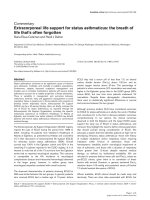
The results of the NIH trial led to a diminished attention to ECMO, but a few
centers continued improving the technique (Fig. 1.2).
In 1978 Kolobow and Gattinoni introduced a modified extracorporeal gas
exchange technique, called extracorporeal carbon dioxide removal (ECCO2R). The
6
F. Sangalli et al.
Fig. 1.2 VV ECMO in Monza, early 1990s
rationale of this technique was to reduce CO2 to decrease ventilation to the minimum
necessary to recruit alveoli. The new ECMO was performed at low extracorporeal
blood flows (20–30 % of cardiac output), so that a venovenous bypass technique
instead of a venoarterial one sufficed, which turned out to be less detrimental to
blood cells, coagulation, and internal organs. Using LFPPV–ECCO2-R, Gattinoni
et al. reported survival rates of up to 49 %. In the following years several centers
corroborated the promising survival rates of around 50 % and higher [14, 24–27].
The need for a coordination between ECMO centers led to the foundation in
1989 in New Orleans of ELSO (Extracorporeal Life Support Organization), a free
community of clinicians and researchers, with the aim to collect data from the
ECMO centers on a unique database and to standardize the procedures.
The evolution of venovenous and venoarterial ECMO diverged over time, with
VV ECMO consolidating its primary role in respiratory support and VA ECMO
assuming an increasing role in the advanced management of circulatory failure.
1.1
VV ECMO
After a period of “disgrace,” mainly due to the relevant complications and to the
appearance on the scene of new promising and – apparently – less invasive strategies, namely, inhaled nitric oxide and prone positioning, VV ECMO was subject of
1
History of Extracorporeal Life Support
7
a renewed interest after the publication of CESAR Trial [15]. This is a multicenter
study comparing conventional therapies to VV ECMO support in ARDS. Results
showed a higher survival and less disability at 6 months in the ECMO group.
Moreover, although not the primary outcome, an actual difference in survival of
around 25 % was observed for patients considered for ECMO treatment at 28 days,
the primary outcome of most ARDS literature.
Even if what this trial actually demonstrated was the importance of centralization of severe ARDS patients to a specialized center, this gave a great thrust to
research, and in the following years the final explosion of the application of this
extracorporeal support was due to the use of ECMO as a rescue therapy in Australia
and New Zealand during the H1N1 influenza pandemic, proving its power in
hypoxemic emergencies [28]. The results obtained during this pandemic, more
than any randomized trial, led to the worldwide acceptance of the use of membrane
lungs.
This led to the creation of ARDS Network, a clinical network initiated by the
National Heart, Lung, and Blood Institute, National Institutes of Health, developed
in order to carry out multicenter trials of ARDS treatment.
Similar experiences, with excellent results both from a clinical and an organizational point of view, were realized in Italy [29] as well as in many other countries [30].
1.2
VA ECMO
Although VA ECMO was originally applied for respiratory support, its main application is nowadays as a circulatory support. In this setting, VA ECMO was employed
almost exclusively as a support for postcardiotomic cardiogenic shock until recent
years.
In the past few decades, VA ECMO gained a place out of the operating theater to
become an advanced treatment for cardiogenic shock. As you will read in the following chapters, it is nowadays widely employed as a circulatory support for cardiogenic shock of any etiology. Its ease of application, which makes it possible to
institute the extracorporeal support virtually anywhere, and the relatively low costs
made it an appealing alternative to other mechanical circulatory support systems,
especially in the emergency setting.
Another emergency application where ECMO gained a pivotal role as a unique
option is that of refractory cardiac arrest. In selected populations, ECMO demonstrated an advantage in survival and neurological outcome in patients with an
expected mortality approaching 100 % [18].
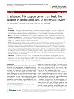
The development of miniaturized systems and more biocompatible circuits
made it possible to bring ECMO everywhere in the hospital, to retrieve patients
from hospitals without ECMO facilities (Fig. 1.3) or even out of the hospital [19,
31]. This was simply unimaginable just two decades ago, as Fig. 1.3 demonstrates
clearly.
8
F. Sangalli et al.
Fig. 1.3 Retrieval of an acute cardiogenic shock patient from a peripheral hospital
1
History of Extracorporeal Life Support
1.3
9
Conclusion
The technological evolution and new directions expand every day the potential of
ECLS.
The relatively short history of ECMO is dotted with great discoveries and forward leaps and hampered with disillusions, but – for sure – most of this story has yet
to be written!
References
1. Zapol WM, Kitz RJ (1972) Buying time with artificial lungs. N Engl J Med 286:657–658
2. Ludwig C, Schmidt A (1868) Das Verhalten der Gase, Welche mit dem Blut durch die reizbaren Säugethiermuskelz strömen. Leipzig Berichte 20:12–72
3. Rendell-Baker L (1963) History of thoracic anaesthesia. In: Mushin WW (ed) Thoracic anaesthesia. Blackwell Scientific Publications, Oxford, pp 598–661
4. Wylie WD, Churchill-Davidson HC (1972) A practice of anaesthesia, 3rd edn. Lloyd-Luke,
London, pp 691–715
5. Hewitt RL, Creech O Jr (1966) History of the pump oxygenator. Arch Surg 93:680–696
6. von Frey M, Gruber M (1885) Studies on metabolism of isolated organs. A respirationapparatus for isolated organs. Untersuchungenuber den stoffwechsel Isolierter organe. Ein
respirations-apparat fur isolierte organe [in German]. Virchows Archiv Physiol 9:519–532
7. Lim MW (2006) The history of extracorporeal oxygenators. Anaesthesia 61:984–995
8. Kirklin JW, Theye RA, Patrick RT (1958) The stationary vertical screen oxygenator. In: Allen
JG (ed) Extracorporeal circulation. Thesis. Charles C Thomas, Springfield, pp 57–66
9. Lee LH, Krumhaar D, Fonkolsrud EW, Schjeide OA, Maloney JV (1961) Denaturation of
plasma proteins as a cause of morbidity and death after intracardiac operations. Surgery
50:29–37
10. Probert WR, Melrose DG (1960) An early Russian heart-lung machine. Br Med J
1:1047–1048
11. Brukhonenko S (1929) Circulation artificielle du sang dans l’organisme entire d’un chin avec
Coeur exclu. J Physiol Pathol Gen 27:251–272
12. Brukhonenko S, Tchetchuline S (1929) Experiences avec la tete isolee du chien. J Physiol
Pathol Gen 27:31–79
13. Miller BJ, Gibbon JH, Fineburg C (1953) An improved mechanical heart and lung apparatus;
its use during open cardiotomy in experimental animals. Med Clin North Am 1:1603–1624
14. Gattinoni L, Pesenti A, Mascheroni D, Marcolin R, Fumagalli R, Rossi F, Iapichino G,
Romagnoli G, Uziel L, Agostoni A (1986) Low-frequency positive pressure ventilation with
extracorporeal CO2 removal in severe acute respiratory failure. JAMA 256:881–886
15. Peek GJ, Mugford M, Tiruvoipati R, Wilson A, Allen E, Thalanany MM, Hibbert CL, Truesdale
A, Clemens F, Cooper N, Firmin RK, Elbourne D (2009) Efficacy and economic assessment of
conventional ventilatory support versus extracorporeal membrane oxygenation for severe adult
respiratory failure (CESAR): a multicentre randomised controlled trial. Lancet 374:1351–1363
16. Gibbon JH Jr (1954) Application of a mechanical heart and lung apparatus to cardiac surgery.
Minn Med 37:171–185
17. Jones RE, Donald DE, Swan JC, Harshbarger HG, Kirklin JW, Wood EH (1955) Apparatus of
the Gibbon type for mechanical bypass of the heart and lungs; preliminary report. Proc Staff
Meet Mayo Clin 30:105–113
18. Avalli L, Maggioni E, Formica F, Redaelli G, Migliari M, Scanziani M, Celotti S, Coppo A,
Caruso R, Ristagno G, Fumagalli R (2012) Favourable survival of in-hospital compared to
out-of-hospital refractory cardiac arrest patients treated with extracorporeal membrane oxygenation: an Italian tertiary care centre experience. Resuscitation 83:579–583
10
F. Sangalli et al.
19. Arlt M, Philipp A, Voekel S, Camboni D, Rupprecht L, Graf BM, Schmid C, Hilker M (2011)
Hand-held minimized extracorporeal membrane oxygenation: a new bridge to recovery in
patients with out-of-centre cardiogenic shock. Eur J Cardiothorac Surg 40:689–694
20. Iwahashi H, Yuri K, Nosè K (2004) Development of the oxygenator: past, present and future.
J Artif Organs 7:111–120
21. Hill JD, O’Brien TG, Murray JJ, Dontigny L, Bramson ML, Osborn JJ, Gerbode F (1972)
Extracorporeal oxygenation for acute post-traumatic respiratory failure (shock-lung syndrome): use of the Bramson Membrane Lung. N Engl J Med 286:629–634
22. Bartlett RH, Gazzaniga AB, Jefferies R, Huxtable RF, Haiduc NJ, Fong SW (1976)
Extracorporeal membrane oxygenation (ECMO) cardiopulmonary support in infancy. Trans
Am Soc Artif Intern Organs 22:80–88
23. Lewandowski K, Metz J, Deutschmann C, Preiss H, Kuhlen R, Artigas A, Falke KJ (1995)
Incidence, severity, and mortality of acute respiratory failure in Berlin, Germany. Am J Respir
Crit Care Med 151:1121–1125
24. Kolobow T, Gattinoni L, Tomlinson T, White D, Pierce J, Iapichino G (1977) The carbon
dioxide membrane lung (CDML): a new concept. Trans Am Soc Artif Intern Organs
23:17–21
25. Kolobow T, Gattinoni L, Tomlinson TA, Pierce JE (1977) Control of breathing using an extracorporeal membrane lung. Anesthesiology 46:138–141
26. Gattinoni L, Pesenti A (2005) The concept of ‘baby lung’. Intensive Care Med 31:776–784
27. Gattinoni L, Agostoni A, Pesenti A, Pelizzola A, Rossi GP, Langer M, Vesconi S, Uziel L, Fox
U, Longoni F, Kolobow T, Damia G (1980) Treatment of acute respiratory failure with lowfrequency positive-pressure ventilation and extracorporeal removal of CO2. Lancet
2:292–294
28. The Australia and New Zealand Extracorporeal Membrane Oxygenation (ANZ ECMO)
Influenza investigators (2009) Extracorporeal membrane oxygenation for 2009 influenza
A(H1N1) acute respiratory distress syndrome. JAMA 302:1888–1895
29. Patroniti N, Zangrillo A, Pappalardo F, Peris A, Cianchi G, Braschi A, Iotti GA, Arcadipane
A, Panarello G, Ranieri VM, Terragni P, Antonelli M, Gattinoni L, Oleari F, Pesenti A (2011)
The Italian ECMO network experience during the 2009 influenza A(H1N1) pandemic: preparation for severe respiratory emergency outbreaks. Intensive Care Med 37:1447–1457
30. Zangrillo A, Biondi-Zoccai G, Landoni G, Frati G, Patroniti N, Pesenti A, Pappalardo F (2013)
Extracorporeal membrane oxygenation (ECMO) in patients with H1N1 influenza infection: a
systematic review and meta-analysis including 8 studies and 266 patients receiving ECMO.
Crit Care 17:R30
31. Lebreton G, Pozzi M, Luyt CE, Chastre J, Carli P, Pavie A, Leprince P, Vivien B (2012) Outof-hospital extra-corporeal life support implantation during refractory cardiac arrest in a halfmarathon runner. Resuscitation 82:1239–1242
2
Developing a New ECMO Program
Antonio F. Arcadipane and Giovanna Panarello
Since the first successful use of an artificial heart/lung apparatus by John Gibbon the
extracorporeal circulation technique has been optimized, and its applicability
expanded to multiple clinical settings, in recent decades, extracorporeal membrane
oxygenation has become the first line of mechanical circulatory support for cases of
severe cardiopulmonary failure not responsive to conventional therapy.
The diffusion of this complex technology in clinical practice and the good results
in terms of morbidity and mortality explain the desire that many hospitals feel to
exploit this treatment, though awareness of the technical skills and clinical competencies required for proper management of such an invasive and high-risk treatment
has limited its application. Only highly specialized centers equipped with specific
infrastructural characteristics, knowledge, experience, and organizational models
are suited to make use of extracorporeal circulation.
Since the H1N1 pandemic influenza in 2009, and following the publication of the
CESAR [1] trials results and the Anzic [2] study, the medical community has felt
the need to increase the availability and the number of centers specialized in extracorporeal circulation.
This chapter is intended for health-care givers already expert in intensive care
and willing to set up an ECMO program.
The Extracorporeal Life Support Organization (ELSO [3], an international organization founded in 1989) has published a list of recommendations and requirements that a center should satisfy in order to be recognized as suitable for managing
extracorporeal support. These guidelines are reviewed and updated every 3 years to
keep pace with the continuous improvement in technique and scientific understanding. All of the 240 international centers adhering to ELSO are required to meet the
standards of ELSO’s prerequisites.
A.F. Arcadipane, MD (*) • G. Panarello, MD
Department of Anesthesia and Critical Care, ISMETT (Mediterranean Institute for
Transplantation and Advanced Specialized Therapies), Via Tricomi 5, Palermo 90127, Italy
e-mail: ;
F. Sangalli et al. (eds.), ECMO-Extracorporeal Life Support in Adults,
DOI 10.1007/978-88-470-5427-1_2, © Springer-Verlag Italia 2014
11
12
A.F. Arcadipane and G. Panarello
Though this treatment is burdened by intrinsic risks, the morbidity and mortality rates can be contained in centers with specific management protocols, careful selection of candidates for extracorporeal circulation support, and
latest-generation technology. A learning curve is unavoidable, but certain technical, clinical, and scientific standards should be met before implementing an
ECMO program.
The support of already experienced centers with recognized competence in the
field is an essential aid, and their assistance during the learning phase, when sharing
decision-making and programs may ensure better results, is crucial.
Specific steps can be identified in the set up of an ECLS program, and all the
passages must be fully analyzed to obtain the best results before the program starts.
2.1
Organization
Ideally, an ECMO center should be located in a tertiary hospital where all ventilation modes and/or rescue therapies can be guaranteed. There should also be availability of rapid consultation by a wide range of specialists, which is often necessary
for critical patients. According to the ELSO guidelines, the regionalization of a
referring system, with predefined centers covering precise geographic areas, is
advisable. Regionalization can have several advantages: from an organizational
standpoint, it facilitates coordination of activity within a geographic area; from a
clinical standpoint, it allows concentration of patient volume in specialized centers
in order to guarantee at least six cases a year, the minimum recognized as sufficient
for maintaining clinical expertise and better outcomes [4, 8, 9]. The relation among
outcome, regionalization, and high-volume programs is even stronger for lowvolume procedures performed in high-risk patients (such as ECMO candidates),
and though the relation between outcome, ECMO, and population volume has never
been formally addressed, we can deduce from studies done in highly specialized
adult and pediatric ICU cases that centralization is an effective tool for optimizing
results and costs.
2.2
Planning
Defining the scope of the program, and the role of the center, in the local health-care
system is the first step in clarifying not only the duties but principally the limits of
this highly complex clinical activity.
An exhaustive plan starts with an assessment of needs, which means verifying
the requests from the medical community and defining which tools (human and
instrumental) the new center should rely on to satisfy the request.
Assessment of needs consists of:
1. Identification of the manageable patient population
2. Identification of the personnel necessary to run the project
3. Evaluation of required equipment
4. Identification of financial support
2 Developing a New ECMO Program
2.3
13
Manageable Patient Population
The demand for the new center should be measured considering currently unmet
needs and the potentially increasing request for treatment as soon as the project
starts. A further consideration is proximity of potential referring hospitals. This is
essential for better defining the volume of patients the referral center might be asked
to respond to. Patients already managed by the referral center can also be the beneficiaries of the new program, though it is possible that an entirely new population of
patients should be included.
At the outset of the program, a center may be not ready to manage all subtypes of
extracorporeal support and all classes of patients. Age, disease requiring ECMO support,
and already consolidated expertise should drive the starting choices. A lack of neonatal/
pediatric expertise should not preclude the development of an adult ECMO service, and
a cardiac surgery center, for example, could start by offering only cardiac ECMO support
for respiratory cases. A wise starting point might be to begin with a select group of
patients suffering from a specific disease with more predictable outcome, and only later
expand the program to include patients affected with more complex clinical conditions,
and with a higher risk of complications and less predictable outcomes [5, 6].
2.4
Identification of Personnel
In setting up a new ECMO program, a steering group must be identified. The components of the steering group are both medical and administrative personnel with
responsibility of:
• Identifying the program’s purpose
• Setting up the program
• Identifying achievable results and defining performance indicators, ideally compared with benchmarks of similar centers
• Implementing the program
• Defining a business plan for predicting expenses and potential revenues, not only
monetary (QUALY adjusted) [11].
2.5
Staff
For an ECMO program to be developed, a dedicated team, led by a coordinator,
must be available daily for 24-h coverage. Supportive personnel is important: consultants and rehabilitation specialists are of extreme value in meeting the needs of
ECMO patients, both during and after the Extracorporeal circulatory support.
2.5.1
Coordinator
At least one ECMO coordinator (the ideal number of leaders will depend on the
volume of ECMO service activity) should be designated. Part of this responsibility