Ebook Toronto notes 2018 (34/E): Part 2
Bạn đang xem bản rút gọn của tài liệu. Xem và tải ngay bản đầy đủ của tài liệu tại đây (35.86 MB, 719 trang )
Medical Genetics
.c
MG
bo
ok
Spencer van Mil, chapter editor
Sheliza Halani and Taraneh Tofighi, associate editors
Arnav Agarwal and Sukhmani Sodhi, EBM editors
Dr. Hanna Faghfoury and Dr. Joyce So, staff editors
Acronyms. . . . . . . . . . . . . . . . . . . . . . . . . . . . . . . . . 2
co
m
Introduction to Genetics. . . . . . . . . . . . . . . . . . . . . 2
Background
Pedigrees
Genetic Testing and Counselling
ee
Dysmorphisms. . . . . . . . . . . . . . . . . . . . . . .
Congenital Anomalies
Approach to the Dysmorphic Child
. . . . 4
Syndromes and Diseases . . . . . . . . . . . . . . . . . . . . 5
Large Genomic Changes
Single Gene Disorders
Metabolic Diseases
References . . . . . . . . . . . . . . . . . . . . . . . . . . . . . . . . 9
Medical Genetics MG1
Toronto Notes 2018
MG2 Medical Genetics
Toronto Notes 2018
c
Acronyms
fr
Acronyms
ONTD open neural tube defect
PKUphenylketonuria
SCID
severe combined immunodeficiency
USultrasound
ok
k
cystic fibrosis
copy number variation
fluorescence in situ hybridization
first trimester screening
integrated prenatal screening
b
CF
CNV
FISH
FTS
IPS
Introduction to Genetics
o
Background
eb
eb
bo
oo
sf
re
ok
sf
re
e
e
Terms
• Penetrance: extent that a gene is observably expressed in an individual that carries it
• Expressivity: extent of gene expression
• Genetic heterogeneity: genetic disorder can arise from different allele/locus mutations
• Phenotypic heterogeneity: mutations in the same gene resulting in multiple diverse clinical
manifestations and degree of severity
• Imprinting: epigenetic process that involves methylation or acetylation of DNA, affecting gene
expression
• Uniparental disomy: two full or partial copies of a chromosome from one parent and no chromosome
from the other parent
m
m
Mendelian Inheritance
• disorders caused by mutation of one or both copies (alleles) of a gene, inherited in one of two patterns
■■ autosomal: when disorder is caused by genes on one of 22 pairs of autosomes (chromosomes 1-22)
■■ X-linked: when disease is caused by a gene on the X chromosome
.
ee
ee
.
Triplet Repeat Expansions
• disorder in which trinucleotide repeats in certain genes exceed the normal number and result in altered
gene expression or production of an abnormal protein (e.g. Fragile X syndrome, Huntington's disease)
bo
k
bo
bo
ok
sf
Imprinting Disorders
• imprinted genes are expressed entirely from either the maternal or paternal allele, depending on the
gene (parent-of-origin gene expression)
• occur when a mutation disrupts the normally expressed allele of imprinted gene (e.g. Prader-Willi
syndrome, Angelman syndrome, Beckwith-Wiedemann syndrome) or through uniparental disomy of
the normally silenced allele
om
co
m
Mitochondrial Disorders
• disorders caused by mutations of the DNA present in mitochondria or nuclear genes whose protein
products are important for mitochondrial function
• inheritance pattern of mitochondrial DNA mutations: mother passes on the defect to all her children;
father cannot pass on defect since embryo only receives mitochondria from the mother (in the egg)
fre
ks
oo
oo
ks
fe
e
Copy Number Variation
• difference in the amount of genetic material
■■ decrease: deletion of a chromosomal region, leaving only one copy of the genetic material in that
region (e g. 22q11.2 deletion syndrome due to deletion on chromosome 22)
■■ increase duplication of a chromosomal region, resulting in more than two copies of the genetic
material in that region (e.g. Potocki-Lupski syndrome due to duplication of chromosome 17p11.2)
• CNVs can be part of normal range of genetic variation
MG3 Medical Genetics
Toronto Notes 2018
c
Introduction to Genetics
fre
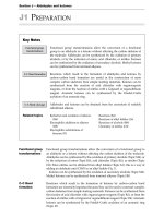
Pedigrees
Married/Partners
Female, unaffected
Divorced/Separated
Spontaneous Abortion
e
Male, unafffected
m
ok
k
• diagrams that show the pattern/distribution of phenotypes for a genetic disorder within a family, often
across multiple generations
Termination of Pregnancy
Consanguinity
Deceased
Infertility
re
Affected Individual
s
Carrier unaffected at
this time but could
manifest disease later
Adopted Sibling
Siblings (listed from
left to right (oldest to
youngest)
No Offspring
by choice
Affected Individual
≥2 conditions
P
Dizygous Twins
(fraternal)
Pregnancy
o
P
bo
Carrier not likely to
manifest disease
Ectopic Pregnancy
ECT
Gender Unknown,
unaffected
SB
Monozyous Twins
(identical)
Stillbirth (write SB
and gestational age
if known)
SB
Figure 1. Common pedigree symbols
co
Genetic Testing and Counselling
re
e.
c
om
Whole-Genome Sequencing Expands
Diagnostic Utility and Improves Clinical
Management in Paediatric Medicine
Genomic Med 2016;1:15012
While the standard of care for neurodevelopmental
and congenital malformations is chromosome
microarray analysis for copy number variations,
whole exome sequencing a lows the identification
of sequence-level mutations across all known
coding genes. Whole genome sequencing has
been previo sly associated with a diagnostic yield
of ~25% for neurological disorders or congenital
anomalies. A recent study published in Genomic
Medicine has demonstrated that whole genome
sequencing exceeds other technologies in detecting
genetic variants with a 34% diagnostic yield, a
four-fold increase in molecular diagnosis relative to
chromosome microarray analysis and a two-fold
increase relative to all genetic testing protocols.
These results suggest that whole genome
sequencing may be used as a first-tier molecular
test in individuals with development delays and
congenital abnormalities, with a higher diagnostic
yield than conventional genetic testing and
decreased time to genetic diagnosis.
m
sf
m
ee
fre
e.
c
o
o
m
e
e
o
oo
s
ks
fre
e
om
om
m
b
bo
bo
ok
ks
sf
fr
re
• microarray analysis
■■ array comparative genomic hybridization (CGH): a collection of DNA probes attached to a solid
surface to which test DNA hybridizes in order to determine copy number of DNA regions
■■ microarray analysis can identify small deletions or duplications of genetic material anywhere in the
genome
■■ commonly indicated when there is developmental delay OR two or more congenital anomalies
• FISH (fluorescence in situ hybridization): a DNA probe used to identify a gain or loss of chromosomal
material
• karyotype: microscopic analysis of chromosomes with a special stain that shows large changes in the
number or structure of chromosomes; can detect large CNVs
• Sanger sequencing: the ‘gold-standard’ method for identification of single nucletotide variants in short
DNA sequences (e.g. the exons of the gene(s) known to cause suspected syndrome)
• next-generation sequencing: high throughput method to sequence exomes or whole-genomes; useful
when genetic syndrome is suspected, but diagnosis is unclear: increasingly used for multi-gene test
panels
• prenatal screening
■■ offer optional prenatal screening before diagnostic testing
■■ first trimester screening (FTS)
◆◆ biochemistry (b-hCG, PAPP-A)
◆◆ US est mate of gestational age and measurement of nuchal translucency
◆◆ screen for trisomy 21 and 18
◆◆ done between 11 and 14 wk, sensitivity=80-85%
■■ integrated prenatal screening (IPS)
◆◆ ONTD, trisomy 21 and 18
◆◆ use results from FTS and combine with additional biomarkers completed between 15-21 weeks
(inhibin A, unconjugated estradiol, AFP, 2nd trimester b-hCG)
◆◆ improved sensitivity, reduced false positive rate compared to FTS
■■ fetal anatomy scan
◆◆ US at 18-20 wk
• newborn screening
■■ detect potentially fatal, treatable disorders before symptoms begin to allow for early therapy
■■ performed on all newborns in Canada
■■ heel puncture to collect blood
■■ screens for CF, congenital hypothyroidism, congenital adrenal hyperplasia, SCID,
hemoglobinopathies, metabolic diseases, etc.
MG4 Medical Genetics
Toronto Notes 2018
c
Dysmorphisms
fr
Dysmorphisms
o
Congenital Anomalies
m
e
e
b
Minor and Major Anomalies
minor anomaly: an unusual anatomic feature that is of no serious medical or cosmetic consequence to
the patient
• major anomaly: anomaly that creates significant medical, surgical, or cosmetic problems for the patient
fre
fre
e.
e.
m
co
m
Mechanism for Anomalies
• malformation: results from an intrinsically abnormal developmental process (e.g. polydactyly)
• disruption: results from the extrinsic breakdown of, or interference with, an originally normal
developmental process (e g. amniotic band disruption sequence)
• deformation: alteration of the final form of a structure by mechanical forces (e.g. Potter deformation
sequence)
• dysplasia: abnormal development that results in abnormal organization of cells into tissues (e.g. bone
dysplasia)
eb
m
om
m
eb
o
bo
o
Multiple Anomalies
• association: non-random occurrence of multiple independent anomalies that appear together more
than would be predicted by chance but are not known to have a single etiology (e.g. VACTERL)
• sequence: related anomalies that come from a single initial major anomaly or precipitating factor that
changes the development of other surrounding or related tissues or structures (e.g. Potter sequence or
Pierre-Robin sequence)
• syndrome: a pattern of anomalies that occur together and are known or thought to have a single cause
(e.g. Down syndrome)
o
Approach to the Dysmorphic Child
e.
e.
• congenital abnormalities are the most common cause of infant death in developed countries
o
ok
sf
r
General Approach to the Dysmorphic Child
• Are the anomalies major or minor?
• What is the mechanism underlying the anomaly?
• Do the anomalies fit as part of an association, sequence, or syndrome?
e
m
m
e
b
History
• prenatal/obstetrical history (see Obstetrics, OB4) with particular attention to potential teratogenic
exposures, developmental history (see Pediatrics, P22), and past medical history
• complete 3 generation family pedigree: health history, consanguinity, stillbirths, neonatal deaths,
specific illnesses, intellectual disability, multiple miscarriages, ethnicity
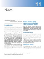
Physical Exam
re
Ears: structure, size,
placement, rotation
Skull: contour and symmetry
Hair: texture, pattern
Eyes: distance apart, brows, lashes,
folds, creases, coloboma, fundus
o
Nose: nasal bridge, nostrils
Philtrum: length, shape
Mouth: lips, palate, tongue, teeth
fre
Face: gestalt
Spine: scoliosis, kyphosis
Chin: size, position
e
Skin: hair tufts, sacral
dimples, sinus
m
Neck: webbed, redundant nuchal skin
Thorax: shape, size, nipple spacing
Limbs: proportions, amputations
Genitalia: ambiguous
ee
co
Hands and Feet: creases,
structure, nails
Growth parameters (head circumference, height, weight)
fr
Figure 2. Physical exam of the dysmorphic child
VACTERL Association
VVertebral dysgenesis
AAnal atresia (imperforate anus)
± fistula
CCardiac anomalies
T-ETracheoEsophageal fistula
± esophageal atresia
RRenal anomalies
LLimb anomalies
MG5 Medical Genetics
Toronto Notes 2018
Syndromes and Diseases
oo
ks
fr
Investigations
• screening for TORCH infections
• serial photographs if child is older
• x-rays for bony abnormalities
• cytogenetic studies
■■ karyotype if recognized aneuploidy syndrome
■■ chromosomal microarray analysis (array comparative genomic hybridization) if developmental
delay OR two or more congenital anomalies
■■ FISH if aneuploidy syndrome (e.g. trisomy 13, 18 or 21) suspected
• biochemistry: various biochemical profiles, specific enzyme assays
• single gene testing, multi-gene panel testing
m
eb
b
Check the umbilical cord for 2 arteries and 1 vein.
The presence of a single umbilical artery may be
associated with other congenital anomalies
fe
sf
re
e.
e.
c
co
Management
• prenatal counselling and assessing risk of recurrence
• referral for specialized pediatric or genetic care for symptomatic management
• specific treatments are available for certain metabolic disorders and genetic syndromes
■■ metabolic disorders: enzyme replacement therapy, substrate reduction therapy, etc. (e.g. low-protein
diet in PKU patients)
■■ genetic syndromes: e.g. mTOR inhibitors in tuberous sclerosis
oo
oo
Syndromes and Diseases
m
e
Large Genomic Changes
Table 1. Trisomy Chromosomal Syndromes
Trisomy 13
Disease
Patau syndrome
Incidence
1:600-800 births
Most common abnormality of autosomal chromosomes
Rises with advanced maternal age from 1:1,500 at age 20
to 1:20 by age 45
co
Trisomy 18
Edwards’ syndrome
1:10,000 live births
c
1:6,000 live births
F:M = 3:1
r
Microcephaly, prominent occiput
Microcephaly, sloping forehead, occipital
scalp defect, holoprosencephaly
s
Mild microcephaly, flat occiput, 3rd fontanelle,
brachycephaly
Microphthalmia, corneal abnormalities
Ears
Low-set, small, overfolded upper helix frequent AOM,
hearing loss
Low-set, malformed
Low-set, malformed
Facial Features
Protruding tongue, large cheeks, low flat nasal bridge,
small nose
Cleft lip/palate
Small mouth, micrognathia
60-80% cleft lip and palate
Skeletal/MSK
Short stature
Excess nuchal skin
Joint hyperflexibility (80%) including dysplastic hips,
vertebral anomalies, atlantoaxial instability
Short stature
Clenched fist with overlapping digits, hypoplastic
nails, clinodactyly, polydactyly
Severe growth retardation
Polydactyly, clenched hand
Cardiac Defect
50%, pa ticularly atrioventricular septal defect
60% (VSD, PDA, ASD)
80% (VSD PDA, ASD)
GI
Duodenal/esophageal/anal atresia, TEF, Hirschsprung’s
disease, chronic constipation
Hernia, TEF
GU
Cryptorchidism, rarely fertile
Polycystic kidneys, cryptorchidism
CNS
Hypotonia at birth
Low IQ, developmental delay, hearing problems
Onset of Alzheimer’s disease in 40s
Hypertonia
Hypo- or hypertonia
Seizures, deafness
Severe developmental delay
Other Features
Transverse palmar crease, clinodactyly, and absent middle
phalanx of the 5th finger
1% lifetime risk of leukemia
Polycythemia
Hypothyroidism
SGA
Rocker-bottom feet
Single umbilical artery
Midline anomalies: scalp, pituitary, palate,
heart, umbilicus, anus
Rocker-bottom feet
Prognosis/
Management
Prognosis: long term management per AAP Guidelines
(Health Supervision of Children with Down syndrome),
recommend chromosomal analysis, CBC, Echo, yearly
thyroid test, atlanto-occipital x-ray at 2 yr, sleep study,
hearing test, and ophthalmology assessment
13% 1-year survival, 10% ten-year survival
Profound intellectual disability in survivors
20% 1-year survival, 13% ten-year survival
Profound intellectual disability in survivors
e
k
Polycystic kidneys
co
co
m
b
e
eb
m
m
co
co
ee
om
Microphthalmia, hypotelorism, iris coloboma,
retinal anomalies
om
Upslanting palpebral fissures, inner epicanthal folds,
speckled iris (Brushfield spots), refractive errors (myopia),
acquired cataracts, nystagmus, strabismus
oo
Eyes
e
Cranium/Brain
re
e
co
Trisomy 21
Down syndrome
MG6 Medical Genetics
Toronto Notes 2018
c
Syndromes and Diseases
fr
e
Table 2. Common Genetic Disorders Involving the Sex Chromosomes
Klinefelter Syndrome
Turner Syndrome
45,X (most common)
f
Fragile X Syndrome
47,XXY (most common)
48,XXXY, 49,XXXXY
Incidence
1:3,600 males, 1:6,000 females
Most common heritable cause of intellectual
disability in boys
1:1,000 live male births
Increased risk with advanced maternal age
1:4,000 live female births
Risk not increased with advanced maternal age
Phenotype
Overgrowth: prominent jaw, forehead, and nasal
bridge with long and thin face, large protuberant
ears, macroorchidism, hyperextensibility, and high
arched palate
Complications: seizures, scoliosis, mitral valve
prolapse
Tall, slim, underweight
No features prepuberty
Postpuberty: male may suffer from developmental
delay, long limbs, gynecomastia, lack of facial
hair
Short stature, short webbed neck, low posterior hair
line, wide carrying angle
Broad chest, widely spaced nipples
Lymphedema of hands and/or feet, cystic hygroma in
newborn with polyhydramnios, lung hypoplasia
Coarctation of aorta, bicuspid aortic valve
Renal and cardiovascular abnormalities, increased
risk of HTN
Less severe spectrum with mosaic
IQ and Behaviour
Mild to moderate intellectual disability, 20% of
affected males have normal IQ
ADHD and/or autism
Female carriers may show intellectual impairment
Male carriers may demonstrate tremor/ataxia
syndrome in later life
Mild intellectual disability
Behavioural or psychiatric disorders – anxiety,
shyness, aggressive behaviour, antisocial acts
Mild intellectual disability to normal intelligence
Gonad and
Reproductive
Function
Premutation carrier females at risk of developing
premature ovarian failure
Infertility due to hypogonadism/hypospermia
Streak ovaries with deficient follicles, infertility,
primary amenorrhea, impaired development of
secondary sexual characteristics
Diagnosis/
Prognosis/
Management
Molecular testing of FMR1 gene: overamplification
of the trinucleotide repeat, length of segment
is proportional to severity of clinical phenotype
(genetic anticipation)
Increased risk of germ cell tumours and breast
cancer
Management: testosterone in adolescence
Normal life expectancy if no complications
Increased risk of X-linked diseases
Management: Echo, ECG to screen for cardiac
malformation
GH therapy for short stature
Estrogen replacement at time of puberty for
development of secondary sexual characteristics
m
co
m
sf
r
s
m
fre
e.
om
m
om
co
m
m
bo
ks
e
om
co
m
m
k
X-linked
Genetic anticipation
CGG trinucleotide repeat on X chromosome
measurable by molecular analysis
m
Genotype
Noonan Syndrome
CHARGE Syndrome
Lack of expression of genes
on paternal chromosome
15q11-13 due to deletion,
maternal uniparental disomy
of chromosome 15, or
imprinting defect
Lack of expression of genes
on maternal chromosome
15q11-13 due to deletion
or inactivation or paternal
uniparental disomy
Autosomal dominant with
variable expression
PTPN11 mutation most
common cause but multiple
genes known
2/3 of children with
CHARGE have been found
to have a CHD7 mutation on
chromosome 8
1 10,000
e
Incidence
1:4000; Second most
common genetic diagnosis
(next to Down syndrome)
1:15,000
1:10,000
1:2,000 male and female
live births
“CATCH 22”
Cyanotic CHD
Anomalies: craniofacial
anomalies, micrognathia
and low set ears
Thymic hypoplasia:
immunodeficiency
Cognitive impairment
Hypoparathyroidism,
hypocalcemia
22q11 microdeletions
High risk for schizophrenia
and other psych disorders
“H3O”: Hypotonia and
weakness, Hypogonadism,
obsessive Hyperphagia,
Obesity
Short stature, almond-shaped
eyes, small hands and feet
with tapering of fingers
Developmental delay
(variable)
Hypopigmentation, type 2 DM
Ataxia with severe intellectual
disability, seizures,
tremulousness, hypotonia
Midface hypoplasia, fair hair,
uncontrollable laughter
Short stature, webbed
neck, triangular facies
hypertelorism, low set ears,
epicanthal fo ds, ptosis,
pectus excavatum
Right sided CHD, pulmonary
stenosis
Increased risk of
hematological cancers,
moderate intellectual
disability, delayed puberty
oo
ks
eb
sf
oo
eb
m
co
ee
.
Clinical Features
fr
Microdeletions of
chromosome region
22q11.2
m
Genotype
m
Angelman Syndrome
o
Prader-Willi Syndrome
o
22q11.2 Deletion
Syndrome
o
ok
Table 3. Other Genetic Syndromes
“CHARGE”
CColoboma
Hcongenital Heart disease
Achoanal Atresia
Rmental Retardation
G GU anomalies
EEar anomalies
MG7 Medical Genetics
Toronto Notes 2018
Syndromes and Diseases
Gene
Associated Cancers
TP53
k
Li-Fraumeni Syndrome
ok
sf
Syndrome
re
Table 4. Familial Cancer Syndromes
Breast, osteosarcoma, leukemia, soft tissue carcinoma, and
numerous other cancers
FAP
APC
Colorectal, small intestine/stomach tumours
Hereditary Breast and Ovarian
Cancer Syndrome
BRCA1, BRCA2
Female: breast, ovarian, pancreatic
Male: prostate, breast, pancreatic
Von Hippel-Lindau Syndrome
VHL
Kidney + tumours (e.g. pheochromocytoma)
PTEN
Breast, thyroid, endometrial
m
m
Cowden Syndrome
Colorectal, endometrial, ovarian, renal, pancreatic, liver/biliary
duct, stomach, brain, breast
bo
MSH2, MLH1, MSH6,
PMS2, EPCAM
bo
Lynch Syndrome (HNPCC)
NF
Astrocytoma, optic glioma, neurofibroma, leukemia
Vestibular schwannoma, meningioma, ependymoma,
astrocytoma
e
NF1
NF2
sf
re
Type 1
Type 2
o
Single Gene Disorders
e
CYSTIC FIBROSIS
• see Respirology, R12 and Pediatrics, P82
SICKLE CELL DISEASE
• see Hematology, H20
o
DUCHENNE MUSCULAR DYSTROPHY
Epidemiology
• 1:4,000 males
oo
oo
k
ks
Etiology
• one type of muscular dystrophy characterized by progressive skeletal and cardiac muscle degeneration
• X-linked recessive: 1/3 spontaneous mutations, 2/3 inherited mutations
• missing structural protein (dystrophin) → muscle fibre fragility → fibre breakdown → necrosis and
regeneration
m
Clinical Presentation
• proximal muscle weakness by age 3, positive Gower’s sign, waddling gait, toe walking
• pseudohypertrophy of calf muscles (muscle replaced by fat) and wasting of thigh muscles
• decreased reflexes
• non-progressive delayed motor and cognitive development (dysfunctional dystrophin in brain)
•cardiomyopathy
ks
fre
e
Diagnosis
• molecular genetic studies of dystrophin gene (DMD) (first line)
• family history (pedigree analysis)
• increased CK (50-100x normal) and lactate dehydrogenase
• elevated transaminases
• muscle biopsy, EMG
m
eb
bo
Management
• supportive (e.g. physiotherapy, wheelchairs, braces , prevent obesity
• cardiac health monitoring and early intervention
• bone health monitoring and intervention (vitamin D, bisphosphonates)
• steroids (e.g. prednisone or deflazacort)
• surgical (for scoliosis)
• gene therapy trials underway
ee
.
ee
.
c
Complications
• patient usually wheelchair-bound by 12 yr of age
• early flexion contractures, scoliosis, osteopenia of immobility, increased risk of fracture
• death due to pneumonia/respiratory failure or CHF in 2nd-3rd decade
Gower’s Sign
Child uses hands to “climb up” the legs to
move from a sitting to a standing position
MG8 Medical Genetics
Toronto Notes 2018
c
Syndromes and Diseases
fre
Metabolic Diseases
b
o
k
k
• inherited disorders of metabolism; often autosomal recessive
• infants and older children may present with FTT or developmental delay
•organelle disorders can present with dysmorphism
universal newborn screening in Ontario includes metabolic disorders
Table 5. Metabolic Disorders
Carbohydrate
Disorders
Fatty Acid Disorders
Organelle Disorders
PKU
Tyrosinemia
Homocystinuria
MSUD
Alkaptonuria
Urea cycle defects
Galactosemia
GSDs: von Gierke’s,
Pompe’s, Cori’s,
Andersen, McArdle
MCAD deficiency
Carnitine deficiency
Mucopolysaccharidosis
Congenital disorders of
glycosylation
Lysosomal storage diseases:
Hurler’s, Niemann-Pick, TaySachs, Gaucher, Fabry, Krabbe
Clinical
Manifestations
Irritability, lethargy, poor
feeding
Seizures
Intellectual disability
Vomiting and acidosis
after feeding initiation
Sweet-smelling urine
(MSUD)
Vomiting and acidosis
after feeding initiation
Growth retardation, FTT
Lethargy, poor feeding
Seizures, coma
Symptoms triggered by
fasting
Liver dysfunction
Sudden infant death
Seizures/early-onset severe
epilepsy
Chronic encephalopathy
Developmental delay
Bone crises (Gaucher)
Deafness, blindness
Laboratory
Findings
Hypoglycemic
hyperammonemia,
high anion gap (organic
acidemia)
Normoglycemic
hyperammonemia,
normal an on gap (urea
cycle defects)
Hypoglycemia,
hyperlipidemia (GSD)
Hypoketotic
hypoglycemia
Elevated free fatty acids
Elevated urine oligosaccharides
(oligosaccharidoses)
and glycosaminoglycans
(mucopolysaccharidoses)
Enzyme deficieny
Infantile cataracts
(galactosemia)
Hepatomegaly
Muscle weakness/
cramping
Hepatomegaly
Hypotonia
m
ks
fr
Dysmorphic facial features
Macrocephaly (Tay-Sachs,
Hurler’s)
Hepatosplenomegaly (NiemannPick type A/B/C, not Tay-Sachs)
Cherry-red spot on macula
(Niemann-Pick type A/B, TaySachs, Gaucher’s)
Corneal clouding (Hurler’s)
Infantile cataract (Fabry)
Peripheral neuropathy (Fabry,
Krabbe)
Spasticity
oo
eb
m
Hypotonia/hypertonia
Microcephaly, musty
odour, eczema,
hypopigmentation (PKU)
Dark urine, pigmented
sclerae, arthralgias
(alkaptonuria)
Lens subluxation,
marfanoid appearance
(homocystinuria)
fre
Physical Exam
ee
.c
o
s
re
e
.c
o
Examples of
Conditions
m
Organic and Amino
Acid Disorders
b
m
eb
oo
ks
ks
fre
o
e.
co
Initial Investigations
• important to send lab studies at initial presentation in order to facilitate immediate diagnosis and
treatment
• check newborn screening results
• electrolytes, ABGs (calculate anion gap, rule out acidosis)
• CBC with differential and smear
• blood glucose (hypoglycemia seen with organic acidemia, fatty acid oxidation defects, and GSDs)
• lactate, ammonium (hyperammonemia with urea cycle defects), plasma Ca2+ and Mg2+
• routine urinalysis: ketonuria must be investigated
• carnitine levels with acylcarnitine profile
• others: urate, urine nitroprusside, plasma amino acid screen, urine organic acids, CSF glycine, free fatty
acids (3-β-hydroxybutyrate ratio >4 in fatty acid oxidation defect)
• storage diseases: urine mucopolysaccharide and oligosaccharide screen
m
m
Treatment
• varies according to inborn error of metabolism
• dietary restrictions, supplementation, enzyme replacement therapy, gene therapy, liver transplant, stem
cell transplant
MG9 Medical Genetics
Toronto Notes 2018
c
References
e
PHENYLKETONURIA
ks
Epidemiology
• 1:10,000; autosomal recessive disease
b
m
b
bo
Etiology
• deficiency of phenylalanine hydroxylase prevents conversion of phenylalanine to tyrosine leading to
build up of toxic metabolites
• mothers who have PKU may have infants with congenital abnormalities
bo
o
ok
ok
sf
re
fre
e
Management
• PKU screening at birth
• dietary restrict on of phenylalanine starting within the first 10 d of life
• duration of dietary restriction controversial – lifelong or until end of puberty; should be resumed
during pregnancy to maintain normal phenylalanine levels
• large neutral amino acid (tyrosine) replacement, BH4 enzyme treatment, phenylalanine lyase treatment
are other options
GALACTOSEMIA
Epidemiology
• 1:60,000; autosomal recessive disease
Etiology
• most commonly due to deficiency of galactose-1-phosphate uridyltransferase leading to an inability to
process lactose/galactose
e
ee
Clinical Presentation
• signs of liver and renal failure, jaundice, FTT, and cataracts with ingestion of lactose/galactose
k
ok
s
Management
• elimination of galactose from the diet (e.g. dairy, breast milk)
• most infants are fed a soy-based diet
m
b
Complications
• increased risk of sepsis, especially E. coli
• if the diagnosis is not made at birth, liver and brain damage may become irreversible
References
o
re
e
o
sf
oo
eb
eb
oo
ks
fre
e.
e.
c
o
Amato RSS. Nelson’s essentials of pediatrics, 4th ed. Philadelphia: WB Saunders, 2002. Human genetics and dysmorphology. 129-146.
Blake KD, Prasad C. CHARGE syndrome, o phanet. J Rare Diseases 2006;1.
Biggar W. Duchenne muscular dystrophy. Pediatr Rev 2006;27:83-88.
Chudley AE, Conry J, Cook JL, et al Fetal alcohol spectrum disorder: Canadian guidelines for diagnosis. CMAJ 2005;172(5 Suppl):S1-21.
Elieff, M. P., Lopez-Beltran, A., Montironi, R., & Cheng, L. (2008). Familial cancer syndromes. In Molecular genetic pathology (pp. 449-466). Humana Press.
Grati, F. R., Malvestiti, F Ferreira, J. C., Bajaj, K., Gaetani, E., Agrati, C., ... & Maggi, F. (2014). Fetoplacen al mosaicism: potential implications for false-positive and
false-negative noninvasive prenatal screening results. Genetics in Medicine, 16(8), 620-624.
Moeschler JB, Sheve l M. Committee on Genetics. Comprehensive evaluation of the child with intellectual disability or global developmental delays. Pediatrics 2014
Sep;134(3):e903 18. doi:10.1542/peds.2014-1839.
Nicholson JF Nelson’s essentials of pediatrics, 4th ed. Philadelphia: WB Saunders, 2002. Inborn errors of metabolism. 153-178.
Sobel, E , & Lange, K. (1996). Descent graphs in pedigree analysis: applications to haplotyping, location scores, and marker-sharing statistics. American journal of human
genetics, 58(6), 1323.
Therrell, B. L., & Adams, J. (2007). Newborn screening in North America. Journal of inherited metabolic disease, 30(4), 447-465.
Vissers LE, van Ravenswaaij CM, Admiraal R, et al. Mutations in a new member of the chromodomain gene family cause CHARGE syndrome. Nat Genet 2004 36:955-957.
Metabolic disease must be ruled out in any
newborn who becomes acut ly ill after a period
of normal behaviour and development or with a
Reffamily history of early infant death even if the
newborn screen is negative
co
m
o
Clinical Presentation
• baby is normal at birth, then develops a musty odour, eczema, hypertonia, tremors, and mental
retardation
• hypopigmentation due to low tyrosine (fair hair, blue eyes)
sf
Notes
Toronto Notes 2018
Syndromes and Diseases
c
MG10 Medical Genetics
________________________________ ______________________________________ ____________________________
_____________________________________________________________________________________________________________________
_____________________________________________________________________________________________________________________
_____________________________________________________________________________________________________________________
__________________________ ______________________________________ ______________________________________ ____________
_____________________________________________________________________________________________________________________
_____________________________________________________________________________________________________________________
_____________________________________________________________________________________________________________________
_____ _____________________________________ _____________________________________ ________________________________
_____________________________________________________________________________________________________________________
__________________________________ ______________________________________ ______________________________________ ____
_____________________________________________________________________________________________________________________
_____________________________________________________________________________________________________________________
_____________________________________________________________________________________________________________________
_____________________________________________________________________________________________________________________
_________ ______________________________________ ______________________________________ _____________________________
___ _ ___________________________________ _ ___________________________________ __ ________________________________
_____________________________________________________________________________________________________________________
_____________________________________________________________________________________________________________________
_____________________________________________________________________________________________________________________
_____________________________________________________________________________________________________________________
.
.
.
_____________________________________________________________________________________________________________________
_____________________________________________________________________________________________________________________
_____________________________________________________________________________________________________________________
_ __ ___________________________________ __ ___________________________________ __ __________________________________
_____________________________________________________________________________________________________________________
_____________________________________________________________________________________________________________________
_________________________ ______________________________________ ______________________________________ _____________
_____________________________________________________________________________________________________________________
_______________________________________________________________________________________________ _____________________
_____________________________________________________________________________________________________________________
_____________________________________________________________________________________________________________________
.c
.c
Medical Imaging
bo
ok
s
s
Mark Barszczyk, Tian Yang (Darren) Liu, Zafir Syed, and Jinhui Yan, chapter editors
Sheliza Halani and Taraneh Tofighi, associate editors
Arnav Agarwal and Sukhmani Sodhi, EBM editors
Dr. Nasir Jaffer and Dr. Eugene Yu, staff editors
Acronyms . . . . . . . . . . . . . . . . . . . . . . . . . . . . . . . . 2
References . . . . . . . . . . . . . . . . . . . . . . . . . . . . . 32
e
co
m
Imaging Modalities. . . . . . . . . . . . . . . . . . . . . . . . . 2
X-Ray Imaging
Ultrasound
Magnetic Resonance Imaging
Positron Emission Tomography Scans
Contrast Enhancement
Breast Imaging. . . . . . . . . . . . . . . . . . . . . . . . . . . . 30
Modalities
Breast Interventional Procedures
Breast Findings
m
e
o
Chest Imaging . . . . . . . . . . . . . . . . . . . . . . . . . . . . 4
Chest X-Ray
Computed Tomography Chest
Lung Abnormalities
Pulmonary Vascular Abnormalities
Pleural Abnormalities
Mediastinal Abnormalities
Tubes, Lines, and Catheters
f
ee
c
.c
om
Abdominal Imaging. . . . . . . . . . . . . . . . . . . . . . . . 10
Abdominal X-Ray
Approach to Abdominal X-Ray
Abdominal Computed Tomography
Approach to Abdominal Computed Tomography
Contrast Studies
Specific Visceral Organ Imaging
“itis” Imaging
Angiography of Gastrointestinal Tract
k
Genitourinary System and Adrenal. . . . . . . . . . 16
Urological Imaging
Gynecological Imaging
Adrenal Mass
m
Neuroradiology . . . . . . . . . . . . . . . . . . . . . . . . . 18
Modalities
Approach to CT Head
Selected Pathology
co
Musculoskeletal System . . . . . . . . . . . . . . . . . . . 21
Modalities
Approach to Bone X-Rays
Trauma
Arthritis
Bone Tumour
Infection
Metabolic Bone Disease
ee
.
Nuclear Medicine. . . . . . . . . . . . . . . . . . . . . . . . . . 25
Brain
Thyroid
Respiratory
Cardiac
Abdomen and Genitourinary System
Bone
Interventional Radiology. . . . . . . . . . . . . . . . . . . . 28
Vascular Procedures
Nonvascular Interventions
co
m
MI
Medical Imaging MI1
Toronto Notes 2018
MI2 Medical Imaging
Toronto Notes 2018
c
Acronyms
fr
Acronyms
POCUS point-of-care ultrasound
PTA
percutaneous transluminal angioplasty
PTC
percutaneous transhepatic cholangiography
RA
right atrium
RAIU radioactive iodine uptake
RV
right ventricle
SPECT single photon emission computed
tomography
SVC
superior vena cava
TBtuberculosis
TNKtenecteplase
tPA
tissue plasminogen activator
TRUS transrectal ultrasound
TVUS transvaginal ultrasound
U/Sultrasound
VCUG voiding cystourethrogram
V/Qventilation/perfusion
co
m
m
LA
left atrium
LV
left ventricle
MAA microaggregated albumin
MAG3mertiatide
MCA middle cerebral artery
MR
magnetic resonance
MRA magnetic resonance angiogram
MRCP magnetic resonance
cholangiopancreatography
MRI
magnetic resonance imaging
MS
multiple sclerosis
MUGA multiple gated acquisition
PAposteroanterior
PBD
percutaneous biliary drainage
PET
positron emission tomography
PFT
pulmonary function test
PICC peripherally-inserted central catheter
co
m
m
eb
oo
k
DTPA diethylene triamine pentaacetic acid
DWI diffusion-weighted image
ECD
ethyl cysteinate dimer
ERCP endoscopic retrograde cholangiopancreatography
FLAIR fluid-attenuated inversion recovery
GIgastrointestinal
GPA
granulomatosis with polyangiitis
HCC
hepatocellular carcinoma
HIDA hepatobiliary iminodiacetic acid
HMPAO hexamethylpropyleneamine oxime
HSGhysterosalpingogram
IBD
inflammatory bowel disease
ICV
ileocecal valve
IPF
interstitial pulmonary fibrosis
IVP
intravenous pyelogram
KUB
kidneys, ureters, bladder
e
ee
c
b
ok
18
FDG18-fluorodeoxyglucose
APanteroposterior
ARDS acute respiratory distress syndrome
AVarteriovenous
AXR abdominal x-ray
BOOP bronchiolitis obliterans
organizing pneumonia
CNS
central nervous system
CSF
cerebrospinal fluid
CT
computed tomography
CTA
computed tomographic angiogram
CVD
collagen vascular disease
CVP
central venous pressure
CXR
chest x-ray
DEXA dual-energy x-ray absorptiometry
DMSA dimercaptosuccinic acid
DSA
digital subtraction angiography
ok
Imaging Modalities
m
om
e
fre
12 d
3 wk
4 mo
6 mo
2d
1d
7 wk
3 mo
3 mo
10 wk
<1 d
1 yr
0.5/2
300
250
400
<1 d/4 d
2 yr
20 mo
2.7 yr
100
150
300
350
750
800
400
300
8 mo
1 yr
2 yr
2.3 yr
5 yr
5.3 yr
2.7 yr
2 yr
705
315
240
95
4.7 yr
2.1 yr
1.6 yr
8 mo
470
640
25
100
3 yr
4 yr
2 mo
8 mo
90 165
105
155
7-13 mo
8.4 yr
1 yr
Radionuclide
Brain (18FDG)
Bone (99mTc)
Thyroid (99mTc)
Thyroid (123I)
Cardiac rest-stress test
(99mTc 1-d)
(99mTc 2-d)
Lung ventilation (133Xe)
Lung perfusion
(99mTc)
Renal (99mTc)
Liver-spleen (99mTc)
B liary tract (99mTc)
m
bo
oo
eb
m
om
Head
Neck
Spine
Chest
Chest (pulmonary embolism)
Cor nary angiography
Abd men
Pelvis
e
m
co
e
s
sf
ok
5
10
50
75
1
0.5
20
35
35
30
0.25
150
CT
*Source: Radiology 2008;248:254-263
**Calculated using average natural background
exposure in Canada (Health Canada: c gc ca/hl-vs/iyh-vsv/environ/expos-eng.php)
c
oo
m
om
re
e
Computed Tomography
• x-ray beam opposite a detector moves in a continuous 360º arc as patient is advanced through the
imaging system
■■ subsequent computer assisted reconstruction of anatomical structures from the axial plane
• attenua ion is quantified in Hounsfield units:
■■ subsequent computer assisted reconstruction of anatomical structures from the axial plane
■■ adjusting the “window width” (range of Hounsfield units displayed) and “window level” (midpoint
value of the window width) can maximally visualize certain anatomical structures (e.g. CT chest can
be viewed using “lung”, “soft tissue”, and “bone” settings)
• contraindications: pregnancy (relative) contraindications to contrast agents (e.g. allergy, renal failure)
• advantages: delineates surrounding soft tissues, excellent at delineating bones and identifying lung/
liver masses, may be used to guide biopsies, spiral/helical multidetector CT has fast data acquisition and
allows 3D reconstruction, CTA is less invasive than conventional angiography
• disadvantages: high radiation exposure, soft tissue characterization is not as good in comparison
with MRI, IV contrast injection, anxiety of patient when going through scanner, higher cost, and less
available than plain film
Skull
Cervical spine
Thoracic spine
Lumbar spine
Chest (single PA film)
Shoulder
Mammography
Abdomen
Hip
Pelvis
Knee
IVU
Dual-energy x-ray
absorptiometry (without/
with CT)
Upper GI series
Small bowel series
Barium enema
fre
e
fe
re
sf
ok
bo
Fluoroscopy
• continuous x-rays used for guiding angiographic and interventional procedures, in contrast
examinations of the GI tract, and in the OR for certain surgical procedures (e.g. orthopedic, urological)
• on the fluoroscopic image, black and white are reversed so that bone and contrast agents appear dark
and radiolucent structures appear light
• advantages: allows for real-time visualization of structures
• disadvantages: increased radiation dose; however, the use of pulsed fluoroscopy has reduced
fluoroscopy time by 76% and radiation dose by 64% as compared with continuous fluoroscopy
Approximate
Equivalent
Period of
Natural
Background
Radiation**
(~3 mSv/yr)
X-Ray
ee
.
om
om
e
Plain Films
• x-rays pass through the patient and interact with a detection device to produce a 2-dimensional
projection mage
• structures closer to the film appear sharper and less magnified
• contraindications: pregnancy (relative)
• advantages: inexpensive, non-invasive, readily available, reproducible, fast
• disadvantages: radiation exposure, generally poor at distinguishing soft tissues
Equivalent
Number
of Chest
X-Rays
m
Diagnostic Procedure Type
m
• x-rays, or Röentgen rays, are a form of electromagnetic energy of short wavelength
• as x-ray photons traverse matter, they can be absorbed (a process known as “attenuation”) and/or
scattered
• the density of a structure determines its ability to attenuate or “weaken” the x-ray beam
■■ air < fat < water < bone < metal
• structures that have high at enuation (e.g. bone) appear white on the resulting images
m
bo
Typical Effective Doses from Diagnostic
Medical Exposures (in adults)*
X-Ray Imaging
MI3 Medical Imaging
Toronto Notes 2018
c
Imaging Modalities
fre
Ultrasound
e
m
Attenuation
Bone (= bright) > grey matter > white
matter (“fatty” myelin) > CSF > air (= dark)
o
eb
bo
oo
ok
sf
r
ks
fre
co
m
co
m
m
e
bo
oo
k
ok
• high-frequency sound waves are transmitted from a transducer and passed through tissues; reflections
of the sound waves are picked up by the transducer and transformed into images
• reflection (or “echo”) occurs when the sound waves pass through tissue interfaces of different acoustic
densities
• structures are described based on their echogenicity; hyperechoic structures appear bright (U/S
reflected) whereas hypoechoic structures appear dark (U/S waves not reflected back but pass through)
• higher U/S frequencies result in greater resolution but greater attenuation (i.e. deeper structures more
difficult to visualize)
• artifacts: acoustic shadowing refers to the echo-free area located behind an interface that strongly
reflects (e.g. tissue/air) or absorbs (e.g. tissue/bone) sound waves; enhancement refers to the increase
in reflection amplitude (i.e increased brightness) from objects that lie below a weakly attenuating
structure (e.g. cyst)
• Duplex scan: grey-scale image that utilizes the Doppler effect to visualize the velocity of blood flow past
the transducer
• Colour Doppler: assigns a colour based on the direction of blood flow (i.e. red = toward transducer,
blue = away)
• advantages: relatively low cost, non-invasive, no radiation, real time imaging, may be used for guided
biopsies many different imaging planes (axial, sagittal), determines cystic versus solid
• disadvantages: highly operator-dependent, air in bowel may prevent imaging of midline structures in
the abdomen, may be limited by patient habitus, poor for bone evaluation
m
Magnetic Resonance Imaging
Remember that water is “white” on T2 as
“World War II”
eb
bo
oo
oo
e.
Methods to Reduce the Risk of ContrastInduced Nephropathy
• Optimal: 0.9% NaCl at 1 ml/kg/hr for 12
hr pre-procedure and 12 hr post-contrast
administration
• For same-day procedure: 0.9% NaCl
or NaHCO3 at 3 ml/kg/hr for 1-3 hr
pre-procedure and for 6 hr post-contrast
administration
re
fre
re
e
e
co
co
m
m
• non-invasive imaging technique that does not use ionizing radiation and able to produce images in
virtually any plane
• patient is placed in a magnetic field; protons (H+) align themselves along the plane of magnetization
due to intrinsic polarity. A pulsed radiofrequency beam is subsequently turned on and deflects all the
protons off their aligned axes due to absorption of energy from the radiofrequency beam. When the
radiofrequency beam is turned off, the protons return to their pre-excitation axis, giving off the energy
they absorbed. This energy is measured with a detector and interpreted by software to generate MR
images
• the MR image reflects the signal intensity picked up by the receiver. This signal intensity is dependent
on:
1.hydrogen density: tissues with low hydrogen density (e.g. cortical bone, lung) generate little to no
MR signal compared to tissues with high hydrogen density (e.g. water)
2.magnetic relaxation times (T1 and T2): reflect quantitative alterations in MR signal strength due to
intrinsic properties of the tissue and its surrounding chemical and physical environment
m
Table 1. Differences Between Diffusion, T1- and T2-Weighted MR Imaging
Main Application
Advantages
Diffusion-Weighted
Imaging
Contrast dependent on the
molecular motion of water
Decreased diffusion is
hyper ntense (bright),
whereas increased diffusion is
hypointense (dark)
Neuroradiology
Sensitive for detection of acute ischemic stroke
and differentiating an acute stroke from other
neurologic pathologies
Acute infarction appears hyperintense
Abscess collections also show restricted
diffusion
T1-Weighted
Fluid is hypointense (dark) and
fat is hyperintense (bright)
Body soft tissues
T2-Weighted
Fluid is hyperintense (bright)
and fat is hypointense (dark)
Body soft tiss es
ee
k
k
e.
co
co
m
Contrast Enhancements
m
Imaging Techniques
Often considered an anatomic scan since they
provide a reference for functional imaging
Often considered a pathologic scan since they
will highlight edematous areas associated with
certain pathologies
ks
ok
sf
re
e
e
Contraindications to IV Contrast
MADD Failure
Multiple myeloma
Adverse reaction previously
DM
Dehydration
Failure (renal severe heart)
o
co
co
m
m
• non-invasive technique that involves exposure to ionizing radiation (~7 mSv)
• nuclear medicine imaging technique that produces images of functional processes in the body
• current generation models integrate PET and CT technologies into a single imaging device (PET-CT)
that collects both anatomic and functional information during a single acquisition
• positron-producing radioisotopes, such as 18FDG, are chemically incorporated into a metabolically
active molecule (e.g. glucose). These are then injected into the patient, travel to the target organ,
and accumulate in tissues of interest. As the radioactive substance begins to decay, gamma rays are
produced, and are then detected by the PET scanner
• contraindications: pregnancy
• advantages shows metabolism and physiology of tissues (not only anatomic); in oncology, allows for
diagnosis staging, and restaging; has predictive and prognostic value; can evaluate cardiac viability
• disadvantages: cost, ionizing radiation
e.
m
e
Positron Emission Tomography Scans
MI4 Medical Imaging
Toronto Notes 2018
c
Chest Imaging
fre
Contrast Enhancement
Disadvantages
Radio-opaque substance
that helps to delineate
intraluminal anatomy; may
demonstrate patency,
lumen integrity, or large
filling defects
U/S
Microbubbles
(IV injection)
eb
Previous adverse reaction
to contrast, renal failure,
DM, pregnancy, multiple
myeloma, severe heart
failure and dehydration
eGFR <60 may require
preventative measures
and follow-up
Shortens T1 relaxation
time, thereby increasing
signal intensity in T1weighted sequences;
gadolinium has some
effect on T2-relaxation
time; highlights highly
vascular structures (e.g.
tumours)
Risk of nephrogenic
systemic fibrosis in
patients with end-stage
renal disease
Previous adverse
reaction to contrast or if
end-stage renal disease
(relative contraindication)
m
eb
co
m
Risk of nephrogenic
systemic fibrosis in
patients with end-stage
renal disease
k
Gadolinium-Chelates
(IV injection)
Delineates intraluminal
anatomy; may
demonstrate patency,
lumen integrity, or large
filling defects; under
fluoroscopy, may also give
information on function of
an organ
o
MRI
m
m
m
2. Iodine (IV injection)
Contraindications
Previous adverse reaction
to contrast; barium
enema is contraindicated
in toxic megacolon, acute
colitis, and suspected
perforation
eb
1. Barium (oral or rectal)
o
Advantages
o
bo
Imaging Modality Types
X-Ray/CT
oo
ok
Table 2. Contrast Agents
Since gas is highly
echogenic, the
microbubbles allow for
echo-enhancement of a
tissue
Contraindicated in
individuals with rightto-left cardiac shunts
or people with known
hypersensitivity reactions
fre
Chest Imaging
ok
Chest X-Ray
m
c
m
co
m
m
eb
bo
Standard Views
• PA: anterior chest against film plate to minimize magnification of the heart size
• lateral: better visualization of retrocardiac space and thoracic spine (more sensitive at picking up pleural
effusions)
■■ helps localize lesions when combined with PA view
• AP: for bedridden patients (generally a lower quality film than PA because of enlarged cardiac
silhouette)
• lateral decubitus: to assess for pleural effusion and pneumothorax in bedridden patients; however,
POCUS can also be utilized for both of these purposes
• lordotic: angled beam allowing better visualization of apices normally obscured by the clavicles and
anterior ribs
o
©Bonnie Tang 2012
Film
Figure 1. CXR views
Anterior-posterior Position
Lateral Decubitus Position Lordotic Position
m
Lateral Position
m
Posterior-anterior Position
Approach to CXR
o
ks
oo
oo
ks
fre
e
c
Basics
• ID: patient name, MRN, sex, age
• date of exam
• markers: right and/or left
• technique: view (e.g. PA, AP, lateral), supine or erect
• indications for the study
• comparison: date of previous study for comparison (if available)
• quality of film: inspiration (6th anterior and 10th posterior ribs should be visible), penetration (thoracic
spine should be visible) and rotation (clavicles vs spinous process)
MI5 Medical Imaging
Toronto Notes 2018
c
Chest Imaging
s
Chest X-Ray Interpretation
Basics ABCDEF
AP, PA or other view
Body position/rotation
Confirm name
Date
Exposure/quality
Films for comparison
e
co
m
Analysis ABCDEF
Airways and hilar Adenopathy
Bones and Breast shadows
Cardiac silhouette and Costophrenic angle
Diaphragm and Digestive tract
Edges of pleura
Fields (lung fie ds)
sf
e
sf
re
e.
om
m
eb
oo
ok
s
ks
fr
Analysis
• tubes and lines: check position and be alert for pneumothorax or pneumomediastinum
• soft tissues: neck, axillae, pectoral muscles, breasts/nipples, chest wall
■■ nipple markers can help identify nipples (may mimic lung nodules)
■■ amount of soft tissue, presence of masses and air (subcutaneous emphysema)
• abdomen (see Abdominal Imaging, MI10)
■■ free air under the diaphragm, air-fluid levels, distention in small and large bowels
■■ herniation of abdominal contents (i.e. diaphragmatic hernia)
• bones: C-spine, thoracic spine, shoulders ribs, sternum, clavicles
■■ lytic and blastic lesions and fractures
• mediastinum: trachea, heart, great vessels
■■ cardiomegaly (cardiothoracic ratio >0.5), tracheal shift, tortuous aorta, widened mediastinum
• hila: pulmonary vessels, mainstem and segmental bronchi, lymph nodes
• lungs: lung parenchyma, pleura, diaphragm
■■ comment on abnormal lung opacity, pleural effusions or thickening
■■ right hemidiaphragm usually higher than left due to liver
■■ right vs. left hemidiaphragm can be discerned on lateral CXR due to heart resting directly on left
hemidiaphragm
• please refer to Toronto Notes website for supplementary material on how to approach a CXR
o
Anatomy
m
c
co
m
m
m
e
eb
b
Localizing Lesions for Parenchymal Lung Disease
• silhouette sign: when two objects of the same radiolucency contact each other, they become
indistinguishable on imaging and result in the loss of normal interfaces. It can be used to identify
lung pathology (consolidation, atelectasis, mass) and localize disease to specific lung segments. The
silhouette sign is not only used in the chest, but can also be an aid to interpreting imaging studies
throughout the body
• spine sign: on lateral films, vertebral bodies should appear progressively radiolucent as one moves down
the thoracic vertebral column; if they appear more radio-opaque, it is an indication of pathology (e.g.
consolidation in overlying left lower lobe)
• air bronchogram: branching pattern of air-filled bronchi on a background of fluid-filled airspaces
e
Table 3. Localization Using the Silhouette Sign
Interface Lost
Location of Lung Pathology
RUL
Right heart border
RML
Right hemidiaphragm
RLL
Aortic knob/left superior mediastinum
LUL
Left heart border
Lingula
Left hemidiaphragm
LLL
Legend
a1
anterior 1st rib
a2
anterior 2nd rib
aa
aort c arch
apw aorto-pulmonary window
as
anterior airspace
ca
carina
cl
clavicle
co
coracoid process
cpa
costophrenic angle
di
diaphragm
g
gastric bubble
ivc
inferior vena cava
la
left atrium
lbr
left mainstem bronchus
lpa
left pulmonary artery
lv
left ventricle
mf
major fissure
mi
minor fissure
p3
posterior 3rd rib
p4
posterior 4th rib
pa
main pulmonary artery
ra
right atrium
rbr
right mainstem bronchus
rpa
right pulmonary artery
rv
right ventricle
sc
scapula
sp
spinous process
st
sternum
svc
superior vena cava
tr
trachea
vb
vertebral body
.c
o
bo
ok
SVC/right superior mediastinum
a2
cl
co
st svc ca aa
apw
rbr
pa lpa
rpa
mi
st
ivc
rv
lv
rv
di
aa
lpa
rpa rbr
g
sc
mf
lbr
la
vb
lv
ivc
di
cpa
as
mi
ra
vb
tr
om
a1
p3
p4
di
cpa
e
cpa
.c
tr
sp
e
cl
co
Lateral view
oo
o
PA view
Figure 2. Location of fissures, mediastinal structures, and bony landmarks on CXR
MI6 Medical Imaging
Toronto Notes 2018
c
Chest Imaging
RUL
RML
LUL
o
RUL
LLL
LLL
RLL
Front AP
Right-Lateral
Back AP
RLL
m
eb
RML
RLL
Left-Lateral
RUL: Right Upper Lobe; RML: Right Middle Lobe; RLL: Right Lower Lobe; LUL: Left Upper Lobe; LLL: Left Lower Lobe
Soft Tissue Window
e
LUL
LLL
© Anas Nader 2009
LUL
RUL
RML
co
m
Figure 3. Location of lobes of the lung
Computed Tomography Chest
Bone Window
High
Resolution
Thinner slices provide high
definition of lung parenchyma
Low Dose
1/5th the radiation
CTA
Iodinated contrast highlights
asculature
sf
Figure 4. CT thorax windows
Indication
±
CXR abnormality
Pleural and mediastinal abnormality
Lung cancer staging
Follow-up metastases
Empyema vs. abscess
Only 5-10% lung is sampled
No
Hemoptysis
Diffuse lung disease (e g. sarcoidosis,
hypersensitivity pneumonitis,
pneumoconiosis)
Pulmonary fibrosis
Normal CXR but abnormal PFTs
Characterize solitary pulmonary nodule
Decreased detail
No
Contrast can cause severe
allergic reaction and is
nephrotoxic
Yes
om
PE
Aortic aneurysms
Aortic dissection
Lung Abnormalities
e
e
b
Atelectasis
• pathogenesis: collapse of alveoli due to restricted breathing, blockage of bronchi, external compression,
or poor surfactant
•findings
■■ increased opacity of involved segment/lobe, vascular crowding, silhouette sign, air bronchograms
■■ volume loss: fissure deviation, hilar/mediastinal displacement, diaphragm elevation
■■ compensatory hyperinflation of remaining normal lung
• differential diagnosis
■■ obstructive (most common): air distal to obstruction is reabsorbed causing alveolar collapse
◆◆ post-surgical, endobronchial lesion, foreign body, inflammation (granulomatous infections,
pneumoconiosis, sarcoidosis, radiation injury), or mucous plug (cystic fibrosis)
■■ compressive
■■ tumour bulla, effusion, enlarged heart, lymphadenopathy
■■ traction (cicatrization): due to scarring, which distorts alveoli and contracts the lung
■■ adhesive: due to lack of surfactant
◆◆ hyaline membrane disease, prematurity
DDx of Airspace Disease
• Pus (e.g. infections such as pneumonia,
non-infectious inflammatory process)
• Fluid (e.g. pulmonary edema)
• Blood (e.g. pulmonary hemorrhage)
• Cells (e.g. bronchioalveolar carcinoma,
lymphoma)
•Protein (e.g. alveolar proteinosis)
ee
ks
ok
sf
re
e.
c
.c
m
m
Figure 5 Atelectasis: RML collapse
e
fre
Screening
Follow-up infections, lung transplant,
metastases
o
e
m
e
Poor at evaluating diffuse
disease
re
Disadvantage
Scans full lung very quickly
(<1 min)
re
Advantage
b
Contrast
eb
Table 4. Types of CT Chest
Standard
Lung Window
o
o
e
oo
ok
ks
fre
Approach to CT Chest
• soft tissue window
■■ thyroid, chest wall, pleura
■■ hea t: chambers, coronary artery calcifications, pericardium
■■ vessels: aorta, pulmonary artery, smaller vasculature
■■ lymph nodes: mediastinal, axillary
bone window
■■ vertebrae, sternum, manubrium, ribs: fractures, lytic lesions, sclerosis
• lung window
■■ trachea: patency, secretions
■■ bronchial trees: anatomic variants, mucus plugs, airway collapse
■■ lung parenchyma: fissures, nodules, fibrosis/interstitial changes
■■ pleural space: effusions
• please refer to Toronto Notes website for supplementary material on how to approach a CT chest
Figure 6. Air bronchograms in right lung
MI7 Medical Imaging
Toronto Notes 2018
c
Chest Imaging
sf
ks
fre
■■ passive (relaxation): a result of air or fluid in the pleural space
◆◆ pleural effusion, pneumothorax
• management: in the absence of a known etiology, persisting atelectasis must be investigated (i.e. CT
thorax) to rule out a bronchogenic carcinoma
m
Figure 7. Consolidation bacterial
pneumonia
f
ok
sf
re
e.
co
co
m
b
bo
Consolidation
• pathogenesis: fluid (water, blood), inflammatory exudates, protein, or tumour in alveoli
•findings
■■ air bronchograms: lucent branching bronchi visible through opacification
■■ airspace nodules: fluffy, patchy, poorly defined margins with later tendency to coalesce, may take on
lobar or segmental distribution
■■ silhouette sign
• differential diagnosis
■■ fluid: pulmonary edema, blood (trauma, vasculitis, bleeding disorder, pulmonary infarct)
■■ inflammatory exudates: bacterial infections, TB, allergic hypersensitivity alveolitis, BOOP, allergic
bronchopulmonary aspergillosis, aspiration, sarcoidosis
■■ protein: pulmonary alveolar proteinosis
■■ tumour: bronchoalveolar carcinoma, lymphoma
• management: varies depending on the pattern of consolidation, which can suggest different etiologies;
should also be done in the context of clinical picture
e
bo
Figure 8. Interstitial disease: fine reticular
pattern
s
e
co
FASSTEN (upper lung disease)
Farmer’s lung (hypersensitivity pneumonitis)
Ankylosing spondylitis
Sarcoidosis
Silicosis
TB
Eosinophilic granuloma (Langerhans cell
histiocytosis)
Neurofibromatosis
BAD RASH (lower lung disease)
BOOP
Asbestos
Drugs (nitrofurantoin, hydralazine, isoniazid,
amiodarone, many chemotherapy drugs)
Rheumatological disease
Aspiration
Scleroderma
Hamman Rich (IPF) and idiopathic pulmonary
fibrosis
DDx for Cavitating Lung Nodule
c
WEIRD HOLES
GPA (Wegener’s)
Embolic (pulmonary septic)
Infection (anaerobes, pneumocystis, TB)
Rheumatoid (necrobiotic nodules)
Developmental cysts (sequestration)
Histiocytosis
Oncological
Lymphangioleiomyomatosis
Environmental, occupational
Sarcoidosis
sf
re
e.
m
co
ee
fre
e.
c
om
m
eb
DDx of Interstitial Lung Disease
re
e
m
oo
ok
s
Pulmonary Nodule
• findings: round opacity ± silhouette sign
■■ note: do not mistake nipple shadows for nodules; if in doubt, repeat CXR with nipple markers
• differential diagnosis
■■ extrapulmonary density: nipple, skin lesion, electrode, pleural mass, bony lesion
■■ solitary nodule
◆◆ tumour: carcinoma, hamartoma, metastasis, bronchial adenoma
◆◆ inflammation: histoplasmoma, tuberculoma, coccidioidomycosis
◆◆ vascular: AV fistula, pulmonary varix (dilated pulmonary vein), infarct, embolism
■■ multiple nodules: metastases, abscess, granulomatous lung disease (TB, fungal, sarcoid, rheumatoid
nodules, silicosis, GPA)
• management: clinical information and CT appearance determine level of suspicion of malignancy
■■ if high probability of malignancy, invasive testing (fine needle aspiration, transbronchial/
transthoracic biopsy) is indicated
■■ if low probability of malignancy, repeat CXR or CT in 1-3 mo and then every 6 mo for 2 yr; if no
change, then >99% chance benign
bo
Figure 9. Interstitial disease: medium
reticular pattern
sf
re
e
e.
co
om
m
m
e
oo
ok
ks
fre
e.
e.
co
co
m
m
m
bo
bo
Interstitial Disease
• pathogenesis: pathological process involving the interlobular connective tissue (i.e. “scaffolding of the
lung”)
•findings
■■ linear: fine lines caused by thickened connective tissue septae
◆◆ Kerley A: long thin lines in upper lobes
◆◆ Kerley B: short horizontal lines extending from lateral lung margin
◆◆ Kerley C: diffuse linear pattern throughout lung
◆◆ seen in pulmonary edema, lymphangitic carcinomatosis, and atypical interstitial pneumonias
■■ nodular: 1-5 mm well-defined nodules distributed evenly throughout lung
◆◆ seen in malignancy, pneumoconiosis, and granulomatous disease (e.g. sarcoidosis, miliary TB)
■■ reticular (honeycomb): parenchyma replaced by thin-walled cysts suggesting extensive destruction
of pulmonary tissue and fibrosis
◆◆ seen in IPF, asbestosis, and CVD
◆◆ watch for pneumothorax as a complication
■■ reticulonodular: combination of reticular and nodular patterns
■■ may also see signs of airspace disease (atelectasis, consolidation)
• differential diagnosis
■■ occupational/environmental exposure
◆◆ inorganic: asbestosis, coal miner’s pneumoconiosis, silicosis, berylliosis, talc pneumoconiosis
◆◆ organic: hypersensitivity pneumonitis, bird fancier’s lung, farmer’s lung (mouldy hay), and other
organic dust
■■ autoimmune: CVD (e.g. rheumatoid arthritis, scleroderma, SLE, polymyositis, mixed connective
tissue disease), IBD, celiac disease, vasculitis
■■ drug-related: antibiotics (cephalosporins, nitrofurantoin), NSAIDs, phenytoin, carbamazepine,
fluoxetine, amiodarone, chemotherapy (e.g. methotrexate), heroin, cocaine, methadone
■■ infections: non-tuberculous mycobacteria, certain fungal infections
■■ idiopathic: hypersensitivity pneumonitis, IPF, BOOP
■■ for Causes of Interstitial Lung Disease Classified by Distribution see Respirology, R13
■■ management: high-resolution CT thorax and biopsy
MI8 Medical Imaging
Toronto Notes 2018
c
Chest Imaging
re
Table 5. Characteristics of Benign and Malignant Pulmonary Nodules
Margin
Ill-defined/spiculated (“corona radiata”)
Well-defined
Contour
Lobulated
Smooth
Calcification
Eccentric or stippled
Diffuse, central, popcorn, concentric
Doubling Time
20-460 d
<20 d or >460 d
Other Features
Cavitation, collapse, adenopathy, pleural effusion, lytic bone
lesions, smoking history
Size
>3 cm
<3 cm
Cavitation
Yes, especially with wall thickness >15 mm, eccentric cavity, and
shaggy internal margins
No
Satellite Lesions
No
Yes
om
Figure 10. Pulmonary nodule:
bronchogenic carcinoma
.
co
b
Benign
bo
Malignant
fr
fre
Pulmonary Vascular Abnormalities
Figu e 11. Peribronchial cuffing
e
r
fre
e.
.c
co
m
m
eb
b
oo
oo
k
Pulmonary Edema
• pathogenesis: fluid accumulation in the airspaces of the lungs
•findings
■■ vascular redistribution/enlargement, cephalization, pleural effusion, cardiomegaly (may be present
in cardiogenic edema and fluid overloaded states)
■■ fluid initially collects in interstitium
◆◆ loss of definition of pulmonary vasculature
◆◆ peribronchial cuffing
◆◆ Kerley B lines
◆◆ reticulonodular pattern
◆◆ thickening of interlobar fissures
■■ as pulmonary edema progresses, fluid begins to collect in alveoli causing diffuse airspace disease
often in a “bat wing” or “butterfly” pattern in perihilar regions with tendency to spare the outermost
lung fields
• differential diagnosis: cardiogenic (e.g. CHF), renal failure, volume overload, non-cardiogenic (e.g.
ARDS)
bo
bo
co
om
m
bo
o
Pulmonary Embolism
• pathogenesis: arterial blockage in the lungs due to emboli from pelvic or leg veins, rarely from PICC
lines, ports, air, fat, or amniotic fluid (difficult to diagnose on imaging except by combination of clinical
history and CXR and CT findings of ARDS)
•findings
■■ CXR: Westermark sign (localized pulmonary oligemia), Hampton’s hump (triangular peripheral
infarct), enlarged right ventricle and right atrium, atelectasis, pleural effusion, and rarely pulmonary
edema
■■ definitive imaging study: CT pulmonary angiography to look for filling defect in contrast-filled
pulmonary arteries (emboli can be seen up to 4th order arterial branching)
■■ V/Q scan: not a diagnostic study
ee
Pleural Abnormalities
s
Pleural Effusion
25 mL: most sensitive
Upright lateral
50 mL: meniscus seen in the posterior costophrenic sulcus
PA
200 mL
Supine
Diffuse haziness
m
Minimum Volume to Visualize
Lateral decubitus
bo
X Ray Projection
o
e.
fre
k
ks
fre
e.
o
• a horizontal fluid level is seen only in a hydropneumothorax (i.e. both fluid and air within pleural
cavity)
• effusion may exert mass effect, shift trachea and mediastinum to opposite side, or cause atelectasis of
adjacent lung
• U/S is superior to plain film for detection of small effusions and may also aid in thoracentesis, and
POCUS is now standard of care in acute situations
• fluid level >1 cm on lateral decubitus film is indication to perform thoracentesis
Figure 12. Pleural effusion in lateral view
s
o
Table 6. Sensitivity of Plain Film Views for Pleural Effusion
MI9 Medical Imaging
Toronto Notes 2018
c
Chest Imaging
m
co
m
co
m
m
b
bo
o
oo
fr
fr
Pneumothorax
• pathogenesis: gas/air accumulation within the pleural space resulting in separation of the lung from the
chest wall
•findings
■■ upright chest film allows visualization of visceral pleura as curvilinear line paralleling chest wall
separating partially collapsed lung from pleural air
■■ more obvious on expiratory (increased contrast between lung and air) or lateral decubitus films (air
collects superiorly)
■■ more difficult to detect on supine film; look for the “deep (costophrenic) sulcus” sign, double
diaphragm” sign (dome and anterior portions of diaphragm outlined by lung and pleural air,
respectively), hyperlucent hemithorax, sharpening of adjacent mediastinal structures
■■ mediastinal shift may occur if tension pneumothorax
• differential diagnosis: spontaneous (tall and thin males, smokers), iatrogenic (lung biopsy, ventilation,
CVP line insertion), trauma (associated with rib fractures), emphysema, malignancy, honeycomb lung
• management: needle decompression or chest tube insertion, repeat CXR to ensure resolution
e
m
b
bo
bo
o
ok
sf
fre
ee
c
.c
m
om
m
e
eb
Mediastinal Mass
the mediastinum is divided into four compartments; this provides an approach to the differential
diagnosis of a mediastinal mass
• anterior border formed by the sternum and posterior border by the heart and great vessels
■■ 4 Ts: see sidebar
■■ cardiophrenic angle mass differential: thymic cyst, epicardial fat pad, foramen of Morgagni hernia
• middle border (extending behind anterior mediastinum to a line 1 cm posterior to the anterior border
of the thoracic vertebral bodies)
■■ esophageal carcinoma, esophageal duplication cyst, metastatic disease, lymphadenopathy (all
causes), hiatus hernia, bronchogenic cyst
• posterior border (posterior to the middle line described above)
■■ neurogenic tumour (e.g. neurofibroma, schwannoma), multiple myeloma, pheochromocytoma,
neurenteric cyst, thoracic duct cyst, lateral meningocele, Bochdalek hernia, extramedullary
hematopoiesis
• superior boundaries (superiorly by thoracic inlet, inferiorly by plane of the sternal angle, anteriorly by
manubrium, posteriorly by T1-T4, laterally by pleura)
• in addition, any compartment may give rise to lymphoma, lung cancer, aortic aneurysm or other
vascular abnormalities, abscess, or hematoma
Depressed Hemidiaphragm Suggests
TALC
Tumour
Asthma
Large pleu al effusion
COPD
DDx Anterior Mediastinal Mass
4 Ts
Thyroid
Thymic neoplasm
Teratoma
Terrible lymphoma
o
e.
c
e.
c
Figure 14. Lateral CXR showing
four mediastinal compartments
m
om
m
eb
b
ok
sf
re
re
co
e.
co
m
Enlarged Cardiac Silhouette
• heart borders
■■ on PA view, right heart border is formed by right atrium; left heart border is formed by left atrium
and left ventricle
■■ on lateral view, anterior heart border is formed by right ventricle; posterior border is formed by left
atrium (superior to left ventricle) and left ventricle
• cardiothoracic ratio = greatest transverse dimension of the central shadow relative to the greatest
transverse dimension of the thoracic cavity
■■ using a good quality erect PA chest film in adults, cardiothoracic ratio of >0.5 is abnormal
■■ differential of ratio >0.5
◆◆ cardiomegaly (myocardial dilatation or hypertrophy)
◆◆ pericardial effusion
◆◆ poor inspiratory effort/low lung volumes
◆◆ pectus excavatum
• ratio <0.5 does not exclude enlargement (e.g. cardiomegaly + concomitant hyperinflation)
• pericardial effusion: globular heart with loss of indentations on left mediastinal border
• RA enlargement: increase in curvature of right heart border and enlargement of SVC
• LA enlargement: straightening of left heart border; increased opacity of lower right side of
cardiovascular shadow (double heart border); elevation of left main bronchus (specifically, the upper
lobe bronchus on the lateral film), distance between left main bronchus and “double” heart border >7
cm, splayed carina (late sign)
• RV enlargement: elevation of cardiac apex from diaphragm; anterior enlargement leading to loss of
retrosternal air space on lateral; increased contact of right ventricle against sternum
• LV enlargement: rounding of the cardiac apex; displacement of left cardiac boarder leftward, inferiorly,
and posteriorly
Elevated Hemidiaphragm Suggests
PAL DIP
Pregnancy
Atelectasis
Lung resection
Diaphragmatic paralysis
Intra-abdominal process
Pneumonectomy
Pleural effusion also may result in apparent
elevation
o
o
o
Mediastinal Abnormalities
Figure 13. Pneumothorax
fre
fr
sf
re
Asbestos
• asbestos exposure may cause various pleural abnormalities including benign plaques (most common;
these may calcify), diffuse pleural fibrosis, effusion, and malignant mesothelioma
MI10 Medical Imaging
Toronto Notes 2018
c
Abdominal Imaging
f
fre
Tubes, Lines, and Catheters
ok
ok
• ensure appropriate placement and assess potential complications of lines and tubes
• avo d mistaking a line/tube for pathology (e.g. oxygen rebreather mask for pneumothoraces)
e
m
co
m
co
m
m
e
b
Central Venous Catheter
• used for fluid and medication administration, vascular access for hemodialysis, and CVP monitoring
• tip must be located proximal to right atrium to prevent inducing arrhythmias or perforating wall of
atrium
■■ if monitoring CVP, catheter tip must be proximal to venous valves
• tip of well-positioned central venous catheter projects over silhouette of SVC in a zone demarcated
superiorly by the anterior first rib end and clavicle, and inferiorly by top of RA
• course should parallel course of SVC; if appears to bend as it approaches wall of SVC or appears
perpendicular, catheter may damage and ultimately perforate wall of SVC
• complications: pneumothorax, bleeding (mediastinal, pleural), air embolism
oo
eb
Figure 15. CXR showing well-positioned
central venous catheter
m
m
eb
bo
oo
k
fr
ok
sf
re
Endotracheal Tube
• frontal chest film: tube projects over trachea and shallow oblique or lateral chest radiograph will help
determine position in 3 dimensions
• progressive gaseous distention of stomach on repeat imaging is concerning for esophageal intubation
• tip should be located 4 cm above tracheal carina (avoids bronchus intubation and vocal cord irritation)
• maximum inflation diameter <3 cm to avoid necrosis of tracheal mucosa and rupture; ensure diameter
of balloon is less than tracheal diameter above and below balloon
• complications: aspiration (parenchymal opacities), pharyngeal perforation (subcutaneous emphysema,
pneumomediastinum, mediastinitis)
re
c
c
m
m
Nasogastric Tube
• tip and sideport should be positioned distal to esophagogastric junction and proximal to gastric pylorus
• radiographic confirmation of tube is mandatory because clinical techniques for assessing tip position
may be unreliable
• complications: aspiration (parenchymal opacities), intracranial perforation (trauma patients),
pneumothorax
m
eb
bo
o
ok
s
sf
Swan-Ganz Catheter
• to monitor pulmonary capillary wedge pressure and to measure cardiac output for suspected LV
dysfunction
• tip should be positioned within right or left main pulmonary arteries or in one of their large, lobar
branches
if tip is located more distally, increased risk of prolonged pulmonary artery occlusion resulting in
pulmonary infarction or, rarely, pulmonary artery rupture
• complications: pneumothorax, bleeding (mediastinal, pleural), air embolism
co
m
Chest Tube
• in dorsal and caudal portion of pleural space to evacuate fluid
• in ventral and cephalad portions of pleural space to evacuate pneumothoraces
• tube may lie in fissure as long as functioning
• complications: lung perforation (mediastinal opacities)
e
Abdominal Imaging
ks
Abdominal X-Ray
b
m
m
b
o
Indications
■■ acute abdomen: bowel perforation, toxic megacolon, bowel ischemia, small bowel obstruction, large
bowel obstruction
■■ chronic symptoms: constipation, calcifications (gallstones, renal stones, urinary bladder stones, etc.)
■■ not useful in: GI bleeds, chronic anemia, vague GI symptoms
ee
.
co
co
m
Anatomy
• abdomen divided into 2 cavities
■■ peritoneal cavity: lined by peritoneum that wraps around most of the bowel, the spleen, and most of
the liver; forms a recess lateral to both the ascending and descending colon (paracolic gutters)
■■ retroperitoneal cavity: contains several organs situated posterior to the peritoneal cavity; the contour
of these can often be seen on radiographs
3 Views of AXR
•Erect/Upright
•Supine
• Left lateral decubitus
MI11 Medical Imaging
Toronto Notes 2018
c
Abdominal maging
Small Bowel
Large Bowel
Mucosal Folds
Uninterrupted valvulae conniventes (or plicae
circularis)
Interrupted haustra extend only partway across
lumen
k
k
Property
o
e
Table 7. Differentiating Small and Large Bowel
Central
Peripheral (picture frame)
Maximum Diameter
3 cm
6 cm (9 cm at cecum)
Maximum Fold Thickness
3 mm
5 mm
Other
Rarely contains solid fecal material
Commonly contains solid fecal material
eb
Location
m
Approach to Abdominal X-Ray
s
m
fr
k
oo
oo
ks
fre
e.
co
m
m
e
eb
bo
oo
k
ok
sf
fre
re
e
o
om
m
e
e
o
k
fr
ks
fr
e
• mnemonic: “Free ABDO”
• “Free”: free air and fluid
■■ free fluid
◆◆ small amounts of fluid: increased distance between lateral fat stripes and adjacent colon may
indicate free peritoneal fluid in the paracolic gutters
◆◆ large amounts of fluid: diffuse increased opacification on supine film; bowel floats to centre of
anterior abdominal wall
◆◆ ascites and blood (hemoperitoneum) are the same density on the radiograph, and therefore,
cannot be differentiated
◆◆ free intraperitoneal air suggests rupture of a hollow viscus (anterior duodenum, transverse
colon), penetrating trauma, or recent (<7 d) surgery
• “A”: air in the bowel (can be normal, ileus, or obstruction)
■■ volvulus – twisting of the bowel upon itself; from most to least common:
◆◆ sigmoid: “coffee bean” sign (massively dilated sigmoid projects to right or mid-upper abdomen)
with proximal dilation
◆◆ cecal: massively dilated bowel loop projecting to left or mid-upper abdomen with small bowel
dilation
◆◆ gastric: rare
◆◆ transverse colon: rare (usually young individuals)
◆◆ small bowel: “corkscrew sign” (rarely diagnosed on plain films, seen best on CT)
■■ toxic megacolon
◆◆ manifestation of fulminant colitis
◆◆ extreme dilatation of colon (>6.5 cm) with mucosal changes (e.g. foci of edema, ulceration,
pseudopolyps), loss of normal haustral pattern
• “B”: bowel wall thickening
■■ increased soft tissue density in bowel wall, thumb-like indentations in bowel wall (“thumbprinting”), or a picket-fence appearance of the valvulae conniventes (“stacked coin” appearance)
■■ may be seen in IBD, infection, ischemia, hypoproteinemic states, and submucosal hemorrhage
• “D”: densities
■■ bones: look for gross abnormalities of lower ribs, vertebral column, and bony pelvis
■■ abnormal calcifications: approach by location
◆◆ RUQ: renal stone, adrenal calcification, gallstone, porcelain gallbladder
◆◆ RLQ: ureteral stone appendicolith, gallstone ileus
◆◆ LUQ: renal stone adrenal calcification, tail of pancreas
◆◆ LLQ: ureteral stone
◆◆ central: aorta/aortic aneurysm, pancreas, lymph nodes
◆◆ pelvis: phleboliths (i.e. calcified veins), uterine fibroids, bladder stones
• “O”: organs
■■ kidney, liver, gallbladder, spleen, pancreas, urinary bladder, psoas shadow
■■ outlines can occasionally be identified because they are surrounded by more lucent fat, but all are
best visualized with other imaging modalities (CT, MRI)
k
ok
Figure 16. Normal AXRs: (left) supine anteroposterior AXR, (m ddle) upright anteroposterior AXR, and (right)
left lateral decubitus AXR
3-6-9 Rule of Dilation
Small bowel (>3 cm)
Large bowel (>6 cm)
Cecum (>9 cm)
MI12 Medical Imaging
Toronto Notes 2018
c
Abdominal Imaging
e
Table 8. Abnormal Air on Abdominal X-Ray
Common Etiologies
Upright film: air under diaphragm
Left lateral decubitus film: air between liver and
abdominal wall
Supine film: gas outlines of structures not normally
seen:
Inner and outer bowel wall (Rigler’s sign)
Falciform ligament
Peritoneal cavity (“football” sign)
Perforated viscus
Post-operative (up to 10 d to be resorbed)
Retroperitoneal
Gas outlining retroperitoneal structures allowing
increased visualization:
Psoas shadows
Renal shadows
Perforation of retroperitoneal segments of bowel:
duodenal ulcer, post-colonoscopy
Intramural
(pneumatosis
intestinalis)
Lucent air streaks in bowel wall, 2 types:
1 Linear
2. Rounded (cystoides type)
1. Linear: ischemia, necrotizing enterocolitis
2. Rounded/cystoides (generally benign):
prima y (idiopathic), secondary to COPD
e
e
Dilated loops of bowel, air-fluid levels
Intraluminal
Adynamic (paralytic) ileus, mechanical bowel
obstruction
Abscess (evaluate with CT)
Bil ary
Air centrally over liver
Sphincterotomy, gallstone ileus, erosive peptic
ulcer, cholangitis, emphysematous cholecystitis
Portal Venous
Air peripherally over liver in branching pattern
Bowel ischemia/infarction
e
m
Table 9. Adynamic Ileus vs. Mechanical Obstruction
bo
Mottled, localized in abnormal position without normal
bowel features
ks
Loculated
o
Biliary vs. Portal Venous Air
“Go with the flow”: air follows the flow of bile
or portal venous blood
Biliary air is most prominent centrally over
the liver
Portal venous air is most prominent
peripherally
c
co
m
m
eb
oo
oo
Appearance
Extraluminal
Intraperitoneal
(pneumoperitoneum)
k
Air
Adynamic Ileus
Mechanical Obstruction
Calibre of Bowel Loops
Normal or dilated
Usually dilated
Same level in the same single loop
Multiple air fluid levels giving “step ladder”
appearance, dynamic (indicating peristalsis
present), “string of pearls” (row of small
gas accumulations in the dilated valvulae
conniventes)
e
Air-Fluid Levels
(erect and left lateral
decubitus films only)
co
om
Feature
Air throughout GI tract is generalized or
localized
In a localized ileus (e.g. pancreatitis,
appendicitis), dilated “sentinel loop remains
in the same location on serial films, usually
adjacent to the area of inflammation
Dilated bowel up to the point of obstruction (i.e.
transition point)
No air distal to obstructed segment
“Hairpin” (180°) turns in bowel
bo
o
fr
Distribution of Bowel Gas
m
Abdominal Computed Tomography
ks
fre
e
m
r
om
m
co
co
m
m
e
• look through all images in gestalt fashion to identify any obvious abnormalities
• look at each organ/structure individually from top to bottom, evaluating size and shape of each area of
increased or decreased density
• evaluate the following:
■■ soft tissue window
◆◆ liver, gallbladder, spleen and pancreas
◆◆ adrenals, kidneys, ureters, and bladder
◆◆ stomach, duodenum, small bowel mesentery, and colon/appendix
◆◆ retroperitoneum: aorta, vena cava, and mesenteric vessels; look for adenopathy in vicinity of
vessels
◆◆ peritoneal cavity for fluid or masses
◆◆ abdominal wall and adjacent soft tissue
■■ lung window
◆◆ visible lung (bases)
■■ bone window
◆◆ vertebrae, spinal cord, and bony pelvis
e.
c
oo
oo
oo
Approach to Abdominal Computed Tomography
Colorectal Cancer: CT Colonography and
Colonoscopy for Detection-Systematic Review
and Meta-Analysis
Radiology 2011;259:393-405
Purpose: To assess the sensitivity of computed
tomography (CT) colonography and optical
colonoscopy (OC) for colorectal cancer (CRC)
detection.
Methods: Systematic review and meta-analysis
of diagnostic studies evaluating CT colonography
detection of CRC based on a priori eligibility criteria,
in particular requiring both OC and histological
confirmation of disease. Studies that also assessed
true-positive and false-negative diagnoses with OC
were used to calculate OC sensitivity. Sensitivity of
CTC and OC for CRC was the main outcome.
Results: 49 studies on 11,151 patients undergoing
diagnostic study for detection of CRC were
included. CTC has a sensitivity of 96.1% (95% CI
93.8%, 97.7%) and OC has a sensitivity of 94.7%
(95% CI 90.4%, 97.2%) for the detection of CRC.
Conclusion: CTC is highly sensitive for the
detection of CRC and may be a better modality for
the initial investigation of suspected CRC, assuming
reasonable specificity.
re
s
sf
e
re
e
co
co
m
m
• indications for plain CT: renal colic, hemorrhage
• indications for CT with contrast
■■ IV contrast given immediately before or during CT to allow identification of arteries and veins
◆◆ portal venous phase: indicated for majority of cases
◆◆ biphasic (arterial and portal venous phases): liver, pancreas, bile duct tumours
◆◆ caution: contrast allergy (may premedicate with steroids and antihistamine)
◆◆ contraindication: impaired renal function (based on eGFR)
■■ oral contrast: barium or water-soluble (water soluble if suspected perforation) given in most cases to
demarcate GI tract
■■ rectal contrast: given for investigation of colonic lesions
MI13 Medical Imaging
Toronto Notes 2018
c
Abdominal maging
Small bowel
ks
f
Gallbladder
Spleen
Head of he pancreas
Left kidney
Liver
c
Right kidney
b
d
a e
Blood vessels
a. Aorta
Right crus of diaphragm
b. Inferior vena cava
Vertebra
c. Superior mesenteric vein
Psoas muscles
d. Superior mesenteric artery
Erector spinae muscles
e. Left renal artery
fr
Figure 17. Axial abdominal computed tomography
o
eb
eb
bo
oo
ok
CT and Bowel Obstruction
• cause of bowel obstruction rarely found on plain films; CT is best choice for imaging
• the “3 6,9” rule is a very useful guide to determining when the bowel is dilated; the maximum diameter
of he bowel is 3 cm for small bowel, 6 cm for large bowel, and 9 cm for cecum; this can also be seful
to distinguish small and large bowel, and to assess for ‘impending’ cecal perforation (e.g. post-untreated
Ogilvie’s syndrome)
• closed-loop obstruction: an obstruction in two locations (usually small bowel) creating a loop of bowel
segment obstructed both proximally and distally; complications (e.g. ischemia, perforation, necrosis)
may occur quickly
s
sf
re
e
co
co
CT Colonography (virtual colonoscopy)
• emerging imaging technique for evaluation of intraluminal colonic masses (i e. polyps, tumours)
• two CT scans of the abdomen (prone and supine) after the instillation of carbon dioxide into a prepped
colon
• computer reconstruction of 2D CT images into a 3D intraluminal view of the colon
• lesions seen on 3D images correlated with 2D axial images
• indications: surveillance in low-risk patients, incomplete colonoscopy, staging of obstructing colonic
lesions
bo
Contrast Studies
Table 10. Types of Contrast Studies
Procedure Description
Assessment
Findings
Cervical esophagus
Contrast agent swallowed
Recorded for later playback
and analysis
Dysphagia, swallowing
incoordination,
recurrent aspiration,
post-operative cleft
palate repair
Aspiration, webs
(partial occlusion),
Zenker’s diverticulum,
cricopharyngeal bar,
laryngeal tumour
Barium Swallow
Thoracic esophagus
Contrast agent swallowed
under fluoroscopy, selective
images captured
Dysphagia, rule out
GERD, post-esophageal
surgery
Achalasia, hiatus
hernia, esophagitis,
cancer, esophageal tear
Upper GI Series
Thoracic esophagus,
stomach, and
duodenum
Double contrast study:
1. Barium to coat mucosa,
then
2. Gas pills for distention
Patient NPO after midnight
Dyspepsia, investigate
possible upper GI bleed,
weight loss/anemia,
post-gastric surgery
Ulcers, neoplasms,
filling defects
Enterography and
Enteroclysis (MRI
or CT)
Entire small bowel
Enterography: patient drinks
1-2 L of sorbitol, psyllium,
or barium solution to
distend small bowel
Enteroclysis: NJ tube used
to pump barium, psyllium,
or sorbitol contrast media
directly into small bowel
IBD, malabsorption,
weight loss/anemia,
Meckel’s diverticulum
Neoplasms, IBD,
malabsorption, infection
m
e
e
re
k
oo
eb
m
e
Organ
Cine
Esophagogram
co
Study
MI14 Medical Imaging
Toronto Notes 2018
c
Abdominal Imaging
fre
fre
Specific Visceral Organ Imaging
e
Ill-defined, irregular margin, hypoechoic contents
Low attenuation lesion with an irregular enhancing
wall
Hydatid Cyst
Simple/multiloculated cyst
Low attenuation simple or multiloculated cyst;
calcification
HCC
Single/multiple masses, or diffuse infiltration
Hypervascular; enhances in arterial and washes out
in venous phase with portal venous tumour thrombus
Metastases
Multiple masses of variable echotexture
Usually low attenuation on contrast-enhanced scan
m
b
m
b
re
e
co
co
o
o
m
co
c
m
Malignant
ks
fr
fr
Spleen
• U/S, CT, nuclear medicine scan (nuclear medicine only to distinguish ectopic splenic tissue from
enhancing tumours)
• CT for splenic trauma (hemorrhage)
c
e
Abscess
m
Hypervascular mass in arterial phase and
isoattenuation to liver in portal venous phase
m
Well-defined mass, central scar seen in 50%
m
Focal Nodular
Hyperplasia
co
Pe ipheral globular enhancement in arterial phase
scans; central filling and persistent enhancement on
delayed scans
e.
Homogeneous hyperechoic mass
co
Hemangioma
e.
Well-defined hypervascular lesion with enlarged
central vessel becoming slightly isoattenuating in
venous phase
Revised Estimates of Diagnostic Test Sensitivity
and Specificity in Suspected Biliary Tract
Disease
Arch Intern Med 1998;154:2573-2581
Purpose: To assess the sensitivity and specificity
of tests used to diagnose cholelithiasis and acute
cholecystitis, including ultrasonography (U/S), oral
cholecystography, radionucleotide scanning with
Technetium, magnetic resonance imaging (MRI) or
computed tomography (CT).
Methods: Meta-analysis of studies evaluating the
use of different imaging modalities in the diagnosis
of biliary tract disease. Main outcomes were
sensitivity and specificity of the different imaging
modalities, using the gold standard of surgery,
autopsy, or 3 mo clinical follow-up for cholelithiasis.
For acute cholecystitis, pathologic findings,
confirmation of an alternate disease, or clinical
resolution during hospitalization for cholecystitis
were used as the standard.
Results: Thirty studies were included. For
evaluating cholelithiasis U/S had the best
unadjusted sensitivity (0.97; 95% CI 0.95-0.99)
and specificity (0.95, 0 88-1.00) and adjusted (for
verification bias) sensitivity (0.84; 0.76-0.92) and
specificity (0.99; 95% CI 0.97-1.00). For evaluating
acute cholecystitis, radionucleotide scanning has
the best sensitivity (0.97; 0.96-0.98) and specificity
(0.90; 0.86-0.95).
Conclusion: U/S is the test of choice for diagnosing
cholelithiasis and radionucleotide scanning is the
superior test for diagnosing acute cholecystitis.
re
Most common in young women taking oral
contraceptives. Well-defined mass with
hyperechoic areas due to hemorrhage
Benign
re
CT
bo
re
o
U/S
Hepatic Adenoma
5 Hs
HCC
Hydatid cyst
Hemangioma
Hepatic adenoma
Hyperplasia (focal nodular)
eb
eb
Mass
Liver Mass DDx
m
Table 11. Imaging of Liver Masses
Normal liver appears denser than spleen on
CT. If less dense, suspect fatty infiltration
m
oo
oo
fre
ks
fre
e
co
co
m
e
o
oo
oo
Liver
• U/S: assessment of cysts, abscesses, tumours, biliary tree
• CT ± IV: most popular procedure for imaging the liver parenchyma (primary liver tumours, metastases,
cysts, abscesses, trauma, cirrhosis)
• MR: also excellent in evaluation of primary liver tumours, liver metastases, and other parenchymal
conditions, and is particularly helpful in differentiating common benign hepatic hemangiomas from
primary liver tumours and metastases
• elastography: measures shear wave velocity by U/S (Fibroscan) or MRI (MR elastography) to noninvasively quantify liver fibrosis
•findings
■■ advanced cirrhosis: liver small and irregular (fibrous scarring, segmental atrophy, regenerating
nodules)
■■ portal HTN: increased portal vein diameter, collateral veins, splenomegaly (≥12 cm), portal vein
thrombosis, recanalization of the umbilical vein
■■ porto-systemic shunts: caput medusa, esophageal varices, spontaneous spleno-renal shunt
■■ U/S: cirrhosis appears nodular and hyperechoic with irregular areas of atrophy of the right lobe and
hypertrophy of the caudate or left lobes
■■ CT: fatty infiltration appears hypodense
• in order to be visualized, some masses require contrast
• upon identifying a liver lesion on imaging (e.g U/S), the follow-up imaging modality should be CT or
MR. CT would be four-phase non-contrast, arterial, venous, and delayed to distinguish the common
benign liver lesion hemangioma from other tumours
om
om
m
e
o
Pancreas
•tumours
■■ U/S: mass is more echogenic than normal pancreatic tissue
■■ CT: preferred modality for diagnosis/staging
• ductal dilation secondary to stone/tumour
■■ MRCP: imaging of ductal system using MRI cholangiography; no therapeutic potential
■■ ERCP: endoscope to inject dye into the biliary tree and x-ray imaging to assess pancreatic and
biliary ducts; therapeutic potential (stent placement, stone retrieval); acute pancreatitis is a
complication in 5% of diagnostic procedures and 10% of therapeutic procedures
o
ks
re
fre
e
e
Biliary Tree
• U/S: bile ducts usually visualized only if dilated, secondary to obstruction (e.g. choledocholithiasis,
benign stricture mass)
• CT: dilated intrahepatic ductules seen as branching, tubular structures following pathway of portal
venous system
• MRCP, ERCP, PTC: further evaluation of obstruction and possible intervention
Figure 18. ERCP: biliary tree
MI15 Medical Imaging
Toronto Notes 2018
c
Abdominal maging
fre
“itis” Imaging
e
m
m
m
e
bo
oo
ok
Acute Cholecystitis
• pathogenesis: inflammation of gallbladder resulting from sustained gallstone impaction in
cystic duct or, in the case of acalculous cholechystitis, due to gallbladder ischemia or
cholestasis (see General Surgery, GS47)
• best imaging modality: U/S (best sensitivity and specificity); nuclear medicine (HIDA scan) can help
diagnose cases of acalculous or chronic cholecystitis
• findings: Most sensitive findings are presence of gallstones and positive sonographic Murphy’s sign
(tenderness from pressure of US probe over visualized gallbladder). Secondary findings include
thickened gallbladder wall (>3 mm), dilated gallbladder and pericholecystic fluid.
• management: admit, NPO, IVF analgesia, cefazolin, early laparoscopic cholecystectomy
Figure 19. Ultrasound: inflamed
gallbladder
o
m
Acute Diverticulitis
• pathogenesis: erosion of the intestinal wall (most commonly rectosigmoid) by increased intraluminal
pressure or inspissated food particles → inflammation and focal necrosis → micro- or macroscopic
perforation (see General Surgery, GS31)
• best imaging modality: CT although U/S is sometimes used
• contrast: oral and rectal contrast given before CT to opacify bowel
•findings
■■ cardinal signs: thickened wall, mesenteric infiltration, gas-filled diverticula, abscess
■■ CT can be used for percutaneous abscess drainage before or in lieu of surgical intervention
■■ sometimes difficult to distinguish from perforated cancer (therefore send abscess fluid for cytology
and follow-up with colonoscopy)
■■ if chronic, may see fistula (most common to bladder) or sinus tract (linear or branching structures)
• management: ranges from antibiotic treatment to surgical intervention; can use imaging to follow
progression
m
m
eb
eb
oo
oo
k
k
sf
r
fr
e
e
c
c
Acute Appendicitis
• pathogenesis: luminal obstruction → bacterial overgrowth → inflammation/swelling → increased
pressure → localized ischemia → gangrene/perforation → localized abscess or peritonitis (see General
Surgery, GS27)
• best imaging modality: U/S or CT
•findings
■■ U/S: thick-walled appendix, appendicolith, dilated fluid-filled appendix, non-compressible; may also
demonstrate other causes of RLQ pain (e.g. ovarian abscess, IBD, ectopic pregnancy)
■■ CT: enlargement of appendix (>6 mm in outer diameter), enhancement of appendiceal wall,
adjacent inflammatory stranding, appendicolith; also facilitates percutaneous abscess drainage
management: admit, NPO, IVF, analgesia, cefazolin + metronidazole, appendectomy
ks
sf
re
o
eb
m
Chronic Pancreatitis
• pathogenesis: (see Gastroenterology, G45)
• best imaging modality: MRCP (can show calcification and duct obstruction)
• findings: U/S, CT scan, and MRI may show calcifications, ductal dilatation, enlargement of the pancreas
and fluid collections (e.g. pseudocysts) adjacent to the gland
m
e.
co
m
m
sf
sf
re
fre
e
co
co
m
Acute Pancreatitis
• pathogenesis: activation of proteolytic enzymes within pancreatic cells leading to local
and systemic inflammatory response (see Gastroenterology, G44); a clinical/biochemical diagnosis
• best imaging modality: imaging used to support diagnosis and evaluate for complications (diagnosis
cannot be excluded by imaging alone)
■■ U/S good for screening and follow-up
■■ CT is useful in advanced stages and in assessing for complications (1st line imaging test)
•findings
■■ U/S: hypoechoic enlarged pancreas (if ileus present, gas obscures pancreas)
■■ CT: enlarged pancreas, edema, stranding changes in surrounding fat with indistinct fat planes,
mesenteric and Gerota’s fascia thickening, pseudocyst in lesser sac, abscess (gas or thick-walled fluid
collection), pancreatic necrosis (low attenuation gas-containing non-enhancing pancreatic tissue),
hemorrhage
• management: supportive therapy
■■ CT-guided needle aspiration and/or drainage done for abscess when clinically indicated
■■ pseudocyst may be followed by CT and drained if symptomatic
oo
Computed Tomography and Ultrasonography
to Detect Acute Appendicitis in Adults and
Adolescents
Ann Intern Med 2004;141:537-546
Purpose: To review the diagnostic accuracy of
computed tomography (CT) and ultrasonography
(U/S) in the diagnosis of acute appendicitis.
Methods: Meta-analysis of prospective studies
evaluating the use of CT or U/S, followed by surgical
or clinical follow-up in patients with suspected
appendicitis. Patients aged ≥14 years with a
clinical suspicion of appendicitis were eligible.
Sensitivity and specificity using surgery or clinical
follow-up as the gold standard were the main
outcomes studied.
Results: Twenty-two tudi s were included. CT (12
studies) had an overall sensitivity of 0.94 (95% CI
0.91-0.95) and a specificity of 0.95 (0.93-0.96). U/S
(14 studies) had an overall sensitivity of 0.86 (0.830.88) and a specificity of 0.81 (0.78-0.84).
Conclusion: CT is more accurate for diagnosing
appendicitis in adults and adolescents, although
verification bias and inappropriate blinding of
reference standards were noted in the included
studies.
re
bo
oo
ok
sf
sf
re
re
e
e
co
Figure 20. Ultrasound: inflamed appendix