Endocrine and metabolic complications in children and adolescents with Sickle Cell Disease: An Italian cohort study
Bạn đang xem bản rút gọn của tài liệu. Xem và tải ngay bản đầy đủ của tài liệu tại đây (1.02 MB, 9 trang )
Mandese et al. BMC Pediatrics
(2019) 19:56
/>
RESEARCH ARTICLE
Open Access
Endocrine and metabolic complications in
children and adolescents with Sickle Cell
Disease: an Italian cohort study
V. Mandese1, E. Bigi2, P. Bruzzi3, G. Palazzi2, B. Predieri3, L. Lucaccioni1, M. Cellini2 and L. Iughetti1,2,3*
Abstract
Background: Children with Sickle Cell Disease (SCD) show endocrine complications and metabolic alterations. The
physiopathology of these conditions is not completely understood: iron overload due to chronic transfusions,
ischemic damage, and inflammatory state related to vaso-occlusive crises may be involved. Aims of this study were
to evaluate the growth pattern, endocrine complications, and metabolic alterations and to detect the relationship
between these conditions and the SCD severity in affected children and adolescents.
Methods: Fifty-two children and adolescents with SCD [38 homozygous sickle hemoglobin (HbSS) and 14
heterozygous sickle hemoglobin (HbSC); age range 3–18 years] were recruited. Anthropometric [height, body mass
index (BMI), arm span, sitting height, target height (TH), and pubertal status] and laboratory [blood cell counts,
hemolysis indices, metabolic and nutritional status indices and hormonal blood levels] data were evaluated. The
SCD severity was defined according to hematological and clinical parameters.
Results: Height-SDS adjusted for TH and BMI-SDS were significantly higher in HbSC children than in HbSS ones.
Forty-eight out of 52 patients (92%) had at least one metabolic and/or endocrine alteration: insufficiency/deficiency
of vitamin D (84.7%), insulin resistance (11.5%), growth hormone deficiency (3.8%), subclinical hypothyroidism (3.8%)
, and hypogonadism (1.9%). Levels of vitamin D were significantly and negatively correlated with clinical indicators
of the SCD severity. Subjects with HbSS genotype show significant lower levels of both insulin-like growth factor-1
(IGF-1) and insulin-like growth factor binding protein 3 than children with HbSC. In the study population IGF-1
values were significantly and positively correlated with Hb and negatively with lactate dehydrogenase.
Conclusions: Metabolic alterations and endocrine complications are very common in children and adolescents
with SCD. A regular follow-up is necessary to identify subjects at risk for complications to precociously start an
appropriate treatment and to improve the quality of life of SCD patients.
Keywords: Sickle cell disease, Metabolism, Endocrine complications, Children and adolescents
Background
Sickle cell disease (SCD) is an inherited disease due
to a single-point mutation on the β-globin subunit of
hemoglobin (Hb) determining polymerization of the
mutant HbS and resulting in sickling of erythrocytes.
* Correspondence:
1
Post Graduate School of Pediatrics, Department of Medical and Surgical
Sciences for Mothers, Children and Adults, University of Modena and Reggio
Emilia, Via del Pozzo 71, 41124 Modena, Italy
2
Oncology and Hematology Pediatric Unit Department of Medical and
Surgical Sciences for Mothers, Children and Adults, University of Modena and
Reggio Emilia, 41124 Modena, Italy
Full list of author information is available at the end of the article
It is characterized by a high clinical variability because of inflammation, hemolysis, and micro-vascular
obstruction leading to unpredictable acute complications and chronic organ damage [1, 2]. The HbS
mutation can be inherited in homozygosis (HbSS) or
in heterozygosis with other β-globin qualitative
(HbSC) or quantitative (HbSβ0 and HbSβ+) defects.
Subjects affected by HbSS and HbSβ0 have the most
severe phenotype while the other forms have milder
clinical manifestations [3].
In high-income countries the great and continuous
rise of the SCD survival rate demonstrated in the last
© The Author(s). 2019 Open Access This article is distributed under the terms of the Creative Commons Attribution 4.0
International License ( which permits unrestricted use, distribution, and
reproduction in any medium, provided you give appropriate credit to the original author(s) and the source, provide a link to
the Creative Commons license, and indicate if changes were made. The Creative Commons Public Domain Dedication waiver
( applies to the data made available in this article, unless otherwise stated.
Mandese et al. BMC Pediatrics
(2019) 19:56
decades was mainly due to newborn screening programs
[4–6], advances in the supportive care, and a better use
of disease modifying agents such as Hydrxyurea (HU)
[7]. However, the reduction of mortality has led to an increase of long-term complications, including also metabolic and endocrine ones.
Specifically, poor growth and delay of pubertal development are the most frequent disorders observed in children
and adolescent with SCD. Children with SCD have lower
height, weight, and body mass index (BMI) than healthy
controls [8]. However, published data on endocrine and
metabolic disorders during childhood and adolescence,
such as gonadal insufficiency, thyroid dysfunction, and
bone and glycemic metabolism, are really few [9–11]. The
pathophysiology of these complications is not yet fully
understood. Endocrine disorders appear to be related to
vaso-occlusive and ischemic events, rather than iron overload resulting from frequent transfusion [9]. According to
the literature, the prevalence of endocrine and metabolic
disorders in children with SCD varies in different populations depending on the literacy rate, socioeconomic status,
and access to appropriate treatment [7, 9, 10, 12]. In children with HbSS, the treatment with HU has been demonstrated to allow growth rates similar to patients with
HbSβ+ or healthy controls [13].
Aims of the present study were to define the growth
pattern, endocrine complications, and metabolic alterations in children and adolescents with SCD and to
evaluate the role of therapeutic regimens in improving
anthropometric, endocrine, and metabolic parameters.
Methods
Study design and setting
This was a cross-sectional population study. We evaluated 52 children and adolescents with SCD (38 with
HbSS and 14 with HbSC) at steady state, aged between
3 and 18 years, who were recruited during the first six
months of 2017.
Patients with acute complications or comorbidities
(genetic disease, congenital heart disease, neurological
disease), lost to follow-up or transferred to other centers
were excluded.
The study was approved by the Ethics Committee of
the University of Modena and Reggio Emilia (Protocol
number 213/16). Written informed consent was
obtained from all parents at the moment of recruitment
in the study and before the first data collection. The
study database was created before the beginning of
patient’s recruitment and was approved by the local EC
before data collection.
Data collection
Anthropometric parameters [height, weight, body mass
index (BMI), arm span, sitting height] were evaluated in
Page 2 of 9
all recruited subjects. Height and sitting height were
measured to the nearest 0.1-cm with a wall-mounted
stadiometer and stadiometer for sitting height (Harpenden, Crymych; UK), respectively. Body weight was measured to the nearest 0.1-kg. Arm span was represented
by the distance, measured in cm, between the end of the
third finger of the two hands and it was measured to the
nearest 0.1-cm with a no extensible meter. We calculated: sitting height/height ratio, arm span/height ratio,
and BMI (weight in kg/height in meters squared).
Height-SDS and BMI-SDS were reported according to
age- and sex- specific World Health Organization
(WHO) growth chart 2007 [14]. Parental height was also
collected to estimate target height (TH) according to the
formula: [(mother’s height + 13) + father’s height]/2 in
males and [(mother’s height - 13) + father’s height]/2 in
females [15]. In all the participants pubertal development was determined using the grading system defined
by Tanner for breast (B) and genital stage (G) according
to gender [16].
Blood and plasma samples were collected in all SCD
children to measure: blood cell counts [red blood cells,
white blood cells (WBC), neutrophils (N), hemoglobin
(Hb), platelets (PTL)], lactate dehydrogenase (LDH) as
hemolysis index, iron levels, metabolic and nutritional status indices [fasting glucose (enzymatic test Gluco-Quant,
Roche), fasting insulin (chemiluminescent immunometric
assay, Immunolite 2000, Siemens healthcare), lipid status
[total cholesterol, high-density lipoprotein (HDL-C),
low-density lipoprotein, tryglicerides) (enzymatic test
Hitachi, Roche Diagnostic)], thyroid hormones [thyroid-stimulating hormone (TSH) and free thyroxine 4 (fT4)
(fluorometric assay AutoDELFIA automatic immune assay
system)], vitamin D (chemiluminescent immunometric
assay, BAYER, Germany), reproductive and growth function [luteinizing hormone (LH), follicle stimulating hormone (FSH), prolactin, estradiol, testosterone, insulin-like
growth factor-1 (IGF-1), (chemiluminescent immunometric assay, BAYER, Germany), and insulin-like growth
factor binding protein-3 (IGFBP-3) (ELISA test)].
Vitamin D insufficiency and deficiency were defined
by 25-hydroxy-vitamin D levels between 10 and 30 ng/
ml and < 10 ng/ml, respectively.
Insulin resistance was estimated using the homeostasis
model assessment (HOMA) model as fasting insulin
(microU/L) x fasting glucose (mmol/L)/22.5. Insulin
resistance was defined by HOMA-IR values of ≥3.16 in
pubertal subjects and HOMA-IR of ≥2.67 in prepubertal
ones [17, 18].
Subclinical hypothyroidism was defined by normal
FT4 values associated to TSH > 5 μIU/ml [19].
Growth hormone (GH) deficiency (GHD) was diagnosed according to both specific anthropometric (height
< − 3 SDS or height < − 2 SDS associated with height
Mandese et al. BMC Pediatrics
(2019) 19:56
growth velocity < -1SDS) and biochemical parameters
(GH peak values < 10 ng/ml in 2 different pharmacologic
stimulation tests) [20].
Hypergonadotropinic hypogonadism was defined by
high serum gonadotropin concentrations in the absence
of pubertal signs at the appropriate age for puberty [21].
Ovarian insufficiency was defined in post pubertal
female with secondary amenorrhea, high concentration of
FSH and low anti-mullerian hormone (AMH) levels [21].
The severity of SCD was evaluated according to the following indices: the average of total Hb and LDH (year
2016), the average number of hospitalizations and days of
hospitalization (year 2016), the average number of hospitalizations of the last five years, and the total number of
lifetime Acute Chest Syndrome (ACS) episodes.
Statistical analysis
Descriptive data are reported as mean ± standard deviation (SD), number of observations, and percentages.
Data were checked for normal distribution using the
Kolmogorov-Smirnov test, so non-parametric statistical
analysis (STATISTICA™ software, StatSoft Inc., Tulsa,
OK, USA) was performed.
Subjects’ data were analyzed according to gender
(males vs. females), SCD genotype (HbSC vs. HbSS),
HDL-C levels (cut-off 40 mg/dl), and HU treatment (HU
> 1 year vs. HU < 1 year).
Between-group comparisons were performed using the
Mann-Whitney U-test. Spearman correlation was used
to evaluate correlations between clinical, anthropometric, and biochemical parameters.
For each test, statistical significance was considered
for p < 0.05.
Results
This study reports data from 52 children and adolescents (29 males, 55.7%) with SCD having mean age of
11.1 ± 4.6 years. Thirty-eight subjects (73%) presented
HbSS genotype while others were HbSC. Among our
population, 50% of patients was pubertal; 9 out of 23
female had menarche (average age of menarche 12.8
years). Analyzing patients according to the genotype
(HbSS and HbSC), 42% of subjects with HbSS and 70%
of subjects with HbSC were pubertal.
HbSS showed lower level of Hb and higher level of
HbS %, WBC, PTL, LDH, and bilirubin than HbSC ones
(Table 1).
Anthropometric parameters
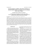
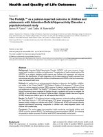
Subjects with HbSC genotype compared to HbSS ones
showed significantly higher values of both height-SDS adjusted for TH (1.0 ± 0.6 vs. 0.3 ± 0.9 SDS, respectively; p =
0.027) and BMI-SDS (0.9 ± 1.1 vs. -0.7 ± 1.4 SDS, respectively; p = 0.004) despite chronological age was not
Page 3 of 9
Table 1 Laboratory data in HbSS patients vs. HbSC patients
Laboratory data
Hb SS
HbSC
P-value
Group (n = 38) Group (n = 14)
HU > 1 years (%)
71% (27/38)
21.4% (3/14)
–
WBCs, 1000s
11.2 ± 4.31
6.76 ± 1.80
0.0001
WBCs, 1000s(mean 2016)
11.2 ± 3.32
7.18 ± 1.96
< 0.0001
Neutrophils, %
50.0 ± 12.1
49.9 ± 10.6
0.9835
47.7 ± 8.3
0.4960
Neutrophils, % (mean 2016) 49.1 ± 9.9
Hb, g/dl
9.0 ± 1.0
11.8 ± 1.2
< 0.0001
Hb, g/dl (mean 2016)
9.1 ± 0.9
11.6 ± 1.2
< 0.0001
Hb S, %
63.3 ± 14.2
46.7 ± 10.4
0.0005
Hb F, %
15.7 ± 7.8
7.0 ± 11.5
0.0003
Platelets, 1000s
421 ± 201
221 ± 100
0.0002
LDH, U/L
951.8 ± 216.5
582.4 ± 144.8
< 0.0001
Data are reported as mean ± SD (standard deviation)
Abbreviations: HbSS homozygous SS patients, HbSC double heterozygous SC
patients, HU hydroxyurea, WBC white blood cells, Hb hemoglobin, LDH
lactate dehydrogenase
P-values statistically significant are printed in bold
different (Table 2; Fig. 1). Analyzing data according to
gender no difference was found in anthropometric parameters (Additional file 1: Table S1).
Height-SDS adjusted for TH was significantly and negatively correlated with clinical severity parameters such as
number of hospital admissions/2016 (Spearman R = − 0.31
p = 0.040), average number of days of hospital admissions/
2016 (Spearman R = − 0.31, p = 0.041), and average number of ACS (Spearman R = − 0.40, p = 0.008).
Two out of 52 of SCD subjects (3.8%) showed
height-SDS < − 2 SDS and 9.6% (5/52) showed BMI-SDS <
− 2 SDS. Patients on treatment with HU for more than
Table 2 Anthropometric parameters in HbSS patients vs. HbSC
patients
Anthropometric data
Hb SS
HbSC
Group (n = 38)
Group (n = 14)
Pvalue
Age (years)
10.44 ± 4.55
13.05 ± 4.47
0.0850
Height
137.1 ± 21.7
150.2 ± 24.5
0.1340
Height-SDS
− 0.2 ± 1.1
0.4 ± 0.7
0.1807
Height-SDS adjusted for TH
0.3 ± 0.9
1.0 ± 0.6
0.0270
Weight
32.6 ± 14.0
51.1 ± 25.0
0.0374
BMI (Kg/m2)
16.5 ± 2.7
21.1 ± 4.9
0.0045
BMI-SDS
−0.7 ± 1.4
0.9 ± 1.1
0.0043
Growth velocity cm/year
4.0 ± 2.3
4.2 ± 3.3
0.7863
Growth velocity -SDS
−1.6 ± 2.2
−2.3 ± 3.5
0.8398
Sitting height
69.3 ± 9.0
75.7 ± 11.8
0.1760
Sitting height/height
0.51 ± 0.02
0.50 ± 0.02
0.8614
Data are reported as mean ± SD (standard deviation)
Abbreviations: HbSS homozygous SS patients, HbSC double heterozygous SC
patients, SDS standard deviation, TH target height, BMI body mass index
P-values statistically significant are printed in bold
Mandese et al. BMC Pediatrics
(2019) 19:56
Page 4 of 9
Fig. 1 Anthropometric parameters according to SCD genotype. BMI-SDS in HbSC group was significantly higher than in HbSS group (p = 0.004).
Height-SDS adjusted for TH in HbSC group was significantly higher than in HbSS group (p = 0.027)
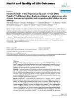
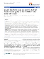
one year (29/52, 56%) respect to those on HU for less time
had lower values of BMI-SDS (− 0.8 ± 1.4 vs. 0.4 ± 1.2
SDS, respectively; p = 0.008) and sitting height/height ratio
(0.50 ± 0.02 vs. 0.52 ± 0.02, respectively; p = 0.004) (Fig. 2).
days of hospital admissions/2016 (Spearman R = − 0.29,
p = 0.034) and average number of hospital admissions in
the last 5 years (Spearman R = − 0.36, p = 0.009).
Glucose and lipid metabolism
The prevalence of metabolic alterations and endocrine
complications among SCDs was high: 48 out of 52
patients show at least one metabolic and/or endocrine
alteration. Among all patients, 41 (79%), 6 (11.5%), and
1 (1.9%) presented respectively one, two, and three alterations at the same time. The most detected conditions
were the vitamin D insufficiency/deficiency (84.7%), the
insulin resistance (11.5%), and to a lesser extent the
GHD (3.8%), the subclinical hypothyroidism (3.8%), and
the hypergonadotropic hypogonadism (1.9%) (Table 3).
Analyzing data according to HDL-C levels, we found
that subjects with HDL-C > 40 mg/dl, respect to those
with HDL-C < 40 mg/dl, had significantly higher levels of
vitamin D (22.4 ± 11.2 vs. 18.2 ± 17.3 ng/ml, respectively;
p = 0.044). The mean values of HDL-C were correlated
with neutrophils (Spearman R = − 0.29, p = 0.041), LDH
(Spearman R = − 0.29, p = 0.037), and serum ferritin
(Spearman R = − 0.40, p = 0.003) (Fig. 3).
The 11.5% of subjects had insulin resistance as suggested
by abnormal HOMA-IR values. However, HOMA-IR was
not different between HbSS and HbSC subjects.
Vitamin D insufficiency/deficiency
Growth and gonadotropin
In particular, in 63.5% of patients vitamin D levels were
between 10 and 30 ng/ml while in 21.2% were < 10 ng/
ml. We found a significant negative relationship between
plasmatic levels of vitamin D and clinical severity of the
disease, represented by number of hospital admissions/
2016 (Spearman R = − 0.29 p = 0.040), average number of
GHD was detected in 2 boys (3.8%) with HbSS genotype,
who have been started human recombinant GH replacement therapy.
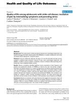
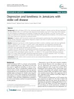
In addition, patients with HbSS genotype compared to
HbSC ones showed lower levels of IGF-1 (211.7 ± 93.2 vs.
315.3 ± 89.3 ng/ml, respectively; p < 0.001) and IGFBP-3
Prevalence of metabolic and endocrine complications
Fig. 2 Anthropometric parameters according to HU therapy groups. Patients in HU > 1-year group, respect to HU < 1-year one, had both
significantly lower BMI-SDS (p = 0.008) and sitting height/height ratio (p = 0.004)
Mandese et al. BMC Pediatrics
(2019) 19:56
Page 5 of 9
Table 3 Prevalence of endocrine and metabolic alterations in
children and adolescents with SCD
Endocrine/metabolic complications
N°/52
%
M/F
SS/SC
Vitamin D insufficiency (10–30 ng/ml)
33
63.5%
16/17
24/9
Vitamin D deficiency (< 10 ng/ml)
11
21.2%
7/4
7/4
GHD
2
3.8%
2/0
2/0
Subclinical hypothyroidism
2
3.8%
1/1
2/0
Hypergonadotropinic hypogonadism
1
1.9%
1/0
1/0
Ovarian insufficiency
1
1.9%
0/1
1/0
Insulin resistance
6
11.5%
2/4
4/2
Abbreviations: GHD growth deficiency hormone
(3267.1 ± 828, 4 vs. 3761.7 ± 773.5 ng/ml, respectively; p <
0.001) (Fig. 4). IGF-1 values were significantly correlated
with both Hb (Spearman R = 0.51, p = 0.0001) and LDH
levels (Spearman R = − 0.44, p = 0.0009) (Fig. 5).
Ovarian insufficiency was detected in one 17-years old
girl with normal secondary sexual characteristics for age,
with secondary amenorrhea, high concentration of FSH
and low levels of AMH.
Diagnosis of hypergonadotropic hypogonadism was also
performed in a 15-years old male with HbSS genotype and
with Tanner Stage 1 (testes 3 ml bilateral).
The mean values of testosterone in our males were
also positively correlated with the mean values of Hb
(Spearman R = 0.40, p = 0.029). No correlation between IGF-1, IGFBP-3, TSH, fT4, testosterone, estradiol, LH, FSH levels and ferritin values [both as a
relative value at the time of enrollment and as the
average value of the last two years (2015–2017)] were
identified in our population both in prepubertal and
in pubertal patients.
Discussion
Survival rate among children with SCD has increased
especially in the recent decades, due to an earlier diagnosis and a better quality of care. Consequently, the incidence of long-term complications, such as metabolic
and endocrine disorders, is increasing in SCD population and it has become a main concern to treat them
properly, improving their prognosis and their quality of
life. In this study we demonstrated a high prevalence
(92%) of endocrine complications and metabolic alterations in the pediatric SCD population, mainly represented by vitamin D insufficiency or deficiency, insulin
resistance, and to a lesser extent GHD, subclinical
hypothyroidism, and hypogonadism. In literature, it is
really difficult to understand the cumulative incidence of
these disorders in subjects with SCD because of
published studies evaluated mainly one single endocrine
alteration. Specifically, growth impairment and delayed
puberty are the most frequent disorders observed among
SCD pediatric patients [10, 11].
Özen et al. reported that 50% of the examined population show endocrine disorders mainly represented, as in
our study, by insufficiency/deficiency of vitamin D and
to a lesser degree of osteopenia, hypoplasia/testicular
atrophy, hypogonadism, hypothyroidism, and insulin
resistance [22].
In our study the prevalence of endocrine complications was even higher that those reported by Özen et al.
Fig. 3 Relationship between HDL-C values and parameters of clinical severity
Mandese et al. BMC Pediatrics
(2019) 19:56
Page 6 of 9
Fig. 4 IGF-1 and IGFBP-3 values according to SCD genotype. In HbSC group both IGF-1 and IGFBP-3 levels were significantly higher respect to
HbSS group (p < 0.0001)
[22], but it is important to consider that the majority of
our subjects were immigrants, coming mainly from
Africa (96%) with socio-economic conditions that may
influence the anthropometric, endocrine and metabolic
parameters.
It has been demonstrated that children with SCD had
a poorer growth compared to matched healthy subjects
[23]. Near two thirds of SCD patients experience a decline in one or more growth parameters (height, weight,
and BMI) and the incidence of growth retardation (defined by the presence of one or more of anthropometric
parameters below the 5th percentile) could reach the
38% during the follow-up [24]. In our population, the
prevalence of growth alterations was about 3.8% when
height was considered <-2DS and 9.6% when BMI-SDS
was considered <-2SD. The discrepancy between our
results and previous published data [24] could be
explained by differences in the study design (longitudinal
vs. transversal study).
The underline mechanism on growth delay in SCD is
very complex and probably influenced by many variables, such as hematologic and cardiovascular status,
socio-economic factors, endocrine function, metabolic
function and nutritional status [25].
It has been shown that the mean height SDS of
children with SCD is comparable to those of children
with constitutional growth delay but it is higher than
those of children with GHD [26, 27]. In agreement with
published data, our study demonstrated that growth was
more affected in subjects with HbSS genotype than in
subjects with HbSC genotype.
According to the therapeutic regimen, significant
differences were found with respect to BMI-SDS and
sitting height/height ratio. Although not expected,
patients treated with HU for more than one year had
lower BMI-SDS and sitting height/height ratio. The reason of these findings is likely related to the more severe
phenotype of patients treated with HU for more than a
year. In fact, in our opinion, the disease severity could
influence these anthropometric parameters. However, it
would be important to continue these evaluations in
order to assess whether treated patients may have an
improvement in growth parameters over time by a
reduction in clinical severity. In fact, the prospective use
of HU can both improve clinical outcome of the disease
and also positively influence growth and development,
reducing the risk of iron overload due to the chronic
transfusion regimen.
Fig. 5 Relationship between values of IGF-1 and parameters of clinical severity
Mandese et al. BMC Pediatrics
(2019) 19:56
Our data showed significant correlations between clinical parameters of disease severity and anthropometric
parameters: children with better control of the disease
(expressed as lower number of hospital admissions in
2016, lower number of days of hospitalization in 2016
and lower ACS) had higher values of height-SDS adjusted for TH. Subjects with HbSS genotype showed a
negative correlation between the number of ACS and
the values of height-SDS adjusted for TH. A good
clinical control of the disease seems not only to affect
the survival but also to reduce the long-term
comorbidity.
In our SCD population, vitamin D insufficiency was
demonstrated in 63.5% while 21.2% had a deficient level.
In a study conducted by Buison et al., 65% of children
with SCD had levels of vitamin D lower than those of
healthy children [28]. Jackson et al [29] reported that
96% of SCD patients had vitamin D level between 10
and 20 ng/ml. Severe vitamin D deficiency (< 10 ng/ml)
was found in 64% of subjects and it was demonstrated
to be associated with age and reduction in lung function
but not with pain and/or ACS episodes. In a Spanish
study on vitamin D status in 78 children with SCD, Garrido et al. [30] report that near to 80% and 56.4% had
vitamin D level < 30 ng/ml and < 20 ng/ml, respectively.
The vitamin D metabolism is complex because of the
involvement of different organ including skin, intestines,
liver, kidney, and parathyroid [9]. Patients with SCD
have some peculiar characteristics that can lead to the
development of vitamin D deficiency such as decreased
appetite or reduction of nutrients absorption due to the
intestinal mucosa damage. Continuous red blood cells
production to compensate anemia characterize SCD and
causes an increase of basal metabolic rate with higher
nutritional demands [8, 31]. Moreover, in SCD patients
with renal impairment conversion of vitamin D to its
active form can be reduced. Finally, vitamin D binding
protein levels can be low being SCD an inflammatory
disease [28]. The importance of vitamin D assessment in
patients with SCD is supported by the demonstration
that vitamin D deficiency is more frequent among children with SCD than in controls [32].
The different prevalence of vitamin D deficiency/insufficiency demonstrated between African Americans and
Caucasians populations can be explained by the
decreased synthesis of vitamin D in the skin [33] and
differences in dietary habits [34]. A better absorption of
dietary calcium and lower levels of vitamin D binding
protein have been demonstrated in African Americans
subjects compared to Caucasians [35, 36]. This suggests
that neither the optimal Vitamin D threshold for Caucasians nor levels suggested for healthy African Americans
are applicable to patients with SCD [32]. It is therefore
really important to identify the optimal level of vitamin
Page 7 of 9
D in children and adults with SCD, in particular in
patients of African origin living in European Countries,
as patients enrolled in our study.
In our population, vitamin D levels showed an inverse
and statistically significant correlation with the number
of admissions and hospitalizations in 2016 and the average number of admissions in the last 5 years, suggesting
that this deficit could adversely affects the clinical severity of the disease.
It was hypothesized that inadequate levels of vitamin
D could be linked to a condition of chronic inflammation, as well as low levels of HDL-C [37]. This finding is
confirmed also in our population study. Dividing our patients into two groups according to HDL-C levels, we
found that vitamin D values were significantly lower in
the group of patients with lower HDL-C values (< 40 mg/
dl). In addition, HDL-C values of our population showed a
negative relationship (p < 0.05) to neutrophils percentage,
LDH and ferritin values, particularly in subjects with HbSS
genotype. Seixas et al. [37] found a negative association
between LDH and HDL-C levels, showing how low HDL-C
levels could be a prognostic marker of hemolysis and endothelial dysfunction in view of their anti-inflammatory,
anti-oxidant,
anti-aggregating,
anti-coagulant
and
pro-fibrinolytic role. Patients with SCD and high HDL-C
levels had fewer reticulocytes, WBC, monocytes, PTL, and
erythroblasts and a lower concentration of HbS as well as a
lower concentration of hemolytic markers. Our data confirm that HDL-C and vitamin D could play an important
role in inflammatory condition such as SCD.
The 3.8% of our population showed GHD. An impairment of the GH-IGF1-IGFBP3 axis was demonstrated in
SCD subjects [38, 39]. Children with SCD have significantly decreased IGF-1 concentrations compared to children with constitutional delay of growth. The poor
synthesis of IGF-1 could depend on a primitive defect of
the axis, but also from malnutrition and hypermetabolic
status of these patients [40]. In some cases, however,
there is a real GHD due to a pituitary vascular insult
during vaso-occlusive crises [41, 42]. These patients
could benefit from a human recombinant GH replacement therapy [43]. Our data showed that mean values of
both IGF-1 and IGFBP-3 were lower in subjects with
HbSS genotype compared to subjects with HbSC genotype and that IGF1-levels had a positive correlation with
Hb and Hb mean values of 2016 and a negative correlation with average LDH and LDH mean values of 2016.
These data underline how the clinical severity of the disease, the number of vaso-occlusive crises and chronic
hemolysis could adversely affect GH-IGF-1-IGFBP-3 axis
in SCD patients.
In our population 11.5% of patients had pathological
HOMA index. In literature there is evidence of insulin
resistance among patients with SCD [10]. A multicenter
Mandese et al. BMC Pediatrics
(2019) 19:56
study by Fung et al. [44] revealed that for every 10 years
of transfusion therapy, subjects with SCD have a 2.5
times greater probability to develop diabetes (while patients with thalassemia have a double risk). However,
there are cases of insulin resistance in patients with normal oral glucose tolerance test. In these patients BMI
values were above 85° percentile and none of the patients with normal weight had an insulin resistance
condition [22]. However, it is important to point out
that our study included a pediatric population with a
normal BMI and this could also explain the reduced
rate of diabetes. There is currently no agreement on
the causes of this complication and further investigations are needed.
We found a case of hypergonadotropic hypogonadism
and one case of ovarian failure in HbSS genotype group.
The etiology of hypogonadism in SCD is not fully understood yet: in some cases, primitive gonadal failure is related to structural anomalies, resulting from chronic
tissue damage associated with chronic anemia condition
and local vaso-occlusive crises [45]. According to this
hypothesis, our study demonstrated a direct correlation
between Hb levels and testosterone average values in
males, demonstrating how clinical control can affect reproductive function.
This study has some important limitations. This is a
single center study with a small sample size. Secondly
there are some confounders factors (i.e. genotype, gender…). The main outcome of this study was to report
clinical features of our patients, in a cross sectional way,
to better understand the actual prevalence of both metabolic alterations and endocrine complications. Surely, a
longitudinal study design of these parameters will provide us more information on the natural history of these
complications in SCD. However, it must be considered
that in our country SCD is a rare disease and, at the best
of our knowledge, this is the first Italian study on these
topics. In literature there are very little data available on
these condition in pediatric patients with SCD mainly in
European Countries. We think that it is important to
evaluate these conditions in different environmental
setting.
Conclusion
Subjects with SCD show a high prevalence of metabolic alterations and endocrine complications. However, our results suggest that through the achievement
of a good clinical control the SCD patients can obtain
a positive impact on growth, metabolic and endocrine
function.
Consequently, it is crucial to perform periodic anthropometric and endocrine evaluations, especially during
puberty, and to have a comprehensive approach to this
disease in order to reduce its long-term complications.
Page 8 of 9
Additional file
Additional file 1: Table S1. Anthropometric parameters in males and
females. (DOCX 30 kb)
Abbreviations
ACS: Acute Chest Syndrome; AIFA: Italian Drugs Agency; AMH: Anti-mullerian
hormone; BMI: Body Mass Index; FSH: Follicle-stimulating hormone; FT4: Free
thyroxine 4; GH: Growth Hormone; Hb: Hemoglobin; HDL: HDL-cholesterol;
HOMA: Homeostasis model assessment; HU: Hydroxyurea; IGF-1: Insulin-like
Growth Factor-I; IGFBP-3: Insulin-like Growth Factor Binding Protein 3;
IR: Insulin resistance; LDH: Lactate dehydrogenase; LH: Luteinizing hormone;
SCD: Sickle Cell Disease; SDS: Standard Deviation Score; TH: Target Height;
TSH: Thyroid stimulating hormone; VOC: Vaso-occlusive crises; WHO: World
Health Organization
Acknowledgements
Not applicable
Funding
Not applicable
Availability of data and materials
The datasets generated and/or analysed during the current study are not
publicly available due to privacy reason but are available from the
corresponding author on reasonable request.
Authors’ contributions
VM, EB and PB collected data. EB, VM, PB and LI conceived the study and its
design, coordinated it and wrote the manuscript. PB performed the statistical
analysis. GP, BP, MC, LL, and LI supervised and reviewed the manuscript
making important intellectual contributions. All authors read and approved
the final manuscript.
Ethics approval and consent to participate
Provincial Ethical Committee approved the protocol study (E.C. n. 213/16),
informed consent was obtained for all enrolled patients. Parents and/or legal
guardians provided the written informed consent for participation on behalf
of the underage participants who were not of legal age to consent for
themselves.
Consent for publication
Not applicable
Competing interests
Lorenzo Iughetti is an Editorial Board Member for BMC Pediatrics. All the
others authors declared that they have no competing interests.
Publisher’s Note
Springer Nature remains neutral with regard to jurisdictional claims in
published maps and institutional affiliations.
Author details
1
Post Graduate School of Pediatrics, Department of Medical and Surgical
Sciences for Mothers, Children and Adults, University of Modena and Reggio
Emilia, Via del Pozzo 71, 41124 Modena, Italy. 2Oncology and Hematology
Pediatric Unit Department of Medical and Surgical Sciences for Mothers,
Children and Adults, University of Modena and Reggio Emilia, 41124
Modena, Italy. 3Pediatric Unit, Department of Medical and Surgical Sciences
for Mothers, Children and Adults, University of Modena and Reggio Emilia,
41124 Modena, Italy.
Received: 8 March 2018 Accepted: 30 January 2019
References
1. Platt OS, Brambilla DJ, Rosse WF, Milner PF, Castro O, Steinberg MH, et al.
Mortality in sickle cell disease. Life expectancy and risk factors for early
death. N Engl J Med. 1994;330:1639–44.
Mandese et al. BMC Pediatrics
2.
3.
4.
5.
6.
7.
8.
9.
10.
11.
12.
13.
14.
15.
16.
17.
18.
19.
20.
21.
22.
23.
24.
25.
26.
(2019) 19:56
Panepinto JA, O'Mahar KM, DeBaun MR, Loberiza FR, Scott JP. Health-related
quality of life in children with sickle cell disease: child and parent
perception. Br J Haematol. 2005;130:437–44.
Meier ER, Miller JL. Sickle cell disease in children. Drugs. 2012;72:895–906.
Lodi M, Bigi E, Palazzi G, Vecchi L, Morandi R, Setti M, et al. Universal
screening program in pregnant women and newborns at-risk for sickle cell
disease: first report from northern Italy. Hemoglobin. 2017;41:230–3.
Quinn CT, Rogers ZR, McCavit TL, Buchanan GR. Improved survival of
children and adolescents with sickle cell disease. Blood. 2010;115:3447–52.
Lobo CL, Ballas SK, Domingos AC, Moura PG, do Nascimento EM, Cardoso
GP, et al. Newborn screening program for hemoglobinopathies in Rio de
Janeiro, Brazil. Pediatr Blood Cancer. 2014;61:34–9.
Iughetti L, Bigi E, Venturelli D. Novel insights in the management of sickle
cell disease in childhood. World J Clin Pediatr. 2016;5:25–34.
Barden EM, Kawchak DA, Ohene-Frempong K, Stallings VA, Zemel BS.
Body composition in children with sickle cell disease. Am J Clin Nutr.
2002;76:218–25.
Rees DC, Williams TN, Gladwin MT. Sickle-cell disease. Lancet. 2010;376:
2018–31.
Smiley D, Dagogo-Jack S, Umpierrez G. Therapy insight: metabolic and
endocrine disorders in sickle cell disease. Nat Clin Pract Endocrinol Metab.
2008;4:102–9.
el-Hazmi MA, Bahakim HM, al-Fawaz I. Endocrine functions in sickle cell
anaemia patients. J Trop Pediatr. 1992;38:307–13.
Hagag AA, El-Farargy MS, Elrefaey S, Abo El-enein AM. Study of gonadal
hormones in Egyptian female children with sickle cell anemia in correlation
with iron overload: single center study. Hematol Oncol Stem Cell Ther.
2016;9:1–7.
Hankins JS, Ware RE, Rogers ZR, Wynn LW, Lane PA, Scott JP, et al. Longterm hydroxyurea therapy for infants with sickle cell anemia: the HUSOFT
extension study. Blood. 2005;106:2269–75.
World Health Organization Growth Reference. 2007. />growthref/en/. Accessed 7 Dec 2018.
Tanner JM, Goldstein H, Whitehouse RH. Standards for children’s height at ages
2–9 years allowing for height of parents. Arch Dis Child. 1970;47:755–62.
Marshall WA, Tanner JM. Variations in pattern of pubertal changes in girls.
Arch Dis Child. 1969;44:291–3.
Keskin M, Kurtoglu S, Kendirci M, Atabek ME, Yazici C. Homeostasis model
assessment is more reliable than the fasting glucose/insulin ratio and
quantitative insulin sensitivity check index for assessing insulin resistance
among obese children and adolescents. Pediatrics. 2005;115:500–3.
Kurtoğlu S, Hatipoğlu N, Mazıcıoğlu M, Kendirici M, Keskin M, Kondolot M.
Insulin resistance in obese children and adolescents: HOMA-IR cut-off levels
in the prepubertal and pubertal periods. J Clin Res Pediatr Endocrinol. 2010;
2:100–6.
Iughetti L, Predieri B, Bruzzi P, Predieri F, Vellani G, Madeo SF, et al. Ten-year
longitudinal study of thyroid function in children with Down's syndrome.
Horm Res Paediatr. 2014;82:113–21.
Grimberg A, DiVall SA, Polychronakos C, Allen DB, Cohen LE, Quintos JB, et
al. Drug and therapeutics committee and ethics Committee of the Pediatric
Endocrine Society. Guidelines for growth hormone and insulin-like growth
factor-I treatment in children and adolescents: growth hormone deficiency,
idiopathic short stature, and primary insulin-like growth factor-1 deficiency.
Horm Res Paediatr. 2016;86:361–97.
Brook C, Clayton P, Brown R. Brook's clinical pediatric endocrinology. 6th
edition. Oxford: Wiley-Blackwell; 2009.
Özen S, Ünal S, Erçetin N, Taşdelen B. Frequency and risk factors of
endocrine complications in Turkish children and adolescents with sickle cell
Anemia. Turk J Hematol. 2013;30:25–31.
Al-Saqladi AW, Cipolotti R, Fijnvandraat K, Brabin BJ. Growth and nutritional
status of children with homozygous sickle cell disease. Ann Trop Paediatr.
2008;28:165–89.
Zemel BS, Kawchak DA, Ohene-Frempong K, Schall JI, Stallings VA. Effects of
delayed pubertal development, nutritional status, and disease severity on
longitudinal patterns of growth failure in children with sickle cell disease.
Pediatr Res. 2007;61:607–13.
Singhal A, Morris J, Thomas P, Dover G, Higgs D, Serjeant GR. Factors
affecting prepubertal growth in homozygous sickle cell disease. Arch Dis
Child. 1996;6:502–6.
Soliman A, el Zalabany M, Amer M, Ansari BM. Growth and pubertal
development in transfusion-dependent children and adolescents with
Page 9 of 9
27.
28.
29.
30.
31.
32.
33.
34.
35.
36.
37.
38.
39.
40.
41.
42.
43.
44.
45.
thalassaemia major and sickle cell disease: a comparative study. J Trop
Pediatr. 1999;45:23–30.
Thomas PW, Singhal A, Hemmings-Kelly M, Serjeant GR. Height and
weight reference curves for homozygous sickle cell disease. Arch Dis
Child. 2000;82:204–8.
Buison AM, Kawchak DA, Schall J, Ohene-Frempong K, Stallings VA,
Zemel BS. Low vitamin D status in children with sickle cell disease. J
Pediatr. 2004;145:622–7.
Jackson TC, Krauss MJ, Debaun MR, Strunk RC, Arbelaez AM. Vitamin D
deficiency and comorbidities in children with sickle cell anemia. Pediatr
Hematol Oncol. 2012;29:261–6.
Garrido C, Cela E, Belendez C, Mata C, Huerta J. Status of vitamin D in
children with sickle cell disease living in Madrid, Spain. Eur J Pediatr. 2012;
171:1793–8.
Singhal A, Parker S, Linsell L, Serjeant G. Energy intake and resting metabolic
rate in preschool Jamaican children with homozygous sickle cell disease.
Am J Clin Nutr. 2002;75:1093–7.
Nolan VG, Nottage KA, Cole EW, Hankins JS, Gurney JG. Prevalence of
vitamin D deficiency in sickle cell disease: a systematic review. PLoS One.
2015;10:e0119908.
Bell NH, Greene A, Epstein S, Oexmann MJ, Shaw S, Shary J. Evidence
for alteration of the vitamin D-endocrine system in blacks. J Clin Invest.
1985;76:470–3.
O'Connor MY, Thoreson CK, Ramsey NL, Ricks M, Sumner AE. The
uncertain significance of low vitamin D levels in African descent
populations: a review of the bone and cardiometabolic literature. Prog
Cardiovasc Dis. 2013;56:261–9.
Heaney RP. The importance of calcium intake for lifelong skeletal health.
Calcif Tissue Int. 2002;70:70–3.
Gutierrez OM, Farwell WR, Kermah D, Taylor EN. Racial differences in the
relationship between vitamin D, bone mineral density, and parathyroid
hormone in the National Health and nutrition examination survey.
Osteoporos Int. 2011;22:1745–53.
Seixas MO, Rocha LC, Carvalho MB, Menezes JF, Lyra IM, Nascimento VM, et
al. Levels of high-density lipoprotein cholesterol (HDL-C) among children
with steady-state sickle cell disease. Lipids Health Dis. 2010;9:91.
Luporini SM, Bendit I, Manhani R, Bracco OL, Manzella L, Giannella-Neto D.
Growth hormone and insulin-like growth factor I axis and growth of
children with different sickle cell anemia haplotypes. J Pediatr Hematol
Oncol. 2001;23:357–63.
Collett-Solberg PF, Fleenor D, Schultz WH, Ware RE. Short stature in children
with sickle cell anemia correlates with alterations in the IGF-I axis. J Pediatr
Endocrinol Metab. 2007;20:211–8.
Mandese V, Marotti F, Bedetti L, Bigi E, Palazzi G, Iughetti L. Effects of
nutritional intake on disease severity in children with sickle cell disease. Nutr
J. 2016;15:46.
Soliman AT, Darwish A, Mohammed SH, Bassiony MR, el Banna N, Asfour M.
Circulating growth hormone (GH), insulin-like growth factor-I (IGF-I) and free
thyroxine, GH response to clonidine provocation and CT scanning of the
hypothalamic-pituitary area in children with sickle cell disease. J Trop
Pediatr. 1995;41:285–9.
Soliman AT, el Banna N, alSalmi I, De Silva V, Craig A, Asfour M. Growth
hormone secretion and circulating insulin-like growth factor-I (IGF-I) and IGF
binding protein-3 concentrations in children with sickle cell disease.
Metabolism. 1997;46:1241–5.
Nunlee-Bland G, Rana SR, Houston-Yu PE, Odonkor W. Growth hormone
deficiency in patients with sickle cell disease and growth failure. J Pediatr
Endocrinol Metab. 2004;17:601–6.
Fung EB, Harmatz PR, Lee PD, Milet M, Bellevue R, Jeng MR, et al. MultiCentre study of Iron overload research group. Increased prevalence of ironoverload associated endocrinopathy in thalassaemia versus sickle-cell
disease. Br J Haematol. 2006;135:574–82.
Casale M, Ciliberti A, Colombatti R. Italian guidelines for the management of
pediatric patients with sickle cell disease. Italian Ematology oncology
pediatric association AIEOP 2012. />uploads/2017/05/tutto-giu12.pdf. Accessed 7 Dec 2018.