Deciphering risk factors for blood stream infections, bacteria species and antimicrobial resistance profiles among children under five years of age in NorthWestern Tanzania: A multicentre
Bạn đang xem bản rút gọn của tài liệu. Xem và tải ngay bản đầy đủ của tài liệu tại đây (4.17 MB, 11 trang )
Seni et al. BMC Pediatrics
(2019) 19:32
/>
RESEARCH ARTICLE
Open Access
Deciphering risk factors for blood stream
infections, bacteria species and
antimicrobial resistance profiles among
children under five years of age in NorthWestern Tanzania: a multicentre study in a
cascade of referral health care system
J. Seni1,2* , A. A. Mwakyoma1, F. Mashuda3, R. Marando3, M. Ahmed3, R. DeVinney2†, J. D. D. Pitout2† and
S. E. Mshana1†
Abstract
Background: Blood stream infections (BSIs) cause a complex cascade of inflammatory events, resulting in significant
morbidity and mortality in children in Tanzania. This study was designed to delineate circulating bacterial species,
antimicrobial resistance (AMR) profiles and risk factors for BSIs and mortality among children in the cascade of referral
health care facilities so as to guide comprehensive BSIs management.
Methods: A multiple cross sectional analytical study was conducted between July 20, 2016 to October 04, 2017
involving 950 children less than five years of age in the North-western part of Tanzania. Children with clinical
features suggestive of BSIs were included. Demographic, clinical and laboratory information on culture and
antimicrobial susceptibility testing was collected from children; and analyzed using STATA version 13.0 software.
Results: The prevalence of BSIs among children was 14.2% (95% CI: 12.1–16.6%), with specific prevalence in the
district, regional and tertiary hospitals being 8.3, 6.4 and 20.0%, respectively. The most common bacterial pathogens
isolated from 135 culture-positive children were Klebsiella pneumoniae (55, 40.4%), Staphylococcus aureus (23, 17.0%),
and Escherichia coli (17, 12.6%). Multi-drug resistance (MDR) was higher in isolates from children at Bugando Medical
Centre (BMC) tertiary hospital than isolates from district and regional hospitals [OR (95% CI): 6.36 (2.15–18.76); p = 0.001].
Independent risk factors for BSIs were neonatal period [OR (95% CI): 1.93 (1.07–3.48); p = 0.003] and admission at BMC
[2.01 (1.08–3.74); p = 0.028)]. Approximately 6.6% (61/932) of children died, and risk factors for mortality were found to
be children attending BMC [OR (95% CI): 4.95 (1.95–12.5); p = 0.001)], neonatal period [OR (95% CI): 2.25 (1.02–5.00);
p = 0.045)], and children who had blood culture positive results [OR (95% CI): 1.95 (1.07–3.56); p = 0.028)].
(Continued on next page)
* Correspondence:
†
R. DeVinney, J. D. D. Pitout and S. E. Mshana contributed equally to this work.
1
Department of Microbiology and Immunology, Weill-Bugando School of
Medicine, Catholic University of Health and Allied Sciences, P.O. Box 1464,
Mwanza, Tanzania
2
Department of Microbiology, Immunology and Infectious Diseases,
Cumming School of Medicine, University of Calgary, 3330 Hospital Dr NW,
Calgary, AB T2N 4N1, Canada
Full list of author information is available at the end of the article
© The Author(s). 2019 Open Access This article is distributed under the terms of the Creative Commons Attribution 4.0
International License ( which permits unrestricted use, distribution, and
reproduction in any medium, provided you give appropriate credit to the original author(s) and the source, provide a link to
the Creative Commons license, and indicate if changes were made. The Creative Commons Public Domain Dedication waiver
( applies to the data made available in this article, unless otherwise stated.
Seni et al. BMC Pediatrics
(2019) 19:32
Page 2 of 11
(Continued from previous page)
Conclusions: The prevalence of BSIs (14.2%) in this multi-centre study is high and predominantly caused by the MDR
K. pneumoniae. Priority interventional measures to combat BSIs and mortality, specifically among neonates at BMC are
urgently recommended.
Keywords: Blood stream infections, Children, Tanzania
Background
Blood stream infections (BSIs) are the most common
causes of morbidity and mortality in children [1, 2].
They constitute a complex cascade of inflammatory processes spanning from systemic inflammatory response
syndrome, sepsis, severe sepsis, septic shock and ultimately death if not promptly managed [3–5].
Introduction of vaccines and the advancements in technology, with more invasive diagnostic and treatment modalities has resulted in a paradigm shift in both implicated
etiological agents as well as the age-groups affected by
BSIs [6–8]. As a result, previously dominant bacteria such
as Streptococcus pneumoniae, Haemophilus influenzae
type b and Neisseria meningitidis, are currently outnumbered by multidrug resistant (MDR) bacteria like Methicillin resistant Staphylococcus aureus (MRSA) and Extended
spectrum beta lactamase (ESBL) producing enterobacteriaceae, which in most cases are of nosocomial origin [6–8].
A recent review of ESBL attributable BSIs in children
across the world showed varying magnitude across countries, ranging from 10 to 15% (Africa, South America and
South-Eastern Asia), and below 5% in Europe [9].
In Tanzania, previous studies which were largely centered
in the tertiary health care facilities showed that the proportion of BSIs ranged from 5 to 15%, with ESBL producing
Klebsiella pneumoniae and Escherichia coli being the most
predominant pathogens [10–14]. In this regard, findings
from these studies cannot be generalized to all levels of
health care facilities in Tanzania [10–14]. Of note, mortality
in these studies was unacceptably high (in some studies up
to 20%), calling for interventional measures in these tertiary
hospitals, along with evaluating the trend in other health
care facilities like regional/referral and district hospitals.
This study evaluated the magnitude of BSIs, bacterial
species, and antimicrobial resistance (AMR) profiles
among children attending different health care facilities
in the North-western part of Tanzania to guide specific
antimicrobial therapies. Moreover, risk factors for BSIs
and mortality were ascertained so as inform specific target groups for preventive and control measures.
four health care facilities in the cascade of referral system in
North-western Tanzania. These health care facilities were
Bugando Medical Centre (BMC), a tertiary hospital, Sekou
Toure Regional Referral Hospital (SRRH), Nyamagana
District Hospital (NDH) to represent an urban setting, and
Sengerema District Designated Hospital (SDDH) to represent a rural setting. All these health care facilities are teaching hospitals for the Catholic University of Health and
Allied Sciences (CUHAS), except NDH (Table 1 and Fig. 1).
Study population, inclusion and exclusion criteria
The study enrolled prospectively children presenting to
the health care facilities with clinical symptoms and signs
suggestive of BSIs [5, 13], and whose parents/guardians
voluntarily consented to participate on their behalf. The
clinical signs and symptoms for enrollment were based on
the WHO Young Infant Study Group and its methodology
paper i.e. temperature (of > 38 °C or < 36 °C), age specific
tachycardia, age specific tachypnoea, convulsions, altered
state of consciousness and abnormal feeding [5]. To ensure consistency, enrolment evaluation was done by
paediatrician and/or experienced registrar who were also
part of this study. A sample size was estimated by the Kish
Leslie formula, using previous prevalence of BSIs among
children of 7.4% in Mwanza. This resulted into a minimum of 106 children per site and 424 children in all four
sites [15]. Taking into account different hospital bed capacities, a total of 1008 children under 5 years of age were
prospectively enrolled during the study period. Fifty eight
(5.7%) children were excluded because of incomplete information in the questionnaires and/or medical records.
Also, using unique identifying numbers, children who
were already enrolled in the lower level health care
facilities and referred to another heath care facility which
was also a study site were excluded. Therefore, this resulted into a total of 950 children under 5 years (Table 1).
This sample size sufficed to estimate the primary study
end-points (i.e. the overall prevalence and health facilitylevel specific prevalence of BSIs, bacterial species and
AMR profiles), and the study secondary end-points (risk
factors for BSIs and mortality).
Methods
Study design and settings
Data collection and laboratory procedures
This was a multiple cross sectional analytical study conducted from July 20, 2016 to October 04, 2017 involving
Socio-demographic and clinical characteristics of children were collected using a structured pre-tested
Seni et al. BMC Pediatrics
(2019) 19:32
Page 3 of 11
Table 1 Demographic descriptions of health facilities involved and respective number of children enrolled
Level/rank of HCF
HCF involved
HCF catchment population
HCF bed capacity
Study participants enrolled (%)
Tertiary
BMC (urban)
16,252,410
950
514 (54.1)
Regional/referral
SRRH (urban)
2,772,509
375
218 (23.0)
District
NDH (urban)
363,452
88
80 (8.4)
SDDH (rural)
663,034
320
138 (14.5)
Sources: Hospital Records; Tanzania Population and Health Census (2012) and Staffing Levels for Ministry of Health Tanzania (2014–2019). HCF: Health care facility;
BMC: Bugando Medical Center; SRRH: Sekou Toure Regional Hospital; NDH: Nyamagana District Hospital; SDDH: Sengerema District Designated Hospital
Ideal bed capacity in health care facilities in Tanzania are 550 to 1500 beds for tertiary hospitals; 176 to 450 beds for regional referral hospitals; and 150 to 175
beds for district hospitals
questionnaire. Absolute age of children (in months) was
collected and then during analysis, three key groups were
delineated i.e. neonates (≤ 1 month), infants (2 to 12
months) and other children (13 to 60 months). Moreover,
clinical information like co-morbidities such as HIV infection, malnutrition, sickle cell disease, pneumonia, anemia
and congenital anomalies (to mention a few) were obtained from patient medical records. Additionally,
calculation of body weight was done to categorize children
into normal weight (z-score between 2 and − 2); underweight (z-score between − 2 and − 3) or overweight
(z-score between 2 and 3) for the respective age using the
WHO Child Growth Standards for boys and girls [http://
www.who.int/childgrowth/standards/cht_wfa_girls_p_0_5.
pdf?ua=1 and />cht_wfa_boys_p_ 0_5.pdf?ua=1].
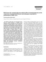
Fig. 1 The map showing North-western part of Tanzania. Africa and Tanzania maps (inserts); Area marked in apple green in the Africa map is Tanzania;
Area marked in pink in the Tanzania map is the catchment area for the study in the North-western part of Tanzania. Bugando Medical Centre (a
tertiary hospital) and eight administrative regions forming its catchment area are labeled. This map was produced using the base map obtained from
the Tanzanian Land Survey Department [48], using Quantum Geographic Information System (Quantum GIS), a software for mapping [49]
Seni et al. BMC Pediatrics
(2019) 19:32
The Tanzania Algorithm for HIV testing among children above 18 months of age employs SD Bioline HIV
1/2 test (Standard Diagnostics Inc., California, USA) as
the first test, and if reactive, it is confirmed by a second
serological test, the Unigold HIV test (Trinity Biotech,
Bray, Ireland). For children below 18 months of age
HIV diagnosis is done by HIV DNA PCR [16, 17].
About two to five milliliters of blood sample from
each child was collected and inoculated into Brain
Heart Infusion broth (OXOID, UK) in a ratio of blood
to Brain Heart Infusion of 1:10. The samples from
SDDH were analysed at SDDH Laboratory, whereas
samples from the rest of the study sites were analysed
at the CUHAS Multipurpose Laboratory as previously
described [18, 19].
AST was done by the conventional Kirby–Bauer disk
diffusion method using the Clinical Laboratory Standard Institute guidelines [20]. The phenotypic screening
of ESBL was done in Muller Hinto agar (OXOID, UK)
along with other disks, using a cut-off zone inhibition
of ≤25 mm for ceftriaxone and ≤ 22 mm for ceftazidime
[20]. Confirmation of ESBL production among E.coli, K.
pneumoniae, and Proteus mirabilis was done in Muller
Hinton agar by double disc synergy method [21].
MRSA was confirmed by the use of cefoxitin disc
(30 μg) and strains showing zone of inhibition of ≤21
mm were labelled as MRSA [20]. A bacterial strain was
confirmed to be MDR when it was resistant to at least
one agent in three or more classes of antimicrobial
agents [22]. E. coli ATCC 25922 and Staphylococcus
aureus ATCC 25923 were used as reference strains for
Gram negative and Gram positive bacteria, respectively
in quality control of culture media, biochemical identification tests and AST.
Page 4 of 11
than 1 month and 60 months, respectively. The most
common age group was children above 1 year of age,
41.6% (n = 395); followed by neonates, 36.4% (n = 346).
The median weight (IQR) for different age categories
were: neonates [2.9 (2.5–3.4) kg], children between 2 to
12 months [7.5 (5.5–8.5) kg] and children above 1 year
of age [10.7 (9.0–13.0) kg]. A total of 392 (41.3%) children had underlying co-morbidities and the majority of
children presented with fever, 86.2% (n = 819) (Table 2).
Of the 950 children enrolled, the proportions of specific
co-morbidities were malnutrition (13.2%), prematurity
(5.3%), HIV (3.9%), and sickle cell disease (3.1%).
Table 2 Socio-demographic and clinical characteristics of children
Characteristic
Sex
Age group
Residence
Mwanza city
Current antibiotic use
The median age (IQR) of the participants was 9 (1–23)
months, with minimum and maximum age being less
392 (41.3)
≤ 1 month
346 (36.4)
2–12 months
209 (22.0)
Normal
539 (56.7)
Underweight
373 (39.3)
Overweight
38 (4.0)
Rural
315 (33.2)
Urban
635 (66.8)
No
313 (33.0)
Yes
637 (67.0)
No
545 (57.4)
405 (42.6)
History of admission in the last 3 monthsb No
525 (86.9)
Yes
Presence of indwelling urinary catheter
79 (13.1)
No
254 (26.7)
Yes
696 (73.3)
No
930 (97.9)
Yes
20 (2.1)
No
558 (58.7)
Yes
392 (41.3)
Fever
Yes
819 (86.2)
Tachypnoea
Yes
235 (24.7)
Tachycardia
Yes
186 (19.6)
Convulsions
Yes
123 (13.0)
Loss of consciousness
Yes
14 (1.5)
Co-morbiditiesc
Presenting symptoms and signs
a
Socio-demographic and clinical characteristics of children
enrolled
558 (58.7)
Girls
Yes
Data management
Results
Boys
13–60 months 395 (41.6)
Weight (kg)a
Presence of i/v line
Data were analyzed by the STATA version 13.0 software
(College Station, Texas, USA). Proportions of children
with culture-confirmed BSIs, bacterial species, and resistance to various antimicrobial agents were determined. Univariate logistic regression analysis was done
to all variables, but only variables with a p-value of less
than 0.05 were subjected to multivariate logistic regression analysis. Independent risk factors for BSIs and mortality among children were determined by multivariate
logistic regression analysis using odds ratios, 95% confidence intervals and p-value cut-off of less than 0.05.
Number (%)
Weight adjusted to age; b Non neonates; c Malnutrition (n = 105), Respiratory
tract infections (n = 86), Prematurity (n = 50), Congenital anomalies (n = 36:
congenital heart diseases, neural tube defects, hydrocephalus and others),
Anemia (n = 31), Sickle cell disease (n = 26), HIV (n = 19), Skin and soft tissue
infections (n = 8); Necrotizing enterocolitis (n = 3); Amoebiasis (n = 2); Burn
injury (n = 2); Rheumatic heart diseases (n = 2); Cerebral malaria (n = 1); Spinal
injury (n = 1); Malnutrition and HIV (n = 17); Malnutrition and sickle cell disease
(n = 3); Premature and HIV (n = 1)
Seni et al. BMC Pediatrics
(2019) 19:32
Prevalence of blood stream infections among children in
North-Western Tanzania
The prevalence of BSIs among children was 14.2%
(95%CI: 12.1–16.6%), with specific prevalence in the district, regional and tertiary hospitals being 8.3, 6.4 and
20.0%, respectively. Also, the age-specific prevalence of
BSIs for neonates, children between 2 to 12 months and
children above 12 months were 25.4% (88/346), 5.7%
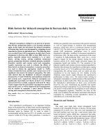
(12/209) and 8.9% (35/395), respectively. The most common bacteria species were K. pneumoniae (55, 40.4%), S.
aureus (23, 17.0%), and E. coli (17, 12.6%). There was an
overall preponderance of BSIs with Gram negative bacteria (78.5%) compared to BSIs attributable to Gram
positive bacteria (21.5%); p < 0.001 (Fig. 2).
Antimicrobial resistance patterns of bacteria causing
blood stream infections
The majority of bacteria were resistant to ampicilllin and
trimethoprim-sulfamethoxazole with resistance rates ranging from 66.6 to 100.0%. All Gram negative bacteria were
sensitive to meropenem, except one Acinetobacter spp isolate. The resistance of Acinetobacter spp. to piperacillin
and piperacillin-tazobactam was 100 and 50.0%, respectively. One Pseudomonas aeruginosa isolate was resistant
to piperacillin and ceftazidime, but sensitive to gentamicin, ciprofloxacin, piperacillin-tazobactam and meropenem. The third generation cephalosporin resistance (3rd
gen Ceph-R) was strikingly high in K. pneumoniae
(95.7%), E. coli (58.8%), and other Gram negative
Page 5 of 11
Enterobacteriaceae (69.6%). E. coli and K. pneumoniae
strains which were 3rd gen Ceph-R were all confirmed to
be ESBL producers. The proportion of MRSA among S.
aureus strains was 34.7%. The distributions of eight
MRSA strains in children with BSIs in health care facilities
were: two in NDH & SDDH, two in SRRH and four in
BMC, nevertheless this distribution was not statistically
significant (p = 0.510). Two MRSA strains (8.7%) were
found to be non-susceptible to vancomycin (Table 3).
Cephalosporin resistant and multi-drug resistant bacterial
strains attributable blood stream infections
The overall proportion of 3rd gen Ceph-R among members of the family Enterobacteriaceae was 79.0% (75/95).
Irrespective of the bacteria species, 3rd gen Ceph-R was
significantly higher in isolates from BMC tertiary hospital [OR (95%CI): 4.95 (1.15–21.32); p = 0.032], than
those from district and regional hospitals (Table 4).
Over three quarters of bacteria strains were found to be
MDR [77.8% (105/135)], with the majority of these being
Gram negative bacteria compared to Gram positive bacteria [81.9% (86/105) versus 18.1% (19/105), p < 0.001].
The distribution of MDR among isolates from children
with BSIs in tertiary hospital, regional/referral hospital
and two district hospitals were 86.4% (89/103), 50.0%
(7/14) and 50.0% (9/18), respectively. MDR was
significantly higher in strains from BMC tertiary hospital
[OR (95% CI): 6.36 (2.15–18.76); p = 0.001], than those
from district and regional hospitals.
Fig. 2 Bacteria species strains from children with blood stream infections. Other Gram negative bacteria (GNB): Citrobacter freundii (5), Salmonella
spp. (1); Serratia marcescens (1); Morganella morganii (1); Pseudomonas aeruginosa (1), Chromobacterium violaceum (1), unidentified GNB (2). Other
Gram positive bacteria (GPB): Enterococcus spp. (3), Streptococcus pyogenes (1) and other Streptococcus spp. (2)
Seni et al. BMC Pediatrics
(2019) 19:32
Page 6 of 11
Table 3 Antimicrobial resistance patterns of bacteria causing blood stream infections
Bacteria (n)
Antimicrobial resistance (%)
AMP
SXT
GEN
CIP
ERY
VAN
AMC
CAZ
CRO
MER
K. pneumoniae (55)
100
96.3
78.2
29.1
NA
NA
94.6
90.9
95.7
0.0
S. aureus (23)
100
82.6
26.1
34.8
65.2
13.0
NA
NA
NA
NA
E. coli (17)
100
94.1
40.1
52.9
NA
NA
94.1
52.9
58.8
0.0
Acinetobacter spp. (10)
NA
90.0
40.0
10.0
NA
NA
NA
100
NA
10.0
Other GNB (23)
95.6
78.3
60.9
17.4
NA
NA
82.6
60.6
69.6
0.0
Other GPB (6)
83.3
66.7
33.3
50.0
50.0
0.0
NA
NA
NA
NA
AMP Ampicillin, SXT Trimethoprim-sulfamethoxazole, GEN Gentamicin, CIP Ciprofloxacin, ERY Erythromycin, VAN Vancomycin, AMC Amoxycillin-clavulanate, CRO
Ceftriaxone, CAZ Ceftazidime, MEM Meropenem, NA Not applicable. Other Gram negative bacteria (GNB): Enterobacter spp. (12), Citrobacter freundii (5), Salmonella
spp. (1); Serratia marcescens (1); Morganella morganii (1); Chromobacterium violaceum (1), Unidentified GNB (2). Other Gram positive bacteria (GPB): Enterococcus spp.
(3), Streptococcus pyogenes (1) and other Streptococcus spp. (2)
Risk factors of blood stream infections among children in
North-Western Tanzania
Children under 5 years of age with low median weight
were significantly more associated with BSIs compared to
those with higher median weight [3.4 (2.5–8.0) kg versus
7.5 (3.3–10.0) kg; p < 0.001]. But when weight was adjusted to age, there was no significant difference between
under-weight and overweight children, compared to those
with normal weight (Table 5). Other factors which were
associated with BSIs on univariate analysis were children
admitted at BMC tertiary hospital, neonates, previous use
of antibiotics, prematurity and malnutrition. On multivariate logistic regression analysis, neonatal period and admission at BMC were found to be the independent risk
factors of BSIs [OR (95% CI): 1.93 (1.07–3.48); p = 0.003
and 2.01(1.08–3.74); p = 0.028), respectively] (Table 5).
Management outcomes among children with blood stream
infections
Out of 950 children, 18 (1.9%) could not be followed to
the end because they were referred to other hospitals
and their respective information could not be traced. Of
the remaining 932 children, 871 (93.4%) were treated
successfully and discharged, and unfortunately 61 (6.6%)
died. The median length of hospital stay (IQR) was 5
Table 4 Cephalosporin resistance among Enterobacteriaceae
causing blood stream infections
Health facility
(N)
Cephalosporin resistant strains attributable blood stream
infections
(n, %)
OR (95%CI)
p-value
NDH & SDDH (9) 5 (55.6)
1
SRRH (7)
2 (28.6)
0.32 (0.04–2.62)
0.288
BMC (79)
68 (86.1)
4.95 (1.15–21.32)
0.032
Total (95)
75 (79.0)
Screening for Ceph-R was done to all Gram negative bacteria belonging to the
family Enterobacteriaceae; BMC: Bugando Medical Center; SRRH: Sekou Toure
Regional Referral Hospital; NDH: Nyamagana District Hospital; SDDH: Sengerema
District Designated Hospital
(3–10) days, minimum and maximum of 1 day and 70
days, respectively. The median length of hospital stay
(IQR) was longer among children who were culture
positive [7 (3–14) days] compared to those who were
culture negative [4 (2–9) days] (p < 0.001). Bacteria
species-specific mortality was: K. pneumonieae (14.8%,
8/54), E. coli (23.5%, 4/17), S. aureus (4.4%, 1/23), Acinetobacter spp. (9.1%, 1/9), Other GNB (22.7%, 5/22) and
other GPB (16.7%, 1/6). Moreover, out of eight children
who had MRSA attributable BSIs, one (12.5%) died.
On univariate analysis, more children with 3rd gen
Ceph-R died compared to those with non-3rd gen
Ceph-R [18.7% versus 16.7%, p = 0.844]. Also, more children with MDR attributable BSIs died compared to
non-MDR BSIs [16.4% versus 10.3%, p = 0.428], although
the difference was not statistically significant. On multivariate logistic regression analysis, the independent risk
factors for mortality were found to be children attending
BMC [OR (95% CI): 4.95 (1.95–12.5); p = 0.001)], neonatal period [OR (95% CI): 2.25 (1.02–5.00); p = 0.045)],
and children who had blood culture positive results [OR
(95% CI): 1.95 (1.07–3.56); p = 0.028)] (Table 6).
Discussion
The magnitude of blood stream infections and bacteria
pathogens among children
This current large multi-centre study has shown a higher
prevalence of children with BSIs (14.2%), compared to
two previous studies in the general pediatric population
in the same region (6.6 and 7.4%), and other countries
like Malawi (7.5%), Cambodia (9.1%), in six countries
across the world (10.6%), Spain and the USA (< 1.5%)
[13, 15, 23–28]. Our results are comparable to another
previous study in the same region among malnourished
children (13.9%) [14]. Similar to the current study, a review of BSIs in developing countries and other previous
studies in Dar es Salaam and Kilimanjaro, Tanzania
reported that more than half of children get BSIs due to
S. aureus, E. coli and Klebsiella spp. (range: 39 to 70%)
Seni et al. BMC Pediatrics
(2019) 19:32
Page 7 of 11
Table 5 Risk factors of blood stream infections among children in North-western Tanzania
Variable
Hospital
Sex
Age category
Weight (kg)
Residence
Mwanza city
Current antibiotic use
Previous admission*
Intravenous line
Urinary catheter
Co-morbidities**
Prematurity
Malnutrition
HIV
SCD
BSIs (n, %)
Univariate OR (95%CI)
NDH & SDDH (218)
18 (8.3)
1
p-value
Multivariate OR (95%CI)
p-value
SRRH (218)
14 (6.4)
0.76 (0.37–1.57)
0.464
0.90 (0.43–1.90)
0.917
BMC (514)
Boys (558)
103 (20.0)
2.78 (1.64–4.72)
< 0.001
2.01 (1.08–3.74)
0.028
80 (14.3)
1
Girls (392)
55 (14.0)
0.97 (0.67–1.41)
13–60 months (395)
35 (8.9)
1
2–12 months (209)
12 (5.7)
0.63 (0.32–1.23)
0.177
0.59 (0.30–1.17)
0.128
≤1 month (346)
88 (25.4)
3.51 (2.30–5.36)
< 0.001
1.93 (1.07–3.48)
0.030
1.42 (0.97–2.08)
0.069
0.005
1.15 (0.58–2.25)
0.691
0.019
0.52 (0.22–1.20)
0.126
0.894
Normal (539)
85 (15.8)
1
Underweight (373)
49 (13.1)
0.81 (0.55–1.18)
0.270
Overweight (38)
1 (2.6)
0.14 (0.02–1.07)
0.058
Rural (315)
39 (12.4)
1
Urban (635)
96 (15.1)
1.26 (0.85–1.88)
No (313)
41 (13.1)
1
Yes (637)
94 (14.8)
1.15 (0.78–1.71)
No (545)
66 (12.1)
1
Yes (405)
69 (17.0)
1.49 (1.03–2.15)
No (525)
39 (7.4)
1
Yes (79)
8 (10.1)
1.40 (0.63–3.13)
No (254)
27 (10.6)
1
Yes (696)
108 (15.5)
1.54 (0.99–2.42)
No (930)
132 (14.2)
1
Yes (20)
3 (15.0)
1.07 (0.31–3.69)
No (558)
85 (15.2)
1
Yes (392)
50 (12.8)
0.81 (0.56–1.18)
No (900)
121 (13.4)
1
Yes (50)
14 (28.0)
2.50 (1.31–4.78)
No (825)
126 (15.3)
1
Yes (125)
9 (7.2)
0.43 (0.21–0.87)
Negative (913)
129 (14.1)
1
Positive (37)
6 (16.2)
1.18 (0.48–2.88)
No (921)
132 (14.3)
1
Yes (29)
3 (10.3)
0.69 (0.21–2.31)
0.256
0.492
0.032
0.406
0.058
0.919
0.282
0.722
0.547
BMC Bugando Medical Center, SRRH Sekou Toure Regional Referral Hospital, NDH Nyamagana District Hospital, SDDH Sengerema District Designated Hospital:* In
the past three months (excluding current admission); **Malnutrition (n = 105), Respiratory tract infections (n = 86), Prematurity (n = 50), Congenital anomalies (n = 36:
congenital heart diseases, neural tube defects, hydrocephalus and others), Anemia (n = 31), SCD: Sickle cell disease (n = 26),HIV (n = 19), Skin and soft tissue infections
(n = 8); Necrotising enterocolitis (n = 3); Amoebiasis (n = 2); Burn injury (n = 2); Rheumatic heart diseases (n = 2); Cerebral malaria (n = 1); Spinal injury (n = 1); Malnutrition and
HIV (n = 17); Malnutrition and sickle cell disease (n = 3); Premature and HIV (n = 1)
[11, 29, 30]. However, in the current study the most
common bacteria species was K. pneumoniae. The study
in Kilimanjaro showed that nearly a quarter of pathogens implicated were Salmonella enterica [11]. The difference can be accounted for by the high prevalence of
HIV infections among children enrolled in the study in
Kilimanjaro (12.2%), as opposed to 3.9% in the current
study. It is well known that HIV/AIDS is an important
risk factor for invasive salmonellosis in both children
and adult febrile patients [11, 31]. Three previous studies
in Kenya, Uganda and Malawi have also shown similar
findings of a predominance of Salmonella enterica and
its association with HIV infections among children [28,
32, 33]. In most developed countries there is low prevalence of BSIs which is largely related to the high vaccine
coverage, stringent IPC and antimicrobial stewardship
measures. In these countries, Gram positive bacteria
causing BSIs predominate among healthy children [8],
Seni et al. BMC Pediatrics
(2019) 19:32
Page 8 of 11
Table 6 Risk factors of mortality among children with blood stream infections
Variable
Hospital
Sex
Age category
Residence
Mwanza city
Co-morbidities
Prematurity
Culture
3rd gen. Ceph-R
MDR
Deaths (n, %)
Univariate OR (95%CI)
NDH & SDDH (217)
2 (0.9)
1
SRRH (203)
0 (0.0)
–
BMC (512)
59 (11.5)
14.0 (3.39–57.84)
Boys (548)
43 (7.9)
1
p-value
Multivariate OR (95%CI)
p-value
–
< 0.001
4.95 (1.95–12.5)
0.001
Girls (384)
18 (4.7)
0.58 (0.33–1.02)
13–60 months (386)
9 (2.3)
1
2–12 months (203)
7 (3.5)
1.50 (0.55–4.08)
0.431
1.32 (0.48–3.66)
0.592
≤1 month (343)
45 (13.1)
6.33 (3.04–13.15)
< 0.001
2.25 (1.02–5.00)
0.045
< 0.001
1.70 (0.77–3.73)
0.186
< 0.001
1.95 (1.07–3.56)
0.028
Rural (308)
17 (5.5)
1
Urban (624)
44 (7.1)
1.30 (0.73–2.31)
No (307)
18 (5.86)
1
Yes (625)
43 (6.88)
1.19 (0.67–2.09)
No (549)
35 (6.4)
1
Yes (383)
26 (6.8)
1.07 (0.63–1.81)
No (883)
51 (5.8)
1
Yes (49)
10 (20.4)
4.18 (1.98–8.86)
Negative (799)
41 (5.1)
1
Positive (133)
20 (15.0)
3.27 (1.85–5.79)
No (18)
3 (16.7)
1
Yes (75)
14 (18.7)
1.15 (0.29–4.51)
No (29)
3 (10.3)
1
Yes (104)
17 (16.4)
1.69 (0.46–6.23)
0.058
0.375
0.556
0.802
0.844
0.428
BMC Bugando Medical Center, SRRH Sekou Toure Regional Referral Hospital, NDH Nyamagana District Hospital, SDDH Sengerema District Designated Hospital; 3rd
gen. Ceph-R Third generation cephalosporin resistance, MDR Multi-drug resistance
whereas Salmonella enterica predominate in children
with underlying risk conditions like sickle cell disease
[26, 27, 34, 35]. On the other hand, low prevalence in a
few studies in Tanzania and other LMICs may be due to
previous use of antibiotics before admission which in
turn lead to culture negative results in the majority of
non-neonatal children with community on-set BSIs or
improved IPC measures in some hospitals.
Antimicrobial resistance profiles of bacteria causing blood
stream infections
The proportion of 3rd gen Ceph-R among members of the
family Enterobacteriaceae in the current study is alarmingly higher (79.0%) than the 25 to 50% reported before in
the same region, and is predominated by K. pneumoniae
[14, 19]. All Gram negative bacteria were sensitive to meropenem, except one Acinetobacter spp. High AMR among
Gram negative bacteria is similar to a previous report involving six countries in Africa, Asia and South America:
gentamicin (43%), ciprofloxacin (35%), 3rd gen Ceph
(61.3%) and meropenem (11.1%) [24]. The predominance
of MDR K. pneumoniae compared to E. coli has also been
reported in an extensive review from developing countries
[30]. The majority of Gram positive bacteria were sensitive
to vancomycin, and over two third were sensitive to gentamicin. The proportion of MRSA among S. aureus strains
in the current study is higher (34.7%), than the 28.0% in
Mwanza and 23.3% in Dar es salaam reported 8 years ago
[19, 36]. As a result, there is an urgent need to introduce
routine culture and AST in hospitals lacking this service
for all children with clinical features suggestive of BSIs to
ensure rational antimicrobial therapies. This is especially
important as the remaining antimicrobial therapeutic options like meropenem for Gram negative bacteria, and
vancomycin for Gram positive bacteria are very expensive,
and have adverse effects in children if not monitored carefully [37–39]. The findings of AMR profiles in different
health care facilities in North-western Tanzania are pivotal
in addressing the WHO global action plan to combat
AMR in the context of a recently launched National
Action Plan on AMR (2017–2022) in the United Republic
of Tanzania [40, 41]. Indeed, these findings can be used as
baseline data to inform interventional measures, and for
future monitoring of AMR trends in different levels of
health care facilities in Tanzania.
Seni et al. BMC Pediatrics
(2019) 19:32
Risk factors for blood stream infections among children
The main two added values of the current study is the fact
that it was a multi-centre study involving four hospitals in
the cascade of referral system in North western Tanzania,
and also involved all children under 5 years of age, contrary
to other previous studies in this country which were
single-centred, and often involving neonates only [12, 19,
29]. In this regard, it allowed stratification of the burden of
BSIs in different ranks of health care facilities, and across
various age-groups. Children in the neonatal period (odds
ratio = 1.93) and those admitted at BMC (odds ratio = 2.10)
had increased odds of having BSIs, as opposed to other
age-groups and children admitted in other hospitals. Moreover, those admitted in BMC tertiary hospital had 4.96 odds
of developing 3rd gen Ceph-R attributable BSIs as opposed
to those in the regional and district hospitals (and predominantly by K. pneumoniae). Similarly, a study in England
and Wales showed 10-fold increase in BSIs among infants
as opposed to older children, and also more common in
boys than girls [8]. These findings have critical treatment
values and policy implications in terms of where stringent
screening criteria for BSIs and more resources should be
directed as previously described in a state-of-the-art review
on current aspects in treatment of sepsis [7].
Other risk factors for BSIs found in this study on univariate analysis were prematurity, unadjusted low median weight and previous exposure to antibiotics.
Similarly, earlier studies in East Africa have shown that
previous exposure to antibiotics and co-morbidities such
as malnutrition, HIV, malaria and anemia were associated with BSIs [11, 13, 14, 28, 32]. Co-existence of malaria in the same area, which is also a febrile illness like
BSIs may pose diagnostic and therapeutic challenges [13,
15, 28, 29, 32], and calls for laboratory guided management to ensure favourable treatment outcomes in children [25]. The current study did not find an association
between BSIs and invasive procedures such as intravenous lines and urinary catheterization, but a previous
study in the USA ascertained the association between
central venous lines and BSIs among children with sickle
cell disease [26]. Therefore, these predictors should be
important factors in raising awareness amongst attending clinicians to take timely blood samples and judiciously start empirical antimicrobial therapies to prevent
negative heath impacts, including mortality.
Management outcomes among children with blood stream
infections
The present study showed that the overall mortality was
6.6%, with neonates from BMC tertiary hospital being the
most vulnerable age-group in over three quarter of these
deaths. This mortality is higher than 1.1% reported from
Spain among healthy children [27], but similar to previous
studies in eight European countries, six countries in three
Page 9 of 11
continents and in Kilimanjaro, Tanzania [11, 24, 35]. However, this mortality is low compared to 13.9 to 34.9% previously reported in four studies in Mwanza and Dar es
salaam between 2005 and 2013 [12, 14, 19, 29]. The reason behind low mortality in the current study may be
partly due to improved IPC in these hospitals. The differences in mortality reiterate the fact that, neonates and
children with underlying co-morbidities like malnutrition
and prematurity should be priority target groups for interventional measures against BSIs. Additionally, the preponderance of BSIs attributable deaths among children at
BMC may be related to the fact that this hospital takes
care of critically ill children as well as children with underlying risky conditions who are referred from other health
care facilities for tertiary care.
In Tanzania, a combination of ampiclox and gentamicin (first line treatment) and cefotaxime and gentamicin
(second line) are antimicrobial therapeutic options [42].
These therapeutic options were compared in a previous
randomised controlled trial in Malawi, and it was found
that, a combination of penicillin and gentamicin had
similar treatment outcomes compared to ceftriaxone
(13.7% versus 16.5% mortality) and both combinations
were shown to be safe for infants [43]. But given the rapidly increasing AMR in the present study and a recent
report from Malawi (15), laboratory guided antimicrobial
therapies should be an enduring next step to ensure
good management outcomes among children with BSIs.
Preventive measures for children with BSIs require
identification of potential sources of pathogens, and especially the MDR pathogens. In a previous study in our research group, we reported higher ESBL gastrointestinal
carriage among delivering mothers (15%) and their newborns (25.4%), with acquisition among neonates occurring
predominantly in the first twenty four hours of life [44].
This was higher than 2.9% reported among pregnant
women in Norway, but of note, four out of 14 women
who remained positive for ESBL strains at delivery transmitted these strains to their newborns as shown by the
PFGE analysis of the five mother-neonate pairs [44, 45].
Our recent study at BMC found that, 10.5% of 304 neonates had ESBL-attributable sepsis, and these infections
were predicted by admission to the intensive care unit and
positive ESBL gastrointestinal carriage by mothers and neonates [46]. This was also higher than the 2.8% reported
previously in the USA, connoting possible differences in
the IPC measures between these two countries [46, 47]. In
both studies the blaCTX-M-15 gene predominated, and
similar strains involved in colonization were found to
cause subsequent invasive infections in neonates.
However, the predominant strains involved were K. pneumoniae ST45 in Tanzania and E. coli ST131 in the USA
[46, 47]. Therefore, similar delineation of potential sources
and dynamics of transmission using genomic approaches
Seni et al. BMC Pediatrics
(2019) 19:32
is urgently required in other hospitals so as to have a
comprehensive interventional strategy in North-western
Tanzania.
Conclusions
The prevalence of BSIs (14.2%) in this multi-centre study
among children under 5 years of age in North-western
Tanzania is comparable to previously reported studies in
developing countries, but higher than studies from developed countries. Multidrug resistant K. pneumoniae is
the predominant pathogen in approximately half of the
patients. The overall mortality was 6.6%, with neonates
remaining the most vulnerable age-group in over three
quarter of these deaths. Strengthening of provision of
routine culture and AST services among children with
clinical symptoms suggestive of BSIs at BMC tertiary
hospital, and introduction of these tests routinely in district and regional hospitals is recommended. Neonates
at BMC tertiary hospital should be a specific target
group for preventive measures against BSIs.
Abbreviations
3rd gen Ceph-R: Third generation cephalosporin resistance; AMR: Antimicrobial
resistance; AST: Antimicrobial susceptibility testing; BMC: Bugando Medical
Centre; BSIs: Blood stream infections; CUHAS: Catholic University of Health and
Allied Sciences; ESBL: Extended spectrum beta lactamases; IPC: Infection
prevention and control; LMICs: Low and middle income countries; MDR: Multidrug resistance; MRSA: Methicillin resistant Staphylococcus aureus;
NDH: Nyamagana District Hospital; SDDH: Sengerema District Designated
Hospital; SRRH: Sekou Toure Regional Hospital
Acknowledgments
The authors are thankful for all medical doctors and pediatricians especially,
Dr. Adolfine Hokororo, Dr. Neema Chami, Dr. Sr. Restituta Muro, Dr. Georgina
Balyoruguru, Dr. Christopher Matiko, Dr. Chuki Sunzu, and Dr. Sr. Marie Jose
Voeten who were involved in managing children; the nurses, Mary Peter and
Rehema Lyakulwa for collecting samples, and Vitus Silago, Japhet Mwihambi,
Betrand Msemwa, Saulo Liho and Hezron Bassu for their technical inputs in
the laboratory analysis of blood samples. Dr. Mariam M. Mirambo and Martha
F. Mushi are thanked for their laboratory expertise and other logistical support
during the study period. We are grateful to Mr. Elias C. Nyanza for his assistance
in the production of the Map showing North-western Tanzania.
Funding
This work was supported by the University of Calgary and CUHAS to JS as
part of Ph.D training research fund.
Availability of data and materials
All data generated or analyses during this study are included in this published
article.
Authors’ contributions
JS, RD, JDDP and SEM conceived and designed the study; RD, JDDP and SEM
supervised execution of the study; FM, RM and MA collected patients’ data,
samples and managed patients; JS and AAM collected patients’ data, samples
and did laboratory procedures; JS analyzed data. RD, JDDP and SEM critically
reviewed study findings. JS wrote the initial draft of the manuscript which was
critically reviewed by all authors. All authors have read and approved the final
version of the manuscript.
Ethics approval and consent to participate
This study was approved by the joint Catholic University of Health and Allied
Sciences/Bugando Medical Centre Research and Ethics Committee (CREC 123/
2016) in Tanzania. Permission to conduct the study in various hospitals was
sought and provided by the Mwanza Regional Administrative Secretary,
Page 10 of 11
through Regional Medical Officer. The Director/Medical Officers in-charge of
BMC, SRRH, NDH and SDDH provided permission for their respective hospitals.
Parents/guardians were informed about the purposes of the research study,
procedures, risks, benefits, confidentiality and rights for participants. Then,
voluntary written informed consent to participate into the study and to
publish study findings was obtained from parents/guardians on behalf of
their respective children. All patients’ information was kept anonymous
and confidential using study codes. Results on culture and AST were timely
reported to the attending doctors for specific management based on the
respective health care facility’s treatment guideline.
Consent for publication
Not applicable.
Competing interests
The authors declare that they have no competing interests.
Publisher’s Note
Springer Nature remains neutral with regard to jurisdictional claims in published
maps and institutional affiliations.
Author details
1
Department of Microbiology and Immunology, Weill-Bugando School of
Medicine, Catholic University of Health and Allied Sciences, P.O. Box 1464,
Mwanza, Tanzania. 2Department of Microbiology, Immunology and Infectious
Diseases, Cumming School of Medicine, University of Calgary, 3330 Hospital
Dr NW, Calgary, AB T2N 4N1, Canada. 3Department of Paediatrics and Child
Health, Bugando Medical Centre, Catholic University of Health and Allied
Sciences, P.O. Box 1370 - 1464, Mwanza, Tanzania.
Received: 2 October 2018 Accepted: 18 January 2019
References
1. Randolph AG, McCulloh RJ. Pediatric sepsis: important considerations for
diagnosing and managing severe infections in infants, children, and
adolescents. Virulence. 2014;5(1):179–89.
2. Liu L, Oza S, Hogan D, Chu Y, Perin J, Zhu J, Lawn JE, Cousens S, Mathers C,
Black RE. Global, regional, and national causes of under-5 mortality in 200015: an updated systematic analysis with implications for the sustainable
development goals. Lancet. 2016;388(10063):3027–35.
3. Goldstein B, Giroir B, Randolph A. International consensus conference on
pediatric S: international pediatric sepsis consensus conference: definitions for
sepsis and organ dysfunction in pediatrics. Pediatr Crit Care Med. 2005;6(1):2–8.
4. Singer M, Deutschman CS, Seymour CW, Shankar-Hari M, Annane D, Bauer
M, Bellomo R, Bernard GR, Chiche JD, Coopersmith CM, et al. The third
international consensus definitions for Sepsis and septic shock (Sepsis-3).
JAMA. 2016;315(8):801–10.
5. Margolis P, Mulholland E, Harallel F. Gove Sea: the WHO young infants study
group. Clinical prediction of serious bacterial infections in young infants in
developing countries. Pediatr Infect Dis J. 1999;18(10 Suppl):S23–31.
6. Pai S, Enoch DA, Aliyu SH. Bacteremia in children: epidemiology, clinical diagnosis
and antibiotic treatment. Expert Rev Anti-Infect Ther. 2015;13(9):1073–88.
7. Candel FJ, Borges Sa M, Belda S, Bou G, Del Pozo JL, Estrada O, Ferrer R,
Gonzalez Del Castillo J, Julian-Jimenez A, Martin-Loeches I, et al. Current
aspects in sepsis approach. Turning things around. Rev Esp Quimioter. 2018;
31(4):298–315.
8. Henderson KL, Johnson AP, Muller-Pebody B, Charlett A, Gilbert R, Sharland
M. The changing aetiology of paediatric bacteraemia in England and Wales,
1998-2007. J Med Microbiol. 2010;59(Pt 2):213–9.
9. Flokas ME, Karanika S, Alevizakos M, Mylonakis E. Prevalence of ESBLproducing Enterobacteriaceae in pediatric bloodstream infections: a
systematic review and meta-analysis. PLoS One. 2017;12(1):e0171216.
10. Blomberg B, Jureen R, Manji KP, Tamim BS, Mwakagile DS, Urassa WK, Fataki
M, Msangi V, Tellevik MG, Maselle SY, et al. High rate of fatal cases of
pediatric septicemia caused by gram-negative bacteria with extendedspectrum beta-lactamases in Dar es Salaam, Tanzania. J Clin Microbiol. 2005;
43(2):745–9.
11. Crump JA, Ramadhani HO, Morrissey AB, Msuya LJ, Yang LY, Chow SC,
Morpeth SC, Reyburn H, Njau BN, Shaw AV, et al. Invasive bacterial and
fungal infections among hospitalized HIV-infected and HIV-uninfected
Seni et al. BMC Pediatrics
12.
13.
14.
15.
16.
17.
18.
19.
20.
21.
22.
23.
24.
25.
26.
27.
28.
29.
30.
(2019) 19:32
children and infants in northern Tanzania. Tropical Med Int Health. 2011;
16(7):830–7.
Mhada TV, Fredrick F, Matee MI, Massawe A. Neonatal sepsis at Muhimbili
National Hospital, Dar es Salaam, Tanzania; aetiology, antimicrobial
sensitivity pattern and clinical outcome. BMC Public Health. 2012;12:904.
Christopher A, Mshana SE, Kidenya BR, Hokororo A, Morona D. Bacteremia
and resistant gram-negative pathogens among under-fives in Tanzania. Ital
J Pediatr. 2013;39:27.
Ahmed M, Mirambo MM, Mushi MF, Hokororo A, Mshana SE. Bacteremia caused
by multidrug-resistant bacteria among hospitalized malnourished children in
Mwanza, Tanzania: a cross sectional study. BMC Res Notes. 2017;10(1):62.
Msaki BP, Mshana SE, Hokororo A, Mazigo HD, Morona D. Prevalence and
predictors of urinary tract infection and severe malaria among febrile
children attending Makongoro health Centre in Mwanza city, North-Western
Tanzania. Arch Public Health. 2012;70(1):4.
Nuwagaba-Biribonwoha H, Werq-Semo B, Abdallah A, Cunningham A,
Gamaliel JG, Mtunga S, Nankabirwa V, Malisa I, Gonzalez LF, Massambu C,
et al. Introducing a multi-site program for early diagnosis of HIV infection
among HIV-exposed infants in Tanzania. BMC Pediatr. 2010;10:44.
NACP. National Guidelines for the Management of HIV and AIDS. Sixth Edition.
Fourth edn. Dar-Es-Salaam: Ministry of Health, Community Development,
Gender, Elderly and Children. The United Republic of Tanzania; 2017.
Koneman EW, Allen SD, Janda WM, Schreckenberger PC, Winn WC. Color
atlas and textbook of diagnostic microbiology. 5th ed. Lippincott. Williams &
Wilkins. USA: Philadelphia, Pa; 1997.
Kayange N, Kamugisha E, Mwizamholya DL, Jeremiah S, Mshana SE.
Predictors of positive blood culture and deaths among neonates with
suspected neonatal sepsis in a tertiary hospital, Mwanza-Tanzania. BMC
Pediatr. 2010;10:39.
CLSI. Perfomance Standards for Antimicrobial Susceptibility Testing; Twentyfifth information supplement. CLSI document M100-S25 (ISBN 1–56238–9904). Clinical and Laboratory Standards Institute. 950 West Valley Road, Suit
2500, Wayne, Pennsylvania19087. USA: Clinical and Laboratory Standards
Institute; 2015.
Drieux L, Brossier F, Sougakoff W, Jarlier V. Phenotypic detection of
extended-spectrum beta-lactamase production in Enterobacteriaceae:
review and bench guide. Clin Microbiol Infect. 2008;14(Suppl 1):90–103.
Magiorakos AP, Srinivasan A, Carey RB, Carmeli Y, Falagas ME, Giske CG,
Harbarth S, Hindler JF, Kahlmeter G, Olsson-Liljequist B, et al. Multidrugresistant, extensively drug-resistant and pandrug-resistant bacteria: an
international expert proposal for interim standard definitions for acquired
resistance. Clin Microbiol Infect. 2012;18(3):268–81.
Davies M. The scourge of avoidable neonatal mortality in Malawi. BMJ. 2018;
362:k3434.
Hamer DH, Darmstadt GL, Carlin JB, Zaidi AK, Yeboah-Antwi K, Saha SK, Ray
P, Narang A, Mazzi E, Kumar P, et al. Etiology of bacteremia in young infants
in six countries. Pediatr Infect Dis J. 2015;34(1):e1–8.
Fox-Lewis A, Takata J, Miliya T, Lubell Y, Soeng S, Sar P, Rith K, McKellar G,
Wuthiekanun V, McGonagle E, et al. Antimicrobial resistance in invasive
bacterial infections in hospitalized children, Cambodia, 2007-2016. Emerg
Infect Dis. 2018;24(5):841–51.
Chang TP, Kriengsoontorkij W, Chan LS, Wang VJ. Predictors for bacteremia
in febrile sickle cell disease children in the post-7-valent pneumococcal
conjugate vaccine era. J Pediatr Hematol Oncol. 2013;35(5):377–82.
Gomez B, Hernandez-Bou S, Garcia-Garcia JJ, Mintegi S, Bacteraemia study
working group from the infectious diseases working group SSoPE.
Bacteremia in previously healthy children in emergency departments:
clinical and microbiological characteristics and outcome. Eur J Clin
Microbiol Infect Dis. 2015;34(3):453–60.
Onchiri FM, Pavlinac PB, Singa BO, Naulikha JM, Odundo EA, Farquhar C,
Richardson BA, John-Stewart G, Walson JL. Low bacteremia prevalence
among febrile children in areas of differing malaria transmission in rural
Kenya: a cross-sectional study. J Pediatric Infect Dis Soc. 2016;5(4):385–94.
Blomberg B, Manji KP, Urassa WK, Tamim BS, Mwakagile DS, Jureen R,
Msangi V, Tellevik MG, Holberg-Petersen M, Harthug S, et al. Antimicrobial
resistance predicts death in Tanzanian children with bloodstream infections:
a prospective cohort study. BMC Infect Dis. 2007;7:43.
Downie L, Armiento R, Subhi R, Kelly J, Clifford V, Duke T. Communityacquired neonatal and infant sepsis in developing countries: efficacy of
WHO's currently recommended antibiotics--systematic review and metaanalysis. Arch Dis Child. 2013;98(2):146–54.
Page 11 of 11
31. Meremo A, Mshana SE, Kidenya BR, Kabangila R, Peck R, Kataraihya JB. High
prevalence of non-typhoid salmonella bacteraemia among febrile HIV adult
patients admitted at a tertiary hospital, North-Western Tanzania. Int Arch
Med. 2012;5(1):28.
32. Kibuuka A, Byakika-Kibwika P, Achan J, Yeka A, Nalyazi JN, Mpimbaza A,
Rosenthal PJ, Kamya MR. Bacteremia among febrile Ugandan children
treated with Antimalarials despite a negative malaria test. Am J Trop Med
Hyg. 2015;93(2):276–80.
33. Musicha P, Cornick JE, Bar-Zeev N, French N, Masesa C, Denis B, Kennedy N,
Mallewa J, Gordon MA, Msefula CL, et al. Trends in antimicrobial resistance
in bloodstream infection isolates at a large urban hospital in Malawi (19982016): a surveillance study. Lancet Infect Dis. 2017;17(10):1042–52.
34. Savlov D, Beck CE, DeGroot J, Odame I, Friedman JN. Predictors of
bacteremia among children with sickle cell disease presenting with fever. J
Pediatr Hematol Oncol. 2014;36(5):384–8.
35. Boeddha NP, Schlapbach LJ, Driessen GJ, Herberg JA, Rivero-Calle I, CebeyLopez M, Klobassa DS, Philipsen R, de Groot R, Inwald DP, et al. Mortality
and morbidity in community-acquired sepsis in European pediatric
intensive care units: a prospective cohort study from the European
childhood life-threatening infectious disease study (EUCLIDS). Crit Care.
2018;22(1):143.
36. Moyo S, Aboud S, Kasubi M, Maselle SY. Bacteria isolated from bloodstream
infections at a tertiary hospital in Dar es Salaam, Tanzania--antimicrobial
resistance of isolates. S Afr Med J. 2010;100(12):835–8.
37. Rivera-Chaparro ND, Cohen-Wolkowiez M, Greenberg RG. Dosing antibiotics
in neonates: review of the pharmacokinetic data. Future Microbiol. 2017;12:
1001–16.
38. Abouelkheir M, Alsubaie S. Pediatric acute kidney injury induced by
concomitant vancomycin and piperacillin-tazobactam. Pediatr Int. 2018;
60(2):136–41.
39. Fuchs A, Bielicki J, Mathur S, Sharland M, Van Den Anker JN. Reviewing the
WHO guidelines for antibiotic use for sepsis in neonates and children.
Paediatr Int Child Health. 2018;38(sup1):S3–S15.
40. URT: The National Action Plan on Antimicrobial Resistance (2017–2022). The
Ministry of Health Community Development Gender Elderly and Children
(MHCDGEC), Ministry of Agriculture, Livestock and Fisheries (MALF). The
United Republic of Tanzania (URT). 2017.
41. WHO. Global Action Plan on Antimicrobial Resistance. Geneva: The World
Health Organization; 2015.
42. STG/NEMLIT. Standard Treatment Guidelines & National Essential Medicines
List Tanzania Mainland. Tanzania; 2017.
43. Molyneux EM, Dube Q, Banda FM, Chiume M, Singini I, Mallewa M,
Schwalbe EC, Heyderman RS. The treatment of possible severe infection in
infants: an open randomized safety trial of parenteral Benzylpenicillin and
gentamicin versus ceftriaxone in infants <60 days of age in Malawi. Pediatr
Infect Dis J. 2017;36(12):e328–33.
44. Nelson E, Kayega J, Seni J, Mushi MF, Kidenya BR, Hokororo A, Zuechner A,
Kihunrwa A, Mshana SE. Evaluation of existence and transmission of
extended spectrum beta lactamase producing bacteria from post-delivery
women to neonates at Bugando medical center, Mwanza-Tanzania. BMC
Res Notes. 2014;7:279.
45. Rettedal S, Lohr IH, Bernhoff E, Natas OB, Sundsfjord A, Oymar K. Extendedspectrum beta-lactamase-producing Enterobacteriaceae among pregnant
women in Norway: prevalence and maternal-neonatal transmission. J
Perinatol. 2015;35(11):907–12.
46. Marando R, Seni J, Mirambo MM, Falgenhauer L, Moremi N, Mushi MF,
Kayange N, Manyama F, Imirzalioglu C, Chakraborty T. Predictors of the
extended-spectrum-beta lactamases producing Enterobacteriaceae neonatal
sepsis at a tertiary hospital, Tanzania. Int J Med Microbiol. 2018;308(7):803–11.
47. Li DX, Sick-Samuels AC, Suwantarat N, Same RG, Simner PJ, Tamma PD. Risk
factors for extended-Spectrum Beta-lactamase-producing
Enterobacteriaceae carriage upon pediatric intensive care unit admission.
Infect Control Hosp Epidemiol. 2018;39(1):116–8.
48. Tanzanian Land and Survey Department. Quarter Degree Sheet (QDS) 46/1
1:50, 0000. Government of Japan (JICA), For the Government of the United
Republic of Tanzania. Dar es Salaam; 1994.
49. QGIS Development Team: QGIS geographic information system. Open
source geospatial foundation. 2009.