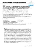
Early features of Kawasaki disease with pyuria in febrile infants younger than 6 months
Bạn đang xem bản rút gọn của tài liệu. Xem và tải ngay bản đầy đủ của tài liệu tại đây (599.76 KB, 5 trang )
Yoon et al. BMC Pediatrics
(2018) 18:389
/>
RESEARCH ARTICLE
Open Access
Early features of Kawasaki disease with
pyuria in febrile infants younger than 6
months
Seo Hee Yoon, Dong Soo Kim and Jong Gyun Ahn*
Abstract
Background: Children with Kawasaki disease (KD) and pyuria have been misdiagnosed with urinary tract infection
(UTI). We compared clinical and laboratory features at admission between two groups of infants under 6 months of
age who showed initial pyuria, to identify the initial clues suggestive of KD.
Methods: We retrospectively reviewed the medical records of children with fever who were under 6 months of age
with pyuria, over a 10-year period (2007–2017). We included infants with sterile pyuria who were finally diagnosed
with KD and those with UTI.
Results: During the period investigated, 12 (9.9%) KD patients with sterile pyuria and 378 infants with UTI were
included in this study. Older age (P < 0.01), a longer duration of fever; total and before admission (P < 0.01), more
negative nitrite test (P < 0.01), higher platelet count (P = 0.04), increased C-reactive protein (CRP) (P < 0.01) and
erythrocyte sedimentation rate (ESR) (P < 0.01), were identified as initial features of infants finally diagnosed with
KD. In the receiver operating characteristic analysis, optimal cut-off values of 509 k/μL for platelet count, 60 mg/L
for CRP, and 68 mm/H for ESR were selected. Patients with ESR > 68 mm/hr had a ninefold higher odds of KD
compared to those with lower ESR levels (odds ratio: 8.963, 95% confidence intervals: 1.936–41.493, P = 0.005),
whereas CRP and platelet count could not significantly increase in the odds of KD at a cut-off point.
Conclusion: Persistent fever, elevated ESR, and negative urine nitrite test can serve as early clues to suspect KD in
febrile infants with pyuria.
Keywords: Differential diagnosis, Fever, Infant, Kawasaki disease, Urinary tract infection
Background
Kawasaki disease (KD) is an acute systemic vasculitis of
unknown aetiology that affects infants and young children [1]. KD is characterized by inflammation of all the
medium-sized arteries and multisystem involvement including the kidney [2]. Renal symptoms in KD include
pyuria, prerenal acute kidney injury, haemolytic uremic
syndrome, acute nephritic syndrome, nephrotic syndrome, and renal tubular abnormalities [3]. Among
them, pyuria is the most frequent renal feature of KD
occurring in 30–80% of the patients [4].
* Correspondence:
Department of Pediatrics, Severance Children’s Hospital, Yonsei University
College of Medicine, 50-1 Yonsei-ro, Seodaemun-gu, Seoul 03722, South
Korea
Some KD children with pyuria have been mistakenly
attributed to urinary tract infection (UTI) [5, 6]. In particular, as infants under the age of 6 months with KD
showed higher rate of incomplete presentation as well as
pyuria [2, 7–9] that is a significant laboratory marker of
UTI with high occurrence rate in these ages [10], it is a
diagnostic challenge to distinguish between UTI and KD
with pyuria in infants aged < 6 month. Considering that
patients with KD younger than 6 months are also known
to have a higher risk of developing coronary complications [11, 12], it is necessary that clinicians, who evaluate
febrile infant under 6 months of age with pyuria, do not
miss the diagnostic potential of KD.
Therefore, it is crucial to find the initial differences
among patients with UTI and KD with pyuria to identify
KD in febrile infants with pyuria during the early phase
© The Author(s). 2018 Open Access This article is distributed under the terms of the Creative Commons Attribution 4.0
International License ( which permits unrestricted use, distribution, and
reproduction in any medium, provided you give appropriate credit to the original author(s) and the source, provide a link to
the Creative Commons license, and indicate if changes were made. The Creative Commons Public Domain Dedication waiver
( applies to the data made available in this article, unless otherwise stated.
Yoon et al. BMC Pediatrics
(2018) 18:389
of the disease. We studied the clinical and laboratory parameters of infants under 6 months of age who showed
fever and pyuria at admission, in order to find initial
clues for the early detection of KD that would allow
timely initiation of therapy.
Methods
We retrospectively reviewed medical records of patients
under 180 days of age with fever and pyuria who were
admitted to Severance Children’s Hospital between January 2007 and January 2017. Among these patients, we
included infants who were finally diagnosed as KD or
UTI. Diagnosis of KD was based on American Heart Association guidelines [13]. We also included incomplete
KD, which showed prolonged unexplained fever, fewer
than four of the major clinical manifestations, and compatible laboratory or echocardiographic findings [2]. The
definition of pyuria was based on urinary white blood
cell (WBC) count > 5 cells/high power field (HPF) and
sterile pyuria is defined as pyuria with a negative urine
culture [14, 15]. The diagnosis of UTI was based on the
culture of a single organism from a catheterized urine
culture with a colony count greater than 10,000 or from
a clean-catch midstream urine culture with greater than
100,000 colonies [15, 16]. Two or more bacterial species
isolated by culture were excluded. We only included patients with KD and with sterile pyuria in the analysis.
This study was approved by the Institutional Review
Board of our institution (Severance Hospital, IRB number: 4–2017-1199).
Page 2 of 5
Results
A total of 121 patients under 6 months of age were diagnosed with KD over the study period. Forty-one (33.9%)
of the 121 KD children had pyuria. Of the 41 pyuria patients, 12 had sterile pyuria, 7 had concomitant UTI, and
16 had combined bacterial growth which had to be excluded. The other 6 patients were unidentified because
they did not perform the urine culture tests. The clinical
presentations of the 12 KD infants with sterile pyuria are
summarized in Table 1. Two patients satisfied the diagnostic criteria of complete Kawasaki disease, whereas the
others were diagnosed with incomplete Kawasaki disease.
During the same period, 378 infants under 6 months of
age were diagnosed with UTI. Clinical and laboratory
findings between KD infants with sterile pyuria and UTI
infants were compared (Table 2). Median age of KD
group (5.2 months; range, 2.5–5.9 months) was older
than that of UTI group (median 3.6 months; range, 0.9–6
months) (P = 0.002). Male-to-female ratios in KD and UTI
infants were 1.4 (7/5) and 2.8 (277/101), respectively.
KD group showed longer duration of fever (before
admission and total) compared to UTI group (P <
0.01). KD group also showed a significantly higher
platelet count (P = 0.04), C-reactive protein (CRP)
level (P < 0.01), and erythrocyte sedimentation rate
(ESR) (P < 0.01) than UTI group.
UTI group showed higher presence of positive urine
nitrite test (P < 0.01). There were no significant differences in white blood cell count, absolute neutrophil
count, haemoglobin, aspartate aminotransferase, alanine
aminotransaminase, albumin, sodium, total bilirubin,
and urine β2-microglobulin (β2-MG) (Table 2).
Statistics
Demographic and clinical data are presented as median
(range) or frequency. Initial laboratory test data at admission were analysed. Categorical variables were compared using chi-squared test or Fisher’s exact test, and
continuous variables were compared by Mann-Whitney
test. To compare the predictive capacity of various laboratory tests for diagnosis of KD, receiver operating
characteristic (ROC) curves were analysed by estimating
the area under the curve (AUC). Optimal cut-off values
in the diagnosis of KD were determined according to
Youden index [17]. Patients were stratified into two
groups according to optimal cut-off values for analyses.
Logistic regression analyses were conducted using these
groups (higher vs. lower than cut-off value) to estimate
odds ratios and 95% confidence intervals. Statistical
analyses were performed using SPSS version 23.0 for
Windows (SPSS Inc., Chicago, IL, USA) and MedCalc
Statistical Software version 18.6 (MedCalc Software,
Ostend, Belgium). P < 0.05 was considered statistically
significant.
Table 1 Clinical characteristics of patients with Kawasaki disease
with sterile pyuria
Variables
n (%) or median (range)
Typical KD/Atypical KD
2/10 (16.7%/83.3%)
Female/male
7/5 (36.1%/63.9%)
Age (months)
5.2 (2.5–5.9)
Total duration of fever (days)
5.5 (3–13)
Duration of fever before admission (days)
4.5 (2–9)
IVIG usage (times)
1
11 (91.7%)
≥2
1 (8.3%)
Presence of coronary artery lesion (CAL)a
3 (25%)
Bilateral coronary ectasia
1 (8.3%)
Left main artery dilatation
1 (8.3%)
LAD dilatation
1 (8.3%)
KD Kawasaki disease, IVIG intravenous immunoglobulin, LAD left anterior
descending coronary artery, Data are presented as case number, percentages
or median (range)
a
CAL is defined according to the Japanese Ministry of Health and Welfare guidelines
Yoon et al. BMC Pediatrics
(2018) 18:389
Page 3 of 5
Table 2 Comparison between patients with Kawasaki disease and patients with urinary tract infection at admission
Parameter (Median, Range)
KD with Sterile Pyuria (n = 12)
UTI (n = 378)
P-value
Age (months)
5.2 (2.5–5.9)
3.6 (0.9–6)
.002
Male/Female
7/5
277/101
.321
Before admission
4.5 (2–9)
2 (1–8)
.000
Total duration
Duration of fever (days)
5.5 (3–13)
2 (1–16)
.000
WBC (/μL)
17,620 (7180–26,820)
15,130 (3390–33,480)
.232
ANC (/μL)
9390 (4010–15,490)
8035 (100–25,150)
.159
Hb (g/dL)
10.8 (9.5–11.7)
10.9 (8.0–13.3)
.293
Platelet count (k/μL)
532.5 (315–772)
442.0 (162–1492)
.040
CRP (mg/L)
74.6 (31.6–294.5)
33.7 (0.3–191.9)
.001
ESR(mm/hr)
80.0 (27–101)
31.0 (2–120)
.000
Na (mmol/L)
137 (135–141)
138 (114–142)
.352
AST (IU/L)
30 (18–178)
28 (13–507)
.642
ALT (IU/L)
21 (7–53)
22 (7–460)
.658
Albumin (g/dL)
3.9 (3.2–4.7)
3.9 (3.0–4.9)
.903
Total bilirubin (mg/dL)
0.3 (0.3–1.5)
0.4 (0.1–7.1)
.546
Urine β2-MG (mg/L)
0.2 (0.14–0.98)
0.2 (0.01–40.06)
.484
Urine nitrite test (+)
0 (0%)
201 (53.2%)
.000
KD Kawasaki disease, UTI urinary tract infection, WBC white blood cell, Hb haemoglobin, ANC absolute neutrophil count, CRP C-reactive protein, ESR erythrocyte
sedimentation rate, Na sodium, AST aspartate aminotransferase, ALT alanine aminotransaminase, β2-MG, β2-microglobulin. Data are presented as case number,
percentages or median (range)
We calculated the optimal cut-off values of platelet
count, CRP, and ESR for differentiating KD with pyuria
from UTI, by drawing ROC curves (Fig. 1). The cut-off
value of platelet count to predict KD with pyuria was 509
k/μL with 58.3% sensitivity, 76.5% specificity, 98.3% negative predictive value, and 7.3% positive predictive value (P
= 0.064). The cut-off value of CRP was 60 mg/L with
66.7% sensitivity, 76.1% specificity, 98.6% negative predictive value, and 8.2% positive predictive value (P < 0.001).
The cut-off value of ESR was 68 mm/H with 72.7% sensitivity, 85.3% specificity, 99.0% negative predictive value,
and 13.8% positive predictive value (P < 0.001). The area
under the curve (AUC) was 0.674 (95% confidence interval [CI]: 0.625–0.720, P = 0.064) for platelet count, 0.749
(95% CI: 0.703–0.791, P < 0.001) for CRP, and 0.846 (95%
CI: 0.804–0.882, P < 0.001) for ESR. Subjects with ESR >
68 mm/hr had a nine-folds higher odds of KD compared
to those with lower ESR level (odds ratio: 8.963, 95% confidence intervals: 1.936–41.493, P = 0.005) (Table 3).
Discussion
This study demonstrates that there are different initial
features at admission between KD and UTI groups.
Older age, longer duration of fever, higher platelet count,
higher level of CRP and ESR, and more urine negative
nitrite test were observed in KD group compared to UTI
Fig. 1 Receiver operating characteristic (ROC) curves for C-reactive
protein (CRP), erythrocyte sedimentation rate (ESR) and platelet
count for discriminating Kawasaki disease with pyuria (n = 12) from
urinary tract infection (n = 378). The area under the curve (AUC)
was 0.674 (95% confidence interval [CI]: 0.625–0.720, P = 0.064) for
platelet count, 0.749 (95% CI: 0.703–0.791, P < 0.001) for CRP, and
0.846 (95% CI: 0.804–0.882, P < 0.001) for ESR
Yoon et al. BMC Pediatrics
(2018) 18:389
Page 4 of 5
Table 3 Odds ratios for diagnosis of Kawasaki disease using
cut-off levels in febrile infants with pyuria
Variables
OR* (95% CI)
P-value
Platelet count (> 509 k μ/L)
2.299 (0.633–8.349)
.206
CRP (> 60 mg/L)
2.169 (0.523–8.998)
.286
ESR (> 68 mm/hr)
8.963 (1.936–41.493)
.005
OR odds ratio, CI confidence interval, CRP C-reactive protein, ESR erythrocyte
sedimentation rate. Odds ratio, 95% confidence interval and P value were
calculated by binary logistic regression analysis
*Higher vs. lower than cut off value
group. Our findings can provide early indicators for the
early detection of KD in febrile infants with pyuria.
In the present study, the median age of KD group (5.2
months; range, 2.5–5.9 months) was older than that of
UTI group (3.6 months; range, 0.9–6 months) (P =
0.002). The age difference could be attributed to the fact
that neonatal KD is extremely rare [18]. In a previous
study, authors examined patients with KD younger than
6 months and reported that the median age was 5
months (range, 2–6) [12], or a mean age was 4.6 ± 3.5
months (range, 2–6) [7]. This is similar to the age
range of the KD group in our results. Alternatively,
the pooled prevalence rates of febrile UTI aged under
3 months ranges 7.5% (female) to 20.1% (uncircumcised males) [10]. Therefore, pyuria occurring in a febrile infant less than at least 2 months is more likely
to be caused by UTI.
The diagnosis of KD is based on clinical criteria, but it
could appear and progress in several days after onset of
fever. Wu et al. [6] reported two cases of persistent fever
and pyuria which were the initial presentation without
signs suggestive of KD, and coronary artery abnormalities were noted in both cases. In our study, KD group
showed longer fever duration of total and before admission compared to UTI group. In the infants under aged
6 months, prolonged fever might be the only clinical
symptom of KD [2]. Thus, early suspicion of KD should
be considered in any infant with prolonged fever and
culture-negative pyuria.
The CRP, ESR, and platelet count were also higher in
KD group than in UTI group in this study. Elevation of
acute phase reactants such as CRP and ESR is characteristic laboratory findings of KD [2]. However, they are
general indicators of an acute inflammatory process and
also increased in UTI, especially upper UTI [19].
Thrombocytosis, classified as a secondary thrombocytosis, is also seen in the upper UTI as well as in KD [2,
20]. Gofrit et al. [21] reported thrombocytosis in a patient with upper UTI is not a random phenomenon, but
a marker of kidney obstruction or perinephric abscess.
Moreover, ESR > 68 mm/hr showed statistically significant increase in the odds of KD, whereas CRP and platelet count could not significantly increase in the odds of
KD at a cut-off point in our study. Therefore, increased
ESR itself could also be a good diagnostic marker for
KD. However, the laboratory markers should be interpreted cautiously and used as a supportive method to
diagnose patients KD using pyuria, in consideration of
nonspecificity.
In urinalysis findings except for pyuria, urine nitrite
test is helpful for diagnosis of UTI for its high specificity
[22]. In this study, UTI group showed higher presence of
positive urine nitrite test than KD group (P < 0.01).
Among UTI patients, 53.2% had positive nitrite test but
none in the KD infant. The findings suggest that urine
nitrite test could be a useful marker to exclude KD if it
is positive.
Sterile pyuria in KD was thought to be due to urethritis caused by a non-specific vasculitis of the urethra
and/or the kidney as a result of mild and sub-clinical
renal injuries [4, 23]. Urinary β2-MG is being used as a
useful indicator to check the function of the renal-urinary tract. In our study, urinary β2-MG is not statistically
different in KD and UTI groups. Choi et al. [24]
reported that urine β2-MG was elevated in patients with
KD and showed no difference between KD with pyuria
and without pyuria groups. This result indicates the
damage on renal function in most patients with KD.
A major limitation of our study is the small population
size, which may have affected our statistical results. Future analysis, with a large sample size, is needed to verify
the results of this study. Despite such shortcomings, our
data can still provide information regarding initial indicators of KD in febrile infants with pyuria, which will be
useful for clinicians.
Conclusions
Prolonged fever duration, elevated ESR, and negative
urine nitrite test can serve as early clues to suspect KD
in febrile infants with pyuria. Therefore, in patients presenting with these features as well as persistent fever
despite appropriate antibiotic treatment for UTI, echocardiography is warranted to identify the possible presence of KD.
Abbreviations
AUC: Area under the curve; CI: Confidence interval; CRP: C-reactive protein;
ESR: Erythrocyte sedimentation rate; HPF: High power field; KD: Kawasaki
disease; OR: Odds ratio; ROC: Receiver operating characteristic; UTI: Urinary
tract infection; WBC: White blood cell; β2-MG: β2-microglobulin
Acknowledgements
Not applicable.
Funding
This study has not been funded.
Availability of data and materials
The datasets used and/or analysed during the current study are available
from the corresponding author on reasonable request.
Yoon et al. BMC Pediatrics
(2018) 18:389
Authors’ contributions
SHY primary responsibility for data collection, preliminary data analysis and
writing the manuscript. DSK participated in the study design and data
evaluation. JGA supervised the design and execution of the study,
performed the final data interpretation and contributed the writing of
the manuscript. All authors have read and approved the final manuscript.
Ethics approval and consent to participate
This study was approved by the Institutional Review Board at Severance
Hospital (approval number: 4–2017-1199). Written consent was not necessary
for this study due to its retrospective nature.
Consent for publication
Not applicable.
Competing interests
The authors declare that they have no competing interests.
Publisher’s Note
Springer Nature remains neutral with regard to jurisdictional claims in published
maps and institutional affiliations.
Received: 28 April 2018 Accepted: 3 December 2018
References
1. Burns JC, Glode MP. Kawasaki syndrome. Lancet. 2004;364(9433):533–44.
2. McCrindle BW, Rowley AH, Newburger JW, Burns JC, Bolger AF, Gewitz M,
et al. Diagnosis, treatment, and long-term Management of Kawasaki
Disease: a scientific statement for health professionals from the American
Heart Association. Circulation. 2017;135(17):e927–e99.
3. Watanabe T. Kidney and urinary tract involvement in Kawasaki disease. Int J
Pediatr. 2013;2013:831834.
4. Watanabe T. Pyuria in patients with Kawasaki disease. World J Clin Pediatr.
2015;4(2):25–9.
5. Ristoska-Bojkovska N, Stavric K, Tasic V. Kawasaki disease misdiagnosed as
acute pyelonephritis. Pediatr Nephrol. 2003;18(8):851–2.
6. Wu CY, Hsieh KS, Chiou YH, Wang RS, Huang IF, Lee WY, et al. Prolonged
fever and pyuria: a urinary tract infection presentation of incomplete
Kawasaki disease. Acta Paediatr. 2005;94(3):375–7.
7. Chang FY, Hwang B, Chen SJ, Lee PC, Meng CC, Lu JH. Characteristics of
Kawasaki disease in infants younger than six months of age. Pediatr Infect
Dis J. 2006;25(3):241–4.
8. Yeom JS, Park JS, Seo JH, Park ES, Lim JY, Park CH, et al. Initial characteristics
of Kawasaki disease with cerebrospinal fluid pleocytosis in febrile infants.
Pediatr Neurol. 2012;47(4):259–62.
9. Liu HC, Lo CW, Hwang B, Lee PC. Clinical manifestations vary with different
age spectrums in infants with Kawasaki disease. ScientificWorldJournal.
2012;2012:210382.
10. Shaikh N, Morone NE, Bost JE, Farrell MH. Prevalence of urinary tract
infection in childhood: a meta-analysis. Pediatr Infect Dis J. 2008;27(4):302–8.
11. Rosenfeld EA, Corydon KE, Shulman ST. Kawasaki disease in infants less than
one year of age. J Pediatr. 1995;126(4):524–9.
12. Yoon YM, Yun HW, Kim SH. Clinical characteristics of Kawasaki disease in
infants younger than six months: a single-center study. Korean Circ J. 2016;
46(4):550–5.
13. Newburger JW, Takahashi M, Gerber MA, Gewitz MH, Tani LY, Burns JC, et al.
Diagnosis, treatment, and long-term management of Kawasaki disease: a
statement for health professionals from the committee on rheumatic fever,
endocarditis, and Kawasaki disease, council on cardiovascular disease in the
young, American Heart Association. Pediatrics. 2004;114(6):1708–33.
14. Hooker JB, Mold JW, Kumar S. Sterile pyuria in patients admitted to the
hospital with infections outside of the urinary tract. J Am Board Fam Med.
2014;27(1):97–103.
15. Jan SL, Wu MC, Lin MC, Fu YC, Chan SC, Lin SJ. Pyuria is not always sterile in
children with Kawasaki disease. Pediatr Int. 2010;52(1):113–7.
16. Robinson JL, Finlay JC, Lang ME, Bortolussi R. Urinary tract infections in
infants and children: diagnosis and management. Paediatr Child Health.
2014;19(6):315–9.
17. Youden WJ. Index for rating diagnostic tests. Cancer. 1950;3(1):32–5.
Page 5 of 5
18. Hangai M, Kubota Y, Kagawa J, Yashiro M, Uehara R, Nakamura Y, et al.
Neonatal Kawasaki disease: case report and data from nationwide survey in
Japan. Eur J Pediatr. 2014;173(11):1533–6.
19. Ayazi P, Mahyar A, Daneshi MM, Jahani Hashemi H, Pirouzi M,
Esmailzadehha N. Diagnostic accuracy of the quantitative C-reactive protein,
erythrocyte sedimentation rate and white blood cell count in urinary tract
infections among infants and children. Malays J Med Sci. 2013;20(5):40–6.
20. Garoufi A, Voutsioti A, Getsi VC, Zeis P, Karpathios T. Platelet changes in
urinary tract infections. Pediatr Res. 1999;45:903.
21. Gofrit ON, Shapiro A, Rund D, Verstandig AG, Landau EH, Katz R, et al.
Thrombocytosis accompanying urinary tract infection suggests obstruction
or abscess. Am J Emerg Med. 2006;24(1):118–21.
22. Roberts KB. Urinary tract infection: clinical practice guideline for the
diagnosis and management of the initial UTI in febrile infants and children
2 to 24 months. Pediatrics. 2011;128(3):595–610.
23. Watanabe T, Abe Y, Sato S, Uehara Y, Ikeno K, Abe T. Sterile pyuria in
patients with Kawasaki disease originates from both the urethra and the
kidney. Pediatr Nephrol. 2007;22(7):987–91.
24. Choi JY, Park SY, Choi KH, Park YH, Lee YH. Clinical characteristics of
Kawasaki disease with sterile pyuria. Korean J Pediatr. 2013;56(1):13–8.