Phân lập, định danh virus viêm gan vịt ở một số tỉnh đồng bằng sông cửu long và sản xuất kháng thể phòng bệnh tt tiếng anh
Bạn đang xem bản rút gọn của tài liệu. Xem và tải ngay bản đầy đủ của tài liệu tại đây (389.05 KB, 16 trang )
MINISTRY OF EDUCATION AND TRAINING
CAN THO UNIVERSITY
------
SUMMARY OF DOCTORAL THESIS
Major: Microbiology
Code: 62 42 01 07
PHẠM CÔNG UẨN
ISOLATION, IDENTIFICATION OF DUCK HEPATITIS VIRUS IN SOME
PROVINCES IN THE MEKONG DELTA AND PRODUCTION
OF ANTIBODIES TO PREVENT DISEASE
Can Tho, 2019
The thesis was completed at Can Tho University.
Scientific supervisors: Assoc. Prof. Dr. Ho Thi Viet Thu
Reviewer 1. Assoc. Prof. Dr ………
Reviewer 2: Assoc. Prof. Dr.............
Reviewer 3: Assoc. Prof. ………
The thesis is defended in front of the University Examination Council in Can Tho
University
Time:…… March ……, ………….
COUNCIL CHAIRMAN
Assoc. Prof. Dr………………….
Further information of the thesis could be found at:
1. Learning Resource Center of Can Tho University
2. National library of Vietnam
PUBLIC WORKS
1. Pham Cong Uan and Ho Thi Viet Thu, 2014. Isolation and identification of duck hepatitis virus type 1 in Hau
Giang Province. Can Tho University Scientific Journal. Special subject of Agriculture, 2:116-121
2. Pham Cong Uan and Ho Thi Viet Thu, 2016. An examination on the virulence and the pathogenicity of a duck
hepatitis A virus strain genotype 3 isolated in Hau Giang province. Can Tho University Scientific Journal. Special
subject of Agriculture, 42:7-10
3. Pham Cong Uan and Ho Thi Viet Thu, 2018. Examination on pathogenicty of duck hepatitis virus isolated from
ducks in Hau Giang Province. Can Tho University Scientific Journal. Special subject of Agriculture, 54 (1B): 1-6
BEGINING PART
CHAPTER 1. INTRODUCTION
1.1 The urgency of the study
Duck viral hepatitis is a highly fatal disease with high mortality, it rapidly spreads in ducking flocks, especially
in the Mekong Delta, but until now there has not had any research about this disease implemented in the Mekong
Delta. So, the disease remains the major threat to the health of ducks as well as affecting the economic result in the
region. So that the determination of major pathogenic viral strains, and using them as antigens in vaccine and antibody
production is necessary requirements in for the strategy of specific prevention and treatment in order to protect duck
health, to respond to the urgent request the study: "Isolation and identification of duck hepatitis virus in some
provinces in the Mekong Delta and production of antibodies to prevent disease" was carried out.
1.2 Objectives of the study
The goals of the study are:
To isolate and determinate viruses causing hepatitis outbreak in ducks from the field.
To study the genetic relationship about the origins of the hepatitis viruses isolated from diseased ducks.
To produce IgY antibodies from chicken egg yolk and examine its efficacy in prevention and treatment of duck
viral hepatitis
1.3 Research content
Contents 1: Isolating and identifying the viruses causing hepatitis for ducks in 5 provinces in the Mekong Delta.
Content 2: Study on the genetic relationship of the hepatitis duck virus strains isolated in 5 provinces in the
Mekong Delta
Contents 3: Surveying of pathogenicity of 1 represented DHAV-3 virus strain, symbol: DHAV-3-HG2.
Content 4: Production of duck hepatitis virus antibodies by IgY antibody production technology from chicken
yolks and its application in prevention and treatment.
1.4 Time and place of study
The study was carried out from 2012 to 2016. Suspicious viral hepatitis ducks were collected from Can Tho city
and Vinh Long, Tra Vinh, Kien Giang, Hau Giang province. Isolation and identification were done in virology
laboratories and molecular biology laboratories of Can Tho university. Nucleotide sequence was carried by Macrogen
cooperation (South Korea).
1.5 New contributions of the thesis
DHAV-3 is determinated the main cause of duck viral hepatitis in the Mekong Delta. DHAV-3 was quite
nucleotide homogeneous and separated into 1 group compared to other DHAV-3 strains in the other countries and in
Asia.
1
Successful production of purified IgY antibodies from strains DHAV-3 isolated in the Mekong Delta and it
showed effective in treatment and prevention.
1.6 The composition of the thesis
The thesis is 174 pages and consists of 5 chapters: introduction, materials and methods, results and
discussions, conclusions and suggestion, references and appendices. There are 31 tables, 38 figures and 119 references
documents.
CONTENT
CHAPTER 2: LITERATURE OVERVIEW
Duck hepatitis is caused by any of three different viruses, namely duck hepatitis type I, type II and type III.
Currently, 3 genotypes of duck hepatitis virus type I (DHAV) were identified. They are belong to Picornaviridae
family, Avihepatovirus genus. There are duck virus hepatitis type I genotype 1 (DHAV-1), duck viral hepatitis type I
genotype 2 (DHAV-2) and duck hepatitis virus type I genotype 3 (DHAV-3). Duck hepatitis virus type II is classified
as Duck Astrovirus type 1 (DAstV-1), viral hepatitis type III is Duck Astrovirus type 2 (DAstV-2) belong to
Astroviridae family. DAstV-1 is mainly reported in the UK, the DAstV-2 is only reported in the United States (OIE,
2010).
Mekong Delta is the most important duck production, duck viral hepatitis occur frequently and cause big loss
for duck raiser but there is not research on this disease and vaccine and antibody production. For proper prevention
and treatment, the identification of the currently circulating strain of the virus is common and is used as an antigen for
the hens to produce IgY antibody from egg yolk is necessary to develop optimal measures.
CHAPTER 3: STUDY METHODS
3.1. Method for isolation, identification and genetic analyzing of virus
3.1.1 Method of isolation of hepatitis virus on duck embryos
Ninety two specimens fluid (HDBP) from 92 duck diseased flocks were collected in the Mekong Delta were
injected into the allantoic cavity of the 12-day-old duck embryo, the specimens fluid of each duck is injected into one
embryo; then follow up: dead time of embryo; gross lesions on the embryo.
3.1.2 Methods of inoculating hepatitis virus on duck embryo fibroblast
Allantoic fluid and embryonic liver was homogenated in a suspension and continued to be inoculation into duck
embryo fibroblasts. Then, inoculated embryo fibrioblasts were collected for duck hepatitis virus identification and
quantification by RT-PCR.
3.2 Genetic analyzation of duck hepatitis virus type I genotype 3
3.2.1 Sequencing of nucleotides
Ten RT-PCR products, denoted DHAV-3-CT2, DHAV-3-CT2, DHAV-3-CT6, DHAV-3-CT6, DHAVDHAV-3-VL8, DHAV-3-VL8 in 63 strain isolates were used for nucleotide sequencing and for genetic analysis.
3.2.2 Genetic studies of isolates of DHAV-3
Genetic analyzation of 10 representative strains was performed by comparing nucleotide analogues, amino
acids with 31 strains in the World Gen Bank, inside 3 strains were isolated from Vietnam with the GenBank number
KU860090.1 , KU860089.1, JF914944.1; 5 strains were isolated from Korea and 23 strains were isolated from China.
Analysis was performed using BioEdit software and genealogy relationships were established using MEGA 5.1
software.
3.3 Virulent examination of 1 strain of DHAV-3-HG2 on embryos and ducklings
3.3.1 Quantification of tissue culture infective dose 50% (TCID50)
Experiment was conducted on 96 wells, virus fluid was diluted from 100 to 10-10, each diluted to 8 wells in the
same column, each well 100 l minimum essential medium, starting from concentration the highest dilution and 8
control wells, each well 100 l minimum essential medium. Calculate the TCID50 by the method of Reed and Muench
(1938). Tracking: TCID 50 /0.1ml.
3.3.2 Quantification of lethal dose 50% on duck embryos and pathological on the embryos.
Experimental design: Uses 50 ducks with 12-day-old embryos divided into 10 lots, each containing 5 duck eggs; 9
lots were injected virus fluid from 10-4 to 10-12 and 1 control were injected physiological aqueous solution. Tracking:
Embryo mortality; ELD50 ; Average embryo death time by concentration; Frequency of appearance of lesions on the
embryo.
2
3.3.3 Quantification of lethal dose 50% on ducks and pathological characteristics of virus.
Experimental design: Complete randomized design with 6 treatments (NT) with 3 replications; There were 5
treatments corresponding to the dilution of 10-1 to 10-5 and 1 control treatment only used physiological aqueous
solution, each using 5 ducks. The follow-up period was 14 days. Tracking: LD50; number of dead ducks; frequency
of appearance of symptoms; frequency of appearance of lesions; microscopic lesions on ducks.
3.4 Production of anti-DHAV-3 antibody by IgY antibody technology in chickens and use in prevention and
treatment
3.4.1 Production of anti-DHAV-3 antibody
3.4.1.1 Create immunity for hens
Experimental setting is as Table 3.1, the interval between 2 injections is 2 weeks
Table 3.1 Experimental design of hens immunity
Experiment
1
2
3
Treatment
Number of
chickens
Viral
dose
(ELD
50/ml)
Dose
(ml)
Route
Number of
injections
104/1
3
104
1
1
10 4/2
3
104
1
104/3
3
104
1
106/1
3
106
1
106/2
3
106
1
106/3
3
106
1
108/1
3
108
1
108/2
3
108
1
10 8/3
3
108
1
Control
3
PBS
1
Pectoral
muscle
Pectoral
muscle
Pectoral
muscle
Pectoral
muscle
Pectoral
muscle
Pectoral
muscle
Pectoral
muscle
Pectoral
muscle
Pectoral
muscle
Pectoral
muscle
2
3
1
2
3
1
2
3
3
3.4.1.2 IgY antibody extraction from egg yolk
IgY antibody from egg yolk is extracted by method of salt deposition (NH4)2SO4 saturation 40%, then
dialysis to remove the salt. Post-dialysis solutions were analyzed for protein concentration by Bradford method and
quantification of antibody titres IgY was determined by virus neutralization.
3.4.1.3 Examination of IgY antibody titres in hen and egg yolk
Antibody titres IgY was tested by viral neutralization.
Follow-up: The antigenic dose generated is best immunized; The number of times the antigen injection causes a
prolonged and stable immune response; The correlation between antibody titres IgY in hen serum and egg yolk.
3.4.1.4 Quantification safe dose of IgY antibody from egg yolk on duck embryo fibrioblast
Experimental design: The experiment was conducted on 96 wells with 10 concentrations of diluted IgY diluted
in 2 (1/2, 1/4, 1/8, 1/16, 1/32, 1/64 , 1/128, 1/256, 1/512, 1/1024), the initial IgY concentration was 13.5 mg/ml. Each
concentration was repeated 8 times. Negative control is a duck embryo fibrioblast that does not provide IgY injections
but only for the minimum essential medium. Follow up: Morphology of cell layer (with or without CPE and CPE
levels); OD value after MMT staining (showed the proportion of living cells).
3.4.1.5 Investigation of DHAV-3 resistance activity of IgY antibody on duck embryo fibrioblast at 24h before
and after infection
Experimental design: The experiment was performed on 96 well wells with four concentrations of diluted IgY
antibody for 10 yr from 100 to 10-3 and injected into DEF medium at 24h before and after inoculation DEF with
DHAV-3 fluid of dose 500TCID50/0.1ml/well. The initial concentration of IgY antibody yolk is x = 1.68 mg / ml, at
subsequent concentrations, diluted IgY antibody concentration is 10. Each concentration was repeated 20 times.
Nagative control is a duck embryo fibrioblast only for the minimum essential medium. Positive control is a duck
3
embryo fibrioblast that does not given IgY antibody but given virus fluid at 500TCID50/0.1ml /well. Follow up:
Morphology of cell layer (with or without CPE and CPE levels); OD value (showed he proportion of living cells).
3.4.1.6 Quanitification protection dose 50% for the 12-day-old duck embryo of yolk IgY antibody
The experiment was carried out on 12 day-old duck embryo and disign as Table 3.2. Embryo protection dose
50% (EPD50) ; Three doses of KT IgY is 10EPD50, 30EPD50, 50EPD50 were selected for the prevention and treatment
of DHAV-3 virus.
Table 3.2 Experimental design quanitification of protection dose 50% duck embryos of IgY antibody
Antibody
dilution
Viral dose
(LD 50 )
Number of duck
eggs have
embryos
Injection dose
(antibody + virus)
(ml)
Route
1 (10 0 )
10 2
5
0.1 + 0.1
Allantoic cavity
1/2 (10 -0.3 )
10 2
5
0.1 + 0.1
1/4 (10 -0.6 )
10 2
5
0.1 + 0.1
1/8 (10 -0.9 )
10 2
5
0.1 + 0.1
1/16 (10
-1,2
10
2
5
0.1 + 0.1
1/32 (10
-1.5
10
2
5
0.1 + 0.1
1/64 (10
-1.8
10
2
5
0.1 + 0.1
10
2
5
0.1 + 0.1
10
2
5
0.1 + 0.1
10
2
5
0.1 + 0.1
10 2
5
0.1 + 0.1
Allantoic cavity
Allantoic cavity
Allantoic cavity
Allantoic cavity
Allantoic cavity
Allantoic cavity
Allantoic cavity
Allantoic cavity
Allantoic cavity
Allantoic cavity
)
)
)
1/128 (10
-2.1
1/256 (10
-2.4
1/512 (10
-2.7
)
)
)
1/1024 (10 -3.0 )
3.4.2 IgY antibody application in prevention and treatment
3.4.2.1 IgY antibody application in prevention.
A stydy on the efficacy of IgY antibody in prevention of DHAV- 3 virus on ducklings was complete randomized
design with 3 dose and 3 repetitions, each treatment used 5 duck. Experiment is arranged as Table 3.3. At 24 hours
after duckling was supplied IgY by oral and intramuscular injection, the ducks were infected by DHAV-3 virus
(DHAV-3-HG2 strain) with dose virus 103.3LD50. Monitoring indicators: Ducks live in treatments after infected by
viral hepatitis.
Table 3.3 Prophylactic experiment from DHAV-3-HG2 with yolk IgY antibody
Repeatimes
Number of
ducks in
treatment
10 EPD 50
Dose of virus
challenge
(0.5ml/unit)
10 3.3 LD 50
3
5
30 EPD 50
10 3.3 LD 50
3
5
50 EPD 50
KTV 0.5 ml
10 3.3 LD 50
3
3
5
5
10 EPD 50
10 3.3 LD 50
3
5
30 EPD 50
10 3.3 LD 50
3
5
50 EPD 50
10 3.3 LD 50
3
5
KTV
0.5 ml
10 3.3 LD 50
3
5
3
5
3
5
Treat
Road level
Dose of antiboy
(0.5ml/unit)
1
Oral
2
Oral
3
4
5
6
7
8
Negative
control
Pasitive
control
Oral
Oral
Pectoral
muscle
Pectoral
muscle
Pectoral
muscle
Pectoral
muscle
Pectoral
muscle
Pectoral
muscle
0.5 ml of IgY(-)
0.5 ml of IgY(-)
10 3.3 LD 50
0.5 ml of PBS
10 3.3 LD 50
4
3.4.2.2 IgY antibody application in treatment
A stydy on the efficacy of IgY antibody in treatment of DHAV- 3 virus on ducklings was complete randomized
design with 3 dose and 3 repetitions, each treatment used 5 duck. Experiment is arranged as Table 3.4.
Table 3.4 DHAV-3 therapeutic experiment by KT IgY yolk and KTV antibody
Treat
The time
of KT
1
After 12
hours
2
After 12
hours
3
After 12
hours
4
After 12
hours
5
After 24
hours
6
After 24
hours
7
After 24
hours
8
After 24
hours
Pasitive
control
After 24
hours
Negative
control
After 24
hours
Route
Pectoral
muscle
Pectoral
muscle
Pectoral
muscle
Pectoral
muscle
Pectoral
muscle
Pectoral
muscle
Pectoral
muscle
Pectoral
muscle
Pectoral
muscle
Pectoral
muscle
Antibody
dose (0.5ml )
Viral dose
(0.5ml/unit)
Times
Repeat
Number of
ducklings
10 EPD 50
103.3 LD 50
3
5
30 EPD 50
103.3 LD 50
3
5
50 EPD 50
103.3 LD 50
3
5
KTV 1 ml
103.3 LD 50
3
5
10 EPD 50
103.3 LD 50
3
5
30 EPD 50
103.3 LD 50
3
5
50 EPD 50
103.3 LD 50
3
5
KTV 1 ml
103.3 LD 50
3
5
0.5ml IgY(-)
103.3 LD 50
3
5
3
5
0.5ml IgY(-)
0.5ml PBS
3.5 Data processing methods
To access world gene bank by the BLAST program. Comparison of nucleotide and amino acid by BioEdit
software. Genealogical scheme set by the MEGA software 5.1. Statistical processing software by Minitab 16.0.
Chapter 4. RESULTS AND DISCUSSION
4.1 Results of isolation and identification of hepatitis virus in some provinces of the Mekong Delta
4.1.1 Results of isolation of duck hepatitis virus on duck embryos
Table 4.1 showed that, out of 92 samples of virus fluid from 92 duck flocks disease, 78 samples killed the
embryos at an average of 84.8%. Time to kill embryos from 48-72 hours.
Table 4.1 Results of isolation of the hepatitis virus on duck embryos
Place
Number
of embryos
monitored
Embryonic Death Time (hours)
Injection
dose
(ml)
Kien
Giang
Hau
Giang
Can
Tho
Vinh
Long
Tra
Vinh
Total
18-24
Embryos
Ratio
dead
(%)
24-48
Embryos
dead
Ratio (%)
48-72
Embryos
Ratio
dead
(%)
72-96
Embr
Ratio
yos
(%)
dead
Total
embryos
dead
Ratio (%)
22
0.2
0
0
5
22.7
11
50.0
3
13.6
19
86.4
25
0.2
0
0
7
28.0
14
56.0
1
4,0
22
88.0
15
0.2
0
0
3
20.0
9
60.0
2
13.3
14
93.3
12
0.2
0
0
3
25.0
6
50.0
1
8.3
10
83.3
18
0.2
0
0
4
22.2
8
44.4
1
5.6
13
72.2
0
0
22
23.9
48
52.2
8
8.7
78
84.8
92
5
4.1.2 Results of transmission of hepatitis virus on duck embryo fibroblast
All 78 samples cause various types of cellular invasions, such as the gradual contraction of the cells, then
clustering or joining together to create intercellular cells and eventually necrotic cells forming melancholy.
4.1.3 Results of identification of hepatitis virus by molecular biology technique
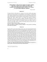
The RT-PCR produce amplifies the 286 bp gene from the nucleotide position of 3,527 to the nucleotide 3,777
of the P2 gene complex in the DHAV-3 genome.
Fig 4.1 Spectrometry of RT-PCR on agarose gel 1.5%
Note: M: Marker size 100bp. Wells 1, 2, 3, 4 (KG1, KG5, KG8, KG12). Wells 5, 6, 7 (HG2, HG4, HG5). Wells 8,
9, 10 (CT2, CT6, CT13). Wells 11, 12, 13 (VL1, VL3, VL8). Wells 14, 15, 16 (TV5, TV5, TV8).
Table 4.2 showed that, in 78 fluid cell cultures, 56 specimens were positive for primer DHAV-3F, DHAV3R, 71.8% (56/78) of which Kien Giang (84.2%), Hau Giang (81.8%), Tra Vinh (61.5%), Vinh Long (60.0%) and
Can Tho (57.1%). This study showed that DHAV-3 is prevalent in duck flocks in 5 provinces in the Mekong Delta.
DHAV-1, DHAV-2 and the type II hepatitis virus have not been detected.
Table 4.2 Percentage of positive dengue virus types
Place
DHAV-1
DHAV-2
(n) (+) %)
(n) (+)
Kien Giang
19
0
0
19
0
Hau Giang
22
0
0
22
Can Tho
14
0
0
Vinh Long
10
0
Tra Vinh
13
Total
78
DHAV-3
(%)
DAsTV-1
(n) (+) (%)
(n)
(+) (%)
0
19
16 84,2
19
0
0
0
0
22
18 81,8
22
0
0
14
0
0
14
8
57,1
14
0
0
0
10
0
0
10
6
60,0
10
0
0
0
0
13
0
0
13
8
61,5
13
0
0
0
0
78
0
0
78
56 71,8
78
0
0
4.2 Results of molecular genetic survey of DHAV-3 strains isolated
4.2.1 Results of nucleotide sequencing of RT-PCR products
Sequencing of 10 RT-PCR products out of a total of 56 products resulted in a 240 nucleotide fragment.
4.2.2 Comparison of nucleotide and amino acid sequences between 10 strains isolated from other strains in
the World Bank
DHA-3-CT2, DHAV-3-CT2, DHAV-3-H3, DHAV-3-HG2, DHAV-3 DHAV-3-TV5, DHAV-3-VL8 have
nucleotide analogues (98-100%) and amino acids (97-100%); DHAV-3-VL3 had nucleotide analogues (93-94%)
and amino acids (89-92%).
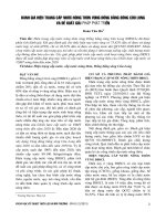
Fig 4.2 showed that 10 strains isolated in the Mekong Delta formed a separate group and closely related
their origin with DHAV-NT isolates in Ninh Thuan province of Vietnam and 3 strains from China DHAV -12-01,
DHAV-BN, DHAV-Du-090905. The DHAV-NT, DHAV-DN2 and DHAV-NC were isolated in different parts of
6
Vietnam outside the Mekong Delta in different branches. While, in the 23 strains of China and 5 strains of Korea
are also classified into different branches of the family tree.
Fig 4.2. The correlation coefficient between DHAV-3 strains varies with different DHAV-3 strains in the World
GenBank.
Note: * are the strains isolated in this study; ** are isolated from Vietnam.
4.3 Results of virulence survey of DHAV-3 on duck embryos and ducklings
4.3.1 Results of virulence and pathogenicity of DHAV-3 on duck embryos
4.3.1.1 Results of quanitification of lethal dose 50% of duck embryos.
Results of the lethal dose 50% of DHAV-3 virus on duck embryos were shown in 0.2 ml of suspension
containing 108.17 LD50 (ELD 50 = 10 8.17/ 0.2 ml)
7
4.3.1.2 Results of average embryo deaths by concentration
Table 4.3 Average embryo death time according to the concentration of virus fluid
Embryos die by time (hours)
Dilutio
Average
downtime
(hours)
Death
rate
(%)
<18
24
30
36
42
48
54
60
66
72
78
10 -4
0
1
2
2
0
0
0
0
0
0
0
24.8 ± 3.3
100
10 -5
10 -6
10 -7
10 -8
10 -9
10 -10
10 -11
10 -12
0
0
0
0
0
0
0
0
0
0
0
0
0
0
0
0
0
0
2
0
0
0
0
0
0
0
0
2
1
1
0
0
0
0
0
0
1
2
0
1
0
0
0
0
0
0
1
1
0
0
0
0
0
0
0
1
2
0
0
0
0
0
0
0
0
0
1
1
0
0
0
0
0
0
0
1
0
0
0
0
0
0
0
0
0
0
0
0
0
0
0
0
0
0
0
0
0
0
0
27.6 ± 3.9
33.4 ± 4.7
36 ± 4.58
39.3 ± 5.3
36.4 ± 5.0
0.0
0.0
0.0
0.0
100
100
80.0
60.0
20.0
0.0
0.0
0.0
0.0
Negative
control
Table 4.3 showed that at high virus concentrations from 10-4 - 10-6, the mortality rate was 100%, at the
concentration of 10-7 - 10-9, the rate of dying embryos gradually decreased and from 10-7 - 10-9 non-lethal embryos.
At the lowest mean embryo mortality level of 24.8 ± 3.3 hours, mean embryo death was highest at 10-8 (39.3 ± 5.3
hours).
4.3.1.3 Testicular lesions on duck embryos
Table 4.4. Frequency of lesions on duck embryos (n = 23)
Lesions
Stunting
embryos
Hemorrhagic
skin embryos
Embryonic
edema
Liver
enlarged and
hemorrhage
Yellowish
liver
Total
Ratio
(%)
Frequency of lesions in duck in concentration
10 -4
10 -5
10 -6
10 -7
10 -8
10 -9
10 -10
10 -11
10 -12
5
4
5
3
2
1
0
0
0
20/23
87.0
5
5
5
4
3
1
0
0
0
23/23
100
3
2
3
1
1
0
0
0
0
10/23
43.5
4
5
4
2
2
1
0
0
0
18/23
78.3
0
0
1
2
2
0
0
0
0
5/23
21.8
The results showed that the hemorrhagic skin embryos for the highest percentage (100%), followed by the
slow progression of stunted embryo (87%), liver enlarged and hemorrhage (78.26%), embryonic edema (43.5%),
yellowish liver (21.8%). These are typical clinical manifestations of DHAV-3 disease by examining embryonic dies
after infection.
4.3.2 Survey results virulence and pathogenicity of DHAV-3 on ducklings
4.3.2.1 Results of quanitification of lethal dose 50% on ducklings
Results of the virulent investigation of DHAV-3 virus in duckling showed that in 0.5ml of fluid there was
3.3
10 LD50 (LD 50/0.5ml = 10 3.3)
8
4.3.2.2. Investigation of duck mortality on average with different virus concentrations
Table 4.5 Average duck mortality according to virus concentration
Phase level
dilute virus
Average
duck dead
time
(hours)
Death
rate (%)
<24
24-48
48-72
72-96
96-120
120-144
10 -1
4
4
2
3
0
-2
0
48 ± 15.5
86.7
4
0
0
0
0
2
2
1
0
0
3
3
2
2
0
2
2
2
2
0
0
1
1
0
0
0
0
0
0
0
48 ± 15.5
60 ± 17.0
60 ± 17.0
48 ± 15.5
0.0
73.3
53.3
40.0
26.7
0.0
Number of dead ducks by time (hours)
10
10 -3
10 -4
10 -5
Negative
control
The highest mortality rate was recorded at 10-1 with a time of 48 ± 15.5 hours. Duck mortality rate diminished at
high dilutions and lowest in the 10 -5 (26.7%).
4.3.2.3 Examination of clinical symptoms in experimental ducks
Table 4.6 Frequency of occurrence of symptoms in ducks(n = 75)
Symptoms
Depression
Dry legs
Moody
Diarrhea with
white feces
Convulsions
Followed by
opisthotonus
and death
Runny nose
Frequency of occurrence of symptom
Total
Ratio
(%)
66.7
13.0
20.0
20.0
58/75
33/75
38/75
25/75
77.3
44.0
50.7
33.3
0
2
0,0
13.3
14/75
36/75
18.7
48.0
1
6.7
16/75
21.3
10-1
Ratio
(%)
10-2
Ratio
(%)
10-3
Ratio
(%)
10-4
Ratio (%)
10 -5
Ratio
(%)
14
12
12
8
93.3
80.0
80.0
53.3
12
8
10
5
80.0
53.3
66.7
33.3
12
7
8
5
80.0
46.7
53.3
33.3
10
4
5
4
66.7
26.7
33.3
26.7
10
2
3
3
5
11
33.3
73.3
5
9
33.3
60.0
3
7
20.0
46.7
1
7
6.7
46.7
6
40.0
5
33.3
3
20.0
1
6.7
Depression symptoms are high (77.3%); moody (50.7%); followed by opisthotonus and death (48%); dry
legs (44.0%); diarrhea with white feces (33.3%); runny nose (21.3%) and convulsions (18.7%).
4.3.2.4 Lesion survey on duck through surgical examination
Table 4.7 Frequency of occurence of lesions in ducks(n = 30 )
Frequency of occurrence of lesions by concentration
10 -2
Ratio
10 -3
Ratio
10 -4
Ratio
(%)
(%)
(%)
Tot
al
Ratio
(%)
100
30/
30
100
2
66.7
73.3
0.0
1
33.3
2
50.0
3
100
16.7
0
0.0
0
0,0
4
66.7
2
50.0
1
33.3
0.0
1
16.7
0
0.0
0
0.0
14.3
1
16.7
0
0.0
0
0.0
22/
30
5/3
0
19/
30
7/3
0
17/
30
2/3
0
3/3
0
Lesions
10 -1
Ratio
(%)
10 -5
Ratio
(%)
Enlarged livers with
ecchymotic
hemorrhages
Swollen bile ducts
10
100
7
100
6
100
4
100
3
8
80.0
6
85.7
4
66.7
2
50.0
Enlargement of kidneys
2
20.0
1
14.3
1
16.7
0
Enlargement of spleens
6
60.0
4
57.1
4
66.7
Discolored heart
muscles
Hemorrhages in lungs
3
30.0
3
42.9
1
5
50.0
5
71.4
Hemorrhages in
gizzards
Hemorrhages in
proventriculus
1
10.0
0
1
10.0
1
16.6
63.3
23.3
56.6
6.7
10.0
The most lesions on the duck were ecchymotic hemorrhages at 100%, swollen bile ducts (73.3%),
enlargement of spleens (63.3%) and hemorrhage in lungs (56.6%); swollen kidneys (16.6%) and the lowest was
discolored heart muscles (23.3%).
9
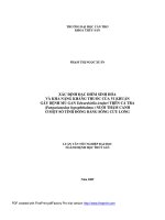
4.3.2.5 Microscopic lesion examination results
The results of the microscopic examination showed that all liver samples had haemorrhage in the surface of
the tissue, necrotizing hepatocyte , there were leukocytes in liver tissue, inflammatory hepatocytes, steatotic liver
cells. The connective tissue around the bile thickens, epithelial cells proliferate, peeling. Hepatic steatosis due to
dysfunction of lipid metabolism in liver, inflammatory reaction occurs at different levels, leading to infiltration of
many inflammatory cells and necrotizing hepatocyte. In the kidney, the Malpighi is small, with some white blood
cells in the kidney tissue, proximal convoluted tubule were necrosis..
4.4 Production of anti-DHAV-3 antibodies by IgY antibody technology in hens
4.4.1 Antibody titres of IgY in hen sera after immunization
Table 4.8 Mean IgY antibody titers in hen serum from experiments
Experiment
10
4
ELD50/ml
10
6
ELD50/ml
10
8
ELD50/ml
Treatment
Antibody titres of IgY over weeks after immunization
(xlog2)
A1
B1
C1
A2
B2
C2
A3
B3
C3
T1
T2
T3
T4
T5
T6
T7
T8
4,2
4,33
4.5
5.00
5,43
5.4
5,67
6.07
6,33
4,67
5,33
5,33
5,67
6.5
6,33
6.0
6.87
7.0
4,83
5.0
6.0
5,75
6.2
6,33
6,67
6,77
8,33
5,33
5,67
6.0
5.72
6,33
7,4
6,67
6,67
8.0
5,07
5,27
6,33
5.7
6.3
7,33
6.7
6.8
8,67
5,33
5.0
6,33
5,67
6,42
7,4
6,67
6.87
8,67
5.6
5,44
7.0
6.0
7,33
7,67
7,33
7,33
8.7
6,05
6.0
6,67
6.3
7.0
7,6
7,3
8.0
8,67
Antibody
titres of
IgY
medium
(xlog2)
5.0 d
5.3 d
6.0 bcd
5.7 cd
6.4 bc
6.9 b
6.62 bc
6.92 b
8.1 a
Note: The numbers in the same column with different caps are statistically significantl (P <0.05).A1, A2, A3: 1 immunogentic
times .B1, B2, B3: 2 immunogentic times.C1, C2, C3: 3 immunogentic times. T: Week
Table 4.8 showed that chickens in all three experiments have good immune response, and chickens are
immunized after 1 week of antibody titration. Chicken treatments were repeated twice, three times, the antibody
titre was significantly (P <0.05) higher than that of chicken administered only once. Particularly, in treatment with
dose injection 108ELD50/ml with 3 replications, the mean antibody titer in blood was 8.1 log2, which was
statistically significant (P <0.05 ).
4.4.2 Correlation between IgY content in chicken and chicken eggs after immunization
Table 4.9 showed that there is a positive correlation between the antibody titres in chicken blood and
corresponding egg yolk, a very positive correlation with correlation coefficient (0.92 r2 0.97).
Table 4.9 Antibody titres of IgY in blood and eggs after immunization
Treatment
G1
T1
G2
T2
G3
T3
G4
T4
G5
T5
G6
T6
G7
T7
G8
T8
G9
T9
Antibody titres over weeks after immunization (xlog2)
1
2
3
4
5
6
7
8
4.2
3.0
4.33
3.0
4.5
3.5
5.00
3.67
5.43
4,0
5.4
4.0
5.67
4.0
6.07
4.0
6.33
5.0
4.67
3.67
5.33
4.0
5.33
4.0
5.67
4.0
6.5
4.67
6.33
4.7
6.0
4.67
6.87
5.0
7.0
5.33
4.83
3.67
5.0
3,67
6.0
4.33
5.75
4.2
6.2
4.33
6.33
4.67
6.67
5.0
6.77
5.0
8.33
7.0
5.33
4,0
5,67
4,0
6.0
4,33
5.72
4,2
6,33
4,67
7.4
6.3
6,67
5.0
6,67
5.0
8.0
7.0
5.07
3.67
5.27
4.0
6.33
5.0
5.7
4.1
6.3
4.67
7.33
6.0
6.7
5.2
6.8
5.2
8.67
7.3
5.33
4.0
5.0
3.67
6.33
5.0
5.67
4.0
6.42
4.67
7.4
6.3
6.67
5.0
6.87
5.2
8.67
7.3
5.6
4.0
5.44
4.0
7.0
5,67
6.0
4.4
7.33
5,27
7,67
6.27
7.33
5.67
7.33
5.4
8.7
7.33
6.05
4.67
6.0
4.67
6.67
5.0
6.3
4,6
7.0
5.33
7.6
6.27
7.3
5.67
8.0
6.67
8.67
7.33
10
Correlation
coefficient
(r 2 )
0.92
0.94
0.92
0.94
0.92
0.97
0.96
0.95
0.97
Note: G1, T1: serum, chicken eggs one time; G2, T2: serum, chicken eggs immunized 2 times; G3, T3: serum, chicken eggs immunized 3
times with 10 4 ELD50/ml; G4, T4: serum, chicken eggs immunized 1 time; G5, T5: serum, chicken eggs immunized 2 times; G6, T6: serum,
chicken eggs immunized 3 times at a dose of 10 6 ELD50 /ml; G7, T7: serum, chicken eggs 1 time; G8, T8: serum, 2 immunogentic times; G9,
T9: serum, 3 immunogenic times at a dose of 10 8 ELD50 /ml
4.4.3 Results of extraction of IgY antibody from egg yolk by fractional salt filtration of ammonium sulphate
40%
After extraction and purification of IgY antibody, the result was an average of 63.2 mg/egg.
4.4.4 To investigate the toxicity of egg yolk IgY antibody to duck embryo fibroblast and DHAV-3 resistance on duck
embryo fibroblast.
4.4.4.1 Results of the quatification of the safe dose of the yolk IgY antibody for the duck embryo fibroblast.
Table 4.10 OD value and survival rate of live stem cells at dilutions of egg yolk IgY
Dilution of IgY
antibody fluid
1/2
1/4
1/8
1/16
1/32
1/64
1/128
1/256
1/512
1/1024
Control
Concentration of
antibody IgY
(mg / ml)
13.5
6,75
3,37
1,68
0,843
0.422
.210
0.105
0,052
0,026
OD value (
)
Average survival
rate (%)
0,088 c 0.006
C
0.107 0.016
C
0.108 0.014
0.109 c 0.015
0,154 b 0.022
0,214 a 0.016
0.221 a 0.032
0.234 a 0.020
0.237 a 0.029
0,238 a 0.018
0.239 a 0.032
36.8
44.8
45.2
45.6
64.5
89.5
92.5
97.9
99.2
99.6
100
Note: Numbers in the same column with different caps are significantly different (P <0.05).
Table 4.10 showed that dilution of 1/2, 1/4, 1/8 and 1/16, IgY fluid is still cytotoxic . From the 1/64
dilution and above, IgY was not affected by cell, the OD value was not statistically significant difference in
comparison with positive control (P> 0.05).
4.4.4.2 Results of the DHAV-3 activity of IgY antibody on duck embryo fibroblasts at 24 h before and after
infection
* The results of the study used IgY at 24 hours before infection
Table 4.11 OD values and survival rate of live cells at dilution concentrations of IgY antibody at 24 hours prior to
DHAV-3 infection
IgY antibody concentration
(mg / ml)
24 hours before DHAV-3 infection
OD value (
1,68
0,168
0,0168
0.00168
Negative control
Positive control
)
Average survival rate (%)
0.137 b 0.054
0.136 c 0.017
0,134 c 0.025
73.3
57.6
56.8
0.117 c 0.011
49.6
0.236 a 0.008
0.134 c 0.005
100
56.8
Note: Numbers in the same column bearing different caps are statistically significant (P <0.05).
Table 4.11 showed that only 0.68 mg / ml of IgY antibodies in the yolk concentration inhibited the growth
of DHAV-3 on duck embryo fibroblast, with OD = 0.137 and overage survival rate was 73.3%, statistically
significant with positive control (P <0.05). From 0.168mg /ml to 0.00168 mg/ml, egg yolk immunoglobulin did not
prevent the development of DHAV-3 on duck embryo fibroblast, because the OD value was not statistically
significant difference with positive control (P> 0.05).
11
* The results of the study used IgY at 24 hours after infection
Table 4.12 OD values and mean survival rates at antibiotic dilutions of IgY antibody at 24 h post DHAV-3 infection
After 24 hours DHAV-3 infection
IgY antibody concentration
Average survival rate
OD value (
)
(mg/ml)
(%)
1,68
56.7
0,108 b 0.033
0,168
41.3
0,081 c 0.025
0,0168
37.8
0,074 c 0.02
0.00168
37.2
0,073 c 0.031
Negative control
100
0.196 a 0.045
Positive control
44.4
0.087 bc 0.025
Note: Numbers in the same column with different caps are statistically significant (P <0.05).
Table 4.12 showed that dilutions of 1.68 mg / ml; 0,168 mg / ml; 0.0168 mg / ml and 0.00168 mg / ml were
not able to inhibit the growth of DHAV-3 on duck embryo fibroblast, OD values was not statistically significant
with positive control (P> 0.05).
Results from Table 4.11 and 4.12 showed that, with only 1.68 mg/ml to introduce IgY before 24 hours, IgY
antibody was able to protect embryonic fibroblast from hepatitis virus.
4.4.5 Application of IgY antibody in prevention and treatment of DHAV-3
4.4.5.1 Result of protection dose 50% duck embryos of IgY antibody
The results showed that in 0.1 ml contains 20 doses of protection (101.3 EPD50/ 0.1ml).
4.4.5.2 Results of duck hepatitis DHAV-3 virus prevention with IgY antibody yolk on 3-day-old ducklings
Table 4.13. Effectiveness of DHAV-3 prevention with IgY antibody
Dose
virus
(0.5ml/duck)
Treatment
Route
Dose
antibody
(0.5ml/duck)
1
2
3
Oral
Oral
Oral
10EPD 50
30 EPD 50
50 EPD 50
10 3.3 LD 50
10 3.3 LD 50
10 3.3 LD 50
6
5
2
4
Oral
KTV 0.5 ml
10 3.3 LD 50
4
10 EPD 50
10 3.3 LD 50
5
30 EPD 50
10 3.3 LD 50
10 3.3 LD 50
3
Negative
control
Intramuscul
ar injection
Intramuscul
ar injection
Intramuscul
ar injection
Intramuscul
ar injection
Intramuscul
ar injection
Positive
control
Intramuscular
injection
5
6
7
8
50 EPD 50
KTV
0.5ml
0.5 ml of
IgY(-)
0.5 ml of
IgY(-)
Number
of dead
ducks
Number
of
survival
ducks
9
10
13
11
Survival
duck rate
(%)
10
66.7 bef
12
80.0 becf
14
93.3 bcf
13
86.7 becf
15
100.0 c
2
15.3 d
`1
2
10 3.3 LD 50
0.5 ml of PBS
10 3.3 LD 50
0
13
60.0 ae
66.7 aef
86.7 acf
73.3 aef
Note: Numbers in the same column bearing different caps are statistically significant (P <0.05)
Results of Table 4.14 showed that the highest preventive effect of IgY fluid was at 50 EPD50 (86.7%)
compared with control but difference was not statistically significant (P>0.05). Providing antibody through the
intramuscular ịnection is more effective than oral.
12
4.4.5.3 Results of DHAV-3 virus treatment of IgY yolk antibody on 3 -days- old duckling
Table 4.14. Effect of DHAV-3 treatment with IgY antibody
Treatment
The time
of
antibody
1
After 12
hours
2
After 12
hours
Intramuscular
injection
Intramuscular
injection
After 12
hours
Intramuscular
injection
4
After 12
hours
Intramuscular
injection
5
After 24
hours
6
Route
IgY
antibody
dose
(0.5ml /
unit)
Viral
dose
(0.5ml /
unit)
Number
of dead
ducks
Number of
survival
duck
Survival
duck rate
(%)
10 EPD 50
10 3.3LD50
4
11
73.3 a
30 EPD 50
10 3.3LD50
3
12
80.0 ac
50 EPD 50
10 3.3LD50
2
13
86.7 ac
KTV
1 ml / child
10 3.3LD50
3
12
80.0 ac
Intramuscular
injection
10 EPD 50
10 3.3LD50
7
8
53.3 b
After 24
hours
Intramuscular
injection
30 EPD 50
10 3.3LD50
5
10
66.7 three
7
After 24
hours
Intramuscular
injection
50 EPD 50
10 3.3LD50
3
12
80.0 bac
8
After 24
hours
Intramuscular
injection
KTV
1 ml / child
10 3.3LD50
4
11
73.3 bac
Positive
control
After 24
hours
0.5ml of
IgY(-)
10 3.3LD50
15
0
0.0 d
Negative
control
After 24
hours
0.5ml of
IgY(-)
0.5ml
PBS
0
15
100.0 c
3
Intramuscular
injection
Intramuscular
injection
Note: Numbers in the same column with different caps are statistically significant (P <0.05)
IgY antibody given at 12 hours after infection with DHAV-3, the 50EPD50 dose is the most effective (86.7%).
Chapter 5. CONCLUSION AND SUGGESTIONS
5.1 Conclusion
Were isolated, the identification is 56 strains of DHAV-3 from 92 samples from 92 suspected viral hepatitis
ducks collected in 5 provinces in the Mekong Delta region accounted for 61.1% rate. In this study has not
discovered is DHAV-1, DHAV-2 and hepatitis virus type II from the isolated model. Duck hepatitis virus type I
genotype 3 is popularly circulated in the natural of the Mekong Delta area.
DHAV-3 strain isolated have close relationships with each other and the same cognate with DHAV-NT strain
(strain isolated from Vietnam) and 3 strains from China as DHAV-12-01, DHAV-B-N, DHAV-090905, nucleotide
similarity rate, reaching from 97-99%.
Highly pathogenic viruses on embryos have 108.17ELD50/0.2ml and 103.3LD50/0.5ml on ducklings. Embryos
were died after inoculation from 24-78 hours and caused embryonic lesions such as skin hemorrhage, liver is
enlarged and hemorrhages, embryos oedematose and palatal myocardial discoloration. The duckling has few
symptoms of spasmodic paddling of legs, lethargic depressionfollowed by opisthotonus and death convulsions and
liver swelling, liver hemorrhage, liver discoloration. .
Microscopic: haemorrhage in surface liver tissue, large leukocytes in hepatic tissue, inflammation of the liver
and severe degeneration of fat. Malpighi's is small, with some leuocyte in kidney tissue, proximal convoluted
tubule cells necrosis.
Successfully produced IgY antibody from egg yolk and effective use in prevention and treatment duck
hepatitis by using DHAV-3 as a darkening antigen for hens with a dose of 108 ELD50/ml and repeated injection 3
times had the best immune response and more stable compared.
Preventing and treating duckling diseases with IgY antibody by intramuscular injection is more effective than
oral administration. Prevention and treatment of DHAV-3 in the best duckling is 50EPD 50 (1 EPD50 with 0.135mg
IgY antibody yolk).
5.2 Suggestions
Continued isolation of other genotypes of the hepatitis D virus type 1, such as DHAV-1, DHAV-2, type II
and type III hepatitis viruses, to better understand the epidemiological and pathogenic nature of these viruses.
Hepatitis virus in the area .
13
Continued testing and prevention of hepatitis with yolk IgY antibody in the study of the topic on wild-type
duck flocks.
Research on the preparation of vaccines by virus strains currently circulating in the Mekong Delta in
particular and Vietnam in general to prevent disease for ducks.
Measures should be taken to prevent and control disease outbreaks through surveillance of offspring from
foreign countries and prevent ducks from contacting wild birds.
14