Transcutaneous bilirubin estimation in extremely low birth weight infants receiving phototherapy: A prospective observational study
Bạn đang xem bản rút gọn của tài liệu. Xem và tải ngay bản đầy đủ của tài liệu tại đây (2.27 MB, 5 trang )
Bhargava et al. BMC Pediatrics (2018) 18:227
/>
RESEARCH ARTICLE
Open Access
Transcutaneous bilirubin estimation in
extremely low birth weight infants
receiving phototherapy: a prospective
observational study
Vidit Bhargava1* , Daniel Tawfik1, Bruce Niebuhr2 and Sunil K. Jain3
Abstract
Background: Measurement of transcutaneous bilirubin (TcB) is a quick, reliable and painless method to guide
management of hyperbilirubinemia. Studies in term and late preterm infants have found that TcB measurements
from covered areas (TcB-C) during phototherapy (PHT) co-relate well with serum bilirubin levels. Limited data exists
in extremely low birth weight (ELBW) infants.
Methods: In this prospective observational study, an opaque patch was placed on the back of an ELBW infant prior
to initiation of PHT. TcB-C and TcB-E (TcB from exposed area) levels were measured at birth and at 24-h intervals for
5 days. Total serum bilirubin (TSB) levels were also measured within 30 min of obtaining TcB levels. A Wilcoxon
signed rank test was used for data analysis. A mixed effect model was used to adjust for repeated measurements
over time. The p value < 0.05 was considered significant.
Results: A total of 19 infants were enrolled in the study, with a mean gestational age of 26 ± 2 weeks and mean
weight 827 ± 127 g. The difference between TcB-C and TSB was 2.68 ± 2.41 mg/dl (mean ± SD, p < 0.001). In contrast,
the difference between TcB-E and TSB was − 0.51 ± 1.74 mg/dl (p = 0.02). TcB-C consistently overestimates TSB, while
TcB-E consistently underestimates TSB.
Conclusions: During PHT exposure, TcB-C does not correlate with TSB values in ELBW infants. TcB-C levels cannot be
used as a surrogate for TSB measurement in ELBW infants.
Keywords: Hyperbilirubinemia, ELBW, Kernicterus, Transcutaneous bilirubinometry
Background
Hyperbilirubinemia is seen in almost two-thirds of term
and more than two-thirds of all preterm infants [1]. The
incidence of kernicterus has dramatically decreased since
the onset of regular screening and aggressive management with phototherapy (PHT). In one post-mortem
series of premature infants, kernicterus was found to be
virtually non-existent [2]. The USA Kernicterus Registry
reported 125 infants ≥35 weeks estimated gestational
age with kernicterus/acute bilirubin encephalopathy
* Correspondence:
1
Department of Pediatrics, Division of Pediatric Critical Care, Lucile Salter
Packard Children’s Hospital, 770 Welch Road, Suite 435, Palo Alto, CA 94304,
USA
Full list of author information is available at the end of the article
between 1992 to 2004. No specific serum bilirubin
values coincided with onset of kernicterus in these
infants [3]. Hence, while the incidence has substantially
decreased, a considerable disease burden still exists in
term and preterm infants with hyperbilirubinemia. The
American Academy of Pediatrics Subcommittee on
Hyperbilirubinemia recommends that every infant be
screened for hyperbilirubinemia by TSB or TcB at 24 h
of life and with subsequent measurements guided by the
bilirubin level at 24 h of life and the presence of other
risk factors [4]. A recent NICHD Neonatal Research
Network study focused exclusively on the management
of hyperbilirubinemia in ELBW infants demonstrated
that aggressive management of hyperbilirubinemia with
© The Author(s). 2018 Open Access This article is distributed under the terms of the Creative Commons Attribution 4.0
International License ( which permits unrestricted use, distribution, and
reproduction in any medium, provided you give appropriate credit to the original author(s) and the source, provide a link to
the Creative Commons license, and indicate if changes were made. The Creative Commons Public Domain Dedication waiver
( applies to the data made available in this article, unless otherwise stated.
Bhargava et al. BMC Pediatrics (2018) 18:227
Page 2 of 5
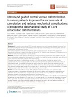
Fig. 1 Demonstrating photo-opaque patch placement in an ELBW neonate
PHT demonstrated significant benefits in neurodevelopmental outcomes [5].
Transcutaneous bilirubinometry is a quick, painless
and reliable alternative to serum bilirubin measurements
in the management of hyperbilirubinemia [6]. Following
PHT, TcB measurements are considered unreliable, as
PHT causes bleaching of the skin [7]. We and others
have identified a moderate correlation between TcB
measurements taken from skin covered by an opaque
patch and TSB levels in term and late preterm infants
following phototherapy [8, 9]; however, there is little
data on the reliability of TcB-C in ELBW infants. We
aim to investigate if TcB-C following PHT exposure is a
suitable surrogate for TSB in ELBW infants.
Methods
A prospective observational study was performed in
NICU at University of Texas Medical Branch (UTMB) at
Galveston after approval from the local institutional review board (IRB). Subjects were enrolled in the study
from January 2014 to June 2016. The study population
consisted of ELBW infants born at UTMB receiving
Table 1 Patient demographics
Sample size
19
Males
13 (68%)
Females
6 (32%)
Gestational age (in weeks)
26 ± 2
Birth weight (in grams)
827 ± 127 g
Race
19 (100%)
Caucasian
6 (31.6%)
African-American
3 (15.8%)
Hispanic
8 (42.1%)
Other
2 (10.5%)
phototherapy for hyperbilirubinemia. ELBW infants were
defined as infants with a birth weight of less than
1000 g. Following IRB approval, subjects were enrolled
in the study with parental assent. The requirement for
written consent was waved by the IRB at our institution.
In our NICU, all ELBW infants get prophylactic PHT for
first 5 days. All ELBW infants were eligible for the study.
Infants with congenital viral infections, conjugated
hyperbilirubinemia, sepsis, major congenital malformations, ABO/Rh incompatibility or gastrointestinal illness
were excluded from the study.
Prior to starting PHT, an opaque patch was placed on the
back of the infant. (Fig. 1) The forehead could not be used
to place the patch as most of these infants required CPAP.
After placing the opaque patch, PHT was started by using a
Giraffe Blue Lite PT system (GE Healthcare, Chicago, IL)
and continued for 5 days. TcB-C, TcB-E and TSB levels
were obtained at birth and every 24 h for 5 days by trained
NICU nurses. TcB-C was obtained from the skin covered
by the opaque patch. TcB-E was obtained from a site not
covered by the patch but adjacent to it. TcB measurements
were obtained using the Respironics™ BiliCheck noninvasive
bilirubin meter. Clinical data was collected from the electronic medical record (EMR) and included gestational age,
birth weight, sex, ethnicity, blood group, maternal blood
group, and ABO/Rh incompatibility, if any.
Serum bilirubin levels (TSB) were used as the gold
standard. The required sample size for each method was
calculated using a power analysis. The power analysis
indicated that 32 subjects (16 in each group) were
needed to have 80% power for detecting a medium-sized
effect (± 0.5 mg/dl) with p < 0.05 for statistical significance. A Wilcoxon signed rank test was used to compare the means of the absolute difference between
transcutaneous and serum bilirubin measurements for
each patient at each time point (24, 48, 72 and 96 h).
Bhargava et al. BMC Pediatrics (2018) 18:227
Page 3 of 5
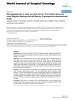
Fig. 2 Graph depicting the difference between TcB-C or TcB-E and TSB on various days
The absolute difference between transcutaneous and
serum bilirubin measurements for each patient is shown
on a Bland-Altman plot for easy visualization. Analysis
was performed using a mixed effect model to account
for repeated measures over time. All analysis was done
using JMP® Pro software.
Results
Twenty subjects were initially enrolled in the study. One
subject was excluded from the study due to neonatal
demise at day 2 of life. The mean birth weight was 827 ±
127 g, and the mean estimated gestational age was 26 ±2
weeks. Caucasians, Hispanics and African-Americans
constituted the majority of study group participants. Patient demographics are shown in Table 1. Differences between TcB-C or TcB-E and TSB are shown in Fig. 2.
A Wilcoxon signed rank test was used to compare the
TcB-C or TcB-E to TSB. The mean of difference between
TcB-C and TSB was 2.68 ± 2.41 mg/dl (p < 0.001, IQR
1.1–4.2) mg/dl. In contrast, the mean of difference between TcB-E and TSB was − 0.51 ± 1.74 mg/dl (p = 0.02,
IQR -1.77 – 0.47, shown in Table 2 and Fig. 3) The absolute differences between the TcB-C or TcB-E and TSB
for each patient were like the trends seen in the
Wilcoxon signed rank test and are depicted as Bland
Altman plots for easy visualization (Fig. 4). TcB-C overestimated serum bilirubin levels for most patients with a
difference of 2.71 ± 0.49 mg/dl (p < 0.001), while TcB-E
underestimated serum bilirubin levels with a difference
of − 0.51 ± 0.26 mg/dl (p = 0.07).
Similar results were obtained following adjustment of
the bilirubin estimation for repeated measures over time.
There was no effect of randomization of the day of bilirubin estimation on the overall results. The TcB-C continued to overestimate the TSB (p < 0.001), while TcB-E
continued to underestimate the TSB (p < 0.001). However, when the bilirubin estimation was randomized for
each patient the overall model improved by 18.9% and
this difference was found to be significant (p = 0.02).
Thus, TcB-C overestimates the estimation of bilirubin
compared to serum bilirubin, while TcB-E underestimates the estimation of bilirubin compared to serum
bilirubin. Hence, TcB-C and TcB-E may not be used as
surrogates for bilirubin estimation in ELBW infants receiving phototherapy.
Discussion
We evaluated the role of TcB estimation in ELBW infants who were receiving PHT. We found TcB-C did not
Table 2 Serum (TSB) and transcutaneous bilirubin
measurements (TcB-C/TcB-E)
SEM
P value
Mean
SD
IQ range (25–75%)
TcB-C – TSB
2.68
2.41
1.1–4.2
0.29
< 0.001
TcB-E – TSB
−0.51
1.74
−1.77 – 0.47
0.21
0.02
Bhargava et al. BMC Pediatrics (2018) 18:227
a
Page 4 of 5
b
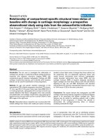
Fig. 3 a The probability density function and the mean of difference between the TcB-C and TSB. b The probability density function and the mean of
difference between the TcB-E and TSB. The vertical bar represents the mean compared to the normal distribution
correlate well with TSB levels in ELBW infants and, in
fact, were often higher than TSB. Within 24 h of PHT
exposure, TSB values decreased significantly compared
to the TcB-C values and thereafter the values decline
gradually. Skin exposure to PHT converts unconjugated
bilirubin into its water-soluble isomer by photoisomerisation. There is a continuous and bidirectional movement of bilirubin isomers between the blood and the
skin leading to skin bleaching [10]. During PHT exposure, TcB drops due to skin bleaching. But if a part of the
skin is covered by a photo-opaque patch bleaching will
be minimal. The lateral movement of bilirubin from exposed to the covered areas of skin is also minimal, thus
accounting for the higher bilirubin levels in the covered
areas of skin [11]. This finding in our study is similar to
a
b
Fig. 4 a A Bland Altman plot depicting the difference between TcBC and TSB for each patient. b A Bland Altman plot depicting the
difference between TcB-E and TSB for each patient
that reported by by Ozkan, et al., who noted that TcB-C
was slower to decline compared to TSB and TcB-E
values. In their study, TcB-E levels declined rapidly in
the first 6 h after starting PHT, while the decline in
TcB-C was not noticed until 12 h after starting PHT.
Serum bilirubin values were noted to decline gradually
during the study period.
In studies performed in term and preterm infants, the
TcB-C levels were found to be comparable with serum
bilirubin levels [8]. This was likely due to a gradual decline in TSB levels, permitting equilibration of TcB-C
with TSB. However, in our study TSB levels were significantly lower than TcB-C levels. This significant decline
in TSB in ELBW infants is likely multifactorial in nature.
First, bilirubin clearance depends on a multiple factors
including wavelength of light used for PHT, irradiance of
light, skin surface area exposed to PHT, and rates at
which bilirubin is removed from skin and blood [12].
Stratum corneum in ELBW is immature [13], allowing
rapid clearance of bilirubin from exposed areas of the
skin (bleaching effect) leading to a significant decline in
TcB-E levels. Second, since bilirubin levels in the skin
rapidly decline following initiation of phototherapy,
equilibration between the skin and serum leads to a
rapid decline in serum bilirubin levels.
Our study has a few limitations. The sample size of
the study population was small and the study was not
adequately powered to reliably predict secondary outcomes such as the correlation between TSB and TcB-E
values. We measured serum bilirubin at 24-h intervals
which may have affected the micro trends in bilirubin
levels via different methods during the first 24 h. More
frequent bilirubin sampling may be helpful in delineating
dermal bilirubin kinetics in ELBW infants.
Conclusions
We suggest that TcB-C is not be a helpful surrogate for
TSB in ELBW infants receiving PHT. Studies in larger
cohorts may be needed to further substantiate these
findings. In the future, we suggest measuring TcB and
TSB at shorter intervals and in larger cohorts to evaluate
the dermal bilirubin kinetics.
Bhargava et al. BMC Pediatrics (2018) 18:227
Abbreviations
NICU: Neonatal intensive care unit; PHT: Phototherapy; TcB-C: Transcutaneous
bilirubin estimation from covered areas of skin; TcB-E: Transcutaneous
bilirubin estimation from exposed areas of skin; TSB: Total serum bilirubin;
UTMB: University of Texas Medical Branch, Galveston
Acknowledgments
The authors would like to acknowledge and thank the NICU transport nurses
at UTMB for their sincere dedication and contribution in data collection for
the study. A special mention to Cheryl Napuli RN, Christie Talbert RN and
Regina Garrison RN for their help in completing the study.
Availability of data and materials
The datasets used and/or analysed during the current study are available
from the corresponding author upon reasonable request.
Authors’ contributions
VB was involved in patient enrollment, data collection, data interpretation
and writing the manuscript. BN was involved in the statistical analysis and
review of the manuscript. SKJ was involved in the study design, and
obtaining approval for the study. He was also involved in data interpretation
and review of the manuscript. DT was involved in the statistical analysis and
review of the manuscript. He was also involved in reviewing the revision of
the manuscript. All authors read and approved the final manuscript.
Ethics approval and consent to participate
The study was approved by the Institutional Review Board (IRB) at University
of Texas Medical Branch, Galveston, TX. The need for written consent was
waived by the IRB. Verbal parental assent was obtained prior to enrolling
infants in the study.
Consent for publication
Not applicable
Competing interests
The authors declare that they have no competing interests.
Publisher’s Note
Springer Nature remains neutral with regard to jurisdictional claims in published
maps and institutional affiliations.
Author details
1
Department of Pediatrics, Division of Pediatric Critical Care, Lucile Salter
Packard Children’s Hospital, 770 Welch Road, Suite 435, Palo Alto, CA 94304,
USA. 2Department of Pediatrics, University of Texas Medical Branch,
Galveston, TX, USA. 3Department of Pediatrics, Division of Neonatology,
University of Texas Medical Branch, Galveston, TX, USA.
Received: 21 December 2017 Accepted: 2 July 2018
References
1. Woodgate P, Jardine LA. Neonatal jaundice: phototherapy. BMJ Clin Evid.
2015;2015
2. Pearlman MA, Gartner LM, Lee K, Morecki R, Horoupian DS. Absence of
kernicterus in low-birth weight infants from 1971 through 1976: comparison
with findings in 1966 and 1967. Pediatrics. 1978;62(4):460–4.
3. Johnson L, Bhutani VK, Karp K, Sivieri EM, Shapiro SM. Clinical report from
the pilot USA kernicterus registry (1992 to 2004). J Perinatol. 2009;29(Suppl
1):S25–45.
4. Hyperbilirubinemia AAoPSo. Management of hyperbilirubinemia in the
newborn infant 35 or more weeks of gestation. Pediatrics. 2004;114(1):297–316.
5. Morris BH, Oh W, Tyson JE, Stevenson DK, Phelps DL, O'Shea TM, McDavid
GE, Perritt RL, Van Meurs KP, Vohr BR, et al. Aggressive vs. conservative
phototherapy for infants with extremely low birth weight. N Engl J Med.
2008;359(18):1885–96.
6. Bajpai PC, Agarwal SS, Kapoor CL, Krishna Murti CR. Skin as the site of
photoconversion of bilirubin in hyperbilirubinaemia of the new born. Indian
J Med Res. 1976;64(8):1214–9.
7. Tan KL, Dong F. Transcutaneous bilirubinometry during and after
phototherapy. Acta Paediatr. 2003;92(3):327–31.
Page 5 of 5
8.
9.
10.
11.
12.
13.
Fonseca R, Kyralessa R, Malloy M, Richardson J, Jain SK. Covered skin
transcutaneous bilirubin estimation is comparable with serum bilirubin
during and after phototherapy. J Perinatol. 2012;32(2):129–31.
Nagar G, Vandermeer B, Campbell S, Kumar M. Effect of phototherapy on the
reliability of transcutaneous bilirubin devices in term and near-term infants: a
systematic review and meta-analysis. Neonatology. 2016;109(3):203–12.
Ozkan H, Oren H, Duman N, Duman M. Dermal bilirubin kinetics during
phototherapy in term neonates. Acta Paediatr. 2003;92(5):577–81.
Hegyi T, Hiatt IM, Indyk L. Transcutaneous bilirubinometry. I. Correlations in
term infants. J Pediatr. 1981;98(3):454–7.
Hegyi T, Hiatt IM, Gertner IM, Zanni R, Tolentino T. Transcutaneous
bilirubinometry II. Dermal bilirubin kinetics during phototherapy. Pediatr
Res. 1983;17(11):888–91.
Kanti V, Bonzel A, Stroux A, Proquitté H, Bührer C, Blume-Peytavi U, Bartels
NG. Postnatal maturation of skin barrier function in premature infants. Skin
Pharmacol Physiol. 2014;27(5):234–41.