Is there an association between vitamin D deficiency and adenotonsillar hypertrophy in children with sleep-disordered breathing?
Bạn đang xem bản rút gọn của tài liệu. Xem và tải ngay bản đầy đủ của tài liệu tại đây (716.78 KB, 8 trang )
Shin et al. BMC Pediatrics (2018) 18:196
/>
RESEARCH ARTICLE
Open Access
Is there an association between vitamin D
deficiency and adenotonsillar hypertrophy
in children with sleep-disordered
breathing?
Ji-Hyeon Shin* , Byung-Guk Kim, Boo Young Kim, Soo Whan Kim, Sung Won Kim and Hojong Kim
Abstract
Background: Low vitamin D levels have been linked to the risk of sleep-disordered breathing (SDB) in children.
Although adenotonsillar hypertrophy (ATH) is the major contributor to childhood SDB, the relationship between
ATH and serum vitamin D is uncertain. We therefore investigated the relationship between vitamin D levels and
associated factors in children with ATH.
Methods: We reviewed data from all children with SDB symptoms who were treated from December 2013 to
February 2014. Of these, 88 children whose serum vitamin D levels were measured were enrolled in the study. We
divided the children into four groups based on adenoidal and/or tonsillar hypertrophy. We conducted a retrospective
chart review to analyze demographic data, the sizes of tonsils and adenoids, serum 25-hydroxy-vitamin D [25(OH)D]
level, body mass index (BMI), and allergen sensitization patterns.
Results: Children in the ATH group had a lower mean 25(OH)D level than did those in the control group (p < 0.05).
Children with vitamin D deficiencies exhibited markedly higher frequencies of adenoidal and/or tonsillar hypertrophy
than did those with sufficient vitamin D (p < 0.05). Spearman’s correlation analysis identified an inverse correlation
between serum 25(OH)D levels and age, tonsil and adenoid size, and height (all p < 0.05). In a multiple regression
analysis, tonsil and adenoid size as well as BMI-z score, were associated with 25(OH)D levels after controlling for age,
sex, height, and mite sensitization (p < 0.05).
Conclusions: Our results suggest that low vitamin D levels are linked to ATH. Both the sizes of the adenoids and
tonsils and the BMI-z score were associated with the 25(OH)D level. Therefore, measurement of the serum 25(OH)D
level should be considered in children with ATH and SDB symptoms.
Keywords: Vitamin D, Adenoids, Tonsils, Sleep-disordered breathing, Body mass index, Child
Background
The spectrum of sleep-disordered breathing (SDB) is characterized by snoring, mouth-breathing, and pauses in
breathing. SDB includes primary snoring, upper airway resistance syndrome, obstructive sleep apnea
(OSA), and obstructive hypoventilation. Children with
SDB not only experience sleep disturbances, but also
neurocognitive impairment and attention problems.
* Correspondence: ;
Department of Otolaryngology-Head and Neck Surgery, College of Medicine,
The Catholic University of Korea, 222 Banpo-daero, Seocho-gu, Seoul 06591,
Republic of Korea
Adenotonsillar hypertrophy (ATH), the primary cause
of OSA, is a common childhood disease that can be
surgically treated [1–4].
Vitamin D, a fat-soluble vitamin, is synthesized in the
skin upon exposure to sunlight and is also obtained from
foods. Low vitamin D levels have been linked to many
risk factors, including obesity, limited exposure to sunlight, prematurity, malabsorption, darkly pigmented skin,
aging, chronic use of steroids or anticonvulsants, and low
socioeconomic status [5–7]. In addition, several studies
have reported that vitamin D deficiency may increase the
risk of numerous acute/chronic otorhinolaryngologic
© The Author(s). 2018 Open Access This article is distributed under the terms of the Creative Commons Attribution 4.0
International License ( which permits unrestricted use, distribution, and
reproduction in any medium, provided you give appropriate credit to the original author(s) and the source, provide a link to
the Creative Commons license, and indicate if changes were made. The Creative Commons Public Domain Dedication waiver
( applies to the data made available in this article, unless otherwise stated.
Shin et al. BMC Pediatrics (2018) 18:196
conditions, including allergic rhinitis, chronic rhinosinusitis with nasal polyps, recurrent otitis media, acute respiratory infections, asthma, and benign paroxysmal positional
vertigo [8–13].
Chronically low vitamin D levels may also be associated with sleep disorders [14, 15]. Recent studies reported that low vitamin D levels were related to OSA,
and that continuous positive airway pressure treatment
increased vitamin D levels in adults with OSA [16, 17].
Vitamin D deficiency has been linked to increases in the
sizes of the tonsils and/or adenoids and thus to OSA development [18–20]. A decrease in vitamin D levels after
an inflammatory insult has also been reported [21, 22],
as has an association of low vitamin D levels and adenotonsillar diseases [23, 24]. In contrast, other studies
found no association between serum vitamin D levels
and such diseases [25, 26]. As the principal cause of vitamin D deficiency is inadequate exposure to sunlight,
these conflicting results may be explained by differences
in latitude and seasonal variations among studies. In
addition, differences in ethnicity and skin color may also
be in play [27–29].
In the present study, all subjects lived at the same latitude, were of the same ethnic group, and were evaluated
only during winter, therefore reducing potential variations attributable to differences in the abovementioned
factors. Our aim was to measure vitamin D levels and
analyze associated factors in children with SDB.
Methods
Subjects
We conducted a retrospective cross-sectional study at a
single, university-based, secondary referral hospital. We
recruited all children with SDB symptoms (e.g., snoring,
mouth- breathing, paused breathing, and excessive daytime sleepiness) who were treated from December 2013
to February 2014.
In 2012, the authors established critical pathways for
the clinical management of SDB, which state that the
work up for SDB includes a physical examination, lateral
plain X-ray of the nasopharynx, a quality of life evaluation using the Korean version [30] of the obstructive
sleep apnea (KOSA)-18 survey [31], allergy evaluation,
and measurement of the serum vitamin D level at our
outpatient clinic.
The inclusion criteria of the present study were: (1)
age 4–12 years; (2) habitual snoring, observed apnea,
and/or mouth- breathing during sleep at least 1 year in
duration; (3) total KOSA-18 score ≥ 60 (4) evaluation of
atopic status using the multiple allergen simultaneous
test (MAST); and (5) 25-hydroxy-vitamin D [25(OH)D]
level measurement. The exclusion criteria were: (1) any
craniofacial anomaly; (2) any anatomical abnormality, including nasal septal deviation, turbinate hypertrophy,
Page 2 of 8
and/or nasal polyps; (3) a recent history of nasal or
upper airway infection; (4) malnutrition; (5) the use of
vitamin D supplements or multivitamin agents; (6) a history of adenoidectomy and/or tonsillectomy; and/or (7)
the use of anti-inflammatory and/or anti-allergic drugs
within 4 weeks prior to enrollment.
We retrieved demographic, height, body weight, body
mass index (BMI), BMI z-score, tonsil and adenoid size,
atopic status, and serum vitamin D level data from medical records. We analyzed retrospectively collected data
without collecting blood samples by our research group.
We described the methods for in vitro IgE sensitization
testing and measurement of serum vitamin D levels to
clarify how these measurements have been obtained.
BMI was the body weight (kg) divided by the height
squared (m2). We used the Korean national 2007 growth
charts to determine BMI z-scores.
Tonsillar hypertrophy (TH) was graded using the
Brodsky scale [32], as follows: grade 0 (tonsils situated in
the tonsillar fossa); grade 1 (tonsils just outside of the
tonsillar fossa and occupying ≤25% of the airway); grade
2 (tonsils occupying 26–50% of the airway); grade 3
(tonsils occupying 51–75% of airway); and grade 4 (tonsils occupying > 75% of the airway). We used the
adenoidal-nasopharyngeal ratio (ANR,) obtained from a
lateral plain X-ray of the nasopharynx, to represent the
adenoidal size. The depths of the adenoids and nasopharynx were measured using the standard landmarks of
Fujioka [33]. The adenoids were measured by drawing
lines perpendicular to lines drawn along the straight region of the anterior margin of the basiocciput to the
point of maximal adenoidal convexity. The nasopharynx
was measured by drawing a line from the anterior inferior edge of the sphenobasioccipital synchondrosis to the
posterior superior margin of the hard palate. The ANR
was then determined by dividing the first measurement
by the second.
We defined grade 3 or 4 tonsils as TH. We defined an
ANR ≥ 0.8 as indicative of adenoidal hypertrophy (AH).
We then divided the children into four groups: control,
AH, TH, and ATH.
The Korean version of the obstructive sleep apnea (KOSA)-18
To assess quality of life, caregivers completed the
KOSA-18 questionnaire, a disease-specific questionnaire
validated in Korea. The 18 items of the KOSA-18 are
grouped within five domains (sleep disturbance, physical
symptoms, emotional distress, daytime function, and
caregiver concerns) and are scored using a 7-point ordinal scale, followed by summing of the scores. Possible
scores range from 18 to 126 points, with a higher score
indicating a worse quality of life. Franco et al. suggested
a clinical classification based on the OSA-18, with scores
< 60 suggesting a small impact on the health-related
Shin et al. BMC Pediatrics (2018) 18:196
quality of life, scores between 60 and 80 a moderate impact, and scores > 80 a large impact [31]. According to
this classification, we used the KOSA-18 as one of the
inclusion criteria and children with total scores of ≥60
were included in this study.
Determination of serum 25-hydroxy-vitamin D levels
To evaluate vitamin D status, serum levels of 25-hydroxyvitamin D (25(OH)D) were measured using a direct competitive chemiluminescence immunoassay (CLIA; LIAISON® 25 OH vitamin D assay; DiaSorin, Saluggia, Italy).
The intra- and interassay coefficients of variation for
25(OH)D were 3–6 and 7–11%, respectively.
Sensitization patterns of the allergens
In vitro IgE sensitization testing was carried out using the
multiple allergen simultaneous test (MAST) (RoboScreen™; Bee Robotics Ltd., Gwynedd, UK). The panel consists of 39 allergens, including foods, tree/grass/weed
pollens, fungi, dogs, cats, cockroaches, and house dust
mites. A score ≥ 2 was interpreted as positive [34].
Statistical analysis
Statistical analyses were performed using SPSS for Windows software (ver. 15.0; SPSS, Inc., Chicago, IL). Qualitative parameters were evaluated with a chi-square test,
and quantitative parameters using a Kruskal-Wallis test.
Factors associated with vitamin D deficiency were evaluated using Spearman’s correlation test. For multivariate
analysis, a multiple regression analysis was used. All statistical tests were two-tailed. A P-value < 0.05 was considered to indicate statistical significance.
Ethics statement
Written informed consent was not obtained because of
the retrospective nature of the study. However, the study
protocol was approved by our Institutional Review Board
(IRB policy NO. UC15RISI0035).
Page 3 of 8
Results
We included 88 patients [59 males (67.0%) and 29 females
(33.0%)] of mean age 8.9 ± 2.5 years. The mean serum
25(OH)D level was 19.4 ± 5.1 ng/mL. A serum 25(OH)D
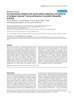
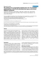
level < 20 ng/mL was considered to reflect a vitamin D deficiency [35]; 52.3% of the children were deficient. The frequency of AH and/or TH in children with vitamin D
deficiency and sufficiency was 91.3 and 71.4%, respectively. Deficient children exhibited markedly higher frequency rates of AH and/or TH than did those exhibiting
vitamin sufficiency (p = 0.035, Fig. 1).
Children with ATH had lower 25(OH)D levels
We compared the clinical characteristics of the control,
AH, TH, and ATH groups. The numbers of children per
group were as follows: control, 16 (18.2%); AH, 18
(20.4%); TH, 19 (21.6%), and ATH, 35 (39.8%). The children in the ATH group were younger than those in the
AH group (p = 0.021). The ATH group had more females
than the control and AH groups (p = 0.002 and 0.042,
respectively). We found no significant difference in
height, body weight, BMI, or BMI z-score among the
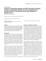
four groups (Table 1). The mean serum 25(OH)D levels
of the four groups were as follows: control, 22.5 ± 4.3;
AH, 18.7 ± 6.5; TH, 19.4 ± 4.5; and ATH, 18.4 ± 4.5 ng/
mL. The children in the ATH group had the lowest
mean 25(OH)D level (i.e., lower than that of the control
group [p = 0.01, Fig. 2]).
Allergen sensitization
A comparison of the atopic status among the four
groups showed that the mean number of sensitized allergens in the control, AH, TH, and ATH groups was 3.0,
2.3, 1.5, and 1.2, respectively. The mean was somewhat
higher in the control group than in the other groups,
but the difference was not significant. The prevalence of
atopy in the control, AH, TH, and ATH groups was
50.0, 77.8, 68.4, and 42.9%, respectively. The higher
prevalence of atopy in the AH group than in the other
groups was also not statistically significant.
Fig. 1 Comparisons of frequencies of adenoid and/or tonsillar hypertrophy by serum 25(OH)D level. Vitamin D-deficient: 25(OH)D < 20 ng/mL;
vitamin D-sufficient: 25(OH)D ≥ 20 ng/mL
Shin et al. BMC Pediatrics (2018) 18:196
Page 4 of 8
Table 1 Characteristics of 88 children with or without adenoid and/or tonsillar hypertrophy
Age (years)
Control
(N = 16)
Adenoid hypertrophy
(N = 18)
Tonsillar hypertrophy
(N = 19)
Adenotonsillar hypertrophy
(N = 35)
9.0 ± 2.3
10.9 ± 1.5*
8.9 ± 2.1
7.8 ± 2.9
*
Gender (male, %)
14 (87.5%)
Height (cm)
136.7 ± 14.2
*
14 (77.8%)
13 (68.4%)
18 (51.4%)
141.5 ± 23.9
137.8 ± 14.9
126.5 ± 21.5
Weight (kg)
35.3 ± 13.3
42.2 ± 24.4
40.1 ± 15.9
31.7 ± 18.8
BMI (kg/m2)
18.2 ± 3.3
19.6 ± 3.8
20.3 ± 4.1
18.2 ± 4.4
BMI z-score
0.1 ± 1.1
0.5 ± 0.8
0.8 ± 1.1
0.3 ± 1.1
25(OH)D
22.5 ± 4.3
18.7 ± 6.5
19.4 ± 4.5
18.4 ± 4.5
BMI body mass index, 25(OH)D serum 25-hydroxy-vitamin D
*
versus adenotonsillar hypertrophy group, p < 0.05
Negative association of age, tonsil size, ANR, and height
with serum 25(OH)D
We used Spearman’s correlation test to explore correlations between the serum 25(OH)D level and other variables (Table 2). Age (r = − 0.26, p = 0.001), tonsil size
(r = − 0.46, p = 0.002), ANR (r = − 0.40, p = 0.001), and
height (r = − 0.33, p = 0.020) were negatively associated with the serum 25(OH)D level. Body weight,
BMI, and BMI z-score also exhibited negative relationships, but these were not statistically significant.
Marked association of tonsil size and ANR with serum
25(OH)D
We used a multiple regression analysis to seek factors
associated with vitamin D level (Table 3). In model 1,
the serum 25(OH)D level was inversely associated
with tonsil size (β = − 0.41, p = 0.001), ANR (β = − 0.21,
p = 0.48), and BMI-z score (β = − 1.07, p = 0.029) after
adjusting for age and sex. These relationships persisted
even after further adjustment in model 2 (tonsil size,
β = − 0.40, p = 0.001; ANR, β = − 0.22, p = 0.043; and
BMI-z score, β = − 1.07, p = 0.001).
Discussion
OSA is associated with an increased risk of vitamin D
deficiency. Low vitamin D level increases the risk of
OSA by promoting ATH, airway muscle myopathy, and/
or chronic rhinitis [23, 36–38]. Recent studies in adults
showed that a large proportion of those with OSA also
had a vitamin D deficiency [39, 40]. ATH is the most
common cause of OSA in children. However, data on
the relationship between vitamin D deficiency and AH
and/or TH are conflicting [23–26]. In the present study,
we used only winter data from children of the same ethnicity (Korean) living at the same latitude (37° 76′ N) to
control for contributions made by these factors to the
extent of sunlight exposure. We found that the
25(OH)D level was reduced in children with ATH, AH,
or TH. The sizes of the adenoids and tonsils, and BMI-z
score predicted the serum 25(OH)D level.
Fig. 2 Serum 25(OH)D levels in children with or without adenoid and/or tonsillar hypertrophy. *: p < 0.05
Shin et al. BMC Pediatrics (2018) 18:196
Page 5 of 8
Table 2 Correlation coefficients for serum 25(OH)D levels by
Spearman’s rank correlation rho
r value
p value
Age
*
− 0.26
0.001
Tonsil size
−0.46*
0.002
ANR
*
−0.40
0.001
Height
−0.33*
0.020
Body weight
−0.26
0.060
BMI
− 0.16
0.270
BMI z-score
−0.07
0.580
The number of sensitized allergens
−0.08
0.61
ANR adenoidal-nasopharyngeal ratio, BMI body mass index
*
p < 0.05
We found that 52.3% of all children were vitamin
D-deficient. In a nationwide Korean cross-sectional survey, the prevalence of vitamin D deficiency in randomly
selected children was 18.4%, thus lower than that in our
study. However, the cited survey was conducted in autumn [41]. Another Korean study, conducted in autumn,
winter, and spring, found that 59.1% of all children were
vitamin D-deficient [42]. These among-study differences
are attributable to seasonal variations, participant age,
and the prevalence of underlying conditions.
We found that the sizes of the tonsils and adenoids
were negatively associated with the serum 25(OH)D
level. Several studies have reported relationships between low vitamin D levels and adenotonsillar diseases
[23, 24]. A Turkish study found that children with recurrent tonsillitis and allergic rhinitis had significantly lower
1,25-dihydroxyvitamin D [1,25(OH)2D] levels than controls [24]. However, it was not clear that the low vitamin
D levels were caused by the tonsillitis or allergic rhinitis,
and the seasons in which blood samples were collected
were not considered. A pilot study performed in the US
found no difference in the vitamin D levels of children
undergoing adenotonsillectomies and controls. However,
the study included children who underwent adenotonsillectomies not only because of obstruction but also to
treat recurrent infections. Again, the seasons in which
blood was collected were not reported [25]. As mentioned above, these conflicting results may be explained
by differences in latitude, season, ethnicity, and skin pigmentation [6, 29].
Vitamin D deficiency may increase ATH via inadequate regulation of the immune system. Vitamin D receptors are found on T cells, B cells, antigen-presenting
cells, macrophages, and dendritic cells. Vitamin D
immunomodulates both innate and adaptive immune responses [18, 43]. In terms of the innate immune system,
vitamin D increases the production of antimicrobial peptides, including defensin ß and cathelicidin [44, 45]. In
the adaptive immune system, the vitamin D inhibits the
proliferation of activated lymphocytes, reduces the production of inflammatory cytokines, and promotes the
development of induced regulatory T cells [46–48]. Vitamin D deficiency increases the risk of upper and lower
airway infections [49, 50]. Many studies have shown that
low vitamin D levels are associated with respiratory tract
infections and that vitamin D supplements exert beneficial effects during the treatment of infectious diseases
[51, 52], although some randomized controlled trials
found that vitamin D afforded no benefit in those
treated for infectious diseases [53–55]. A recent systematic review and meta-analysis reported that vitamin D
supplements had a protective effect against acute respiratory infection, particularly in patients with profound
vitamin D deficiency [12]. In terms of the effects of the
vitamin D on the adenoids and tonsils, a deficiency may
increase recurrent infections. In addition, vitamin D regulates human tonsillar T cells and a deficiency may trigger TH [18, 56]. Interestingly, recent studies suggested
that low vitamin D levels are the result rather than the
cause of the inflammatory process, as bacterial infection
may induce the intracellular conversion of 25(OH)D to
1,25(OH)2D, resulting in high 1,25(OH)2D and low
25(OH)D [57–59]. Therefore, the low vitamin D levels
in ATH patients may be a consequence of recurrent adenotonsillitis by bacterial infections.
Many studies have found that increased BMI is associated with vitamin D insufficiency in children [60, 61].
Table 3 Multiple regression models of serum 25(OH)D level
Parameter
Model 1a
Model 2b
Adjusted OR
95% CI
p value
Height
−0.40
−1.35 ~ 0.54
0.408
Sensitization to mites
−0.13
−0.33 ~ 0.08
0.238
Tonsil size
−0.41
−0.63 ~ − 0.19
0.001
ANR
− 0.21
−0.43 ~ − 0.01
0.048
BMI-z score
−1.07
−2.38 ~ − 0.23
0.029
OR odds ratio, CI confidence interval, ANR adenoidal-nasopharyngeal ratio, BMI body mass index
a
Adjusted for age and sex
b
Adjusted for age, sex, height and sensitization to mites
95% CI
p value
−0.40
− 0.62 ~ − 0.18
0.001
−0.22
− 0.43 ~ − 0.11
0.043
− 1.07
− 1.56 ~ − 0.58
0.001
Adjusted OR
Shin et al. BMC Pediatrics (2018) 18:196
Holick et al. [35] reported that the bioavailability of vitamin D in obesity was reduced because the vitamin was
deposited in the body fat. The 2003–2006 USA National
Health and Nutrition Examination Survey (which
assessed children and adolescents) found that vitamin D
deficiency was very prevalent in overweight and obese
children [62]. A study of Korean children also revealed
that the 25(OH)D level was lower in an overweight compared to a normal-weight group [63]. Consistent with
the results of these previous studies, we found that the
BMI-z score was negatively associated with the serum
25(OH)D level.
In terms of allergen sensitization, we found no significant difference in either the numbers of allergens to
which children were sensitized or the prevalence of
atopy among the four groups. Two explanations are possible. One is that the sensitivity of the MAST is low.
The other is that children with both allergic rhinitis and
turbinate hypertrophy were excluded. Thus, not all children with allergic rhinitis were included. Many studies
have found that low vitamin D levels are associated with
childhood allergic diseases, including allergic rhinitis,
asthma, and atopic dermatitis [64, 65]. A recent Australian study found that a low vitamin D level in early
childhood was associated with an increased risk of
asthma and early allergic sensitization [65]. In Korea, a
recent study showed that low vitamin D levels were associated with symptoms of allergic rhinitis and atopic
dermatitis [41]. However, some studies yielded different
results [66, 67]. A study of two large birth cohorts found
that vitamin D had no protective effect against asthma
or allergic rhinitis, and was positively associated with eczema, in 10-year-old children [66]. Thus, no conclusive
association has been demonstrated between vitamin D
and allergic disease.
A strength of our study is that it was conducted during
one season in children of the same ethnicity and living at
the same latitude. We thus controlled for several possible
confounders. Second, we evaluated allergen sensitization
patterns; other similar studies did not [20, 25]. Many studies have reported associations between vitamin D levels
and allergic diseases [68, 69]; an evaluation of atopic status
is essential when studying the effects of variations in vitamin D levels. Third, we defined clinical features predictive
of vitamin D deficiency. Physicians can easily measure the
sizes of the tonsils and adenoids, body weight, and BMI in
children with SDB.
However, there are some limitations to our study.
First, the sample size was too small to allow detailed
generalizations to be made. Second, we did not use polysomnography (PSG) for the evaluation of SDB. However,
although PSG is the gold standard for the diagnosis of
SDB, in practice, the test is time-consuming and cannot
be easily performed in all patients. A study in the USA
Page 6 of 8
showed that only 10% of children who underwent adenotonsillectomy also underwent a PSG evaluation [70].
In addition, Franco et al. reported that OSA-18 scores
correlated significantly with the respiratory distress
index determined by PSG [31]. We used the KOSA-18
score [30] as one of the inclusion criteria in our study
and included children whose health-related quality of life
was moderately to severely affected by OSA. Third, we
used the MAST rather than the skin prick test (SPT).
However, although the SPT remains a major diagnostic
tool, the MAST has the advantage that many allergens
can be tested simultaneously. Also, MAST data correlate
well with those of the SPT in rhinitis patients, which
suggests that the MAST can serve as an alternative to
the SPT [71]. Finally, we performed only a retrospective
chart review. Additional, larger studies incorporating
polysomnographic data may be required before general
conclusions can be drawn.
Conclusions
Approximately half of all children with SDB were vitamin D-deficient. The sizes of the adenoids and tonsils,
and BMI-z score were negatively associated with the
serum 25(OH)D level. Our results suggest that SDB children with vitamin D deficiencies may need to be evaluated in terms of AH and/or TH, and vice versa.
Abbreviations
1,25(OH)2D: 1,25-dihydroxyvitamin-D; 25(OH)D: Serum 25-hydroxy-vitamin D;
AH: Adenoidal hypertrophy; ANR: Adenoidal-nasopharyngeal ratio;
ATH: Adenotonsillar hypertrophy; BMI: Body mass index; KOSA-18
survey: Korean version of the obstructive sleep apnea-18 survey;
MAST: Multiple allergen simultaneous test; OSA: Obstructive sleep apnea;
PSG: Polysomnography; SDB: Sleep-disordered breathing; SPT: Skin prick test;
TH: Tonsillar hypertrophy
Acknowledgements
We thank Dr. Daeyoung Roh for contributing to the statistical analysis.
Availability of data and materials
The datasets used and/or analysed during the current study are available
from the corresponding author on reasonable request.
Authors’ contributions
JHS and BGK conceived and designed the study. JHS, BYK and HK
contributed to acquisition of the data. JHS, BYK, SWhK and SWoK analyzed
and interpreted the data. JHS drafted and revised the manuscript. All authors
involved in drafting the manuscript or revising it and approved the final
manuscript.
Ethics approval and consent to participate
The study protocol was approved by the Institutional Review Board of
Uijeongbu St. Mary’s Hospital (IRB policy No. UC15RISI0035). Since this study
is a retrospective chart review study the need for written consent was
formally waved by the IRB of Uijeongbu St. Mary’s Hospital.
Consent for publication
Not applicable
Competing interests
The authors declare that they have no competing interests.
Shin et al. BMC Pediatrics (2018) 18:196
Page 7 of 8
Publisher’s Note
Springer Nature remains neutral with regard to jurisdictional claims in
published maps and institutional affiliations.
22.
Received: 24 July 2017 Accepted: 14 June 2018
23.
References
1. Kobayashi R, Miyazaki S, Karaki M, Hoshikawa H, Nakata S, Hara H, et al.
Obstructive sleep apnea in Asian primary school children. Sleep Breath.
2014;18:483–9.
2. Kaditis AG, Alonso Alvarez ML, Boudewyns A, Alexopoulos EI, Ersu R,
Joosten K, et al. Obstructive sleep disordered breathing in 2- to 18-year-old
children: diagnosis and management. Eur Respir J. 2016;47:69–94.
3. Sedky K, Bennett DS, Carvalho KS. Attention deficit hyperactivity disorder
and sleep disordered breathing in pediatric populations: a meta-analysis.
Sleep Med Rev. 2014;18:349–56.
4. Waters KA, Chawla J, Harris MA, Dakin C, Heussler H, Black R, et al. Rationale
for and design of the “POSTA” study: evaluation of neurocognitive
outcomes after immediate adenotonsillectomy compared to watchful
waiting in preschool children. BMC Pediatr. 2017;17:47.
5. Bouillon R. Comparative analysis of nutritional guidelines for vitamin D.
Nat Rev Endocrinol. 2017;13:466–79.
6. Wacker M, Holick MF. Sunlight and Vitamin D: a global perspective for
health. Dermatoendocrinol. 2013;5:51–108.
7. Ariganjoye R. Pediatric Hypovitaminosis D: molecular perspectives and
clinical implications. Glob Pediatr Health. 2017; />2333794X16685504.
8. Akbar NA, Zacharek MA. Vitamin D: immunomodulation of asthma, allergic
rhinitis, and chronic rhinosinusitis. Curr Opin Otolaryngol Head Neck Surg.
2011;19:224–8.
9. Carroll WW, Schlosser RJ, O'Connell BP, Soler ZM, Mulligan JK. Vitamin D
deficiency is associated with increased human sinonasal fibroblast
proliferation in chronic rhinosinusitis with nasal polyps. Int Forum Allergy
Rhinol. 2016;6:605–10.
10. Walker RE, Bartley J, Camargo CA Jr, Flint D, Thompson JMD, Mitchell EA.
Higher serum 25(OH)D concentration is associated with lower risk of
chronic otitis media with effusion: a case-control study. Acta Paediatr. 2017;
106(9):1487–92.
11. Lee SB, Lee CH, Kim YJ, Kim HM. Biochemical markers of bone turnover in
benign paroxysmal positional vertigo. PLoS One. 2017;12:e0176011.
12. Martineau AR, Jolliffe DA, Hooper RL, Greenberg L, Aloia JF, Bergman P,
et al. Vitamin D supplementation to prevent acute respiratory tract
infections: systematic review and meta-analysis of individual participant
data. BMJ. 2017;356:i6583.
13. Martineau AR, Cates CJ, Urashima M, Jensen M, Griffiths AP, Nurmatov U,
et al. Vitamin D for the management of asthma. Cochrane Database Syst
Rev. 2016;9:Cd011511.
14. Ozgurhan G, Vehapoglu A, Vermezoglu O, Temiz RN, Guney A,
Hacihamdioglu B. Risk assessment of obstructive sleep apnea syndrome in
pediatric patients with vitamin D deficiency: a questionnaire-based study.
Medicine (Baltimore). 2016;95:e4632.
15. Zicari AM, Occasi F, Di Mauro F, Lollobrigida V, Di Fraia M, Savastano V, et al.
Mean platelet volume, vitamin D and C reactive protein levels in normal
weight children with primary snoring and obstructive sleep apnea
syndrome. PLoS One. 2016;11(4):e0152497.
16. Liguori C, Izzi F, Mercuri NB, Romigi A, Cordella A, Tarantino U, et al. Vitamin
D status of male OSAS patients improved after long-term CPAP treatment
mainly in obese subjects. Sleep Med. 2017;29:81–5.
17. Liguori C, Romigi A, Izzi F, Mercuri NB, Cordella A, Tarquini E, et al. Continuous
positive airway pressure treatment increases serum vitamin D levels in male
patients with obstructive sleep apnea. J Clin Sleep Med. 2015;11(6):603–7.
18. McCarty DE, Chesson AL Jr, Jain SK, Marino AA. The link between vitamin D
metabolism and sleep medicine. Sleep Med Rev. 2014;18:311–9.
19. Bozkurt NC, Cakal E, Sahin M, Ozkaya EC, Firat H, Delibasi T. The relation of
serum 25-hydroxyvitamin-D levels with severity of obstructive sleep apnea
and glucose metabolism abnormalities. Endocrine. 2012;41:518–25.
20. Kheirandish-Gozal L, Peris E, Gozal D. Vitamin D levels and obstructive sleep
apnoea in children. Sleep Med. 2014;15:459–63.
21. Reid D, Toole BJ, Knox S, Talwar D, Harten J, O'Reilly DS, et al. The relation
between acute changes in the systemic inflammatory response and plasma
24.
25.
26.
27.
28.
29.
30.
31.
32.
33.
34.
35.
36.
37.
38.
39.
40.
41.
42.
43.
44.
45.
25-hydroxyvitamin D concentrations after elective knee arthroplasty. Am J
Clin Nutr. 2011;93(5):1006–11.
Henriksen VT, Rogers VE, Rasmussen GL, Trawick RH, Momberger NG,
Aguirre D, et al. Pro-inflammatory cytokines mediate the decrease in serum
25(OH)D concentrations after total knee arthroplasty? Med Hypotheses.
2014;82(2):134–7.
Reid D, Morton R, Salkeld L, Bartley J. Vitamin D and tonsil disease–
preliminary observations. Int J Pediatr Otorhinolaryngol. 2011;75:261–4.
San T, Muluk NB, Cingi C. 1,25(OH)(2)D-3 and specific IgE levels in children
with recurrent tonsillitis, and allergic rhinitis. Int J Pediatr Otorhinolaryngol.
2013;77:1506–11.
Esteitie R, Naclerio RM, Baroody FM. Vitamin D levels in children undergoing
adenotonsillectomies. Int J Pediatr Otorhinolaryngol. 2010;74:1075–7.
Aydın S, Aslan I, Yıldız I, Ağaçhan B, Toptaş B, Toprak S, et al. Vitamin D
levels in children with recurrent tonsillitis. Int J Pediatr Otorhinolaryngol.
2011;75:364–7.
Schramm S, Lahner H, Jöckel KH, Erbel R, Führer D, Moebus S, et al.
Impact of season and different vitamin D thresholds on prevalence of
vitamin D deficiency in epidemiological cohorts-a note of caution.
Endocrine. 2017;56:658–66.
Fuleihan Gel H, Bouillon R, Clarke B, Chakhtoura M, Cooper C, McClung M,
et al. Serum 25-Hydroxyvitamin D levels: variability, knowledge gaps, and
the concept of a desirable range. J Bone Miner Res. 2015;30:1119–33.
Mazahery H, von Hurst PR. Factors affecting 25-Hydroxyvitamin D concentration
in response to vitamin D supplementation. Nutrients. 2015;7:5111–42.
Park CS, Guilleminault C, Hwang SH, Jeong JH, Park DS, Maeng JH.
Correlation of salivary cortisol level with obstructive sleep apnea syndrome
in pediatric subjects. Sleep Med. 2013;14(10):978–84.
Franco RA Jr, Rosenfeld RM, Rao M. First place–resident clinical science
award 1999. Quality of life for children with obstructive sleep apnea.
Otolaryngol Head Neck Surg. 2000;123:9–16.
Brodsky L, Moore L, Stanievich JF. A comparison of tonsillar size and
oropharyngeal dimensions in children with obstructive adenotonsillar
hypertrophy. Int J Pediatr Otorhinolaryngol. 1987;13:149–56.
Fujioka M, Young LW, Girdany BR. Radiographic evaluation of adenoidal size in
children: adenoidal-nasopharyngeal ratio. AJR Am J Roentgenol. 1979;133:401–4.
Nepper-Christensen S, Backer V, DuBuske LM, Nolte H. In vitro diagnostic
evaluation of patients with inhalant allergies: summary of probability
outcomes comparing results of CLA- and CAP-specific immunoglobulin E
test systems. Allergy Asthma Proc. 2003;24:253–8.
Holick MF, Chen TC. Vitamin D deficiency: a worldwide problem with health
consequences. Am J Clin Nutr. 2008;87:1080S–6S.
Atan Sahin O, Kececioglu N, Serdar M, Ozpinar A. The association of
residential mold exposure and adenotonsillar hypertrophy in children living
in damp environments. Int J Pediatr Otorhinolaryngol. 2016;88:233–8.
Dogru M, Suleyman A. Serum 25-hydroxyvitamin D3 levels in children with
allergic or nonallergic rhinitis. Int J Pediatr Otorhinolaryngol. 2016;80:39–42.
Prabhala A, Garg R, Dandona P. Severe myopathy associated with vitamin D
deficiency in western New York. Arch Intern Med. 2000;160(8):1199–203.
Piovezan RD, Hirotsu C, Feres MC, Cintra FD, Andersen ML, Tufik S, et al.
Obstructive sleep apnea and objective short sleep duration are
independently associated with the risk of serum vitamin D deficiency. PLoS
One. 2017;12(7):e0180901.
Salepci B, Caglayan B, Nahid P, Parmaksiz ET, Kiral N, Fidan A, et al. Vitamin
D deficiency in patients referred for evaluation of obstructive sleep apnea.
J Clin Sleep Med. 2017;13(4):607–12.
Yang HK, Choi J, Kim WK, Lee SY, Park YM, Han MY, et al. The association
between hypovitaminosis D and pediatric allergic diseases: a Korean
nationwide population-based study. Allergy Asthma Proc. 2016;37:64–9.
Roh YE, Kim BR, Choi WB, Kim YM, Cho MJ, Kim HY, et al. Vitamin D
deficiency in children aged 6 to 12 years: single center's experience in
Busan. Ann Pediatr Endocrinol Metab. 2016;21:149–54.
Rosendahl J, Holmlund-Suila E, Helve O, Viljakainen H, Hauta-Alus H,
Valkama S, et al. 25-hydroxyvitamin D correlates with inflammatory markers
in cord blood of healthy newborns. Pediatr Res. 2017;81(5):731–5.
Liu PT, Stenger S, Li H, Wenzel L, Tan BH, Krutzik SR, et al. Toll-like receptor
triggering of a vitamin D-mediated human antimicrobial response. Science.
2006;311(5768):1770–3.
Wang TT, Nestel FP, Bourdeau V, Nagai Y, Wang Q, Liao J, et al. Cutting
edge: 1,25-dihydroxyvitamin D3 is a direct inducer of antimicrobial peptide
gene expression. J Immunol. 2004;173(5):2909–12.
Shin et al. BMC Pediatrics (2018) 18:196
46. Xie Z, Chen J, Zheng C, Wu J, Cheng Y, Zhu S, et al. 1,25-dihydroxyvitamin Dinduced dendritic cells suppress experimental autoimmune encephalomyelitis
by increasing proportions of the regulatory lymphocytes and reducing T
helper type 1 and type 17 cells. Immunology. 2017;152(3):414–24.
47. Mansouri L, Lundwall K, Moshfegh A, Jacobson SH, Lundahl J, Spaak J.
Vitamin D receptor activation reduces inflammatory cytokines and plasma
MicroRNAs in moderate chronic kidney disease - a randomized trial. BMC
Nephrol. 2017;18(1):161.
48. Zhou Q, Qin S, Zhang J, Zhon L, Pen Z, Xing T. 1,25(OH)2D3 induces
regulatory T cell differentiation by influencing the VDR/PLC-γ1/TGF-β1/
pathway. Mol Immunol. 2017;91:156–64.
49. Ginde AA, Mansbach JM, Camargo CA Jr. Vitamin D, respiratory infections,
and asthma. Curr Allergy Asthma Rep. 2009;9:81–7.
50. Mora JR, Iwata M, von Andrian UH. Vitamin effects on the immune system:
vitamins a and D take Centre stage. Nat Rev Immunol. 2008;8:685–98.
51. Ginde AA, Mansbach JM, Camargo CA Jr. Association between serum 25hydroxyvitamin D level and upper respiratory tract infection in the third
National Health and nutrition examination survey. Arch Intern Med.
2009;169:384–90.
52. Ginde AA, Blatchford P, Breese K, Zarrabi L, Linnebur SA, Wallace JI, et al.
High-dose monthly vitamin D for prevention of acute respiratory infection
in older long-term care residents: a randomized clinical trial. J Am Geriatr
Soc. 2017;65(3):496–503.
53. Rees JR, Hendricks K, Barry EL, Peacock JL, Mott LA, Sandler RS, et al. Vitamin
D3 supplementation and upper respiratory tract infections in a randomized,
controlled trial. Clin Infect Dis. 2013;57(10):1384–92.
54. Li-Ng M, Aloia JF, Pollack S, Cunha BA, Mikhail M, Yeh J, et al. A
randomized controlled trial of vitamin D3 supplementation for the
prevention of symptomatic upper respiratory tract infections. Epidemiol
Infect. 2009;137:1396–404.
55. Somnath SH, Biswal N, Chandrasekaran V, Jagadisan B, Bobby Z. Therapeutic
effect of vitamin D in acute lower respiratory infection: a randomized
controlled trial. Clin Nutr ESPEN. 2017;20:24–8.
56. Nunn JD, Katz DR, Barker S, et al. Regulation of human tonsillar T-cell proliferation
by the active metabolite of vitamin D3. Immunology. 1986;59:479–84.
57. Mangin M, Sinha R, Fincher K. Inflammation and vitamin D: the infection
connection. Inflamm Res. 2014;63(10):803–19.
58. Waldron JL, Ashby HL, Cornes MP, Bechervaise J, Razavi C, Thomas OL, et al.
Vitamin D: a negative acute phase reactant. J Clin Pathol. 2013;66(7):620–2.
59. Custódio G, Schwarz P, Crispim D, Moraes RB, Czepielewski M, Leitão CB, et
al. Association between vitamin D levels and inflammatory activity in brain
death: a prospective study. Transpl Immunol. 2018; />trim.2018.02.014. Epub ahead of print
60. Szlagatys-Sidorkiewicz A, Brzezinski M, Jankowska A, Metelska P, SlominskaFraczek M, Socha P. Long-term effects of vitamin D supplementation in
vitamin D deficient obese children participating in an integrated weightloss programme (a double-blind placebo-controlled study) - rationale for
the study design. BMC Pediatr. 2017;17:97.
61. Barja-Fernández S, Aguilera CM, Martínez-Silva I, Vazquez R, Gil-Campos M,
Olza J, et al. 25-Hydroxyvitamin D levels of children are inversely related to
adiposity assessed by body mass index. J Physiol Biochem. 2018;74(1):111–8.
62. Turer CB, Lin H, Flores G. Prevalence of vitamin D deficiency among
overweight and obese US children. Pediatrics. 2013;131:e152–61.
63. Chung IH, Kim HJ, Chung S, Yoo EG. Vitamin D deficiency in Korean
children: prevalence, risk factors, and the relationship with parathyroid
hormone levels. Ann Pediatr Endocrinol Metab. 2014;19:86–90.
64. Aryan Z, Rezaei N, Camargo CA Jr. Vitamin D status, aeroallergen
sensitization, and allergic rhinitis: a systematic review and meta-analysis. Int
Rev Immunol. 2017;36:41–53.
65. Hollams EM, Teo SM, Kusel M, et al. Vitamin D over the first decade and
susceptibility to childhood allergy and asthma. J Allergy Clin Immunol.
2017;139:472–81.
66. Wawro N, Heinrich J, Thiering E, Kratzsch J, Schaaf B, Hoffmann B, et al.
Serum 25(OH)D concentrations and atopic diseases at age 10: results from
the GINIplus and LISAplus birth cohort studies. BMC Pediatr. 2014;14:286.
67. Cairncross C, Grant C, Stonehouse W, Conlon C, McDonald B, Houghton L,
et al. The relationship between vitamin D status and allergic diseases in
New Zealand preschool children. Nutrients. 2016;8:6.
68. Arikoglu T, Kuyucu S, Karaismailoglu E, Batmaz SB, Balci S. The association of
vitamin D, cathelicidin, and vitamin D binding protein with acute asthma
attacks in children. Allergy Asthma Proc. 2015;36(4):51–8.
Page 8 of 8
69. Chiu CY, Su KW, Tsai MH, Hua MC, Liao SL, Lai SH, et al. Longitudinal
vitamin D deficiency is inversely related to mite sensitization in early
childhood. Pediatr Allergy Immunol. 2017; />Epub ahead of print
70. Mitchell RB, Pereira KD, Friedman NR. Sleep-disordered breathing in
children: survey of current practice. Laryngoscope. 2006;116:956–8.
71. Cho JH, Suh JD, Kim JK, Hong SC, Park IH, Lee HM. Correlation between
skin-prick testing, individual specific IgE tests, and a multiallergen IgE assay
for allergy detection in patients with chronic rhinitis. Am J Rhinol Allergy.
2014;28:388–91.