Caffeine is a risk factor for osteopenia of prematurity in preterm infants: A cohort study
Bạn đang xem bản rút gọn của tài liệu. Xem và tải ngay bản đầy đủ của tài liệu tại đây (485.84 KB, 7 trang )
Ali et al. BMC Pediatrics (2018) 18:9
DOI 10.1186/s12887-017-0978-6
RESEARCH ARTICLE
Open Access
Caffeine is a risk factor for osteopenia of
prematurity in preterm infants: a cohort
study
Ebtihal Ali1,4* , Cheryl Rockman-Greenberg2,4, Michael Moffatt1,2,4, Michael Narvey2,4, Martin Reed3
and Depeng Jiang1
Abstract
Background: Caffeine, the most commonly used medication in Neonatal Intensive Care Units, has calciuric and
osteoclastogenic effects.
Methods: To examine the association between the cumulative dose and duration of therapy of caffeine and
osteopenia of prematurity, a retrospective cohort study was conducted including premature infants less than
31 weeks and birth weight less than 1500 g. Osteopenia of prematurity was evaluated using chest X-rays on a
biweekly basis over 12 weeks of hospitalization.
Results: The cohort included 109 infants. 51% had osteopenia of prematurity and 8% had spontaneous rib
fractures. Using the generalized linear mixed model, caffeine dose and duration of caffeine therapy showed a
strong association with osteopenia of prematurity. Steroids and vitamin D were also significantly correlated with
osteopenia of prematurity while diuretic use did not show a statistically significant effect.
Conclusion: The cumulative dose and duration of therapy of caffeine, as well as steroid are associated with
osteopenia of prematurity in this cohort. Future studies are needed to confirm these findings and determine the
lowest dose of caffeine needed to treat effectively apnea of prematurity.
Keywords: Premature infants, Osteopenia of prematurity, Metabolic bone disease, Caffeine
Background
Approximately 80% of bone mineralization of the newborn takes place during the third trimester of pregnancy
because of the high rate of intrauterine growth [1]. Thus,
preterm infants whom deprived of that period, are born
with less bone mineral content. In addition, physiological adaptation of bone to extra-uterine life leads to
an increase in bone resorption. This process occurs earlier in preterm than in term infants and can be accompanied by high risk of bone fragility and fractures [2].
Bone resorption appears to be more important than
* Correspondence:
1
Community Health Sciences Department, Faculty of Health Sciences,
University of Manitoba, MS361K, 820 Sherbrook St, Winnipeg, MB R3A 1R9,
Canada
4
Child Health Program, Winnipeg Regional Health Authority, Winnipeg, MB,
Canada
Full list of author information is available at the end of the article
decreased bone formation in the pathogenesis of osteopenia of prematurity (OP) [3].
Almost 10% of infants are born prematurely worldwide,
representing more than 15 million births every year. The
incidence and severity of osteopenia of prematurity increase as the birth weight (BW) and gestational age (GA)
decrease [4]. Preterm infants are known to have a lower
bone density (BMD) and bone mineral content (BMC) [2]
at the corrected age of term, as well as a lower weight and
Ponderal index [5]. Moreover, preterm infants have lower
bone strength at the distal tibia and radius compared to
age and sex-matched controls, when assessed with
computerized tomography as young adults [6].
In 1989, the incidence of OP was 55% of infants
<1000 g and 23% of infants <1500 g at birth. A notable
finding at this time was that OP risk showed an inverse
relationship to lower GA and a direct relationship to
duration of parenteral nutrition [7]. In 2009, a study
© The Author(s). 2018 Open Access This article is distributed under the terms of the Creative Commons Attribution 4.0
International License ( which permits unrestricted use, distribution, and
reproduction in any medium, provided you give appropriate credit to the original author(s) and the source, provide a link to
the Creative Commons license, and indicate if changes were made. The Creative Commons Public Domain Dedication waiver
( applies to the data made available in this article, unless otherwise stated.
Ali et al. BMC Pediatrics (2018) 18:9
Page 2 of 7
reported pathological fractures in 30% of preterm infants
with osteopenia [8].
Caffeine is the most commonly consumed pharmacologically active compound in the world [9]. In the neonatal
intensive care units (NICU), it is one of the most commonly prescribed drugs to treat apnea of prematurity [10].
The half-life in neonates is 72–96 h (range: 40–230 h) and
the time to peak serum concentration after oral administration ranges from 30 min to 2 h, whereas 86% of caffeine
is excreted unchanged in urine [11]. The liver enzymes responsible for caffeine metabolism mature progressively
with increasing GA. Girls were reported to have a higher
rate of caffeine metabolism than boys [12]. Clearance of
caffeine in infants born prematurely is markedly lower
and the volume of distribution is higher than infants at
term-equivalent age and beyond. Elimination of caffeine is
initially depressed in extremely premature infants and
then increases nonlinearly to final assessment at 6 weeks
postnatal age [13]. It is well established that caffeine
causes calciuria and creates negative calcium balance in
preterm rats especially after prolonged use with compensatory increase in PTH to normalize serum calcium at the
expense of bone [14–16]. Tolerance to the renal effects of
caffeine does not develop with chronic use [17].
In a study in mice, it was found that caffeine effectively enhanced the osteoclastogenesis from bone marrow
hematopoietic cells and bone resorption activity as
assessed by the pit formation assay [18]. In another
study, BMD was significantly lower in growing rats
supplemented with 0.2% caffeine in diets for 20 weeks
compared with the control group. Additionally, the calcium content in tibiae and femora of caffeine-treated
rats was also lower, and the osteoclastogenesis of bone
marrow cells isolated from caffeine-treated rats was
markedly enhanced as compared with that in the control
group. Taken together, these results suggest that caffeine
reduces BMD through the enhancement of osteoclastogenesis and its calciuric effect [19].
Based on existing studies we hypothesize that caffeine
usage, cumulative dose or duration of usage are associated with OP. and this association exists even when controlled for the effects of other neonatal risk factors.
The primary outcome of this study was to determine
the effect of the cumulative dose and the duration of caffeine on OP. Other covariates of interest were included
in the analysis, steroids and diuretics cumulative dose
vitamin D intake, and maternal parity.
least 12 weeks of hospital stay. It is difficult to implement case control study having infants with no caffeine
intake as all admitted infants less than 33 weeks are on
caffeine by hospital guidelines. We excluded infants with
congenital anomalies, infants with gut surgery affecting
feeding, infants with non-osteopenic fractures, and
infants with insufficient data to analyze. The data were
collected from the charts in the medical record. The
study included 109 infants who met the inclusion
criteria. Cases of osteopenia were defined if they have
radiological evidence of osteopenia of prematurity.
The data included: GA in weeks, gender, birth weight,
average biweekly weight, total parenteral nutrition
(TPN) days, and maternal parity level. The later was recorded as categorical data; high if >5, moderate if 3 or 4
and low parity if 1 or 2. Average biweekly vitamin D intake was included as longitudinal data. Serum phosphate
measurements were collected on biweekly basis
+/−1 week. The phosphate level was recorded as categorical data; high if >2.5 mmol/l, normal if between 1.8
to 2.5 mmol/l, low if between 1.3 to 1.8 mmol/l and very
low if <1.3 mmol/l. The radiological data (X rays) were
reviewed and interpreted, by a pediatric radiologist and
the writer, (the Cohen’s kappa was 0.83 and 95% CI 0.82
to 0.084, which indicates very good interrater agreement) [20] both did not know the infants ‘clinical status
or biochemical data at the time of the interpretation, on
a biweekly basis at least for the first 12 weeks of life,
using Koo et al. criteria [21]. Table 1.
The descriptive statistics (means and standard deviations) or (median and quartile) were used to summarize
the characteristics of the sample. As the grade level of
bone of newborn infants was measured fortnightly from
birth to 12 weeks old, the binary outcome variables (OP)
(0, 1), are longitudinal with up to 7 time points. It was
preferable to include grade 1 and 2 of OP together, as
the differentiation between the two grades is very subjective. Grade 3 OP was easier to distinguish, as callus
formation was indicative of previous underlying spontaneous fracture. Due to the limited sample size, we dichotomized the radiological grading of OP by collapsing
grades 1, 2 and 3 together as OP. At the same time, we
Methods
This retrospective quantitative descriptive pilot cohort
study was conducted at Health Sciences Centre in Winnipeg, Manitoba, Canada, from October 2007 to June
2012. Premature infants <31 weeks gestation and birth
weight < 1500 g infants were included, all infants had at
Table 1 Koo et al. Criteria for osteopenia of prematurity
Grades
Description
Grade 0:
Normal density of bone cortex along shaft with normal
dense white line at metaphysis and normal band of
lucency, and thinning of cortex.
Grade 1:
Loss of dense white line at the metaphysis, increased
sub-metaphyseal lucency and thinning of cortex.
Grade 2:
Changes in grade 1 plus irregularity and fraying of
metaphysis, with splaying and cupping that is indicative
of rickets.
Grade 3:
Indications of rickets with evidence of fractures.
Ali et al. BMC Pediatrics (2018) 18:9
Page 3 of 7
considered grade 0 as normal. We assessed the OP status for every two weeks, Therefore, the generalized linear mixed model was used for repeated measures of
binary outcome (OP status) [22].
The cumulative dose of caffeine were included in the
generalized linear mixed model as covariates. Other
covariates added to the generalized linear mixed model
included doses of steroids, diuretics, vitamin D intake,
and other demographic variables such as GA in weeks
and gender. Vitamin D intake, average biweekly weight,
and serum phosphate were treated as time-varying covariates. To examine whether the effect of duration of
caffeine treatment on OP, a generalized linear mixed
model was fitted by including the interaction between
caffeine dosage and duration of therapy, and other covariates. The statistical analyses were carried out using
SAS 9.3 (SAS Institute, Cary, NC). All p-values are twosided, and significance was set at a value of 0.05.
Results
The initial cohort included 335 preterm infants, with
GA of less than 31 weeks and birth weight less than
1500 g, who were admitted to the NICU between July
2007 and July 2012. Of these 335 infants, 35 infants died,
5 infants were transferred to other facilities and 3 others
who had surgical necrotizing enterocolitis with short
bowel syndrome were also excluded. Out of the
remaining 292 infants, the final study group included
109 infants who had the required 12 weeks of hospital
stay, radiological data and laboratory data to analyze.
The raw data were examined for any outliers and
influential points before the start of the analysis. The
results of GA, birth weight, sex, maternal parity and
(TPN) duration are shown in Table 2 as mean ± 2SD,
and average biweekly weight and vitamin D intake in
Table 3 as mean ± 2SD.
There were 8 infants with bone fractures (8%). The
fractures involved the right and left lower ribs and none
Table 2 The cohort biometric data
Variables
Gestational Age (weeks) (mean ± 2SD)
27 ± 1.6
Birth Weight (grams)
Mean ± 2SD
665 ± 229
Male/Female
54 male/55 female
Maternal Parity
Low parity < 2
85 (77.9%)
Moderate parity2–4
16 (14.6%)
High parity > 4
8(7.5%
TPN days
(Median)
21
Quantiles
11, 32
of them had a spontaneous fracture of the humerus. The
prevalence of OP based on Koo et al. in this cohort was
51.3%.
All the infants received caffeine during their hospital
stay, starting day one. The mean ± 2SD dose of caffeine
was 425.33 ± 235.2 mg as a cumulative dose and the
mean ± 2SD duration of caffeine therapy was 60 ±
45.8 days. The mean ± 2SD dose of caffeine was 7.95 ±
2.7 mg per kg per day and the range of caffeine dose was
(4.1–15.6 mg/kg/day) including the loading, the maintenance dose and the mini-load doses. The usual starting
load was 10 mg/kg followed by maintenance of 5–7 mg/
kg/day and the infant received mini-loads of caffeine inbetween according to the severity of apnea of prematurity as long as the heart rate was less than 180 beat/min.
During the study time, there was no systematic protocol
to monitor the serum caffeine level.
There were 79 infants who received diuretics (73%).
The median diuretic dose was 5.9 mg with 1st and 3rd
quartiles of 1, 25.8 during the hospital stay. The steroids
were calculated as dexamethasone dose or equivalent as
100 mg of hydrocortisone are equal to 20 mg of dexamethasone. In this cohort, the median steroid dose was
2 mg and the 1st and 3rd quartiles were 0, 42 mg during
the hospital stay.
We first fitted a logistic regression model to examine
each individual variable associated with the probability
of OP, including gestational age, average biweekly birth
weight, maternal parity, TPN duration, vitamin D intake,
and serum phosphate level, duration of caffeine treatment and the cumulative doses of caffeine, steroids, and
diuretics. The results are presented in Table 4. Table 4
shows that lower gestational age and average biweekly
weight are correlated with OP. Similarly, higher caffeine
cumulative dose and longer caffeine duration of therapy
showed a statistically significant correlation with OP (p*
< 0.05). In the univariate model; steroids doses, TPN
days and average biweekly intake of vitamin D displayed
significant correlation with OP. On the contrary, maternal parity, serum phosphate and diuretics were not associated with OP (p > 0.05) in this study. The maternal
parity was analyzed as low parity if less than 2 and moderate parity if more than 2. Similarly, serum phosphate
was categorized as very low if less than 1.3 mmol/l and
low if between 1.3 and 1.8 mmol/l and normal if more
than 1.8 mmol/l.
Then we fitted a logistic multivariable generalized linear mixed model with gestational age, average biweekly
weight, cumulative dose of caffeine, cumulative steroids
dose and vitamin D considering the clinical importance
and statistical significance at univariate analysis. The
results are showed in Table 5.
Table 5 indicates that higher cumulative dose of caffeine is associated with an increase in the probability of
Ali et al. BMC Pediatrics (2018) 18:9
Page 4 of 7
Table 3 The average biweekly weight and vitamin D intake of the study cohort
Week1–2
Week3–4
Week5–6
Week7–8
Week9–10
Week11–12
Average weight in grams (mean ± 2SD)
993 ± 23
1108 ± 2
1335 ± 29
1660 ± 4
1984 ± 4
2348 ± 5
Average Vitamin D in units (mean ± 2SD)
392 ± 35
555 ± 37
737 ± 33
834 ± 29
947 ± 29
1034 ± 32
OP. The effect of caffeine was true even when we
controlled the effect of other variables (average weight,
the gestational age, steroid and vitamin D). The odds of
OP is 1.10 times (95%CI: 1.05–1.15) higher for every
5 mg/kg increase in cumulative caffeine dose when other
factors are controlled.
The steroid dosage has a statistically significant result
in predicting OP with (p* < 0.0001) (estimated Odds
ratio = 1.1 and CI: 1.005–1.20).
The results showed that the average biweekly vitamin
D intake, both included in the diet and supplemented,
had a negative correlation with the OP (p* < 0.0001).
The probability of OP is decreased by 0.4% when vitamin D increased from 400 to 800 units.
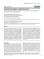
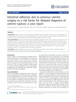
Figure 1 shows the effect of increasing caffeine dosage
on the probability of OP over time in different gestational age (25 weeks GA = 15 infants and 30 weeks GA
= 25 infants) based on the above fitted logistic generalized linear mixed model.
To examine whether the effect of duration of caffeine
treatment, we fitted another generalized linear mixed
model by including the interaction between caffeine
dosage and duration of therapy, and other covariates,
the results are showed in Table 6. This table shows that,
the average caffeine dose, caffeine duration of therapy as
Table 4 Factors associated with OP: Results of univariate
analysis
well as the interaction between caffeine dose and duration of caffeine treatment has a statistical significant
correlation with OP even when controlling for the effects of gestational age, weight and vitamin D (p < 0.05).
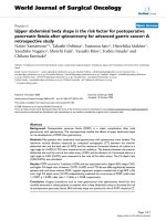
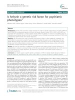
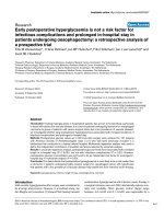
Based on the model in Table 6, Figs. 2 and 3 show the
effect of duration of caffeine usage on the probability of
OP based on the logistic model. The probability of OP
increased in 25 weeks preterm infants (15 infants), is
higher than the 30 weeks preterm infants (25 infants).
The figure exhibited that the lower the gestational age
the higher the probability of osteopenia over prolonged
caffeine use, even when controlling caffeine dose, steroid
dose, birth weight, and vitamin D.
Discussion
Although the overall survival of extreme low birth
weight infants has improved over the past 2 decades,
these infants continue to have significant comorbidities.
The prevalence of OP in our study is similar to that previously reported in the literature and suggests that OP
remains a significant comorbidity in extreme low birth
weight infants and puts them at increased risk for spontaneous fractures during the NICU stay. Our results are
consistent with this concept, the younger and smaller
the babies, the higher the incidence of OP.
The results of this study revealed a strong correlation
between caffeine treatment and the presence of OP. Despite caffeine’s effect on treating apnea of prematurity
with favorable long-term outcomes [23], our study revealed a strong association between cumulative dosage
and duration of treatment with caffeine and OP even
when controlling for the effect of other risk factors. The
results show that the adverse effect of caffeine is more
evident in lower gestational age infants, which may be
Variables
Estimate
Standard Error
P value
Gestational age (weeks)
−0.645
0.147
<0.001*
Average biweekly weight (grams)
0.0006
0.0002
0.006*
Caffeine cumulative dose (mg)
0.005
0.001
<0.001*
Caffeine duration (days)
0.051
0.013
<0.001*
Steroids cumulative dose (mg)
0.09
0.046
0.038
TPN duration (days)
0.034
0.012
0.005*
Table 5 Results from Multivariable generalized linear mixed
model
Vitamin D (units)
−1.863
0.36
<0.001*
Effect
0.003
0.002
0.20
Estimate
(logit)
Standard
Error
P value
Diuretics cumulative dose (mg)
Intercept
5.63
5.59
0.321
Serum phosphate (mmol/l)
Phosphate <1.3
Phosphate (1.3–1.8)
−0.09
0.11
0.16
0.33
0.57
Caffeine Cumulative Dose (mg)
0.39
0.05
0.007*
0.74
Steroid Cumulative Dose
(mg)
0.17
0.05
0.035*
Vitamin D (units)
−1.64
0.47
0.006*
Average Biweekly Weight
(grams)
−0.01
0.0001
<0.0001*
Gestational age (weeks)
−0.41
0.19
0.0408*
Phosphate >1.8 (ref)
Maternal Parity
Low parity
Moderate Parity (ref)
* Means significant
−0.016
0.42
0.96
p* = significant value
Ali et al. BMC Pediatrics (2018) 18:9
Page 5 of 7
Probability of OP with Prolonged Caffeine Usage
0.9
Probability of OP
0.8
0.7
0.6
0.5
0.4
25 weeks gestation
0.3
30 weeks gestation
0.2
0.1
0
0
5
10
1
0.9
0.8
0.7
0.6
0.5
0.4
0.3
0.2
0.1
0
Probability (n=55)
0
15
Caffeine Dosage (mg)
Fig. 1 Probability of OP with increasing caffeine dosage at 25 weeks
and at 30 weeks gestational age based on the logistic model
explained by the prolonged half-life of caffeine in their
bodies due to diminished kidney abilities to eliminate
the caffeine. Furthermore, extreme preterm infants have
immature liver enzymes and are unable to catabolize
caffeine leading to a prolonged effect causing calciuria
and osteoclastogenesis [14, 19].
In contrast to the current study results, a retrospective
study done by Viswanathan et al. (2014), showed that
there was no difference in duration of caffeine use between cases of OP and the control group. Viswanathan
et al. did not calculate caffeine dose, only caffeine duration was tracked between cases and controls. Additionally, in the Viswanathan et al. (2014) study, infants with
spontaneous rib fractures were included in the control
group if there was no radiological evidence of OP. In
our study, the osteopenic fractures were encompassed in
the cohort data and identified as having severe grade
osteopenia. The average duration of caffeine treatment
in both groups in the Viswanathan et al. study was
40 days, while in our study, the average duration of caffeine treatment was 60 days [24].
Our current study was a retrospective one and
there was no accurate documentation of maternal
1
2
3
4 5 6 7 8 9
Weeks of Caffeine
10 11 12
Fig. 2 Probability of OP with prolonged caffeine use based on the
logistic model
caffeine intake during pregnancy and lactation time.
However, it is worth mentioning that in an animal
study, maternal caffeine intake negatively affected
bone formation and development [25]. Thus our results may still imply an effect of maternal caffeine exposure either in utero or through mother’s breast
milk and donor breast milk. However, the high doses
of caffeine prescribed for apnea of prematurity have
paramount contribution to OP.
In this study, there was no difference between male
and female infants regarding OP, which is in agreement
with another comparable study [26]. But our results do
differ from other published studies which found that
male infants have higher bone density than females
when comparing preterm male and female infants with
male and female full term newborns. Such an observation may follow a recognizable trend for testosterone
hormone in utero [27, 28].
This study showed significant effect of TPN duration
on the development of OP but this effect disappeared
when we controlled for other risk factors. This can be
explained by considering that other factors contribute
more to OP, and that TPN contains the maximum
amount of calcium and phosphate according to the
Table 6 Estimates with interaction of caffeine and duration of
treatment
Estimate
(logit)
Standard
Error
P
Intercept
3.39
5.99
0.57
Average Caffeine dose (mg/kg/d)
0.24
0.09
0.029*
Duration of caffeine treatment (days)
0.64
0.27
0.02*
Caffeine dose* Duration of caffeine
treatment (days)
0.07
0.04
0.05*
Steroid cumulative dose (mg)
0.09
0.05
0.07
Vitamin D (units)
−1.86
0.36
0.04*
Average biweekly Birth Weight
(grams)
−0.06
0.02
0.001*
Gestational age (weeks)
−0.64
0.15
0.001*
p* Indicates significant level
Effect of Prolonged Use of Caffeine
Over a 12 week Period
1
Probability of OP
Effect
0.8
0.6
Gestational Age 30
Weeks (n=25)
0.4
Gestational Age of 25
Weeks (n=15)
0.2
0
1 2 3 4 5 6 7 8 9 10 11 12
Number of Weeks of Caffeine Use
Fig. 3 Probability of OP with same Caffeine dosage at 25 weeks and
30 weeks gestational age over the weeks of treatment based on the
logistic model
Ali et al. BMC Pediatrics (2018) 18:9
maximum solubility allowed [29]. In this study, TPN
duration count included the null per os days as well as
partial feeding days. During the study time, TPN is provided till the infant can tolerate the full enteral feeding.
Although Backström et al. suggested that serum phosphate levels lower than 1.8 mmol/L (5.5 mg/dl) may
have a diagnostic sensitivity of 100% and specificity of
70% for OP [30], in our study, serum phosphate on
biweekly basis did not show a statistically significant correlation with OP. No other published studies have examined serum phosphate as a longitudinal marker over the
hospital stay. Yet, serum phosphate is among the minerals that are regulated tightly, and the average biweekly
record may not represent the real situation of serum
phosphate in infants on TPN for the first week at least
and partial feeding for another week. In agreement with
our results, Aly et al., (2005) found that serum phosphate as a single reading at birth was not correlated with
OP in preterm infants [27]. In another study serum
phosphate and serum alkaline phosphatase were correlated with OP later in infancy, which could be explained
by the other confounding factors and medications received that affect premature bone in early life in NICUs
[31, 32].
While it is documented that the number of previous
pregnancies of a healthy mother correlated negatively
with BMD measurements, the effect of previous pregnancies did not show the same effect on infants’ bone
formation. This supports the fact that an infant acquires
the needed minerals and vitamin from the mother’s body
with active transport against the concentration gradient
ignoring the mother’s general status [33]. In our study,
there was no significant effect of maternal parity on OP.
On the other hand, this cohort study with limited sample size did not have enough high parity mothers to detect a correlation, and thus further research is needed
that includes high parity mothers.
Our study results show a statistically significant correlation between OP and steroid cumulative dose, while diuretics did show a positive trend in relation to OP. This
correlation did not reach statistical significance. This result can be explained by the short duration of diuretics
use and the relative small sample size. The use of high
dose of caffeine that has a diuretic effect might explain
the lower need for the diuretic use.
Conclusions
We conclude that caffeine has a strong association with
OP. As limit of viability continues to decrease with 70%
survival of infants between 24 and 26 weeks, OP will
continue to increase and will results in significant morbidity in childhood and adulthood unless strategies to
mitigate risk factors are developed. Our study was limited by the small sample size. The study was conducted
Page 6 of 7
at one center, and thus the results may not be
generalizable on a wider scale. Further studies are
needed to determine effective lower caffeine dosage, different ventilation strategies, adequate vitamin D intake,
and passive movement as all these can provide protection against OP.
Abbreviations
BMC: Bone mineral content; BMD: Bone mineral density; BW: Birth weight;
GA: Gestational age; NICU: Neonatal intensive care unit; OP: Osteopenia of
prematurity; PTH: Parathyroid hormone; TPN: Total parenteral nutrition
Acknowledgements
I acknowledge Mr. Lin Xue and Miss. Aliaa El Tobgy for their help in the data
management.
Funding
This study was not funded from any source.
Availability of data and materials
Data will not be shared. The data will be used for other studies.
Authors’ contributions
Dr. EA have made the acquisition of data, analysis and interpretation of data
and discussion writing. Dr. CRG and MN have been involved in drafting the
manuscript and revising it critically for important intellectual content. Dr. MM
have made substantial contributions to conception and design ensuring that
questions related to the accuracy or integrity of any part of the work are
appropriately investigated and resolved. Dr. MR agreed to be accountable for
all aspects of the work related to the radiological data interpretation and
drafting the manuscript. Dr. DJ have been involved in all stages of this study
and drafting the manuscript and given final approval of the version to be
published. All authors read and approved the final manuscript.
Ethics approval and consent to participate
The study was approved by the Health Research Ethics Board (HREB) at
University of Manitoba number# H2013: 231, and the Health Sciences Center
Research Impact Approval from the Health Science Center. Number# RI2013:
088. The included data were retrospective data from medical records and
did not include any identifying information. Consent to participate is not
applicable for this study.
Consent for publication
Not applicable.
Competing interests
The authors declare that they have no competing interests.
Publisher’s Note
Springer Nature remains neutral with regard to jurisdictional claims in
published maps and institutional affiliations.
Author details
1
Community Health Sciences Department, Faculty of Health Sciences,
University of Manitoba, MS361K, 820 Sherbrook St, Winnipeg, MB R3A 1R9,
Canada. 2Department of Pediatrics and Child Health, Faculty of Health
Sciences, University of Manitoba, Winnipeg, MB, Canada. 3Department of
Radiology, Faculty of Health Sciences, University of Manitoba, Winnipeg, MB,
Canada. 4Child Health Program, Winnipeg Regional Health Authority,
Winnipeg, MB, Canada.
Received: 9 September 2015 Accepted: 28 December 2017
References
1. Specker B. Nutrition influences bone development from infancy through
toddler years. J Nutr. 2004;134(3):691S–5S.
2. Bowden LS, Jones CJ, Ryan SW. Bone mineralisation in ex-preterm infants
aged 8 years. Eur J Pediatr. 1999;158(8):658–61.
Ali et al. BMC Pediatrics (2018) 18:9
3.
4.
5.
6.
7.
8.
9.
10.
11.
12.
13.
14.
15.
16.
17.
18.
19.
20.
21.
22.
23.
24.
25.
26.
27.
Tsukahara H, Takeuchi M, Fujisawa K, Miura M, Hata K, Yamamoto K, Mayumi
M. High-turnover osteopenia in preterm infants: determination of urinary
pyridinium cross-links of collagen. Metabolism. 1998;47(3):333–5.
Backstrom MC, Kuusela AL, Maki R. Metabolic bone disease of prematurity.
Ann Med. 1996;28(4):275–82.
Embleton N, Wood CL. Growth, bone health, and later outcomes in infants
born preterm. J Pediatr. 2014;90(6):529–32.
Backstrom MC, Kuusela AL, Koivisto AM, Sievanen H. Bone structure and
volumetric density in young adults born prematurely: a peripheral
quantitative computed tomography study. Bone. 2005;36(4):688–93.
Koo WW, Sherman R, Succop P, Krug-Wispe S, Tsang RC, Steichen JJ,
Crawford AH, Oestreich AE. Fractures and rickets in very low birth weight
infants: conservative management and outcome. J Pediatr Orthop. 1989;
9(3):326–30.
Vachharajani AJ, Mathur AM, Rao R. Metabolic bone disease of prematurity.
NeoReviews. 2009;10(8):e402–11.
Heaney RP. Effects of caffeine on bone and the calcium economy. Food
Chem Toxicol. 2002;40(9):1263–70.
Erenberg A, Leff RD, Haack DG, Mosdell KW, Hicks GM, Wynne BA. Caffeine
citrate for the treatment of apnea of prematurity: a double-blind, placebocontrolled study. Pharmacotherapy. 2000;20(6):644–52.
Natarajan G, Botica ML, Thomas R, Aranda JV. Therapeutic drug monitoring
for caffeine in preterm neonates: an unnecessary exercise? Pediatrics. 2007;
119(5):936–40.
Fredholm BB, Battig K, Holmen J, Nehlig A, Zvartau EE. Actions of caffeine in
the brain with special reference to factors that contribute to its widespread
use. Pharmacol Rev. 1999;51(1):83–133.
Charles BG, Townsend SR, Steer PA, Flenady VJ, Gray PH, Shearman A.
Caffeine citrate treatment for extremely premature infants with apnea:
population pharmacokinetics, absolute bioavailability, and implications for
therapeutic drug monitoring. Ther Drug Monit. 2008;30(6):709–16.
Glajchen N, Ismail F, Epstein S, Jowell P, Fallon M. The effect of chronic
caffeine administration on serum markers of bone mineral metabolism and
bone histomorphometry in the rat. Calcif Tissue Int. 1988;43(5):277–80.
Yeh JK, Aloia JF, Semla HM, Chen SY. Influence of injected caffeine on the
metabolism of calcium and the retention and excretion of sodium,
potassium, phosphorus, magnesium, zinc and copper in rats. J Nutr. 1986;
116(2):273–80.
Zanardo V, Dani C, Trevisanuto D, Meneghetti S, Guglielmi A, Zacchello G,
Cantarutti F. Methylxanthines increase renal calcium excretion in preterm
infants. Biol Neonate. 1995;68(3):169–74.
Bergman EA, Massey LK, Wise KJ, Sherrard DJ. Effects of dietary caffeine on
renal handling of minerals in adult women. Life Sci. 1990;47(6):557–64.
Harvey NC, Javaid MK, Arden NK, Poole JR, Crozier SR, Robinson SM, Inskip
HM, Godfrey KM, Dennison EM, Cooper C. Maternal predictors of neonatal
bone size and geometry: the Southampton Women's survey. J Dev Orig
Health Dis. 2010;1(1):35–41.
Bosley AR, Verrier-Jones ER, Campbell MJ. Aetiological factors in rickets of
prematurity. Arch Dis Child. 1980;55(9):683–6.
Blackman NJM, Koval JJ. Interval estimation for Cohen's kappa as a measure
of agreement. Stat Med. 2000;19(5):723–41.
Koo WW, Gupta JM, Nayanar VV, Wilkinson M, Posen S. Skeletal changes in
preterm infants. Arch Dis Child. 1982;57(6):447–52.
Booth JG, Hobert JP. Maximizing generalized linear mixed model likelihoods
with an automated Monte Carlo EM algorithm. Journal of the Royal
Statistical Society Series B, Statistical Methodology. 1999:265–85.
Ofek-Shlomai N, Berger I. Inflammatory injury to the neonatal brain - what
can we do? Front Pediatr. 2014;2:30.
Viswanathan S, Khasawneh W, McNelis K, Dykstra C, Amstadt R, Super DM,
Groh-Wargo S, Kumar D. Metabolic bone disease: a continued challenge in
extremely low birth weight infants. JPEN J Parenter Enteral Nutr. 2014;38(8):
982–90.
Schneider PE, Miller HI, Nakamoto T. Effects of caffeine intake during
gestation and lactation on bones of young growing rats. Res Exp Med
(Berl). 1990;190(2):131–6.
Littner Y, Mandel D, Mimouni FB, Dollberg S. Bone ultrasound velocity
curves of newly born term and preterm infants. J Pediatr Endocrinol Metab.
2003;16(1):43–7.
Aly H, Moustafa MF, Amer HA, Hassanein S, Keeves C, Patel K. Gestational
age, sex and maternal parity correlate with bone turnover in premature
infants. Pediatr Res. 2005;57(5 Pt 1):708–11.
Page 7 of 7
28. Namgung R, Tsang RC. Factors affecting newborn bone mineral content: in
utero effects on newborn bone mineralization. Proc Nutr Soc.
2000;59(1):55–63.
29. Pereira-da-Silva L, Costa A, Pereira L, Filipe A, Virella D, Leal E, Moreira A,
Rosa M, Mendes L, Serelha M. Early high calcium and phosphorus intake by
parenteral nutrition prevents short-term bone strength decline in preterm
infants. J Pediatr Gastroenterol Nutr. 2011;52(2):203–9.
30. Backstrom MC, Kouri T, Kuusela AL, Sievanen H, Koivisto AM, Ikonen RS,
Maki M. Bone isoenzyme of serum alkaline phosphatase and serum
inorganic phosphate in metabolic bone disease of prematurity. Acta
paediatrica (Oslo, Norway : 1992). 2000;89(7):867–73.
31. Yesiltepe Mutlu G, Kirmizibekmez H, Ozsu E, Er I, Hatun S. Metabolic bone
disease of prematurity: report of four cases. J Clin Res Pediatr Endocrinol.
2014;6(2):111–5.
32. Hellstern G, Poschl J, Linderkamp O. Renal phosphate handling of
premature infants of 23-25 weeks gestational age. Pediatr Nephrol. 2003;
18(8):756–8.
33. Ghannam NN, Hammami MM, Bakheet SM, Khan BA. Bone mineral density
of the spine and femur in healthy Saudi females: relation to vitamin D
status, pregnancy, and lactation. Calcif Tissue Int. 1999;65(1):23–8.
Submit your next manuscript to BioMed Central
and we will help you at every step:
• We accept pre-submission inquiries
• Our selector tool helps you to find the most relevant journal
• We provide round the clock customer support
• Convenient online submission
• Thorough peer review
• Inclusion in PubMed and all major indexing services
• Maximum visibility for your research
Submit your manuscript at
www.biomedcentral.com/submit