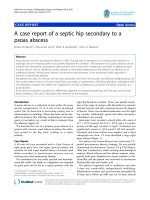
Case report of a central venous access deviceassociated thrombosis with aortic embolism in a preterm infant
Bạn đang xem bản rút gọn của tài liệu. Xem và tải ngay bản đầy đủ của tài liệu tại đây (2.12 MB, 5 trang )
Biermayr et al. BMC Pediatrics (2016) 16:154
DOI 10.1186/s12887-016-0691-x
CASE REPORT
Open Access
Case report of a central venous access
device-associated thrombosis with aortic
embolism in a preterm infant
Marlene Biermayr1, Barbara Brunner1, Kathrin Maurer2, Rudolf Trawoeger1, Ursula Kiechl-Kohlendorfer1
and Vera Neubauer1*
Abstract
Background: Thrombosis in neonates is commonly a central venous access device (CVAD) associated complication.
Furthermore, a patent foramen ovale (PFO) is frequently seen in preterm infants. Even though a coincidence of
both is not unusual, detaching of the thrombus and organisation of an aortic embolism has not been described
until now. Treatment recommendations of CVAD-associated thrombosis in neonates do not consider frequently
seen complications of preterm infants e.g. intraventricular haemorrhage.
This is the first case of a very preterm infant with pre-existing intraventricular haemorrhage, who developed a
CVAD-associated thrombosis and thromboembolic complications.
Case presentation: The authors report on a very preterm girl with a pre-existing intraventricular haemorrhage and
a CVAD-associated thrombus that, after removal of the CVAD, led to assumed pulmonary embolism and to an
extended aortic embolism with consequent cerebral stroke. The girl was treated with unfractionated heparin (UFH)
for about 50 days. During the further in-hospital stay the girl developed a mild bronchopulmonary dysplasia.
Follow-up revealed clinical signs of cerebral palsy.
Conclusion: Even though preterm infants are often diagnosed with a PFO which constitutes the risk for paradoxical
embolism, such complications do not occur frequently due to the physiological heart pressure proportion.
Nevertheless, it is important to monitor vital parameters and cerebral perfusion after removing a CVAD with
confirmed associated thrombosis, because thromboembolic complications are possible. If practicable, patients with
a confirmed CVAD-associated thrombosis should be anticoagulated before removing the CVAD. However, in our
patient it was rational to remove the CVAD without prior anticoagulation due to the pre-existing intraventricular
haemorrhage.
There are various treatment recommendations for thrombosis or embolism in infants. However, there are no clear
recommendations in very preterm infants with a high risk of cerebral bleeding respectively a pre-existing
intraventricular haemorrhage. We decided to treat our patient with unfractionated heparin until the affected vessels
were recanalised.
Finally, it remains a case-by-case decision how to treat CVAD-associated thrombosis and consequent embolism
depending on the patient’s medical history.
Keywords: Preterm, Central venous access device, Thrombosis, Aortic embolism, Paradoxical stroke, Case report
(Continued on next page)
* Correspondence:
1
Department of Paediatrics II, Neonatology, Medical University of Innsbruck,
Anichstrasse 35, 6020 Innsbruck, Austria
Full list of author information is available at the end of the article
© 2016 The Author(s). Open Access This article is distributed under the terms of the Creative Commons Attribution 4.0
International License ( which permits unrestricted use, distribution, and
reproduction in any medium, provided you give appropriate credit to the original author(s) and the source, provide a link to
the Creative Commons license, and indicate if changes were made. The Creative Commons Public Domain Dedication waiver
( applies to the data made available in this article, unless otherwise stated.
Biermayr et al. BMC Pediatrics (2016) 16:154
Page 2 of 5
(Continued from previous page)
Abbreviations: CVAD, Central venous access device; FFP, Fresh frozen plasma; fig., Figure; IVH, Intraventricular
haemorrhage; LMWH, Low molecular weight heparin; MRI, Magnetic resonance imaging; PFO, Patent foramen ovale;
PICC, Peripherally inserted central catheter; PTT, Partial thromboplastin time; PVHI, Periventricular venous
haemorrhagic infarction; UFH, Unfractionated heparin
Background
Central venous access devices (CVAD) are a known risk
factor for thrombosis in neonates. Furthermore, many
preterm infants are diagnosed with a patent foramen
ovale (PFO). Even though the combination of both is
common in preterm neonates, development of pulmonary complications and paradoxical aortic embolism has
not been described yet.
Existing guidelines for the treatment of thrombotic or
thromboembolic complications do not consider the gestational age of the affected child and thereby disregard
prevalent complications in relation to immaturity, such
as intraventricular haemorrhage (IVH) [1]. This is a limiting factor in the applicability of recommended therapeutic
measures for very preterm infants.
In this report we present the first case of a preterm
infant with a pre-existing IVH with periventricular venous haemorrhagic infarction (PVHI), who developed a
CVAD-associated thrombosis with thromboembolic
complications after removal of the CVAD. The patient
suffered from an extensive aortic embolism, which led
to seizures caused by ischaemic brain damage due to
an occlusion of large brain supplying arteries and, or,
cerebral embolism. In addition, the patient presented
with symptoms of pulmonary embolism.
Moreover, we discuss treatment recommendations of
thrombotic complications and their applicability in sick
preterm neonates.
Case presentation
The girl was born at the age of 30 weeks of gestation by
a Caesarean section due to pre-eclampsia of the mother.
Birth weight was appropriate for gestational age, umbilical pH was 7,27 and Apgar score was 6-8-9. Because of
respiratory distress she was treated with surfactant and
extubated on nasal continuous positive airway pressure
ventilation after 8 h. A cranial ultrasound on postnatal
day three revealed a right-sided IVH/PVHI.
During a routine echocardiography on postnatal day
six, a PFO but no other morphologic or functional abnormality was observed. Additionally, a large thrombus
(3,5 mm × 8,0 mm) on the tip of the peripherally inserted
central venous catheter (PICC (premicath©, Vygon,
Germany), which was inserted from the right ankle as part
of the initial care, floating next to the atrial septum was
detected. The catheter was removed immediately. Twelve
hours later the girl’s condition suddenly deteriorated. She
showed fits, skin colour was pale and her limbs were cold.
Furthermore, she presented with respiratory insufficiency.
In the course of intubation, blood, which apparently originated from a lung haemorrhage, was seen. Ultrasound
examination showed a long embolus in the aortic arch,
which extended to the innominate artery, the left carotid
artery and to the descending aorta to just above the celiac
trunk (Fig. 1). Additionally to the pre-existing IVH/PVHI
an ischaemic infarction of the majority of the left hemisphere was seen. We assumed that the thrombus not only
shifted to the aorta but also to the pulmonary artery and
thereby caused a pulmonary embolism that led to the lung
haemorrhage, as echocardiography showed no reopening
of the ductus arteriosus.
Regarding recurrent sanguinary secretions in the
ventilation tube heparin treatment was started at a low
dosage (5 units/kg/hour) and was progressively increased,
depending on the partial Thromboplastin time (PTT),
signs of haemorrhage and size of the thrombus during the
next 25 days up to a maximum of 15 units/kg/hour. Besides, a supportive treatment with repeated administration
of fresh frozen plasma (FFP) was started. Nonetheless,
Antithrombin III levels were low (40 % after first administration of FFP), therefore substitution was started to reach
levels >100 % to improve heparin effectiveness. Protein C
levels were normal. Unfractionated heparin (UFH) was administered for 51 days (48 days >10 units/kg/hour). After
this period the innominate artery was recanalised and the
blood flow in the left carotid artery was normal. Detailed
diagnostic work-up to exclude causes for thrombophilia
did not reveal any abnormality neither in the infant nor in
her mother.
Extubation was possible 4 days after the initial event.
During the further in-hospital stay the girl developed a
mild bronchopulmonary dysplasia but no further pulmonary complications. She exhibited persistent muscular
hypotonia and pronounced myoclonuses, but no persisting
seizures. She was discharged at a postmenstrual age of
39 weeks. A cerebral magnetic resonance imaging (MRI) at
term equivalent age showed a postischaemic cystic encephalomalacia of the left hemisphere and posthaemorrhagic
cysts on the right side (Fig. 2). Follow-up with a corrected
age of 3 months revealed a hypertonic lower extremity and
functional deficits, especially on the left side. Moreover, the
patient had no fidgety movements in the general movements assessment. These findings are highly associated
with the development of cerebral palsy [2].
Biermayr et al. BMC Pediatrics (2016) 16:154
Page 3 of 5
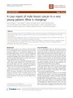
Fig. 1 Vascular ultrasound. Upper row showing sagittal images of the embolus in the a aortic arch (arrowheads) with its extention into the
innominate artery and the left carotid artery (white arrows); b descending aorta (arrows) and c abdominal aorta (arrows) with its end just above
the celiac trunk. Lower row showing axial images of the upper mediastinum d with the embolus in the aortic arch (white arrows) and e the
innominate artery (arrowhead) and in the left carotid artery (arrow); sagittal images of the neck f with the embolus within the supraclavicular part
of the left carotid artery (arrows), g which is no longer visible 2 days later. Ao – aorta, H – heart, T – trachea, JV – jugular vein, LCA – left carotid
artery, C - clavicula
Conclusion
To the best of our knowledge this is the first case of a preterm infant with an IVH/PVHI and a PFO, who developed
a CVAD-associated thrombosis and subsequently suffered
from assumed pulmonary embolism and aortic embolism
with extensive cerebral ischaemic infarction.
As mentioned above, many preterm infants have a
PFO and it is known that a venous or cardiac thrombus
may cause a paradoxical stroke and, or, an arterial embolism [3–6]. Even though our patient exhibited a thrombus
in the right atrium and a PFO was observed during the
echocardiography, an aortic embolism was not to be expected due to the physiological heart pressure proportion.
Furthermore, it is remarkable that even though emboli
detached and led to an assumed pulmonary embolism and
to a cerebral stroke, there were no further clinical complications due to emboli after the initial event.
A Canadian study reported that almost 90 % of all
thromboses in newborns were related to a CVAD [7].
According to recommendations for the maintenance of
CVAD patency, we administer 0.5 units/kg/hour UFH to
all infants with PICC in our neonatal intensive care unit
[1]. A Cochrane Review showed that this heparin
prophylaxis reduces the risk of PICC occlusion, but not
the risk of PICC-associated thrombosis [8]. In the case
of a confirmed CVAD-associated venous thrombosis,
Monagle et al. suggest to remove the CVAD (grade 1B)
and recommend anticoagulation with UFH or low molecular weight heparin (LMWH) for three to five days
(grade 2C). They also regard the case of right atrial
thrombosis related to a CVAD and suggest to remove
the catheter with or without anticoagulation, depending
on individual risk factors [1]. At that time-point treatment with heparin at dosages affecting the PTT in our
patient was hazardous due to the IVH/PVHI, for which
reason the catheter was removed without prior anticoagulation. After PICC removal and demarcation of the
embolus in the aorta and adjacent large arterial vessels,
a thrombolysis was not feasible due to the cerebral and
pulmonary haemorrhagic complications. A thrombectomy was considered as not feasible, because of the
small dimensions of the vascular system and the necessity of post-interventional effective anticoagulation. After
careful appraisal of the benefit risk ratio in the situation
of extensive aortic embolism with partial occlusion of
major brain supplying arteries with concomitant acute
cerebral and pulmonary haemorrhage, we started an
intravenous UFH treatment at 5 units/kg/hour based on
the suggestion of Monagle et al. to treat “neonates with
a first acute ischemic stroke and a documented cardioembolic source” with heparin [1]. Under permanent
monitoring of haemorrhagic complications the dosage
was increased with extreme caution. Fortunately no further bleedings occurred.
After initial anticoagulation, Monagle et al. suggest
further treatment with subcutaneous LMWH for a total
duration of 6 weeks to 3 months [1]. A daily subcutaneous drug administration is challenging in infants with
low weight and little subcutaneous fat tissue. Therefore,
we decided to treat our patient with intravenous UFH
until the affected vessels were recanalised after about
50 days.
In conclusion, removal of the PICC without prior
anticoagulation was rational in this case. Nevertheless, if
Biermayr et al. BMC Pediatrics (2016) 16:154
Page 4 of 5
Fig. 2 Cerebral MRI scan at term equivalent age showing a postischaemic cystic encephalomalacia of the left hemisphere and the intraventricular
haemorrhage (arrow) with posthaemorrhagic cysts (*) on the right side with a consequent e vacuo dilatation of the lateral ventricles
possible, patients with a confirmed thrombosis should
be anticoagulated before removing a CVAD. After removal, monitoring of vital parameters and cerebral
perfusion should be performed because, even though
these complications are uncommon, pulmonary embolism is possible and a PFO constitutes a risk for
paradoxical embolism. Obviously, it is not possible to
give a general applicable recommendation for treating
CVAD-associated thrombosis or aortic embolism in
preterm infants. Thus, it remains a case-by-case decision
depending on the patient’s condition, thrombophilic
factors and previous complications such as (intracerebral)
haemorrhage.
Availability of data and materials
There are no more case specific data that could be shared.
Authors’ contributions
All authors have made substantial contributions to analysis and
interpretation of data, have been revising the manuscript critically for
important intellectual content and read and approved the final manuscript.
Competing interests
The authors declare that they have no competing interests.
Consent for publication
Written informed consent was obtained from the patient’s legal guardian for
publication of this Case report and any accompanying images. A copy of the
written consent is available for review by the Editor of this journal.
Ethics approval and consent to participate
Not applicable.
Author details
1
Department of Paediatrics II, Neonatology, Medical University of Innsbruck,
Anichstrasse 35, 6020 Innsbruck, Austria. 2Department of Radiology, Medical
University of Innsbruck, Innsbruck, Austria.
Biermayr et al. BMC Pediatrics (2016) 16:154
Page 5 of 5
Received: 17 March 2016 Accepted: 27 August 2016
References
1. Monagle P, Chan AKC, Goldenberg NA, Ichord RN, Journeycake JM,
Nowak-Göttl U, et al. Antithrombotic Therapy in Neonates and Children.
Chest J. 2012;141:e737S. Available from: stnet.
org/article.aspx?doi=10.1378/chest.11-2308.
2. Einspieler C, Prechtl HFR. Prechtl’s assessment of general movements: A
diagnostic tool for the functional assessment of the young nervous system.
Ment Retard Dev Disabil Res Rev. 2005;11:61–7.
3. Parker MJ, Joubert GI, Levin SD. Portal vein thrombosis causing neonatal
cerebral infarction. Arch Dis Child Fetal Neonatal Ed. 2002;87:F125–7.
Available from: />fcgi?artid=1721458&tool=pmcentrez&rendertype=abstract.
4. Filippi L, Palermo L, Pezzati M, Dani C, Matteini M, De Cristofaro MT,
et al. Paradoxical embolism in a preterm infant. Dev Med Child Neurol.
2004;46:713–6. Available from: />elink.fcgi?dbfrom=pubmed&id=15473178&retmode=ref&cmd=prlinks\
npapers2://publication/doi/10.1111/j.1469-8749.2004.tb00987.x.
5. Beattie LM, Butler SJ, Goudie DE. Pathways of neonatal stroke and
subclavian steal syndrome. Arch Dis Child Fetal Neonatal Ed.
2006;91:F204–7.
6. Amlie-Lefond CM, Basir MA, Franciosi RA. Fatal Neonatal Stroke From a
Prenatal Cardiac Thrombus. Pediatr Neurol. 2008;38:140–2.
7. Schmidt B, Andrew M. Neonatal thrombosis: report of a prospective
Canadian and international registry. Pediatrics. 1995;96:939–43. Available
from: />8. Shah PS, Shah VS. Continuous heparin infusion to prevent thrombosis and
catheter occlusion in neonates with peripherally placed percutaneous
central venous catheters. In: Shah PS, editor. Cochrane Database Syst.
Rev. [Internet]. Chichester: Wiley; 2008. p. 3–5. Available from:
/>
Submit your next manuscript to BioMed Central
and we will help you at every step:
• We accept pre-submission inquiries
• Our selector tool helps you to find the most relevant journal
• We provide round the clock customer support
• Convenient online submission
• Thorough peer review
• Inclusion in PubMed and all major indexing services
• Maximum visibility for your research
Submit your manuscript at
www.biomedcentral.com/submit