Effects of low-dose clonidine on cardiovascular and autonomic variables in adolescents with chronic fatigue: A randomized controlled trial
Bạn đang xem bản rút gọn của tài liệu. Xem và tải ngay bản đầy đủ của tài liệu tại đây (885.19 KB, 12 trang )
Fagermoen et al. BMC Pediatrics (2015) 15:117
DOI 10.1186/s12887-015-0428-2
RESEARCH ARTICLE
Open Access
Effects of low-dose clonidine on
cardiovascular and autonomic variables in
adolescents with chronic fatigue: a
randomized controlled trial
Even Fagermoen1,2*, Dag Sulheim3,4, Anette Winger5, Anders M. Andersen6, Johannes Gjerstad7,8, Kristin Godang9,
Peter C. Rowe10, J. Philip Saul11, Eva Skovlund12,13 and Vegard Bruun Wyller1,14
Abstract
Background: Chronic Fatigue Syndrome (CFS) is a common and disabling condition in adolescence with few
treatment options. A central feature of CFS is orthostatic intolerance and abnormal autonomic cardiovascular
control characterized by sympathetic predominance. We hypothesized that symptoms as well as the underlying
pathophysiology might improve by treatment with the alpha2A–adrenoceptor agonist clonidine.
Methods: A total of 176 adolescent CFS patients (12–18 years) were assessed for eligibility at a single referral center
recruiting nation-wide. Patients were randomized 1:1 by a computer system and started treatment with clonidine
capsules (25 μg or 50 μg twice daily, respectively, for body weight below/above 35 kg) or placebo capsules for
9 weeks. Double-blinding was provided. Data were collected from March 2010 until October 2012 as part of The
Norwegian Study of Chronic Fatigue Syndrome in Adolescents: Pathophysiology and Intervention Trial
(NorCAPITAL). Effect of clonidine intervention was assessed by general linear models in intention-to-treat analyses,
including baseline values as covariates in the model.
Results: A total of 120 patients (clonidine group n = 60, placebo group n = 60) were enrolled and started treatment.
There were 14 drop-outs (5 in the clonidine group, 9 in the placebo group) during the intervention period. At 8 weeks,
the clonidine group had lower plasma norepinephrine (difference = 205 pmol/L, p = 0.05) and urine norepinephrine/
creatinine ratio (difference = 3.9 nmol/mmol, p = 0.002). During supine rest, the clonidine group had higher heart rate
variability in the low-frequency range (LF-HRV, absolute units) (ratio = 1.4, p = 0.007) as well as higher standard deviation
of all RR-intervals (SDNN) (difference = 12.0 ms, p = 0.05); during 20° head-up tilt there were no statistical differences in
any cardiovascular variable. Symptoms of orthostatic intolerance did not change during the intervention period.
Conclusions: Low-dose clonidine reduces catecholamine levels in adolescent CFS, but the effects on autonomic
cardiovascular control are sparse. Clonidine does not improve symptoms of orthostatic intolerance.
Trial registration: Clinical Trials ID: NCT01040429, date of registration 12/28/2009.
* Correspondence:
1
Institute of Clinical Medicine, Medical Faculty, University of Oslo, P.O.Box
1171, Blindern 0318Oslo, Norway
2
Department of Anaesthesiology and Critical Care, Oslo University Hospital,
P.O.Box 4950, Nydalen 0424Oslo, Norway
Full list of author information is available at the end of the article
© 2015 Fagermoen et al. Open Access This article is distributed under the terms of the Creative Commons Attribution 4.0
International License ( which permits unrestricted use, distribution, and
reproduction in any medium, provided you give appropriate credit to the original author(s) and the source, provide a link to
the Creative Commons license, and indicate if changes were made. The Creative Commons Public Domain Dedication waiver
( applies to the data made available in this article, unless otherwise stated.
Fagermoen et al. BMC Pediatrics (2015) 15:117
Background
Chronic Fatigue Syndrome (CFS) is a disabling condition
with unknown pathophysiology. In adolescents, prevalence has been estimated from 0.1 to 2.4 % depending
on definition of CFS and method of estimation [1, 2].
Apart from a single trial of intravenous immunoglobulin
in adolescents with CFS [3], no pharmacotherapy has
proven beneficial in this patient population.
Orthostatic intolerance is common with a prevalence
of more than 25 % in adults with CFS [4], and more than
90 % in children with CFS [5, 6]. Previously, dysregulation of autonomic cardiovascular control has been demonstrated in adults as well as adolescents, characterized by
increased sympathetic and decreased parasympathetic
nervous activity [7–10]. This autonomic imbalance might
reflect alteration of central control mechanism [11, 12],
and provide a target for pharmacotherapy [7, 13].
Clonidine is a centrally acting agonist to the presynaptic alpha2A receptor, thereby attenuating sympathetic nervous activity and enhancing parasympathetic
activity, even in low doses [14–16]. Thus, clonidine
has well-known antihypertensive properties. A pilot study
suggested normalization of cardiovascular variables in
adolescent CFS patients receiving low-dose clonidine [17].
However, a single nucleotide polymorphism (SNP) of the
alpha2A receptor gene might possible modify the effect of
clonidine treatment [18].
The aim of this study was to investigate the effects
of low-dose clonidine on autonomic cardiovascular
control in adolescent CFS. We hypothesized that clonidine would improve symptoms of orthostatic intolerance and normalize cardiovascular variables and
indices of autonomic nervous activity at rest as well
as during orthostatic challenges. The study is part of
the NorCAPITAL-project (The Norwegian Study of
Chronic Fatigue Syndrome in Adolescents: Pathophysiology and Intervention Trial; ClinicalTrials ID:
NCT01040429, date of registration 12/28/2009).
Methods
Patients
All hospital pediatric departments in Norway (n = 20) as
well as primary care pediatricians and general practitioners were invited to refer patients aged 12 – 18 years
to the national referral center for young CFS patients at
Oslo University Hospital. The referring units were
equipped with written information for distribution to potential study participants and their parents/next-of-kin. If
consent was given, a standard form required the referral
unit to confirm the result of clinical investigations considered compulsory to diagnose pediatric CFS according to
national Norwegian recommendations (pediatric specialist
assessment, comprehensive hematology and biochemistry
analyses, chest x-ray, abdominal ultrasound, and brain
Page 2 of 12
magnetic resonance imaging). Also, the referring units
were required to confirm that the patient a) was unable to
follow normal school routines due to fatigue; b) was not
permanently bedridden; c) did not have any concurrent
medical or psychiatric disorder that might explain the fatigue; d) did not experience any concurrent demanding
life event (such as parents’ divorce) that might explain the
fatigue; e) did not use prescribed pharmaceuticals (including hormone contraceptives) regularly. A previous demanding life event was not an exclusion criterion.
Completed forms were consecutively conveyed to the
study center and carefully evaluated by either of two authors (DS or EF). Patients considered eligible to this study
were invited to a clinical encounter at our study center
after which a final decision on inclusion was made.
In agreement with clinical guidelines [19, 20], this study
applied a “broad” case definition of CFS, requiring three
months of unexplained, disabling chronic/relapsing fatigue
of new onset. We did not require that patients meet any
other accompanying symptom criteria. Details of inclusion
and exclusion criteria are provided in Table 1.
Study design
All included patients underwent a baseline investigational
program at our research unit. Thereafter, they were randomized to 9 weeks of treatment with oral clonidine
capsules or placebo capsules in a 1:1 ratio, using a
computer-based routine for stratified randomization
(block size: 4); 18 months disease duration (the median disease duration in a previous follow-up study [21]) served as
the stratification criterion. Because of practical issues,
randomization was performed prior to final decision on enrolment; the procedure was carried out by a research nurse
not otherwise affiliated with the study. Outcome was
assessed by an investigational program identical to the
baseline program at week 8 and week 30; in this article,
only results from week 8 are reported. Patients and researchers were blinded to treatment allocation at all stages.
Clonidine dosages were 50 μg B.I.D for body
weight >35 kg, and 25 μg B.I.D for body weight <
35 kg. Catapresan® 25 μg clonidine hydrochloride tablets (Boehringer Ingelheim, Germany) were enclosed in
orange opaque, demolition-restraint lactose capsules
(Apoteket Produktion & Laboratorier, Kungens Kurva,
Sweden). Identical capsules without Catapresan® were
used as placebo comparator. Half the dose was given for
the first 3 days of the intervention period in order to
minimize adverse introductory effects. Blood samples for
clonidine concentration analyses were taken approximately two weeks after start of the intervention, and at
the second visit.
NorCAPITAL was approved by the Norwegian National Committee for Ethics in Medical Research and
the Norwegian Medicines Agency. Data were collected
Fagermoen et al. BMC Pediatrics (2015) 15:117
Page 3 of 12
Table 1 Criteria for inclusion and exclusion
CFS patients
Inclusion criteria
Exclusion criteria
Persisting or constantly relapsing fatigue lasting
3 months or more.
Another current disease process or demanding life event
that might explain the fatigue
Functional disability resulting from fatigue to a
degree that prevent normal school attendance
Another chronic disease
Age ≥ 12 years and < 18 years
Permanent use of drugs (including hormones) possibly
interfering with measurements
Permanently bed-ridden
Positive pregnancy test
Pheocromocytoma
Evidence of reduced cerebral and/or peripheral circulation
due to vessel disease
Polyneuropathy
Renal insufficiency
Known hypersensitivity towards clonidine or inert substances
(lactose, saccarose) in capsule
Abnormal ECG (apart from ectopic beats)
Supine heart rate < 50 beats/min
Supine systolic blood pressure < 85 mmHg
Upright systolic blood pressure fall > 30 mmHg
Healthy control subjects
Age ≥ 12 years and < 18 years
Another chronic disease
Permanent use of drugs (including hormones)
in the period March 2010 until October 2012. Written
informed consent was obtained from all participants,
and from parents/next-of-kin if required.
Investigational program
A one-day in-hospital assessment included clinical
examination, blood sampling (antecubital venous puncture), and 20° head-up tilt test (HUT), and always commenced between 7.30 and 9.30 a.m. Patients were
instructed to fast overnight and abstain from tobacco
products and caffeine for at least 48 h, to bring a morning spot urine sample in a sterile container, and to apply
the local anesthetic lidocaine (Emla®) on the skin in the
antecubital area one hour in advance. At week 8, CFS
patients were told to postpone their prescribed morning
study drug dose (clonidine/placebo) until after blood
sampling and HUT. All procedures were undertaken in
a quiet, warm room in a fixed sequence and by three researchers only (DS, EF and AW). Blood samples were
obtained in a fixed sequence from antecubital venous
puncture after at least five minutes supine rest in calm
surroundings. Samples of oral mucosa were collected for
genetic analyses. Following the in-hospital assessment, a
self-administered questionnaire was completed.
Laboratory analyses
The blood samples for plasma norepinephrine (NA) and
epinephrine (A) analyses were obtained in vacutainer
tubes treated with ethylene glycol tetraacetic acid
(EGTA)–Glutathione. The samples were placed on ice for
approximately 30 min; thereafter, plasma was separated by
centrifugation (3000 rpm, 15 min, 4 °C) and frozen at –
80 °C until assayed. Samples were analyzed for plasma NA
and A by high-performance liquid chromatography
(HPLC) with a reversed-phase column and glassy carbon
electrochemical detector (Antec, Leyden Deacade II SCC,
Zoeterwoude, The Netherlands) using a commercial kit
(Chromsystems, München, Germany) [22–24]. All samples were measured in singlet, with serial samples from a
given individual run at the same time to minimize run-torun variability. The intra- and interassay coefficient of
variation (CV) were 3.9 and 10.8 %, respectively. The detection limit was 5.46 pM.
Urine samples for NA and A analyses were collected
in 10 ml universal containers. Immediately after collection the urine was acidified to pH ≈ 2.5, thereafter, stored
at 2–8 °C until assayed. Urine treated this way is stable
at least 5 days. The analyses were performed consecutively. The same HPLC protocol as for plasma measurement was used for the measurement of urin NA/A. The
intra- and interassay coefficient of variation (CV) for
urine were 3.9 and 5.2 %, respectively.
The blood samples for clonidine determinations were
collected in 4 mL heparin tubes. After centrifugation for
12 min at 1000 g at room temperature, the plasma
fraction was frozen at −20 °C until analysis. A slight
Fagermoen et al. BMC Pediatrics (2015) 15:117
modification of the method described by Müller et al.
[25] was used for plasma clonidine assaying. The assay
was validated based on FDA guidelines [26]. The samples were separated on an Alliance HT 2795 HPLC
system and detected by a Micromass Quattro micro
API MS/MS-instrument. System control, data acquisition and integration were performed by Masslynx
software Ver 4.1.2008 (all from Waters, Milford, MA,
USA). The MS/MS conditions were optimized by
manual tuning during pump-infusion of neat solutions. The assay was set up to quantify from 0.10 μg/
L to 5.00 μg/L clonidine in plasma. Quality control
samples were included in all sample series, and placed
both before and after the patient samples in each
analytical run. The median intra assay CV was 1 % at
5 μg/L, 5 % at 0.75 μg/L and 10 % at 0.10 μg/L. The
inter assay CV was 6 % at 5 μg/L, 5 % at 0.75 μg/L
and 12 % at 0.10 μg/L. Limit of detection, defined as
a peak-to-peak signal to noise ratio of 5:1, verified by
the Masslynx software, was 0.025 μg/L. Accuracy was
97 % (median) at 5 μg/L, 97 % at 0.75 μg/L, and
107 % at 0.10 μg/L.
The genotyping of the alpha2A receptor single nucleotide polymorphism (SNP) rs1800544 was carried out by
predesigned TaqMan SNP genotyping assay (Applied
Biosystems, Foster City, CA, USA), using the SDS 2.2
software (Applied Biosystems). As previously described,
approximately 10 % of the samples were re-genotyped,
and the concordance rate was 100 % [27]. Genotyping
was also performed in 68 healthy individuals having the
same distribution of gender and age as the CFS patients.
Head-up tilt-test
Head-up tilt-test (HUT) was performed using an electronically operated tilt table with foot-board support
(Model 900–00, CNSystems Medizintechnik, Graz,
Austria). Patients were connected to the Task Force Monitor (TFM) (Model 3040i, CNSystems Medizintechnik,
Graz, Austria), a combined hardware and software device
for noninvasive recording of cardiovascular variables.
5 min was used for supine recordings, after which the participants were head-up tilted to 20° for 15 min. Details of
the HUT protocol have been described elsewhere [9]. The
feasibility of this protocol for studying adolescent CFS patients has been demonstrated in several previous studies
[9, 28]. In particular, the low tilt angle (20°) does not normally precipitate syncope, which is otherwise a common
problem among adolescents being subjected to stronger
orthostatic challenges [29]. Still, 20° head-up tilt is sufficient to demonstrate hemodynamic alterations and compensatory autonomic responses.
Instantaneous RR intervals (RRI) and heart rate (HR)
were obtained from the electrocardiogram (ECG). Continuous arterial blood pressure was obtained noninvasively
Page 4 of 12
using photoplethysmography on the right middle finger.
Mean arterial blood pressure (BP) was calculated by numerical integration of the recorded instantaneous BP.
The recorded value was calibrated against conventional
oscillometric measurements of arterial BP on the left
arm every five minutes according to the TFM manufacturer’s recommendation. Impedance cardiography with
electrodes placed on the neck and upper abdomen was
used to obtain a continuous recording of the temporal
derivative of the transthoracic impedance (dZ/dt). Beatto-beat stroke volume was calculated from the impedance signal [30].
Power spectral analysis (frequency-domain method) of
HR variability and systolic blood pressure (SBP) variability was automatically provided by the TFM, using an
adaptive autoregressive model [31]. Power was calculated in the Low Frequency (LF) range (0.05 to 0.17 Hz),
and High Frequency (HF) range (0.17 to 0.4 Hz). In
addition, time-domain indices of variability were computed from the RRIs: The standard deviation of all RRintervals (SDNN), the proportion of successive RRIs with
a difference greater than 50 ms (pNN50), and the square
root of the mean square differences of successive RRIs
(r-MSSD).
Heart rate variability (HRV) is considered an index of
autonomic cardiac modulation. In the frequency-domain,
vagal (parasympathetic) activity is the main contributor to
HF variability, whereas both vagal and sympathetic activity
contributes to LF variability [32]. The LF/HF ratio is considered an index of sympathovagal balance. SBP variability
is regarded an index of sympathetic modulation of peripheral resistance vessels [33]. For time-domain indices, vagal
(parasympathetic) activity is the main contributor to
pNN50 and r-MSSD, whereas SDNN is a measure of total
variability, analogous to the Total Power index in the frequency domain.
Data from each HUT procedure was exported to
Microsoft Excel for further calculations. Beat-to-beat
stroke index (SI) was calculated dividing stroke volume
by body surface area, and beat-to-beat total peripheral
resistance index (TPRI) was calculated as mean BP divided by the product of SI and HR. For each participant,
the following epochs of the recordings were chosen:
Baseline (270 to 30 s before tilt up) and Early tilt (30 to
270 s after tilt). In each epoch we computed the median
value for the conventional cardiovascular variables as
well as the indices of HR and SBP variability; this procedure reduces the influence of erroneous outliers, such
as ectopic heart beats. Thereafter, the delta values (Early
Tilt – Baseline) which are considered indices of the cardiovascular response to orthostatic challenge were computed for each participant. This analytic approach has
been proven feasible in several previous report from our
group [9–11].
Fagermoen et al. BMC Pediatrics (2015) 15:117
Questionnaire
The participants received a comprehensive questionnaire
consisting of several validated inventories, as has been
described in detail elsewhere [28].
The Autonomic Symptom Profile (ASP) [34], which
has been used in previous Norwegian CFS studies but
which is not validated for the Norwegian language, was
slightly modified in order to fit our age group. A composite score reflecting orthostatic symptoms was constructed from 8 single items from the ASP, addressing
experiences of dizziness in specific situations (such as
rising suddenly from supine position, taking a shower,
etc.). The total sum score is from 0 to 8; higher values reflect more pronounced orthostatic problems. In addition,
other symptoms related to autonomic cardiovascular control, such as palpitations and pale and cold hands, were
charted on a 1–5 Likert scale.
The questionnaire also included the CFS symptom inventory for adolescents [28, 35]. This inventory was used
to subgroup the CFS patients according to the 1994 CFS
case definition [36].
Statistics
Determination of sample size is described elsewhere
[28]. Outcome of clonidine intervention was assessed by
general linear models (ANCOVA) in intention-to-treat
analyses, including baseline values as covariates in the
model [37]. The net intervention effect was calculated
from the parameters of the fitted general linear model.
Differential effects in subgroups adhering to the 1994
CFS case definition, genotype of the alpha2A receptor
single nucleotide polymorphism (SNP) rs1800544, and
sex, were explored by including these variables as interaction terms. Dose–response relationships for patients
allocated to clonidine were explored by linear regression analyses. Missing values were imputed as last observation carried forward from the pre-medication
test. In order to obtain near-normally distributed variables, ln-transformation was carried out for supine
values of LF-HRV, HF-HRV, Total Power-HRV, LF/HF
ratio and LF-SBP. Square root transformation was
carried out for 20° head-up tilt values of LF-HRV,
HF-HRV and Total Power-HRV. Genotype frequency
among patients and healthy controls were explored
with chi-square analyses.
SPSS statistical software (SPSS Inc., Chicago, IL, USA)
was applied for all statistical analyses, and all tests were
carried out two-sided. A p-value ≤ 0.05 was considered
statistically significant. Corrections for multiple comparisons were not applied.
Results
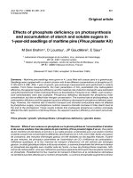
A total of 176 CFS patients were referred to the study,
of which 151 were eligible for randomization (Fig. 1). A
Page 5 of 12
total of 120 patients were enrolled and started treatment;
60 patients in the clonidine group and 60 patients in the
placebo group. At week 8, there were 5 dropouts in the
clonidine group and 9 dropouts in the placebo group
(Fig. 1). Further baseline demographic and clinical characteristics are given in Table 2.
At week 8, the clonidine group had statistically significantly lower plasma norepinephrine (p = 0.05) and urine
norepinephrine/creatinine ratio (p = 0.002) as compared
to the placebo group (Table 3). At supine rest, the clonidine group had higher heart rate variability in the lowfrequency band (LF-HRV, absolute unites) (p = 0.007)
and as well as higher SDNN (p = 0.05) (Table 4). No
other significant differences were observed. In particular,
symptoms of orthostatic intolerance did not change during the intervention period.
Urine norepinephrine/creatinine ratio was negatively
related to plasma clonidine concentration (B = −14.5,
p = 0.004). TPRI supine (B = 4.1, p = 0.01), heart rate
variability in the low-frequency band supine (LF-HRV,
absolute unites) (B = 1423, p = 0.02) and HRV-Total
Power supine (B = 4353, p = 0.04) were positively related to plasma clonidine concentration. No other
dose response-relationships were found.
Subgrouping according to the 1994 CFS case definition, genotype frequency of the alpha2A receptor SNP
rs1800544 and sex did not reveal any differential response to the intervention. Also, the genotype frequency was equal among CFS patients and healthy
controls (p = 0.75).
Discussion
This study shows that clonidine reduces catecholamine levels in adolescent CFS. However, the effects
on cardiovascular autonomic control are sparse, and
clonidine does not improve symptoms of orthostatic
intolerance.
Previous studies have documented that adult as well as
adolescent CFS patients are characterized by enhanced
sympathetic and attenuated parasympathetic nervous activity [7, 9, 38, 39]. In particular, CFS patients have
increased levels of catecholamines [40, 41] and a sympathetic predominance of cardiovascular autonomic control possibly due to central alterations [9, 11, 42]. In this
study, clonidine lowered catecholamine levels as expected. Of note, urine norepinephrine, which is considered an index of sympathetic nervous activity over time
[43], decreased dose-dependently.
Clonidine had limited impact on standard cardiovascular variables, both at rest and during orthostatic challenge. This finding was surprising. In previous studies of
healthy individuals as well as hypertensive patients, clonidine dosages similar to those applied in this study have
been shown to decrease both blood pressures and heart
Fagermoen et al. BMC Pediatrics (2015) 15:117
Page 6 of 12
Fig. 1 Study flowchart. Study flowchart. A total of 176 adolescents with CFS were assessed for eligibility. Of these, 151 fulfilled randomization criteria,
whereas 120 started treatment. At week 8, 106 participants were still participating in the intervention program, 55 in the clonidine group and 51 in the
placebo group
Table 2 Background characteristics
Clonidine (n = 60)
Placebo (n = 60)
13 (22)
21 (35)
Gender - no. (%)
Male
47 (78)
39 (65)
Age - years, mean ± SD
Female
15.3 ± 1.5
15.5 ± 1.6
BMI - kg/m2, mean ± SD
21.6 ± 4.4
21.5 ± 4.0
Adheres to 1994 CFS case definition - no. (%)
No
14 (24)
15 (26)
Yes
45 (76)
43 (74)
C/C
32 (53)
35 (58)
C/G
25 (42)
19 (32)
G/G
3 (5)
6 (10)
Genotype a – no. (%)
Disease duration - months, median (range)
18 (4 to 72)
18 (5 to 104)
Disease duration – months, mean ± SD
19.4 ± 13.0
23.5 ± 17.0
School absenteism - %, mean ± SD
66 ± 29
64 ± 31
Smokers – more than once a week – no.
1
0
a
The alpha2A receptor single nucleotide polymorphism (SNP) rs1800544. C = Cytosine, G = Guanine
Fagermoen et al. BMC Pediatrics (2015) 15:117
Page 7 of 12
Table 3 Outcome of clonidine intervention – symptom scores and catecholamines
Baseline
Week 8 (during treatment)
Clonidine group, mean
3.8
3.5
Placebo group, mean
3.5
Symptoms scores
Orthostatic symptoms – total score
3.5
Difference (95 % CI)
−0.05 (−0.5 to 0.4)
p-value (clonidine vs. placebo)
0.84
Palpitations - score
Clonidine group, mean
2.4
2.2
Placebo group, mean
2.2
2.2
Difference (95 % CI)
0.06 (−0.3 to 0.4)
p-value (clonidine vs. placebo)
0.73
Pale and cold hands - score
Clonidine group, mean
3.0
Placebo group, mean
3.0
2.7
2.8
Difference (95 % CI)
−0.1 (−0.5 to 0.3)
p-value (clonidine vs. placebo)
0.62
Catecholamines
Plasma norepinephrine - pmol/L
Clonidine group, mean
2040
Placebo group, mean
1942
1557
1761
Difference (95 % CI)
−205 (−406 to −4)
p-value (clonidine vs. placebo)
0.05
Plasma epinephrine - pmol/L
Clonidine group, mean
327
291
Placebo group, mean
415
299
Difference (95 % CI)
−8 (−44 to 29)
p-value (clonidine vs. placebo)
0.68
Urine norepinephrine/creatinine ratio - nmol/mmol
Clonidine group, mean
13.3
Placebo group, mean
13.7
9.6
13.6
Difference (95 % CI)
−3.9 (−6.4 to −1.5)
p-value (clonidine vs. placebo)
0.002
Urine epinephrine/creatinine ratio - nmol/mmol
Clonidine group, mean
1.7
1.2
Placebo group, mean
1.6
1.6
Difference (95 % CI)
−0.4 (−0.8 to 0.1)
p-value (clonidine vs. placebo)
0.11
Missing values were imputed based on the principle of last observation carried forwards. Thus, all calculations are based on 120 individuals (60 in each
intervention group except one to two in each group with missing values at baseline). Means and differences at week 8 are estimated from the parameters of the
general linear model
rate, and these alterations of hemodynamics were paralleled by a decrement of catecholamines [15, 44–47]. Furthermore, in healthy subjects, clonidine also attenuates
indices of cardiovascular sympathetic nervous modulation (such as LF-HRV), both in supine and sitting
positions [44]. In this study, there was a clonidinemediated increase in LF-HRV at supine rest, as well as a
positive relationship between LF-HRV and clonidine
plasma concentration. The interpretation of LF-HRVindices is not straight forward; these results, however,
Fagermoen et al. BMC Pediatrics (2015) 15:117
Table 4 Outcome of clonidine intervention – cardiovascular
variables
Baseline
Week 8 (during treatment)
Page 8 of 12
Table 4 Outcome of clonidine intervention – cardiovascular
variables (Continued)
Placebo group, mean
31
38
Supine
Difference (95 % CI)
2.2 (−3.0 to 7.3)
Heart rate - beats/min
p-value (clonidine vs. placebo)
0.40
Clonidine group, mean
70
67
Placebo group, mean
72
69
Clonidine group, mean
40
Difference (95 % CI)
−2.0 (−4.1 to 0.1)
Placebo group, mean
43
p-value (clonidine vs. placebo)
0.06
Difference (95 % CI)
3.7 (−0.5 to 8.0)
p-value (clonidine vs. placebo)
0.08
SBP – mmHg
Clonidine group, mean
103
Placebo group, mean
107
104
LF-HRV – nu
42
38
HF-HRV – nu
103
Clonidine group, mean
60
58
Difference (95 % CI)
1.4 (−1.0 to 3.9)
Placebo group, mean
57
62
p-value (clonidine vs. placebo)
0.25
Difference (95 % CI)
−3.7 (−8.0 to 0.5)
p-value (clonidine vs. placebo)
0.08
MBP – mmHg
Clonidine group, mean
77
78
Placebo group, mean
80
77
Clonidine group, mean
628
Difference (95 % CI)
1.3 (−0.7 to 3.4)
Placebo group, mean
451
p-value (clonidine vs. placebo)
0.19
Ratio (95 % CI)
1.4 (1.1 to 1.8)
p-value (clonidine vs. placebo)
0.007
DBP – mmHg
Clonidine group, mean
65
Placebo group, mean
66
64
LF-HRV* - ms
2
679
487
HF-HRV* - ms2
63
Clonidine group, mean
962
961
Difference (95 % CI)
0.8 (−1.0 to 2.7)
Placebo group, mean
600
825
p-value (clonidine vs. placebo)
0.37
Ratio (95 % CI)
SI - ml/m2
1.2 (0.9 to 1.5)
p-value (clonidine vs. placebo)
0.28
Clonidine group, mean
47
46
Placebo group, mean
46
46
Clonidine group, mean
1991
Difference (95 % CI)
0.2 (−2.1 to 2.4)
Placebo group, mean
1352
p-value (clonidine vs. placebo)
0.86
Ratio (95 % CI)
1.3 (1.0 to 1.6)
p-value (clonidine vs. placebo)
0.06
TPRI - mmHg/L/min/m2
Clonidine group, mean
9.1
Placebo group, mean
8.9
9.4
Total Power-HRV* - ms
2
2053
1638
LF/HF-ratio*
8.9
Clonidine group, mean
0.65
0.70
Difference (95 % CI)
0.5 (−0.1 to 1.1)
Placebo group, mean
0.75
0.59
p-value (clonidine vs. placebo)
0.11
Ratio (95 % CI)
1.2 (1.0 to 1.4)
p-value (clonidine vs. placebo)
0.09
SDNN – ms
Clonidine group, mean
74
78
Placebo group, mean
66
66
Clonidine group, mean
39.3
Difference (95 % CI)
12.0 (−0.2 to 23.7)
Placebo group, mean
38.1
p-value (clonidine vs. placebo)
0.05
Difference (95 % CI)
1.1 (−3.0 to 5.2)
p-value (clonidine vs. placebo)
0.60
r-MSSD – ms
Clonidine group, mean
79
Placebo group, mean
65
83
LF-SBP – nu
38.0
36.9
LF-SBP* - mmHgs2
70
Clonidine group, mean
3.8
3.7
Difference (95 % CI)
13.1 (−3.2 to 29.5)
Placebo group, mean
3.0
3.2
p-value (clonidine vs. placebo)
0.11
Ratio (95 % CI)
1.1 (0.9 to 1.5)
p-value (clonidine vs. placebo)
0.34
pNN50 - %
Clonidine group, mean
40
40
Response to 20° head-up tilt
Fagermoen et al. BMC Pediatrics (2015) 15:117
Table 4 Outcome of clonidine intervention – cardiovascular
variables (Continued)
Heart rate - beats/min
Page 9 of 12
Table 4 Outcome of clonidine intervention – cardiovascular
variables (Continued)
p-value (clonidine vs. placebo)
0.59
Clonidine group, mean
5.2
4.9
Placebo group, mean
4.8
4.9
Clonidine group, mean
8.3
Difference (95 % CI)
0.0 (−1.1 to 1.2)
Placebo group, mean
6.7
p-value (clonidine vs. placebo)
0.97
Difference (95 % CI)
−3.1 (−7.4 to 1.1)
p-value (clonidine vs. placebo)
0.15
SBP – mmHg
Clonidine group, mean
0.74
−0.59
Placebo group, mean
0.15
LF-HRV - nu
6.1
9.2
HF-HRV - nu
−0.01
Clonidine group, mean
−8.3
−6.1
Difference (95 % CI)
−0.58 (−2.2 to 1.0)
Placebo group, mean
−6.7
−9.2
p-value (clonidine vs. placebo)
0.48
Difference (95 % CI)
3.1 (−1.1 to 7.4)
p-value (clonidine vs. placebo)
0.15
MBP - mmHg
#
Clonidine group, mean
1.19
0.61
Placebo group, mean
0.94
1.23
Clonidine group, mean
−320
−161
Difference (95 % CI)
−0.63 (−2.1 to 0.8)
Placebo group, mean
−176
−171
p-value (clonidine vs. placebo)
0.39
n.a.
n.a.
p-value (clonidine vs. placebo)
0.87
DBP - mmHg
Clonidine group, mean
1.13
Placebo group, mean
1.58
1.2
LF-HRV - ms
2
HF-HRV# - ms2
1.8
Clonidine group, mean
−828
−640
Difference (95 % CI)
−0.59 (−2.0 to 0.8)
Placebo group, mean
−523
−629
p-value (clonidine vs. placebo)
0.40
n.a.
SI - ml/m2
n.a.
p-value (clonidine vs. placebo)
0.99
Clonidine group, mean
−5.9
−4.5
Placebo group, mean
−5.1
−5.3
Clonidine group, mean
−1107
−790
Difference (95 % CI)
0.9 (−0.4 to 2.1)
Placebo group, mean
−668
−736
p-value (clonidine vs. placebo)
0.17
n.a.
n.a.
p-value (clonidine vs. placebo)
0.78
TPRI - mmHg/L/min/m
2
Clonidine group, mean
0.66
Placebo group, mean
0.60
0.44
#
2
Total Power-HRV - ms
LF/HF-ratio
0.62
Clonidine group, mean
0.35
0.34
Difference (95 % CI)
−0.18 (−0.47 to 0.11)
Placebo group, mean
0.44
0.55
p-value (clonidine vs. placebo)
0.22
Difference (95 % CI)
−0.21 (−0.46 to 0.04)
p-value (clonidine vs. placebo)
0.09
SDNN - ms
Clonidine group, mean
−5.1
−7.9
Placebo group, mean
−4.4
−0.7
Clonidine group, mean
2.5
Difference (95 % CI)
−7.2 (−16.0 to 1.6)
Placebo group, mean
3.2
p-value (clonidine vs. placebo)
0.11
Difference (95 % CI)
0.7 (−2.4 to 3.8)
p-value (clonidine vs. placebo)
0.66
r-MSSD - ms
Clonidine group, mean
−18
−24
Placebo group, mean
−16
−17
Difference (95 % CI)
−7.6 (−19.6 to 4.4)
p-value (clonidine vs. placebo)
0.11
pNN50 - %
Clonidine group, mean
−14
−11
Placebo group, mean
−9
−13
Difference (95 % CI)
1.2 (−3.1 to 5.4)
LF-SBP - nu
4.4
3.7
LF-SBP - mmHgs2
Clonidine group, mean
−2.6
−1.0
Fagermoen et al. BMC Pediatrics (2015) 15:117
Table 4 Outcome of clonidine intervention – cardiovascular
variables (Continued)
Placebo group, mean
−0.6
−0.2
Difference (95 % CI)
−0.7 (−1.7 to 0.3)
p-value (clonidine vs. placebo)
0.17
Missing values were imputed based on the principle of last observation carried
forwards. Thus, all calculations are based on 120 individuals (60 in each
intervention group). Means and differences at week 8 are estimated from the
parameters of the general linear model
For variables annotated with a *, modeling was performed on ln-transformed
variables; all means are based on back-transformation of the variables, and
ratios instead of differences are reported. For variables annotated with a #,
modeling was performed on square root-transformed variables; all means are
based on back-transformation of the variables, but neither differences nor
ratios can be computed, as indicated with the label n.a. (not applicable). CI =
Confidence Interval; SBP = Systolic Blood Pressure; MBP = Mean arterial Blood
Pressure; DBP = Diastolic Blood Pressure; SI = Stroke Index; TPRI = Total Periferal
Resistance Index; RRI = R-R Interval; HRV = heart rate variability; HF = High
Frequency; LF = Low Frequency; SDNN = standard deviation of all RR-intervals;
pNN50 = the proportion of successive RRIs with adifference greater than 50 ms;
r-MSSD = the square root of the mean square differences of successive RRIs; nu =
normalized units; n.a. = not applicable because of square root transformation of
variables; n = number of patients, for most variables equal to 60 because
of imputation
might suggest an enhancement of sympathetic heart
rate modulation, resembling the effects of clonidine in
essential hypertension [48]. This is in contrast to effects of clonidine in healthy subjects [44]. A previous
study suggests early sympathetic baroreceptor activation and diminished baroreceptor reserve in CFS [11].
We speculate that clonidine, by way of reducing sympathetic tone (as evident from the catecholamine-lowering
effect), might in fact increase the sympathetic nervous system modulatory effects [49].
Taken together, the findings presented in this study
suggest an alteration of clonidine pharmacodynamics in
CFS. One possible explanation is genetically determined
differences of the alpha2A receptor protein, which is the
ligand for clonidine. A single nucleotide polymorphism
(SNP) (rs1800544) in the alpha2A receptor gene implies
substitution of guanine (G) for cytosine (C) at position
1291, and has functional consequences [18]. However, the
genotype frequencies among CFS patients and a comparable group of healthy controls were almost identical, and
subgroup analysis based on genotype revealed no differences in response to treatment. Another possible explanation is altered expression of adrenoceptors, as has
previously been demonstrated in CFS [50] as well as in
other conditions with high levels of catecholamines [51].
The possibility of increased long-term cardiovascular
risk in CFS patients remains a concern [52]. In addition
to increased sympathetic nervous activity, CFS patients
are also characterized by slight inflammatory activation
[28] and elevated nocturnal blood pressure and heart
rate [53], which in turn are associated with development
of atherosclerosis. Further research is warranted to clarify the eventual need of prophylactic measures.
Page 10 of 12
A possible limitation of this study is the wide inclusion
criteria and no a priori-definition of the degree of school
absenteeism necessary to fulfil the diagnostic criteria,
which might have obscured results applying to a subgroup only. However, the study population corresponds
closely to the population who is diagnosed as CFS by pediatricians; thus, we assume the external validity to be
strong. Furthermore, subgrouping based upon the 1994
CFS case definition did not change the results. We have
not done subgrouping based on caffeine use. Another
limitation of this study is the 4 min epochs used for
time-domain analyses of heart rate variability, as opposed to the 5 min epochs recommended [32]. It is considered inappropriate to compare time-domain indices
(especially SDNN) obtained from recordings of different
durations; while the present study does not violate this
principle, caution should be shown when comparing our
results to other studies. Strengths of this study include
high compliance and low drop-out-rates, and the successful blinding of all (staff and patients) clinically involved in the study.
Conclusions
Low-dose clonidine reduces catecholamine levels in adolescent CFS. However, the effects on cardiovascular
autonomic control are sparse, and clonidine does not
improve symptoms of orthostatic intolerance.
Abbreviations
BP: Blood pressure; CFS: Chronic fatigue syndrome; HF: High frequency;
HR: Heart rate; HRV: Heart rate variability; HUT: Head-up tilt test; LF: Low
frequency; RRI: Instantaneous RR intervals; SBP: Systolic blood pressure;
SNP: Single nucleotide polymorphism.
Competing interests
The authors declare that they have no competing interests.
Authors’ contributions
EF, DS and AW collected clinical data, contributed to study design and
participated in data analyses. AMA, JG and KG carried out laboratory
analyses. PCR and JPS contributed to study design. ES supervised data
analyses. VBW conceived of the study, contributed to study design and
participated in data analyses. All authors contributed to data interpretation
and drafting of the manuscript. All authors approved the final manuscript as
submitted.
Acknowledgements
We thank Kari Gjersum for secretary assistance; Hamsana Chandrakumar,
Esther Gangsø, Anne Marie Halstensen, Adelheid Holm, Berit Widerøe
Njølstad, Pelle Rohdin, and Anna Marie Thorendal Ryenbakken for practical
assistance; Berit Bjelkåsen for development of the computerized
randomization procedure; Liv Thrane Bjerke for pharmacy services; Gaute
Døhlen, Bjørn Bendz, Knut Engedal, and Ola Didrik Saugstad for study
monitoring; all referring units; and finally all participants and their parents/
next-of-kin.
The study was funded by: Health South–East Hospital Trust; The University of
Oslo; Oslo and Akershus University College of Applied Sciences; The
Norwegian Competence Network of Paediatric Pharmacotherapy; Simon
Fougner Hartmann’s Family Foundation; Eckbo’s Family Foundation.
Author details
1
Institute of Clinical Medicine, Medical Faculty, University of Oslo, P.O.Box
1171, Blindern 0318Oslo, Norway. 2Department of Anaesthesiology and
Fagermoen et al. BMC Pediatrics (2015) 15:117
Critical Care, Oslo University Hospital, P.O.Box 4950, Nydalen 0424Oslo,
Norway. 3Department of Paediatrics, Oslo University Hospital, P.O.Box 4950,
Nydalen 0424Oslo, Norway. 4Department of Paediatrics, Lillehammer County
Hospital, P.O.Box 1042381 Brumunddal, Norway. 5Institute of Nursing
Sciences, Oslo and Akershus University College of Applied Sciences, P.O. Box
4 St., Olavs plass 0130Oslo, Norway. 6Department of Pharmacology, Oslo
University Hospital, P.O.Box 4950, Nydalen 0424Oslo, Norway. 7National
Institute of Occupational Health, P.O Box 8149, Dep 0033Oslo, Norway.
8
Department of Biosciences, University of Oslo, P.O.Box 1066, Blindern
0316Oslo, Norway. 9Section of Specialized Endocrinology, Department of
Endocrinology, Oslo University Hospital Rikshospitalet, P.O.Box 4950, Nydalen
0424Oslo, Norway. 10Department of Paediatrics, Johns Hopkins University
School of Medicine, 200 N. Wolfe Street, Baltimore, MD 21287, USA.
11
Department of Paediatrics, Medical University of South Carolina, 169 Ashley
Avenue, Charleston, SC 29425, USA. 12Department of Pharmaceutical Science,
University of Oslo, P.O.Box 1068, Blindern 0316Oslo, Norway. 13Norwegian
Institute of Public Health, P.O.Box 4404, Nydalen 0403Oslo, Norway.
14
Department of Paediatrics, Akershus University Hospital, P.O.Box 10001478
Lørenskog, Norway.
Received: 7 September 2014 Accepted: 20 August 2015
References
1. Nijhof SL, Maijer K, Bleijenberg G, Uiterwaal CS, Kimpen JL, van der Putte
EM. Adolscent chronic fatigue syndrome: prevalence, incidence, and
morbidity. Pediatrics. 2011;127:e1169–75.
2. Crawley E. The epidemiology of chronic fatigue syndrome/myalgic
encephalitis in children. Arch Dis Child. 2014;99:171–4.
3. Rowe KS. Double-blind randomized controlled trial to assess the efficacy of
intravenous gammaglobulin for the management of chronic fatigue
syndrome in adolescents. J Psychiatr Res. 1997;31:133–47.
4. Hoad A, Spickett G, Elliott J, Newton J. Postural orthostatic tachycardia
syndrome is an under-recognized condition in chronic fatigue syndrome.
QJM. 2008;101:961–5.
5. Stewart JM, Gewitz MH, Weldon A, Arlievsky N, Li K, Munoz J. Orthostatic
intolerance in adolescent chronic fatigue syndrome. Pediatrics. 1999;103:116–21.
6. Stewart JM, Gewitz MH, Weldon A, Munoz J. Patterns of orthostatic
intolerance: The orthostatic tachycardia syndrome and adolescent chronic
fatigue. J Pediatrics. 1999;135:218–25.
7. Okamoto LE, Raj SR, Peltier A, Gamboa A, Shibao C, Diedrich A, et al.
Neurohumoral and haemodynamic profile in postural tachycardia and
chronic fatigue syndromes. Clin Sci. 2012;122:183–92.
8. Bou-Holaigah I, Rowe PC, Kan J, Calkins H. The relationship between
neurally mediated hypotension and the chronic fatigue syndrome. JAMA.
1995;274:961–7.
9. Wyller VB, Due R, Saul JP, Amlie JP, Thaulow E. Usefulness of an abnormal
cardiovascular response during low-grade head-up tilt-test for
discriminating adolescents with chronic fatigue from healthy controls. Am J
Cardiol. 2007;99:997–1001.
10. Wyller VB, Barbieri R, Thaulow E, Saul JP. Enhanced vagal withdrawal during
mild orthostatic stress in adolescents with chronic fatigue. Ann Noninvasive
Electrocardiol. 2008;13:67–73.
11. Wyller VB, Barbieri R, Saul P. Blood pressure variability and closed-loop
baroreflex assessment in adolescent chronic fatigue syndrome during
supine rest and orthostatic stress. Eur J Appl Physiol. 2011;111:497–502.
12. Boneva RS, Decker MJ, Maloney EM, Lin JM, Jones JF, Helgason HG, et al.
Higher heart rate and reduced heart rate variability persist during sleep in
chronic fatigue syndrome: a population-based study. Auton Neurosci.
2007;137:94–101.
13. Lewis I, Pairman J, Spickett G, Newton JL. Clinical characteristics of a novel
subgroup of chronic fatigue syndrome patients with postural orthostatic
tachycardia syndrome. J Intern Med. 2013;273:501–10.
14. Szabo B. Imidazoline antihypertensive drugs: a critical review on their
mechanism of action. Pharmacol Ther. 2002;93:1–35.
15. Anavekar SN, Jarrott B, Toscano M, Louis WJ. Pharmacokinetic and
pharmacodynamic studies of oral clonidine in normotensive subjects. Eur J
Clin Pharmacol. 1982;23:1–5.
16. Cividjian A, Toader E, Wesseling KH, Karemaker JM, McAllen R, Quintin L.
Effect of clonidine on cardiac baroreflex delay in humans and rats. Am J
Physiol Regul Integr Comp Physiol. 2011;300:949–57.
Page 11 of 12
17. Fagermoen E, Sulheim D, Winger A, Andersen AM, Vethe NT, Saul JP, et al.
Clonidine in the treatment of adolescent chronic fatigue syndrome: a pilot
study for the NorCAPITAL trial. BMC Res Notes. 2012;5:418.
18. Small KM, Liggett SB. Identification and functional characterixation of
alpha2-adrenoceptor polymorphisms. Trend Pharm Sci. 2001;22:471–7.
19. National Institute for Health and Clinical Excellence. Chronic fatigue
syndrome/myalgic encephalomyelitis (or encephalopathy). Diagnosis and
management of CFS/ME in adults and children. NICE clinical guideline 2007,
no. 53. London, England: Royal College of Pediatrics and Child Health.
20. Royal College of Paediatrics and Child Health. Evidence Based Guideline for
the Management of CFS/ME in Children and Young People. London
England: National Institute for Health and Clinical Excellence; 2004.
21. Sulheim D, Hurum H, Helland IB, Thaulow E, Wyller VB. Concurrent
improvement of circulatory abnormalities and clinical symptoms in
adolescent chronic fatigue syndrome. Biopsychosoc Med. 2012;6:10.
22. Tsunoda M. Recent advances in methods for the analysis of catecholamines
and their metabolites. Anal Bioanal Chem. 2006;386:506–14.
23. Kågedal B, Goldstein DS. Catecholamines and their metabolites.
J Chromatogr. 1988;29:177–233.
24. Hjemdahl P. Catecholamine measurements by high-performance liquid
chromatography. Am J Physiol. 1984;247:E13–20.
25. Müller C, Ramic M, Harlfinger S, Hünseler C, Theisohn M, Roth B. Sensitive
and convenient method for the quantification of clonidine in serum of
pediatric patients using liquid chromatography/tandem mass spectrometry.
J Chromatogr A. 2007;1139:221–7.
26. US Department of Health and Human Services, Food and Drug Administration.
Guidance for Industry. Bioanalytic method validation. MD, USA, 2001.http://
www.fda.gov/downloads/Drugs/GuidanceComplianceRegulatoryInformation/
Guidances/UCM070107.pdf (2015.02.18).
27. Olsen MB, Jacobsen LM, Schistad EI, Pedersen LM, Rygh LJ, Røe C, et al. Pain
intensity the first year after lumbar disc herniation is associated with the
A118G polymorphism in the opioid receptor mu 1 gene: evidence of a sex
and genotype interaction. J Neurosci. 2012;32:9831–4.
28. Sulheim D, Fagermoen E, Winger A, Andersen AM, Godang K, Müller F, et al.
Disease mechanisms and clonidine treatment in adolescent chronic fatigue
syndrome: a combined cross-sectional and randomized clinical trial. JAMA
Pediatr. 2014;168:351–60.
29. de Jong-de Vos van Steenwijk CC, Wieling W, Johannes JM, Harms MP, Kuis W,
Wesseling KH. Incidence and hemodynamic characteristics of near-fainting in
healthy6- to 16-year old subjects. J Am Coll Cardiol. 1995;25:1615–21.
30. Fortin J, Habenbacher W, Heller A, Hacker A, Grüllenberger R, Innerhover J,
et al. Non-invasive beat-to-beat cardiac output monitoring by an improved
method of transthoracic bioimpedance measurement. Comput Biol Med.
2006;36:1185–203.
31. Bianchi AM, Mainardi LT, Meloni C, Chierchia S, Cerutti S. Continuous
monitoring of the sympatho-vagal balance through spectral analysis. Eng
Med Biol Mag. 1997;16:64–73.
32. Task force of the European society of cardiology and the North American
society of pacing electrophysiology. Heart rate variability. Standards of
measurement, physiological interpretation, and clinical use. Circulation.
1996;93:1043–65.
33. Malpas S. Neural influences on cardiovascular variability: possibilities and
pitfalls. Am J Physiol Heart Circ Physiol. 2002;282:H6–20.
34. Suarez GA, Opfer-Gehrking TL, Offord KP, Atkinson EK, O’Brien PC, Low PA.
The autonomic symptom profile: a new instrument to assess autonomic
symptoms. Neurology. 1999;52:523–8.
35. Wagner D, Nisenbaum R, Heim C, Jones JF, Unger ER, Reeves WC.
Psychometric properties of the CDC symptom inventory for assessment for
Chronic Fatigue Syndrome. Popul Health Metr. 2005;3:8.
36. Fukuda K, Straus SE, Hickie I, Sharpe MC, Dobbins JG, Komaroff A. The
chronic fatigue syndrome: a comprehensive approach to its definition and
study. Ann Int Med. 1994;121:953–9.
37. Vickers AJ, Altman DG. Statistics notes: Analysing controlled trials with
baseline and follow up measurements. BMJ. 2001;323:1123–4.
38. Pagani M, Lucini D, Mela GS, Langewitz W, Malliani A. Sympathetic
overactivity in subjects complaining of unexplained fatigue. Clin Sci.
1994;87:655–61.
39. Stewart J, Weldon A, Arlievsky N, Li K, Munoz J. Neurally mediated
hypotension and autonomic dysfunction measured by heart rate variability
during head-up tilt testing in children with chronic fatigue syndrome. Clin
Auton Res. 1998;8:221–30.
Fagermoen et al. BMC Pediatrics (2015) 15:117
Page 12 of 12
40. Timmers HJ, Wieling W, Soetekouw PM, Bleijenberg G, Van Der Meer JW,
Lenders JW. Hemodynamic and neurohumoral responses to head-up tilt in
patients with chronic fatigue syndrome. Clin Auton Res. 2002;12:273–80.
41. Wyller VB, Saul JP, Walløe L, Thaulow E. Sympathetic cardiovascular control
during orthostatic stress and isometric exercise in adolescent chronic
fatigue syndrome. Eur J Appl Physiol. 2008;102:623–32.
42. De Becker P, Dendale P, De Meirleir K, Campine I, Vandenborne K, Hagers Y.
Autonomic testing in patients with chronic fatigue syndrome. Am J Med.
1998;105:22S–6.
43. Grouzmann E, Lamine F. Determination of catecholamines in plasma and
urine. Best Pract Res Clin Endocrinol Metab. 2013;5:713–23.
44. Lazzeri C, La Villa G, Mannelli M, Janni L, Franchi F. Effects of acute clonidine
administration on power spectral analysis of heart rate variability in healthy
humans. J Auton Pharmacol. 1998;18:307–12.
45. Anavekar SN, Howes LG, Jarrott B, Syrjanen M, Conway EL, Louis WJ.
Pharmacokinetics and antihypertensive effects of low dose clonidine during
chronic therapy. J Clin Pharmacol. 1989;29:32.
46. Arndts D, Doevendans J, Kiersten R, Heintz B. New aspects of the
pharmacokinetics and pharmacodynamics of clonidine in man. Eur J Clin
Pharmacol. 1983;24:21–30.
47. Veith RC, Beset JD, Halter JB. Dose-dependent supression of norepineprhine
appearance rate in plasma by clonidine in man. J Clin Endocrinol Metab.
1984;59:151.
48. Lazzeri C, La Villa G, Mannelli M, Janni L, Barletta G, Montano N, et al. Effects
of clonidine on power spectral analysis of heart rate variability in mild
essential hypertension. J Auton Nerv Syst. 1998;74:152–9.
49. Saul JP. Beat-to-beat variations of heart rate reflect modulation of cardiac
autonomic outflow. News Physiol Sci. 1990;5:32–7.
50. Light AR, Bateman L, Jo D, Hughen RW, Vanhaitsma TA, White AT, et al.
Gene expression alterations at baseline and following moderate exercise in
patients with Chronic Fatigue Syndrome and Fibromyalgia Syndrome. J Int
Med. 2012;271:64–81.
51. Streeten DH, Anderson Jr GH. Mechanisms of orthostatic hypotension and
tachycardia in patients with pheochromocytoma. Am J Hypertens.
1996;9:760–9.
52. Zhou Y, Xie G, Wang J, Yang S. Cardiovascular risk factors significantly
correlate with autonomic nervous system activity in children. Can J Cardiol.
2012;28:477–82.
53. Hurum H, Sulheim D, Thaulow E, Wyller VB. Elevated nocturnal blood
pressure and heart rate in adolescent chronic fatigue syndrome. Acta
Paediatr. 2011;100:289–92.
Submit your next manuscript to BioMed Central
and take full advantage of:
• Convenient online submission
• Thorough peer review
• No space constraints or color figure charges
• Immediate publication on acceptance
• Inclusion in PubMed, CAS, Scopus and Google Scholar
• Research which is freely available for redistribution
Submit your manuscript at
www.biomedcentral.com/submit