Synergistic effect of combined transcranial direct current stimulation/constraintinduced movement therapy in children and young adults with hemiparesis: Study protocol
Bạn đang xem bản rút gọn của tài liệu. Xem và tải ngay bản đầy đủ của tài liệu tại đây (1.16 MB, 10 trang )
Gillick et al. BMC Pediatrics (2015) 15:178
DOI 10.1186/s12887-015-0498-1
STUDY PROTOCOL
Open Access
Synergistic effect of combined transcranial
direct current stimulation/constraintinduced movement therapy in children and
young adults with hemiparesis: study
protocol
Bernadette Gillick1*, Jeremiah Menk2, Bryon Mueller3, Gregg Meekins4, Linda E. Krach5, Timothy Feyma6
and Kyle Rudser7
Abstract
Background: Perinatal stroke occurs in more than 1 in 2,500 live births and resultant congenital hemiparesis
necessitates investigation into interventions which may improve long-term function and decreased burden of care
beyond current therapies ( Constraint-Induced Movement Therapy
(CIMT) is recognized as an effective hemiparesis rehabilitation intervention . Transcranial direct current stimulation as
an adjunct treatment to CIMT may potentiate neuroplastic responses and improve motor function. The
methodology of a clinical trial in children designed as a placebo-controlled, serial –session, non-invasive brain
stimulation trial incorporating CIMT is described here. The primary hypotheses are 1) that no serious adverse events
will occur in children receiving non-invasive brain stimulation and 2) that children in the stimulation intervention group
will show significant improvements in hand motor function compared to children in the placebo stimulation control
group.
Methods/design: A randomized, controlled, double-blinded clinical trial. Twenty children and/or young adults
(ages 8–21) with congenital hemiparesis, will be enrolled. The intervention group will receive ten 2-hour sessions
of transcranial direct current stimulation combined with constraint-induced movement therapy and the control
group will receive sham stimulation with CIMT. The primary outcome measure is safety assessment of transcranial
direct current stimulation by physician evaluation, vital sign monitoring and symptom reports. Additionally, hand
function will be evaluated using the Assisting Hand Assessment, grip strength and assessment of goals using the
Canadian Occupational Performance Measure. Neuroimaging will confirm diagnoses, corticospinal tract integrity
and cortical activation. Motor cortical excitability will also be examined using transcranial magnetic stimulation
techniques.
Discussion: Combining non-invasive brain stimulation and CIMT interventions has the potential to improve motor
function in children with congenital hemiparesis beyond each intervention independently. Such a combined intervention
has the potential to benefit an individual throughout their lifetime.
Trial registration: Clinicaltrials.gov, NCT02250092Registered 18 September 2014
Keywords: Constraint-induced movement therapy, Non-invasive brain stimulation, Electrical stimulation, Hemiparesis,
Pediatrics, Hand function
* Correspondence:
1
University of Minnesota, 420 Delaware Street SE, MMC 388, Minneapolis, MN
55455, USA
Full list of author information is available at the end of the article
© 2015 Gillick et al. Open Access This article is distributed under the terms of the Creative Commons Attribution 4.0
International License ( which permits unrestricted use, distribution, and
reproduction in any medium, provided you give appropriate credit to the original author(s) and the source, provide a link to
the Creative Commons license, and indicate if changes were made. The Creative Commons Public Domain Dedication waiver
( applies to the data made available in this article, unless otherwise stated.
Gillick et al. BMC Pediatrics (2015) 15:178
Background
Based on data and statistics reported by the Center for
Disease Control, " Population-based studies from around
the world report prevalence estimates of CP ranging from
1.5 to more than 4 per 1,000 live births or children of a
defined age range. " Additionally, the lifetime costs of care
for an individial diagnosed with cerebral palsy is over 1
million dollars. ( />[1]. Specific to children, hemiparesis or paralysis on one
side of the body, is most commonly caused by an ischemic
stroke or vascular disorder, and is often associated with
CP [1].
Page 2 of 10
inhibitory effects of the non-lesioned hemisphere; an
exaggerated interhemispheric inhibition. No longer does
the competition between the two sides exist equally, and
an imbalance of use and integrity of CS system occurs.
Additionally, in children with congenital hemiparesis who
incurred a stroke at or around the time of birth, “developmental disuse” may ensue, wherein the child utilizes
the unaffected hand primarily as the preferred hand,
with the affected hand utilized in more of an assisting
capacity [7, 8]. Without intervention compelling the affected limb to engage in activity, the disability can
become continuously more profound and future recovery less possible.
Significance of diagnosis
Many children with hemiparesis receive rehabilitation,
but our current interventions have a limited impact on
restoring function. The extended cost and utilization of
lengthy formal therapies such as bracing, casting, pharmacologic interventions and surgery can be painful, energy
consuming and resource depleting. Treatments such as
constraint-induced movement therapy (CIMT) have shown
significant improvements in motor function, yet the optimal electrical current dosing in this population has yet to
be established [2] Impacting the recovery of a child during
critical periods of development with achievement of motor
milestones progressing into adulthood is imperative to
positively influence an individual who faces the challenges
of living with CP. Corticospinal system (CS) development
continues postnatally over the first few years of life, and
congenital impact or damage to the system before, during
or one year after birth can cause detriment to function
throughout the individual lifetime [2, 3] Although initially
this CS system develops bilaterally in typical development,
for those with a perinatal stroke, the integrity of the ipsilateral projections is compromised and control of the limbs
occurs from the contralateral hemisphere. This loss is
driven by activity-dependent competition, between the two
hemispheres [4]. As an individual moves and explores the
environment through bimanual and unimanual activity,
the crossed corticospinal tract integrity gains strength.
Typical interhemispheric inhibition is progressively established with potent interaction between the two hemispheric motor cortices and accompanying corticospinal
activation and distinct unimanual function.
Promising potential
Encouragingly, the nervous system of a child is plastic
[9]."The ability to reestablish a balance between the two
hemispheres can therefore be exploited by the following
therapeutic interventions of 1) voluntary activation of the
involved hemiparetic limbs, 2) electrophysiologic decrease
in the exaggerated inhibitory activity of the nonlesioned
hemisphere upon the lesioned hemisphere, or 3) direct
electrophysiologic activation of the lesioned hemisphere
[3] Such potential has been seen, using combined interventions of CIMT and non-invasive brain stimulation
(NIBS) and as demonstrated through cortical excitability,
imaging and motor mapping studies [10]. This work
shows promise for outcomes in the pediatric population
with the positive potential to influence function throughout the lifespan. Considerations of safety, cost and efficacy
are paramount, with further translational consideration of
clinical applicability.
CIMT
CIMT involves constraining the less-affected or nonparetic upper extremity while activating the clinically
more-affected or paretic upper extremity through intensive, structured manual therapy [11]. Day camp models,
wherein subjects engage in continuous training while
wearing a constraint, have been published previously
[12, 13] Such a design allows scheduling of therapy
content based on themes and activities within a group
setting. Subjects perform activities under the guidance of
a therapist or trained/supervised interventionist, with
incorporation of established goal setting and attainment.
Critical barriers to progress in pediatric hemiparesis
If a child incurs a congenital unilateral infarct, the ipsilesional hemisphere may lose the developing crossedcorticospinal tract integrity, while control of bilateral
movement may be dominated by the contralesional hemisphere. This adaptation, however, can have a negative
impact on the quality and timing of hand function [5, 6].
Surviving neurons in the area of the stroke undergo an
imposed dormancy, influenced by the GABA-induced
NIBS- transcranial direct current stimulation
Transcranial direct current stimulation (tDCS) is currently being investigated as a neuromodulation intervention [14] tDCS has shown beneficial motor behavioral
effects in adults [15] and is more cost effective and more
portable than another form of NIBS using electromagnetic current called repetitive Transcranial Magnetic
Stimulation(rTMS). The mechanism of tDCS involves
Gillick et al. BMC Pediatrics (2015) 15:178
changing the spontaneous neuronal firing rate and
therefore the resting membrane threshold, influencing
polarization. Dependent upon the electrode montage
and dosing parameters, tDCS involves down-regulating
or inhibiting the excitability of the motor cortex in the
contralesional hemisphere in an effort to upregulate or
disinhibit the ipsilesional hemisphere. Applying cathodal tDCS to the contralesional hemisphere has been
shown to provide this inhibitory component in adults
with stroke, while anodal stimulation is excitatory,
leading to improved motor function. Importantly, children with diagnoses such as Rasmussen’s Encephalitis,
and Schizophrenia have tolerated the use of tDCS well
with few minor adverse events such as the sensation of
tingling and no reports of pain [16, 17] tDCS has recently been investigated in children with CP and more
specifically in our lab in children with unilateral spastic
CP with only minor adverse events found [18, 19]. No
serious adverse events, such as seizure, have occurred
with its use in child or adult populations [14, 20].
Adverse effects of tDCS have been minimized by maintaining current intensity under 2.0 mA and current
duration under 20 min. A number of studies have
shown that the weak electrical currents applied across
the scalp in tDCS are not sufficient to cause tissue
damage in the cortex [ 21-23].
Modes of brain stimulation
As a point of clarity, tDCS should be distinguished from
other forms of brain stimulation. For example, traditional electroconvulsive therapy (ECT) induces convulsive activity by delivery of large electrical currents for
the treatment of medically refractory depression. In
contrast, tDCS influences cortical neurons through the
process of neuromodulation, not by the induction of
action potentials. Thus, tDCS does not involve the major
risks associated with ECT, affecting memory and consciousness. As compared to other non-invasive brain
stimulation techniques such as rTMS which carries a
low risk of seizure, there have been no reported cases of
tDCS inducing seizures either in healthy pediatric or
adult human subjects or in child and adult subjects with
various neurologic diagnoses[20, 22, 24].
Change in practice
The significance of this proposed study is that combined
CIMT and tDCS could translate research into unprecedented clinical gains in motoric function for individuals
with pediatric hemiparesis as 1) the brain during developmental years has a high capacity for plasticity, possibly
higher than in adults, 2) neuroplastic change can occur
in the injured brain, 3) we are applying a novel
neuromodulatory intervention specifically targeted for
brain reorganization, and 4) we are combining this
Page 3 of 10
intervention with a current clinical behavioral approach
(CIMT) that has been shown to influence neuronal excitability and make meaningful clinically important differences.. This could potentially lead to a transformation
of rehabilitation practice for this condition Furthermore,
even if only a small percentage of the lifetime costs associated with rehabilitating children with hemiparesis were
reduced, the aggregate savings would be significant.
Our protocol proposes to non-invasively apply an electrophysiologic intervention with the clinically mature
therapy of CIMT. Changes in functional brain connectivity after CIMT have been found in adults and children[10, 25, 26]. However, little research has been
performed using non-invasive brain stimulation in
pediatric rehabilitation and even less with combined interventions. This project combines a unique form of
noninvasive brain stimulation (tDCS) with a behavioral
treatment (CIMT) to promote a combined intervention
that could achieve higher recovery in pediatric hemiparesis than current treatment alone provides. Both modalities impact excitability of the brain, yet this synergistic
approach is novel in children with hemiparesis. The protocol includes measurement of functional and morphologic
changes in the brain with neuroimaging and TMS testing
to show “proof of principle” data, enriching our understanding of connectivity and responders to intervention.
The expected outcomes are no serious adverse effects, improved hand function, and improved functional connectivity of the affected hemisphere.
Methods/design
Ethical considerations
This study has been approved by the University of
Minnesota and Gillette Children’s Specialty Healthcare
Institutional Review Boards. Approval by the University of
Minnesota Clinical and Translational Science Institute
(UMN CTSI) and its Scientific Review Committee, as well
as Center for Magnetic Resonance Research (CMRR), was
also obtained. All caregivers and children will be given
oral and written information about the study before a
request for formal informed consent/assent is signed.
Objectives
The primary objective of the proposed project is to determine the feasibility of the study design as well as to assess
the synergistic effect of combined tDCS/CIMT on safely
improving hand motor function in children and young
adults between the ages of 8 and 21 with hemiparesis.
Hypotheses
1. Children with hemiparesis randomized to intervention
(tDCS/CIMT) or control (sham tDCS/CIMT) will not
display any seizure activity or other serious adverse effect.
Gillick et al. BMC Pediatrics (2015) 15:178
2. The intervention group will show greater improvement
in paretic hand function (force production, speed,
quality of movement) than will controls.
3. The intervention group will show greater
improvements in self-reported levels of participation
and satisfaction with rehabilitation goals as evidenced
by the COPM.
Page 4 of 10
2. Changes in brain excitability and reorganization
will correlate with positive changes in motor
function.
Study design
The secondary objective is to examine the influence
of combined tDCS/CIMT on brain excitability and
reorganization.
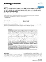
The study will use a randomized, sham-controlled,
pretest-posttest-follow up design involving two groups
of 10 children with hemiparesis and unilateral infarct or
periventricular leukomalacia as confirmed by MRI (total
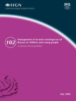
sample size = 20). (Fig. 1). This protocol is registered on
clinicaltrials.gov (# NCT02250092)
Hypotheses
Recruitment
1. Children in the tDCS/CIMT group will show
significantly greater excitability in the ipsilesional
hemisphere than will controls.
We will use our established database of over 200 families
to begin recruitment of children with hemiparesis, plus
talks at local pediatric facilities, mailings, posting on
related websites and newsletters. Incorporation of CIMT
Fig. 1 Flowchart of tDCS/CIMT study adapted from the Consolidated Standards of Reporting Trials (CONSORT). AHA: Assisting Hand Assessment,
CIMT: Constraint-Induced Movement Therapy, COPM: Canadian Occupational Performance Measure, DTI: Diffusion Tensor Imaging, MRI: Magnetic
Resonance Imaging, rs-fMRI: resting-state functional Magnetic Resonance Imaging, tDCS: transcranial Direct Current Stimulation, TMS: Transcranial
Magnetic Stimulation
Gillick et al. BMC Pediatrics (2015) 15:178
provides a motor training component to the study, and
families are more willing to participate with the promise
of receiving what would be an otherwise expensive intervention now covered by the study.
Participants- inclusion and exclusion criteria
Children and young adults, ages 8–21, with hemiparesis
due to perinatal stroke or periventricular leukomalacia
will be recruited for the study based on the following
eligibility criteria:
Page 5 of 10
Exit criteria
Subjects will exit the study if any of the following conditions exist:
1.
2.
3.
4.
Subject voluntarily withdraws from the study.
Subject acquires any of the listed exclusion criteria.
Subject completes the protocol.
Subject’s well-being, in the opinion of the Investigator,
would be compromised by study continuation.
5. Subject experiences a serious adverse event or
seizure.
6. Medical monitor and/or IRB recommendation
Inclusion criteria
Subjects will be eligible to participate in the study if the
following conditions exist:
1. Hemispheric Stroke or Periventricular Leukomalacia
confirmed by most recent MRI or CT radiologic
report with resultant congenital hemiparesis
2. ≥ 10 degrees of active motion at the
metacarpophalangeal joint
3. No evidence of seizure activity within the last
2 years
4. Presence of a motor evoked potential from at least
the contralesional hemisphere if not both
hemispheres
5. Ages 8–21 years
6. If ages 8–17, able to give informed assent along with
the informed consent of the legal guardian
7. Children who have had surgeries, which may
influence motor function e.g.- tendon transfer, will
be included, yet surgical history will be documented
Exclusion criteria
Subjects will be excluded from participation in the study
if any of the following conditions exist:
1.
2.
3.
4.
5.
6.
7.
8.
9.
Metabolic Disorders
Neoplasm
Epilepsy
Disorders of Cellular Migration and Proliferation
Acquired Traumatic Brain Injury
Pregnancy
Indwelling metal or incompatible medical devices
Evidence of skin disease or skin abnormalities
Botulinum toxin or Phenol block within the last sixmonths
Prior and concomitant therapy
Behavioral therapies including occupational, physical or
speech therapies will be allowed both during both the
intervention and follow-up period. These therapies will
be documented and reported.
Randomization
Each child will be randomized to either the real (intervention) or sham (control) tDCS arm of the study. The
randomization will be done by means of sealed envelopes
constructed by the study biostatistician using a random
number generator. The study coordinator will assign envelopes, in numerical order, to the subjects upon their
randomization.
Blinding
The investigator who performs the testing, the CIMT
interventionists and the physician who does the
evaluations will be blinded to the treatment arm as
will the child/caregiver/family. The study biostatistician, study coordinator and principal investigator (PI)
will be unblinded. The following procedure will be
employed:
1. The study biostatistician will provide sealed
envelopes to the study coordinator.
2. The study coordinator will share the group
assignment with the principal investigator in a
secure private room, in the absence of the physician,
research tester, subject legal guardian and subject.
The Medical Monitor to the study will have access
to the group assignment.
3. Blinding is applied through designated settings on
the tDCS machine of sham or real tDCS. The
unblinded investigator who administers the tDCS
(PI: BTG) will, during the intervention, switch the
setting on the tDCS device to the designated
placebo setting, hidden from view of the subject. For
the first 30-seconds of either setting, a gradual
“ramp-up” sensation of the stimulation occurs. This
is built into the machine for both settings. However,
for the sham stimulation, the sensation then abates,
and not until the end of the 20-minute session does
the subject receiving the 30-seconds of “ramp-down”
sham once again experience the sensation. The
sensation then occurs and “ramps-down” the
amperage until the machine turns off.
Gillick et al. BMC Pediatrics (2015) 15:178
4. Information on subject group assignment will be
logged and stored in a designated locked cabinet at
the study coordinators office.
Outcome Assessments. We incorporated the body function/structure and activity/participation domains of the
International Classification of Functioning, Disability and
Health [2727].
Primary outcome measure
The Assisting Hand Assessment (AHA) scaled score will
be used as the primary outcome measure as a test of
unilateral limb dysfunction; it is based on a child’s usual
performance in relevant activities. This test uses a standardized video-recorded play session. Activity is assessed
on 22 items using a 4-point rating scale. The range of
raw scores is 22–88 points, with higher scores indicating
better ability. Excellent interrater (0.97) and intrarater
(0.99) reliability has been found using this tool and it
has been found to have high validity in use with children[28-30]
Secondary outcome measures
The Canadian Occupational Performance Measure
(COPM) is an individualized outcome measure used to
detect changes in the self-perception of the client’s performance and satisfaction over time by identifying difficulty in performance of specific activities [31, 32]. The
tool is a self-reported ordinal scale score which encompasses domains in impairments, body structure, activity,
activity limitations, participation restrictions and environmental factors which an individual experiences. Testretest reliability has been found to be strong in the domains of performance (r = 0.89) and satisfaction (0.88).
Grip strength will also be measured using hand
dynamometry[33].
Sample size/power
Power calculations for the primary outcome measure
AHA scaled score difference between pre- and post-test
measurements used the sample size formula for normally distributed statistics with a type I error level of α
(2-sided test) and power of (1-β):
n ¼ V ðZ ð 1− ∝= 2Þ þ Z ð1−βÞ Þ2
Δ2
where Δ denotes the minimal clinically important difference (MCID) to detect, n denotes the sample size per
group, and V denotes the variance of the test statistic.
For ANCOVA analyses, V = 2(σ^2)(1-ρ^2) where σ^2 is
the (average) variability and ρ is the (average) correlation
between pre- and post-treatment measurements [34, 35].
Power (1-β) was computed for 10 patients per treatment
Page 6 of 10
group across a range of possible values for the correlation with alpha equal to 0.05. These are computed for
an MCID of 2.4, and for the rTMS/CIMT study treatment effect of 5.4, which is considered a reasonable estimate for this tDCS exploratory study [36]. The standard
deviation from the rTMS/CIMT study was used as an
estimate for the power calculations and was equal to 4.7.
In addition, 1.5 times the standard deviation of the
rTMS/CIMT study was also used in the power calculations to provide power estimates within a conservative
standard deviation. The correlation between pre- and
post-treatment AHA scores in the rTMS study was 0.97.
Based on a correlation of 0.8 and using the conservative
standard deviation of 7.1, we will have 80 % power to
detect the difference of 5.4 between treatment groups.
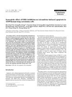
Study protocol
Children will be randomized into two groups: real tDCS/
CIMT (intervention) or sham tDCS/CIMT (control). The
children will receive 10 continuous weekday sessions of
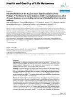
tDCS and CIMT. (Fig. 2) To clarify, we will include only
children in whom we can elicit a motor evoked potential
(MEP) using TMS applied to the ipsilesional hemisphere.
Although by doing so we limit greatly the number of children whom we can include, such conservatism is necessary to proceed in a safe and informed manner with
sufficient awareness of the functional topography of each
subject’s brain.
Rationale for use of tDCS Our previous research incorporated rTMS, which although promising as an intervention, has limitations especially with children [36, 37].
Our recent results demonstrate not only safety and but
also reveal significant improvements in hand function
with rTMS combined with constraint-induced movement therapy, yet we foresee the possible greater benefits from using tDCS over this type of non-invasive brain
stimulation in pediatric hemiparesis. (REF) First, both of
the interventions, tDCS and CIMT, applications of tDCS/
CIMT can occur concurrently whereas it is difficult to
incorporate the use of rTMS at the same time as performing behavioral therapy with the affected upper extremity.
Applying tDCS/CIMT simultaneously could optimize neuroplastic principles of concurrent firing of neurons and
strengthening of neuronal networks [38, 39]. Second,
tDCS may reduce costs. As rTMS can be ten times the
current cost of tDCS, cost may become prohibitive
when considering rTMS. Additionally, if the application
of tDCS reduces the need for additional therapies, a
further costs savings could be realized. Third, use of
rTMS has resulted in reports, albeit rare, of adverse
events such as seizures and syncopal episodes, in both
adults and children [40, 41] .
Gillick et al. BMC Pediatrics (2015) 15:178
Page 7 of 10
Fig. 2 Study design. CIMT, Constraint-Induced Movement Therapy; mos, months; tDCS, Transcranial Direct Current Stimulation
In addition to its economy and portability, tDCS has
shown improvements in motor function in adults with
and without the concurrent use of CIMT [42, 43]. The
rationale for the investigation of tDCS in the pediatric
population is that the use of non-invasive brain stimulation could translate more efficiently into clinical applications thus improving the quality of life for children with
hemiparesis. And tDCS safety, when using standard
guidelines, is supported in the literature as to having
common side effects limited to mild and reversible skin
irritation. Specific to the tDCS model we are using
(Soterix LTE 1×1 tDCS, NY, NY), the design reduces
skin irritation by conditioning the skin prior to stimulation and allowing the device operator to incorporate
subject feedback and adjust the controls without stopping. This model was specifically chosen for our project
because of its adaptability to resistance changes between
the surface electrodes and the subject. The model uses
two 9 V batteries to administer stimulation. As a safety
feature, the unit will automatically reduce the applied
voltage so as to maintain a low voltage level. The builtin ramp-up and ramp-down features allow for gradual
administration of the current. Another component of
this device specifically designed for children is the
adjustable strap that allows for optimal contact of the
surface electrodes with the skin for reduced irritation.
Safety. Safety testing includes a physician screening
with a modified pediatric stroke outcome measure at
pretest, interim day 5, posttest and follow-up sessions.
Additionally, vital signs are assessed before and after
each tDCS/CIMT session and subjects and caregivers
complete a subject report of symptoms and a tolerance
survey.
Corticospinal tract integrity using MRI Sequences.
Resting state fMRI (rs-fMRI) and Diffusion Tensor
Imaging (DTI) techniques are becoming alternatives to
more traditional task fMRI and structural MRI scans to
demonstrate functional and structural connectivity in
the brain, particularly in pediatric patients who may be
challenged by participating in task based fMRI studies.
Our previous work revealed cortical volume differences
in children with hemiparesis; further investigation using
connectivity metrics may provide a better understanding
of the integrity of the corticospinal tract in congenital
hemiparesis. Subjects will be scanned at UMN Center
for Magnetic Resonance Research on a Siemens 3 T
system to obtain a) T1-weighted structural MPRAGE
with a 1 mm isotropic spatial resolution; b) High angular
resolution multi-band diffusion imaging (HARDI) with
whole-brain coverage, 90 slices with 1.5 mm isotropic
spatial resolution, b-values of,1000 and 2000 s/mm2 with
64 gradient directions per b value, and 16 additional nondiffusion weighted images and c) Resting-state multi-band
based functional MRI will be obtained with whole
brain coverage, 72 slices with 2 mm isotropic slices,
TR = 730 ms, and 500 time points in 6 min. The
complete dataset will be obtained in less than 45 min,
taking into account time required for setup and comfortable installation of the child in the scanner. Resting-state
fMRI data will be processed using independent component analysis (ICA) to identify the sensory-motor area. Activation areas on the baseline scans will be used to refine
the site of tDCS and identify seed areas for tractography.
Excitability Measurements using TMS. TMS (Magstim,
Dyfed, UK) will be used to measure corticospinal excitability in the ipsilesional and contralesional hemispheres.
TMS will be delivered with a 70-mm figure-of-eight coil,
tangential to head with handle aligned 45 degrees
lateral. The assessment will incorporate single-pulse
motor threshold, 1 mV and cortical silent period testing. TMS measurements of cortical excitability guided
by stereotactic neuronavigation (Brainsight, Rogue Research, Montreal, QC, Canada), will be employed to create
a map of the motor cortex from anatomical MRI images
obtained at the CMRR. The co-registration of the TMS
coil position on the head with the MRI obtained image of
the brain anatomy will allow precision in children who
have neurologic lesions. All TMS testing will occur in the
UMN CTSI by the PI. For this, the child will be seated in
a child-sized reclining chair. Surface electromyographic
(EMG) electrodes will be attached over the first dorsal
interosseous muscles bilaterally, which will record the
motor evoked potentials (MEP) resulting from the magnetic stimulation to the M1 region of each hemisphere.
Next, the resting motor threshold (RMT) for TMS activation of the target muscle will be determined, and the location of the hotspot defined. If a threshold cannot be
elicited within at least the contralesional hemisphere (i.e.,
Gillick et al. BMC Pediatrics (2015) 15:178
no hotspot), the subject will be excluded from the study as
position of the tDCS electrodes will be influenced. The
RMT will be defined as the lowest amount of stimulation
inducing an MEP of at least 50uV on at least 3 of 5 stimulations Cortical excitability will also be tested by administration of 10 TMS pulses, at the minimum stimulus
intensity to produce MEPs of 1 mV [44].
CSP will be determined by the subject exerting finger
extensor contraction at approximately 30 % of maximum,
as guided by a force tracing on a computer display, while
a single TMS pulse at an intensity of 150 % of RMT is
delivered to the ipsilesional M1 cortex [45, 46]. The resultant CSP will be measured from the onset of the first peak
of the MEP to resumption of surface EMG activity with a
threshold value of 50 % mean prestimulus amplitude [47].
Intervention
tDCS/CIMT intervention group Children randomly
assigned to this group will receive 10 sessions of both
tDCS and CIMT on concurrent days over a span of two
weeks. As supported by results from our previous computerized modeling pilot, the subject will receive tDCS
at the motor hotspot at 0.7 mA current intensity for
20 min [19]. In a primary motor cortex (M1)/supraorbital (SO) montage, the anode will be positioned over
the ipsilesional supraorbital region and the cathode positioned over contralesional motor hotspot to induce
contralesional cortical inhibition. During this 20 min
session, the subject will be involved in CIMT for the
paretic hand and arm using a sling. The sling will be
applied for a 2-hour period, which incorporates the
20 min tDCS session. The CIMT will be given to the
subject on an individual basis for two hours each CIMT
treatment day by a CIMT-trained therapist/interventionist. The CIMT treatments will be standardized, consisting of shaping activities for function, range of motion
and strengthening of the paretic upper extremity. In
addition, children will continue to use their paretic limb
during functional activities at home and during a caregiversupervised home program. This combined treatment of
tDCS and CIMT will continue until 10 weekday sessions
are completed over the two-week period.
tDCS/CIMT control group Children in this group will
receive the same procedures and home program as the
intervention tDCS/CIMT group but, the tDCS device
will be set to a specific placebo setting which extinguishes the current after a 30 s to 1 min ramp-up phase
and gradually reintroduces the ramp-down at the end of
the 20 min session. This feature allows for placebo
control and blinding.
Statistical Analysis. There are 3 analysis populations
planned. Intent-to-treat (ITT) will include any subject randomized according to their treatment assignment. Per-
Page 8 of 10
protocol (PP) will include randomized subjects without
major protocol violations and who were compliant (at least
80 % of planned sessions with their treatment assignment.
A detailed list of the major protocol violations warranting
exclusion from the PP analysis will be determined prior to
trial commencement. The Safety population will include all
subjects who receive treatment, according to treatment received. We do not anticipate these groups to differ. The
safety profile will use the safety population and be primarily descriptive in nature reporting the number and percentage of adverse events for the two treatment groups and
include an aggregate breakdown by severity, seriousness,
and frequency (within a patient). Within-group analysis of
continuous outcomes comparing posttest to pretest will be
done with paired t-tests. Between-group analysis of continuous outcomes comparing the mean change from pretest to posttest between treatment groups will be adjusted
for baseline values in the fashion of ANCOVA for added
precision. The primary analysis will be based on the ITT
population with complementary analyses using the PP
population. The association between changes in brain
excitability/reorganization and motor function will also be
evaluated and based on linear regression. P-values less than
0.05 will be considered statistically significant.
Discussion
We outline the background and design of a trial with two
intervention groups comparing the effects of tDCS on
children and young adults with hemiparesis receiving
CIMT. The design of this study is predicated upon positive outcomes previously established with CIMT. Additionally, we incorporate consideration of trials we have
completed with neuromodulation interventions of repetitive transcranial magnetic stimulation and a tDCS pilot
with this population [19, 36, 48]. The proposed study design pursues investigation of a synergistic effect achieved
by combining rehabilitation (CIMT) and neuromodulatory
(tDCS) interventions. The study hypotheses reflect the importance of safety, feasibility and efficacy surrounding this
dual intervention. As non-invasive brain stimulation in
pediatric hemiparesis is in a nascent investigational phase,
understanding the potential value of such interventions is
paramount.
Abbreviations
AHA: Assisting-Hand Assessment; CP: Cerebral Palsy; CIMT: Constraint-Induced
Movement Therapy; COPM: Canadian Occupational Performance Measure;
MRI: Magnetic Resonance Imaging; NIBS: Non-Invasive Brain Stimulation;
rTMS: Repetitive Transcranial Magnetic Stimulation; tDCS: Transcranial
Direct Current Stimulation; TMS: Transcranial Magnetic Stimulation.
Competing interests
The authors report no competing interests.
Authors’ contributions
The authors listed on this manuscript were directly involved in the protocol
development. BTG conceived the study and developed the study design
Gillick et al. BMC Pediatrics (2015) 15:178
collaboratively with KR, JM and BM. BTG and TF are primarily responsible for
implementation of neuromodulation. TF, MW and GM are responsible for
neurologic assessment and MRI review. KR and JM will conduct the primary
statistical analysis. LK serves as medical monitor. BTG is primarily responsible
for the proposal and this protocol paper. All authors helped refine the study
protocol, and contributed to development and preparation of this
manuscript. All authors read and approved the final manuscript.
Acknowledgments
We wish to acknowledge Sally Jones for her critical revisions of this manuscript.
We thank also the families and children involved in our initial pilot testing to
establish this protocol, and the future participants and families to come.
Funding
This project is supported by the National Institutes of Health (NIH) Eunice
Kennedy Shriver National Institutes of Child Health and Development K01
Award (#HD078484-01A1), the Cerebral Palsy. Foundation and the Foundation
for Physical Therapy Magistro Family Grant. The project described was also
supported in part by Award Number UL1TR000114 and KL2TR000113 from the
National Center for Advancing Translational Sciences (NCATS) of the NIH. The
content is solely the responsibility of the authors and does not necessarily
represent the official views of the National Center For Research Resources or
the NIH. Study data were collected and managed using REDCap electronic data
capture tools hosted at the University of Minnesota. The University of
Minnesota Center for Magnetic Resonance Research funding supported the
imaging work number P41 EB015894.
Author details
1
University of Minnesota, 420 Delaware Street SE, MMC 388, Minneapolis, MN
55455, USA. 2Biostatistical Design and Analysis Center, University of
Minnesota, Minneapolis, MN 55455, USA. 3Department of Psychiatry,
University of Minnesota, Minneapolis, MN 55455, USA. 4Department of
Neurology, University of Minnesota, Minneapolis, MN, USA. 5Courage Kenny
Rehabilitation Institute, part of Allina Health, 800 East 28th Street,
Minneapolis, MN 55407, USA. 6Department of Neurology, Gillette Children’s
Specialty Healthcare, 200 University Ave E, Saint Paul, MN 55101, USA.
7
Division of Biostatistics, University of Minnesota, Minneapolis, MN, USA.
Received: 8 June 2015 Accepted: 27 October 2015
References
1. Agrawal N, Johnston SC, Wu YW, Sidney S, Fullerton HJ. Imaging data reveal
a higher pediatric stroke incidence than prior US estimates. Stroke.
2009;40(11):3415–21. doi:10.1161/STROKEAHA.109.564633.
2. DeLuca S, Case Smith J, Stevenson R, Ramey S. Constraint-induced movement
therapy (CIMT) for young children with cerebral palsy: Effects of therapeutic
dosage. Journal of Pediatric Rehabilitation Medicine. 2012;5(2):133–42.
doi:10.3233/PRM-2012-0206.
3. Eyre J, Smith M, Dabydeen L, Clowry G, Petacchi E, Battini R, et al. Is
hemiplegic cerebral palsy equivalent to amblyopia of the corticospinal
system? Ann Neurol. 2007;62(5):493–503. doi:10.1002/ana.21108.
4. Friel K, Chakrabarty S, Martin J. Pathophysiological mechanisms of impaired limb
use and repair strategies for motor systems after unilateral injury of the developing
brain. Dev Med Child Neurol. 2013;55 Suppl 4:27–31. doi:10.1111/dmcn.12303.
5. Martin JH. The corticospinal system: from development to motor control.
Neuroscientist. 2005;11:161–73.
6. Staudt M, Gerloff C, Grodd W, Holthausen H, Niemann G, Krageloh-Mann I.
Reorganization in congenital hemiparesis acquired at different gestational
ages. Ann Neurol. 2004;56(6):854–63. doi:10.1002/ana.20297.
7. Holmefur M, Krumlinde-Sundholm L, Bergstrom J, Eliasson AC. Longitudinal
development of hand function in children with unilateral cerebral palsy.
Developmental Medicine & Child Neurology. 2010;52(4):352–7.
8. Charles J, Wolf S, Schneider J, Gordon A. Efficacy of a child-friendly form of
constraint-induced movement therapy in hemiplegic cerebral palsy: A
randomized control trial. Dev Med Child Neurol. 2006;48(8):635–42.
doi:10.1017/S0012162206001356.
9. Charles J, Gordon AM. A critical review of constraint-induced movement
therapy and forced use in children with hemiplegia. Neural Plast.
2005;12:245–61. discussion 263–72.
Page 9 of 10
10. Juenger H, Linder-Lucht M, Walther M, Berweck S, Mall V, Staudt M. Cortical
neuromodulation by constraint-induced movement therapy in congenital
hemiparesis: an FMRI study. Neuropediatrics. 2007;38:130–6.
11. Reiss A, Wolf S, Hammel E, McLeod E, Williams E. Constraint-induced
movement therapy (CIMT): Current perspectives and future directions. Stroke
Research and Treatment. 2012;2012:159391–1. doi:10.1155/2012/159391.
12. Gordon AM, Hung Y, Brandao M, Ferre CL, Kuo H, Friel K, et al. Bimanual
training and constraint-induced movement therapy in children with
hemiplegic cerebral palsy: A randomized trial. Neurorehabil Neural Repair.
2011;25(8):692–702. doi:10.1177/1545968311402508.
13. Eliasson AC, Shaw K, Ponten E, Boyd R, Krumlinde-Sundholm L. Feasibility of
a day-camp model of modified constraint-induced movement therapy with
and without botulinum toxin A injection for children with hemiplegia. Phys
Occup Ther Pediatr. 2009;29(3):311–33.
14. Nitsche MA, Cohen LG, Wassermann EM, Priori A, Lang N, Antal A, et al.
Transcranial direct current stimulation: State of the art 2008 RID A-50342009 RID A-3544-2009. Brain Stimul. 2008;1(3):206–23. doi:10.1016/j.brs.
2008.06.004.
15. Nair DN. Improving motor function in chronic stroke patients using
simultaneous occupational therapy and tDCS. Stroke. 2008;39(2):542.
doi:10.1161/STROKEAHA.107.496935.
16. San-Juan D, Calcaneo Jde D, Gonzalez-Aragon MF, Bermudez Maldonado L,
Avellan AM, Argumosa EV, et al. Transcranial direct current stimulation in
adolescent and adult rasmussen’s encephalitis. Epilepsy Behav.
2011;20(1):126–31. doi:10.1016/j.yebeh.2010.10.031.
17. Mattai A, Miller R, Weisinger B, Greenstein D, Bakalar J, Tossell J, et al. Tolerability
of transcranial direct current stimulation in childhood-onset schizophrenia. Brain
Stimulation. 2011;4(4):275–80. doi:10.1016/j.brs.2011.01.001.
18. Young S, Bertucco M, Sheehan Stross R, Sanger T. Cathodal transcranial
direct current stimulation in children with dystonia: A pilot open-label trial.
J Child Neurol. 2013;28(10):1238–44. doi:10.1177/0883073812460092.
19. Gillick B, Feyma T, Menk J, Usset M, Vaith A, Wood T, et al. Safety and
feasibility of transcranial direct current stimulation in pediatric hemiparesis:
Randomized controlled preliminary study. Phys Ther. 2015;95(3):337–49.
doi:10.2522/ptj.20130565.
20. Krishnan C, Santos L, Peterson M, Ehinger M. Safety of noninvasive brain
stimulation in children and adolescents. Brain Stimulation. 2015;8(1):76–87.
doi:10.1016/j.brs.2014.10.012.
21. Edwards D, Cortes M, Datta A, Minhas P, Wassermann EM, Bikson M.
Physiological and modeling evidence for focal transcranial electrical brain
stimulation in humans: A basis for high-definition tDCS. Neuroimage.
2013;74:266–75. doi:10.1016/j.neuroimage.2013.01.042.
22. Bikson M, Rahman A, Datta A. Computational models of transcranial direct
current stimulation. Clinical EEG and Neuroscience. 2012;43(3):176–83.
doi:10.1177/1550059412445138.
23. Peterchev AV, Wagner TA, Miranda PC, Nitsche MA, Paulus W, Lisanby SH,
et al. Fundamentals of transcranial electric and magnetic stimulation dose:
Definition, selection, and reporting practices. Brain Stimulation.
2012;5(4):435–53. doi:10.1016/j.brs.2011.10.001.
24. Fusco A, De Angelis D, Morone G, Maglione L, Paolucci T, Bragoni M,
et al. The ABC of tDCS: Effects of anodal, bilateral and cathodal
montages of transcranial direct current stimulation in patients with
stroke-A pilot study. Stroke Res Treat. 2013;2013:837595–5. doi:10.1155/
2013/837595.
25. Manning KY, Fehlings D, Mesterman R, Gorter JW, Switzer L, Campbell C,
et al. Resting state and diffusion neuroimaging predictors of clinical
improvements following constraint-induced movement therapy in children
with hemiplegic cerebral palsy. Journal of Child Neurology. 2015.
doi:10.1177/0883073815572686.
26. Gauthier LV, Mark VW, Taub E, McCullars A, Barghi A, Rickards T, et al. Motor
recovery from constraint induced movement therapy is not constrained by
extent of tissue damage following stroke. Restorative Neurology and
Neuroscience. 2014;32(6):755–65. doi:10.3233/RNN-130366.
27. McDougall J, Wright V, Rosenbaum P. The ICF model of functioning and
disability: Incorporating quality of life and human development.
Developmental Neurorehabilitation. 2010;13(3):204–11. doi:10.3109/1751
8421003620525.
28. Eliasson AC, Krumlinde-Sundholm L, Rosblad B, Beckung E, Arner M, Ohrvall
AM, et al. Using the MACS to facilitate comunication about manual abilities
of children with cerebral palsy. Developmental Medicine & Child Neurology.
2007;49:156–7.
Gillick et al. BMC Pediatrics (2015) 15:178
29. Krumlinde Sundholm L, Holmefur M, Kottorp A, Eliasson A. The assisting
hand assessment: Current evidence of validity, reliability, and responsiveness
to change. Dev Med Child Neurol. 2007;49(4):259–64. doi:10.1111/j.14698749.2007.00259.x.
30. Holmefur M, Krumlinde-Sundholm L, Eliasson AC. Interrater and intrarater
reliability of the Assisting Hand Assessment. American Journal of
Occupational Therapy. 2007;61:79–84.
31. Law M, Baptiste S, McColl M, Opzoomer A, Polatajko H, Pollock N. The
canadian occupational performance measure: An outcome measure for
occupational therapy. Can J Occup Ther. 1990;57(2):82–7.
32. Cup E, REIMER W, Thijssen M, VAN KM. Reliability and validity of the canadian
occupational performance measure in stroke patients. Clin Rehabil.
2003;17(4):402–9.
33. Mathiowetz V, Weber K, Volland G, Kashman N. Reliability and validity of
grip and pinch strength evaluations. J Hand Surg. 1984;9(2):222–6.
34. Frison L, Pocock SJ. Repeated measures in clinical trials: Analysis using mean
summary statistics and its implications for design. Stat Med.
1992;11(13):1685–704. doi:10.1002/sim.4780111304.
35. Senn S. Change from baseline and analysis of covariance revisited. Stat
Med. 2006;25(24):4334–44. doi:10.1002/sim.2682.
36. Gillick BT, Krach LE, Feyma T, Rich TL, Moberg K, Thomas W, et al. Primed
low-frequency repetitive transcranial magnetic stimulation and constraintinduced movement therapy in pediatric hemiparesis: A randomized
controlled trial. Dev Med Child Neurol. 2014;56(1):44–52. doi:10.1111/
dmcn.12243.
37. Gillick B, Krach L, Feyma T, Rich T, Moberg K, Menk J, et al. Safety of
primed repetitive transcranial magnetic stimulation and modified
constraint-induced movement therapy in a randomized controlled trial in
pediatric hemiparesis. Arch Phys Med Rehabil. 2014. doi:10.1016/j.apmr.
2014.09.012.
38. Hahn C, Rice J, Macuff S, Minhas P, Rahman A, Bikson M. Methods for extra-low
voltage transcranial direct current stimulation: Current and time dependent
impedance decreases. Clinical Neurophysiology. 2013;124(3):551–6.
doi:10.1016/j.clinph.2012.07.028.
39 Kessler SK, Minhas P, Woods AJ, Rosen A, Gorman C, Bikson M. Dosage
considerations for transcranial direct current stimulation in children: A
computational modeling study. PLoS ONE. 2013;8(9):e76112–2. doi:10.1371/
journal.pone.0076112.
40. Kirton A, Deveber G, Gunraj C, Chen R. Neurocardiogenic syncope
complicating pediatric transcranial magnetic stimulation. Pediatr Neurol.
2008;39(3):196–7. Available.
41. Rossi S, Hallett M, Rossini PM, Pascual-Leone A, Safety of TMS
Consensus Group. Safety, ethical considerations, and application
guidelines for the use of transcranial magnetic stimulation in clinical
practice and research. Clin Neurophysiol. 2009;120(12):2008–39.
doi:10.1016/j.clinph.2009.08.016.
42. Bolognini N, Vallar G, Casati C, Latif LA, El-Nazer R, Williams J, et al.
Neurophysiological and behavioral effects of tDCS combined with
constraint-induced movement therapy in poststroke patients. Neurorehabil
Neural Repair. 2011;25(9):819–29. doi:10.1177/1545968311411056.
43. Schlaug G, Renga V, Nair D. Transcranial direct current stimulation in
stroke recovery. Arch Neurol. 2008;65(12):1571–6. doi:10.1001/archneur.
65.12.1571.
44. Kirton A, Deveber G, Gunraj C, Chen R. Cortical excitability and
interhemispheric inhibition after subcortical pediatric stroke: Plastic
organization and effects of rTMS. Clin Neurophysiol. 2010;121(11):1922–9.
doi:10.1016/j.clinph.2010.04.021.
45. Kobayashi Alvaro MPL. Transcranial magnetic stimulation in neurology.
Lancet Neurology. 2003;2(3):145–56. doi:10.1016/S1474-4422(03)00321-1.
46. Kobayashi M, Hutchinson S, Theoret H, Schlaug G, Pascual-Leone A.
Repetitive TMS of the motor cortex improves ipsilateral sequential simple
finger movements. Neurology. 2004;62:91–8.
47. Kimberley TJ, Borich MR, Prochaska KD, Mundfrom SL, Perkins AE, Poepping
JM. Establishing the definition and inter-rater reliability of cortical silent
period calculation in subjects with focal hand dystonia and healthy controls.
Neurosci Lett. 2009;464(2):84–7. Available.
48. Carey JR, Deng H, Gillick BT, Cassidy JM, Anderson DC, Zhang L, et al. Serial
treatments of primed low-frequency rTMS in stroke: Characteristics of
responders vs. nonresponders. Restorative Neurol Neurosci. 2014;32(2):323–35.
doi:10.3233/RNN-130358.
Page 10 of 10
Submit your next manuscript to BioMed Central
and take full advantage of:
• Convenient online submission
• Thorough peer review
• No space constraints or color figure charges
• Immediate publication on acceptance
• Inclusion in PubMed, CAS, Scopus and Google Scholar
• Research which is freely available for redistribution
Submit your manuscript at
www.biomedcentral.com/submit