Abnormal movements associated with oropharyngeal dysfunction in a child with Chiari I malformation
Bạn đang xem bản rút gọn của tài liệu. Xem và tải ngay bản đầy đủ của tài liệu tại đây (368.83 KB, 4 trang )
Berthet et al. BMC Pediatrics 2014, 14:294
/>
CASE REPORT
Open Access
Abnormal movements associated with
oropharyngeal dysfunction in a child with Chiari I
malformation
St?phanie Berthet 1, Louis Crevier2 and Colette Deslandres1,3*
Abstract
Background: Chiari I malformations (CM I) are rare hindbrain herniations. Dysphagia and other oropharyngeal
dysfunctions may be associated with CM I, but to our knowledge, no clinical presentation similar to ours has ever
been reported. The purpose of this communication is to draw attention to a unique and atypical clinical
presentation of a child with CM I.
Case presentation: A 7-year-old boy was evaluated for a two month history of atypical movements which would
occur in the evening, and last for an hour after eating. These stereotypical movements with the head and chest
bending forward and to the left side, accompanied by a grimace, were associated with sensation of breath locking
without cyanosis. Pain and dysphagia were absent. The neurological examination was normal. The possibility of
Sandifer syndrome posturing occurring with gastroesopageal reflux disease was considered but neither pain nor
back hyperextension were associated with the atypical movements. Neither proton pump inhibitors (PPI) nor
prokinetic agents improved his symptoms.
Upper endoscopy and esophageal biopsy did not reveal eosinophilic esophagitis nor reflux esophagitis. Ear, throat and
nose (ENT) exam was normal. A severe gastroparesis was demonstrated on milk scan study. Two 24 hour oesophageal
pH probe studies pointed out severe gastroesophageal reflux (GER). High resolution manometric evaluation of the
oesophagus revealed normal sphincter pressures and relaxations with no dysmotility of the esophageal body.
Electroencephalography and polysomnography were normal. A brain magnetic resonance imaging (MRI) was
performed and revealed a CM I: cerebellar tonsils extending to 12 mm, with syringomyelia (D4-D5).
For a long period of time, the child? s abnormal movements were considered to be nothing but tics and the CM I a
fortuitous finding. Since the child remained symptomatic despite medical treatment, it was decided to proceed with
surgery. One year after the onset of his symptoms, he underwent posterior fossa decompression with upper cervical
laminectomy and expansion duroplasty. Postoperative MRI confirmed adequate decompression. His atypical posture
and dyspnea completely resolved after surgery and he remains asymptomatic two years later.
Conclusion: Children may have atypical presentations of CM I. Thus, CM I diagnosis should be considered in
unexplained atypical oropharyngeal dysfunctions.
Keywords: Chiari I malformation, Oropharyngeal dysfunction, Abnormal movements, Gastroesophageal reflux (GER),
Gastroesophageal reflux disease (GERD)
* Correspondence:
1
Department of Pediatric Gastroenterology Hepatology and Nutrition, CHU
Sainte Justine, University of Montreal, Montreal, QC, Canada
3
CHU Sainte Justine, 3175, C?te Sainte Catherine, H3T1C5 Montr?al, QC,
Canada
Full list of author information is available at the end of the article
? 2014 Berthet et al.; licensee BioMed Central. This is an Open Access article distributed under the terms of the Creative
Commons Attribution License ( which permits unrestricted use, distribution, and
reproduction in any medium, provided the original work is properly credited. The Creative Commons Public Domain
Dedication waiver ( applies to the data made available in this article,
unless otherwise stated.
Berthet et al. BMC Pediatrics 2014, 14:294
/>
Page 2 of 4
Background
Chiari I malformations (CM I) are rare hindbrain herniations that may be present in children or adults. CM I is
characterized by an abnormal position of the cerebellar
tonsils, which herniate outside the cranial cavity into the
upper cervical canal: this is associated with an obliteration
of the subarachnoid spaces at the level of the foramen
magnum [1,2]. Anomalies associated with CM I include
syringomyelia. CM I can be easily identified on magnetic
resonance imaging (MRI) of the cranio-vertebral junction
[3]. Tonsillar herniation of 5 mm below the foramen magnum is the most common cut off for radiological diagnosis
of CM I [4]. More recently, because of the ease of diagnosis and increased clinical awareness, pediatric cases are
increasingly reported [5]. Many studies have reported
symptoms such as headaches, scoliosis or neurological troubles which were attributed to compression of neural structures. Dysphagia and other oropharyngeal dysfunctions have
also been reported but, to our knowledge, no clinical presentation similar to ours has ever been reported.
The purpose of this communication is to draw attention
to a unique and atypical clinical presentation of a child
with CM I.
Case presentation
A 7-year-old boy was evaluated for a two month history of
atypical movements presenting in the evening, and lasting
an hour after eating. These stereotypical movements with
the head and chest bending forward and to the left side,
accompanied by a grimace were associated with sensation
of breath locking without cyanosis. Pain and dysphagia
were absent. The neurological examination was normal.
The possibility of Sandifer syndrome posturing occurring with gastroesophageal reflux disease (GERD) was
considered but neither pain nor back hyperextension
were associated with the atypical movements. PPI did
not improve his symptoms. Various prokinetic agents
(metoclopramide, motilium, cisapride and erythomycin)
were also inefficient.
Upper endoscopy and esophageal biopsy did not reveal
eosinophilic esophagitis or other abnormalities. ENT exam
was normal. A severe gastroparesis was demonstrated on
milkscan study. Two 24 hour esophageal pH probe studies
pointed out severe GER. High resolution manometric
evaluation of the oesophagus revealed normal sphincter
pressures and relaxations with no dysmotility of the
esophageal body. Electroencephalography and polysomnography were normal. Because of the unexplained
dyspnea associated with this abnormal posture, a head
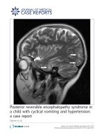
MRI was performed and revealed a CM I: cerebellar tonsils extending to 12 mm, with syringomyelia (D4-D5)
(Figure 1).
For a long period of time, the child abnormal movements were only considered to be tics and the CM I was
Figure 1 Cerebellar tonsils herniation on magnetic resonance
imaging: Chiari malformation type I.
considered a fortuitous finding. Since the child remained
symptomatic despite medical treatment, it was eventually
decided to proceed with surgery. The operative procedure
was done one year after the onset of his symptoms. He
underwent posterior fossa decompression with upper
cervical laminectomy and expansion duroplasty. No postoperative complication occurred. Postoperative MRI confirmed adequate decompression. His atypical posture and
dyspnea completely resolved in the week after surgery.
More than two years after surgery, the child remains
asymptomatic. The patient and parents have refused any
further invasive testing (such as a control esophageal pH
probe study) as the patient was symptom free.
Discussion
Although CM I is increasingly detected in children [3,5],
much remains unknown about its natural history. The
pathophysiology of CM I and its associated anomalies
have been the subject of considerable debate [1]. Chiari I
is a multifactorial condition that is thought to result from
a congenital small posterior fossa. Neurologic signs and
symptoms may be related directly to a tight foramen
magnum associated with the cerebellar tonsillar herniation,
with compression and/or distorsion of the medulla and
lower cranial nerves.
In adults, the most common clinical symptoms are posterior headaches and/or neck pain exacerbated by Valsava
maneuvers [4]. The clinical presentation of young children
Berthet et al. BMC Pediatrics 2014, 14:294
/>
Page 3 of 4
with CM I differs from that of older children and adults.
Albert et al. showed that patients aged 0 to 2 years were
much more likely to have oropharyngeal dysfunction,
whereas those aged 3 to 5 years were more likely to have
syringomyelia, frequently associated with scoliosis [6]. CM
I pediatric presentations from published series are reported
in Table 1. These retrospective series include both operated
and non operated CM I patients: 26 to 37% of the patients
[4,7] were asymptomatic with a fortuitous discovery, while
the remaining had a variety of neurological symptoms
including headaches, ataxia, sensory or motor deficits and
lower cranial abnormalities. The most common symptoms
were headaches and scoliosis [5].
Oropharyngeal dysfunction is not frequently reported.
In fact, in Tubbs? study on 500 cases of pediatric CM I,
oropharyngeal dysfunction only represented 4% of the
symptoms [5]. In some studies involving over 100 patients
no esophageal symptoms were reported [1,4,7]. Moreover,
these dysfunctions are often poorly described, and can
manifest with cough, stridor, dysphagia, abnormal vocal
cord movement, GERD, aspiration, prolonged feeding,
vomiting, sleep apnea or failure to thrive [3,6]. Perkin et al.
[8] have reported common dysphagia in patients with
CM1 malformation by traction of the lower cranial nerves
secondary to the herniation by the CM1 malformation.
Dysphagia is associated with a global impairment of all
phases of swallowing on videofluoroscopy. As they mention dysphagia may be the presenting symptom in some
patients.
Cardi et al. [9] described gastroparesis as a cause of Sandifer syndrome. Indeed gastroparesis may enhance GERD and
thus subsequently induce a Sandifer syndrome. Our patient
had a very unusual presentation and we initially thought that
he presented with an atypical case of Sandifer syndrome as
he had well documented severe gastroparesis and GERD.
We do not exclude that he might have had pre-existing
asymptomatic gastroparesis. Deterioration of his CMI might
have worsened his gastroparesis. We were unable to obtain
invasive diagnostic procedures (as esophageal pH probe
study) after the patient? s surgery but we did obtain a milk
scan study a year after surgery which showed improved but
persistent gastroparesis in an absolutely symptom free
patient. Neurological and gastrointestinal symptoms are
frequently associated in different neurological conditions.
Table 1 Clinical presentations of Chiari malformation type I in children
Study period
Tubbs et al. [5]
Alabama
Benglis et al.
[4] Miami
Caldarelli et al.
[2] Roma
Greenlee et al. Albert et al.
[3] Iowa
[6] Iowa
Aitken et al. [7] Park et al. [1]
San Francisco Boston
1989-2010
1999-2008
1993-2005
1987-2001
1984-2007
1997-1998
1988-1996
No. of children
500
124
30
31
39
51
68
Inclusion criteria
-
no surgery
symptomatic
age <6
age <6
age <20
-
Retrospective study
yes
yes
yes
yes
yes
yes
yes
Age at diagnostis
11
7
5,5
3,5
3,5
11
12
Male %
54
-
40
42
39
-
48
Surgery
500
0
30
25
39
8
68
Asymptomatic %
-
35
0
-
-
37
-
Serious manifestations %
-
-
-
-
-
-
-
Loss of consciousness %
1,2
1,6
-
-
-
4
-
Headache %
40
39
57
23
46
55
63
Neck pain %
-
11
-
-
-
12
-
Ataxia %
4
-
20
-
-
8
16
Sleep apnea %
5
-
20
29
-
-
-
Motor deficit %
10
4
70
3
-
20
45
Sensory deficit %
-
12
36
6
-
6
-
Scoliosis %
18
4
7
23
28
2
16
Dysphagia, oropharyngeal 4
dysfunction %
-
7
35
74
-
-
Vomiting %
16
3
-
-
-
-
3
Dyspnea %
1,2
-
-
-
-
-
-
Dysarthria %
5
-
7
-
-
4
-
Abnormal movement
-
-
-
19
-
-
-
- = no value.
Berthet et al. BMC Pediatrics 2014, 14:294
/>
Greenlee et al. reported abnormal movements in 6
children with CM I but did not describe them [3]. To our
knowledge, no study has reported abnormal movements
related to eating in association with CM I.
In conclusion, our patient? s presentation is clearly
unique and the total resolution of symptoms following
posterior fossa decompression surgery confirms the link
between the abnormal postures and CM I. Again, we
were unable to perform control studies following surgery due to the patient and the parents? decision to not
perform any further invasive testing.
In the literature, complications occur in only 2.4% of
the patients undergoing decompressive surgery for CM I
[5]. Spontaneous resolution of childhood CM I has been
described in several cases [10]. The Benglis study represents the largest series of pediatric patients with CM I
followed without surgery. No new neurological deficits
were observed during the follow up period in this population [4], adding to the controversy regarding the indication for surgery [7]. Therefore, pediatric patients with
CM I who are not clearly symptomatic and do not have
a syrinx or scoliosis, should not undergo surgery [4].
Conclusion
Symptomatic CM I are being increasingly recognized in
young children. The availability of MRI has certainly contributed to this phenomenon. As shown in our case, children may present with atypical manifestations, making CM
I a complex clinical diagnostic challenge. CM I should be
considered in the differential diagnosis of atypical oropharyngeal dysfunction.
Consent
Written informed consent was obtained from the patient? s
parents and assent was obtained from the patient (as he
was too young for a written consent) for publication of
this case report and any accompanying images. A copy of
the written consent is available for review by the Editor of
this journal. The study was approved by the local institutional board.
Abbreviations
CM 1: Chiari I; PPI: Proton pump inhibitors; ENT: Ear, throat and nose;
MRI: Magnetic resonance imaging; GERD: Gastroesophageal reflux disease;
GER: Gastroesophageal reflux.
Page 4 of 4
Acknowledgements
We acknowledge the pediatric gastroenterology, hepatology and nutrition
service of the CHU Sainte-Justine, Universit? de Montr?al, Montr?al, and the
pediatric neurosurgery department of CHU Sainte-Justine. We also acknowledge Mrs Nicole Th?riault for all her secretarial help and Mrs H?l?ne Restieri
for her nursing support to the patient and his family.
Author details
1
Department of Pediatric Gastroenterology Hepatology and Nutrition, CHU
Sainte Justine, University of Montreal, Montreal, QC, Canada. 2Department of
Pediatric Neurosurgery, CHU Sainte Justine, University of Montreal, Montreal,
QC, Canada. 3CHU Sainte Justine, 3175, C?te Sainte Catherine, H3T1C5
Montr?al, QC, Canada.
Received: 7 May 2014 Accepted: 11 November 2014
References
1. Park JK, Gleason PL, Madsen JR, Goumnerova LC, Scott RM: Presentation
and management of Chiari I malformation in children. Pediatr Neurosurg
1997, 26(4):190? 196.
2. Caldarelli M, Novegno F, Vassimi L, Romani R, Tamburrini G, Di Rocco C: The
role of limited posterior fossa craniectomy in the surgical treatment of
Chiari malformation type I: experience with a pediatric series. J Neurosurg
(3 suppl pediatrics) 2007, 106(3 suppl):187? 195.
3. Greenlee J, Donovan KA, Hasan DM, Menezes AH: Chiari I malformation in
the very young child: the spectrum of presentations and experience in
31 children under age 6 years. Pediatrics 2002, 110(6):1212? 1219.
4. Benglis D Jr, Covington D, Bhatia R, Elhammady MS, Ragheb J, Morrison G,
Sandberg DI: Outcomes in pediatric patients with Chiari malformation type
I followed up without surgery. J Neurosurg Pediatrics 2011, 7(4):375? 379.
5. Tubbs RS, Beckman J, Naftel RP, Chern JJ, Wellons JC 3rd, Rozzelle CJ, Blount
JP, Oakes WJ: Institutional experience with 500 cases of surgically treated
pediatric Chiari malformation type I. J Neurosurg Pediatrics 2011, 7(3):248? 256.
6. Albert GW, Menezes AH, Hansen DR, Greenlee JD, Weinstein SL: Chiari
malformation type I in children younger than age 6 years: presentation
and surgical outcome. J Neurosurg Pediatrics 2010, 5(6):554? 561.
7. Aitken LA, Lindan CE, Sidney S, Gupta N, Barkovich AJ, Sorel M, Wu YW:
Chiari type I malformation in a pediatric population. Pediatr Neurol 2009,
40(6):449? 454.
8. Perkin GD, Murray-Lyon I: Neurology and the gastrointestinal system.
J Neurol Neurosurg Psychiatry 1998, 65(3):291? 300.
9. Cardi E, Corrado G, Cavaliere M, Capocaccia P, Matrunola M, Rea P,
Pacchiarotti C: Delayed gastric emptying in an infant with Sandifer
syndrome. Ital J Gastroenterol 1996, 28(9):518? 519.
10. Sun PP, Harrop J, Sutton LN, Younkin D: Complete spontaneous resolution
of childhood Chiari I malformation and associated syringomyelia.
Pediatrics 2001, 107:182? 184.
doi:10.1186/s12887-014-0294-3
Cite this article as: Berthet et al.: Abnormal movements associated with
oropharyngeal dysfunction in a child with Chiari I malformation. BMC
Pediatrics 2014 14:294.
Submit your next manuscript to BioMed Central
and take full advantage of:
? Convenient online submission
Competing interests
The authors declare that they have no competing interests.
? Thorough peer review
? No space constraints or color ?gure charges
Authors? contributions
SB: Conducted a critical analysis of the case reviewed the literature and
wrote the manuscript. LC: Revised the manuscript and added a critical
review to the surgical part of the manuscript. CD: Conceived of the study,
participated in its design and coordinated and helped to draft and edit the
final manuscript. All authors read and approved the final manuscript.
? Immediate publication on acceptance
? Inclusion in PubMed, CAS, Scopus and Google Scholar
? Research which is freely available for redistribution
Submit your manuscript at
www.biomedcentral.com/submit