Detection of porcine cysticercosis in Nagaland of North East, India
Bạn đang xem bản rút gọn của tài liệu. Xem và tải ngay bản đầy đủ của tài liệu tại đây (377.13 KB, 7 trang )
Int.J.Curr.Microbiol.App.Sci (2019) 8(8): 2590-2596
International Journal of Current Microbiology and Applied Sciences
ISSN: 2319-7706 Volume 8 Number 08 (2019)
Journal homepage:
Original Research Article
/>
Detection of Porcine Cysticercosis in Nagaland of North East, India
Acheenta G. Barua*, Koushik Kakoty, Pranjal M. Nath and Nur Abdul Kader
Department of Veterinary Public Health, College of Veterinary Science, Assam Agricultural
University, Khanapara, Guwahati-781022, India
*Corresponding author
ABSTRACT
Keywords
Pig, Nagaland,
Cysticercuscellulos
ae, PCR,
Taeniasolium
Article Info
Accepted:
22 July 2019
Available Online:
10 August 2019
A total of 360 pig carcasses were examined through meat inspection in
different market places of Dimapur and Kohima district of Nagaland, India
in the month of March, 2019. Out of which, 6 (1.67%) were found positive
for porcine cysticercosis with visible cysts. The serum samples were
collected from 300 pigs and out of which 10 (3.00%) serum samples were
found positive for Cysticercus cellulosae antibody. Polymerase chain
reaction (PCR) assay was performed to confirm Cysticercus cellulosae and
to validate the results of meat inspection. Oligonucleotide primers targeting
against the large subunit rRNA gene (TBR primers) of Taenia solium were
used in this study. On reactivity in PCR test, the TBR primers yielded
products of 286bp in cysticercosis positive cases.
Introduction
Cysticercus cellulosae, the metacestode stage
of Taenia solium is an underrated and a
neglected zoonotic disease involving pig and
man (Willingham et al., 2008). Porcine
cysticercosis plays a crucial role in the
transmission and maintenance of human
taeniosis and cysticercosis (Pinto et al., 2000).
In Indian context, meat consumption is greatly
influenced by culture, traditions, customs and
taboos especially in the rural societies. In
north eastern states of India pork is considered
as a traditional food item (Anonymous. 2012)
and has much higher pork consumption than
the other parts of the country. The demand for
pork was increasing along with prices in both
Assam and Nagaland according to the traders.
Thus, accurate inspection of pork is very
important.
Although the pork consumption is highest in
North East India but the scientific procedure
of diagnosis and meat inspection is still
lacking. While meat inspection is the preferred
diagnostic tool to detect heavily infected
carcasses but it is not reliable in detecting
lightly infected carcasses (Cai et al., 2006). So
detection of antibodies by ELISA (Ab-ELISA)
can be used as an alternative tool for the
diagnosis of porcine cysticercosis (Deckers et
al., 2008).
2590
Int.J.Curr.Microbiol.App.Sci (2019) 8(8): 2590-2596
Moreover, meat inspection requires expertise
of the meat inspector; otherwise cysticerci
may be confused with T. hydatigena cysticerci
(Kedra et al., 2001), hydatid cysts (Deplazes
et al., 2005) and left over of muscle fasciae
(Geysen et al., 2007). To overcome such type
of difficulties application of ELISA and
molecular tools like Polymerase chain reaction
(PCR) can be effective in diagnosis and
validation of meat inspection results.
So considering the public health significance
of the disease, the purpose of this study was to
evaluate the performance of ELISA for antemortem diagnosis in pigs and estimating the
prevalence of porcine cysticercosis in
slaughtered pig through meat inspection and
PCR for validation of results.
Materials and Methods
Study area
The study was carried out between January
2019 to February 2019 in Dimapur and
Kohima district of Nagaland, India. Three
blocks from each district were selected for
collection of sera sample and for carcass
examination.
Sample collection
Collection of blood samples
A total of 300blood samples were collected
from the anterior vena cava of pigs by using
BDV accutainer®needles (gauge 19) and
BDV acutainer®plain tubes (10 ml). The
blood samples were kept standing in an ice
box at +4◦C to ensure no haemolysis occurred
while in the field.
At the laboratory, blood was centrifuged to
separate serum from blood clot. Serum was
harvested into barcoded2 ml vials that were
stored at −20◦C until processing.
Carcass Inspection
A total of 360 pig carcasses were examined as
per the standard method [10] for the presence
of Cysticercus cellulosae from different
market places of Dimapur and Kohima
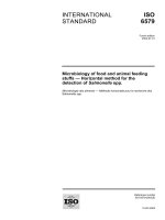
district. Majority of the pigs were brought
from Uttar Pradesh, India to Dimapur and
distributed to the rest of the districts of
Nagaland on weekly basis (Fig.1). Fifty gram
(g) of tissue each from brain, tongue, liver and
skeletal muscles of infected pig carcasses were
brought to the laboratory in ice-box
Serological tests
ELISA was performed as per the
manufacturer’s protocol of RIDASCREEN®
Taenia solium IgG kit (RBiopharm AG,
Germany). The absorbance was read at 450
nm with an ELISA reader (Lab systems
Multiskan Plus, Thermo Fisher Scientific,
USA). Samples having percent positivity
value 0.50 or above (%P ≥ 0.05) were
categorized as positive and below 0.50 as
negative.
Extraction of DNA from cysts
Extraction of DNA from cysts/suspected
lesion was made possible using a
commercially available QIAamp tissue kit
(QIAGEN, Hilden, Germany) according to the
manufacturer’s instructions (Kolesarova et al.,
2012). Briefly, 200 μl of cyst/lesion
homogenate, 20 μl of proteinase K stock
solution, and 200 μl of lysing buffer were
pipetted into 1.5 ml Eppendorf tube. The
mixture was incubated at 37 °C for 1 h and
then at 70 °C for 30 min before the addition of
200 μl of absolute alcohol and mixing by
vortexing. The mixture was then transferred to
the QIAamp spin column placed in a clean 2
ml collection tube and centrifuged at 8000
RPM in MiniSpin centrifuge (Eppendorf,
Wesseling-Berzdorf, Germany) for 1 min at
2591
Int.J.Curr.Microbiol.App.Sci (2019) 8(8): 2590-2596
room temperature. The QIAamp spin column
was washed twice with 500 μl of the washing
buffers by spinning for 1 min. The QIAamp
spin column was placed in a clean 1.5 ml
eppendorf tube and the DNA was eluted with
100 μl of elution buffer preheated at 70 °C.
Maximum DNA yield was obtained by
spinning at 12,000 rpm for 1 min at room
temperature. From the suspended nucleic acid
5 μl was used in the PCR amplification. The
extracted DNA was quantified using
spectrophotometer at 260 nm wave length
Oligonucleotide primers
The oligonucleotide primers specific to T.
solium were adopted from already published
sequences. The primers TBR3 (5′-GGC TTG
TTT GAATGG TTT GAC G-3) and TBR-6
(5′-GCT ACTACA CCT AAA TTC TAA CC3) against large subunit rRNA gene (Jardim et
al., 2006).
PCR amplification and detection of PCR
product
The PCR reaction was performed in a thermal
cycler (Eppendorf, Hamburg, Germany) in 20
μL volume containing 2μl DNA sample (100
ng/ μl), 1 μl (10 pmol) of each forward and
reverse primer, 10 μL of DreamTaq Green
PCR Master Mix (2X) (Thermo Scientific™,
USA) and 7 μL of nuclease free water
(Thermo Scientific™, USA). A total of 40
PCR cycles were run with the following
conditions: one initial denaturation cycle at
94°C for 3 min, followed by 40 repeated
cycles with temperatures at 94°C for 30 s
(denaturation), 59°C for 30 s (annealing,
specific for primers) and 72°C for 1 min. After
the final cycle, the preparations were kept at
72°C for 5 min for final elongation, and the
PCR products were stored at 4°C in thermal
cycler for further use (Sreedevi et al., 2012).
Five microliters of PCR amplicons was
analyzed on ethidium bromide stained 2%
agarose gel (In Genius Gel documentation
system, Syngene, UK). The sizes and
quantities of PCR products were verified by
comparison with a 100 bp plus quantitative
ladder (Thermo Scientific™, USA)
Results and Discussion
Overall
sero-prevalence
of
porcine
cysticercosis in Dimapur and Kohimadistict
was recorded as3.00 % (Table 2). Higherseroprevalence of porcine cysticercosis was
recorded in Dimapur (3.33 %) compared to
Kohima district (2.67%) of Nagaland. These
finding were in close conformity of Barua et
al., (2018) where they recorded seroprevalence of porcine cysticercosis in 4 states
of North EastIndiaas2.72 %.
This might be because, in our study area, the
vast majority of pigs are confined in pens,
even among smallholders, compared to more
extensive (i.e., free roaming and scavenging)
pig husbandry practices which may
predominate in the other study settings.
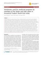
All the 360 pigs inspected at slaughter were
from different market places of Dimapur and
Kohima district of Nagaland, India (Fig. 2&
Fig. 3). The overall prevalence of porcine
cysticercosis based on post-mortem inspection
was 1.67% (Table 1). The prevalence was
higher in Dimapur (2.22 %) compared to
Kohima district (1.67%) of Nagaland.
Previously, Sarma (1977) recorded 6.64 %
positive cysticercosis from greater Guwahati
of Assam. Plain (1991) recorded highest
infection 11.90 % infection from North
Eastern region of Assam. Borkataki et al.,
(2012) conduct study in three districts of
Assam for a period of one year and found 93
pigs (9.50%) positive for cysticercosis out of
978 pigs. Although the previous workers
recorded higher prevalence of porcine
cysticercosis, Barua et al., (2018) recorded
2592
Int.J.Curr.Microbiol.App.Sci (2019) 8(8): 2590-2596
prevalence as 0.92 %in 4 states of North East
India. This might be due to importing of
scavenging pigs from other parts of India as
reported by butchers.
Table.1 Prevalence of porcine cysticercosis pigs of Dimapur and Kohima district of Nagaland
District
Study area
Medziphema
Dimapur
Khuhboto
Nihokhu
Total
Sechu-Zubza
Kohima
Kohima
Zakhama
Total
Overall Total
Animals inspected(No.)
60
60
60
180
60
60
60
180
360
Cyst positive pig (%)
1 (1.67%)
2 (3.33%)
1 (1.67%)
4 (2.22%)
1 (1.67%)
0 (0.00%)
1 (1.67%)
2(1.11%)
6 (1.67%)
Table.2 Sero-Prevalence of porcine cysticercosis in Dimapur and Kohima district of Nagaland
District
Study area
Medziphema
Dimapur
Khuhboto
Nihokhu
Total
Sechu-Zubza
Kohima
Kohima
Zakhama
Total
Overall Total
No. of Pig Serum
50
50
50
150
50
50
50
150
300
Sero Positive (%)
2 (4%)
2 (4%)
1 (2%)
5 (3.33%)
2 (4%)
1 (2%)
1 (2%)
4 (2.67%)
9 (3%)
Fig.1 Unloading of pigs in Dimapurbrought from Uttar Pradesh, India
2593
Int.J.Curr.Microbiol.App.Sci (2019) 8(8): 2590-2596
Fig.2 Cysts of Cysticercus cellulosae in skeletal muscle of pig carcass
Fig.3 Cysts of Cysticercus cellulosae collected from tissue samples
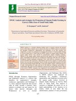
Fig.4 PCR asssay with TBR primers to detect Cysticercus cellulosae from pigcarcasses. Lane L:
100-bp DNA ladder; Lane 1 to 4: DNA samples extracted from cysticercosis positive pigs. Lane
P: Positive control and Lane N: Negative control
2594
Int.J.Curr.Microbiol.App.Sci (2019) 8(8): 2590-2596
Identification of T. Solium cysticerci from the
infected pig carcasses and suspected carcasses
was based on amplification of large subunit
rRNA gene (TBR) gene with a product size of
286 bp. All the positive cases show positive
amplification of TRB gene (Fig. 4). Dalmasso
et al., (2004) extracted DNA from
degenerated and calcified cysts/lesions and
found positive amplification for TRB gene.
Lino Junior (2004) also reported the
importance of PCR test with TBR primers as
a reliable method for detection of cysticerci in
tissues from human autopsies that are in
advanced evaluative stages.
From the above study it was found that
porcine cysticercosis is still a major public
health concern. However there is less
information regarding the prevalence of this
disease has been recorded from North Eastern
states of India. Therefore, an extensive study
regarding the prevalence, transmission, risk
factors and prevention of this neglected
zoonotic disease is utmost concern.
Acknowledgement
Authors are thankful to ICAR, New Delhi for
funding the “Outreach Project on Zoonotic
diseases” and Director of Research
(Veterinary) for necessary facilities to carry
out the research. Due acknowledgement is
also extended to the abattoir workers for
providing samples.
References
Anonymous, 2012. Pork meat in India.
www.g-e-f
a.de/fileadmin/termine.../
Pork_Meat_in_India_-_Report.pdf.
Barua A., Raj H., Goswami C., Sonowal D.
And Rajkhowa U., 2018. Prevalence of
Porcine Cysticercosis in Four States of
North East India. International Journal
of Livestock Research, 8(10): 212-218.
Cai X.P., Zheng Y.D., Luo X.N., Jing Z.Z.,
Hu Z.M. and Lu C.P., 2006.
Immunodiagnosis of cysticercosis in
China. The Journal of Applied
Research, 6(1): 69-76.
Dalmasso A., Fontanella E., Piatti P., Civera
T., Rosati S., Bottero M.T., 2004. A
multiplex PCR assay for the
identification of animal species in
feedstuffs. Molecular and Cellular
Probes, 18: 81–87.
Deckers, N., Kanobana, K., Silva, M.,
Gonzalez, A.E., Garcia, H.H., Gilman,
R.H. and Dorny, P., 2008. Serological
responses in porcine cysticercosis: a
link with the parasitological outcome
infect. Int. J. Parasitol., 38: 1191–1198.
Deplazes P., Grimm F., Sydler T., Tanner I.
and Kapel C.M.O., 2005. Experimental
alveolar echinococcosis in pigs, lesion
development and serological follow up.
Veterinary Parasitology, 130: 213-222.
Geysen D., Kanobana K., Victor B.,
Rodriguez-Hidalgo R., De Borchgrave
J. and Brandt J., 2007. Validation of
meat inspection results for Taenia
saginata
cysticercosis
by
PCR
restriction
fragment
length
polymorphism. Journal of Food
Protection, 70(1): 236-240.
Jardim E.A.G.V., Linhares G.F.C., Torres
F.A.G., Araujo J.L.B. and Barbosa
S.M., 2006. Diferenciacaoe specifica
entre Taenia saginata e Taenia solium
porensaio de PCR e duplex PCR.
Specific
discrimination
between
Taeniasaginata and Taenia soliumby
one step PCR assay and duplex PCR.
Ciencia Rural, Santa Maria, 36(1):166172.
Kedra A.H., Tkach V.V., Swiderski Z. and
Pawowski Z., 2001. Intraspecific
variability
among
NADH
dehydrogenase subunit 1sequences of
Taenia
hydatigena.
Parasitology
International, 50: 145-148.
2595
Int.J.Curr.Microbiol.App.Sci (2019) 8(8): 2590-2596
Kolesarova M., Herich R., LevkutJr. M.,
Curlik J. and Levkut M., 2012.
Suitability of different tissue fixatives
for subsequent PCR analysis of
Cysticercusovis.
Helminthologia.,
49(2): 67-70.
Lino Junior R.S., 2004. Cysticercosis
diagnostic methods in autopsies. Revista
do Instituto de Medicina Tropical de
São Paulo, 46(3): 138.
Pinto P.S.A., Vaz A.J., Germano P.M.L. and
Nakamura P.M., 2000. ELISA test for
the diagnosis of cysticercosis in pigs
using antigens of Taenia solium and
Taenia crassiceps cysticerci. Revista do
Instituto de Medicina Tropical de Sao
Paulo, 42(2): 71-79.
Sreedevi C., Hafeez M., Kumar P.A., Rayulu
V.C., Subramanyam K.V. and Sudhakar
K., 2012. PCR test for detecting Taenia
solium cysticercosis in pig carcasses.
Tropical Animal Health Production, 44:
95-99.
Willingham III A.L., Harrison L.J., Fevre
E.M.
and
Parkhouse
M.E.,
2008.Inaugural
meeting
of
the
cysticercosis working group in Europe.
EID J Home, 14(12).
How to cite this article:
Acheenta G. Barua, Koushik Kakoty, Pranjal M. Nath and Nur Abdul Kader. 2019. Detection
of Porcine Cysticercosis in Nagaland of North East, India. Int.J.Curr.Microbiol.App.Sci. 8(08):
2590-2596. doi: />
2596