A prospective observational study to assess the diagnostic accuracy of clinical decision rules for children presenting to emergency departments after head injuries (protocol): The
Bạn đang xem bản rút gọn của tài liệu. Xem và tải ngay bản đầy đủ của tài liệu tại đây (362.08 KB, 10 trang )
Babl et al. BMC Pediatrics 2014, 14:148
/>
STUDY PROTOCOL
Open Access
A prospective observational study to assess the
diagnostic accuracy of clinical decision rules for
children presenting to emergency departments
after head injuries (protocol): the Australasian
Paediatric Head Injury Rules Study (APHIRST)
Franz E Babl1,2,3,4*, Mark D Lyttle1,5,6, Silvia Bressan1,2,7, Meredith Borland8, Natalie Phillips9, Amit Kochar10,
Stuart R Dalziel11,12, Sarah Dalton13, John A Cheek1,2,14, Jeremy Furyk15, Yuri Gilhotra16, Jocelyn Neutze17,
Brenton Ward2, Susan Donath2,3, Kim Jachno2,3, Louise Crowe2,3, Amanda Williams2,3, Ed Oakley1,2,3
On behalf of the PREDICT research network
Abstract
Background: Head injuries in children are responsible for a large number of emergency department visits. Failure
to identify a clinically significant intracranial injury in a timely fashion may result in long term neurodisability and
death. Whilst cranial computed tomography (CT) provides rapid and definitive identification of intracranial injuries, it
is resource intensive and associated with radiation induced cancer. Evidence based head injury clinical decision
rules have been derived to aid physicians in identifying patients at risk of having a clinically significant intracranial
injury. Three rules have been identified as being of high quality and accuracy: the Canadian Assessment of
Tomography for Childhood Head Injury (CATCH) from Canada, the Children’s Head Injury Algorithm for the
Prediction of Important Clinical Events (CHALICE) from the UK, and the prediction rule for the identification of
children at very low risk of clinically important traumatic brain injury developed by the Pediatric Emergency Care
Applied Research Network (PECARN) from the USA. This study aims to prospectively validate and compare the
performance accuracy of these three clinical decision rules when applied outside the derivation setting.
Methods/design: This study is a prospective observational study of children aged 0 to less than 18 years
presenting to 10 emergency departments within the Paediatric Research in Emergency Departments International
Collaborative (PREDICT) research network in Australia and New Zealand after head injuries of any severity. Predictor
variables identified in CATCH, CHALICE and PECARN clinical decision rules will be collected. Patients will be
managed as per the treating clinicians at the participating hospitals. All patients not undergoing cranial CT will
receive a follow up call 14 to 90 days after the injury. Outcome data collected will include results of cranial CTs
(if performed) and details of admission, intubation, neurosurgery and death. The performance accuracy of each of
the rules will be assessed using rule specific outcomes and inclusion and exclusion criteria.
(Continued on next page)
* Correspondence:
1
Department of Emergency Medicine, Royal Children’s Hospital, Flemington
Rd, Parkville, Vic 3052, Australia
2
Murdoch Childrens Research Institute, Parkville, VIC, Australia
Full list of author information is available at the end of the article
© 2014 Babl et al.; licensee BioMed Central Ltd. This is an Open Access article distributed under the terms of the Creative
Commons Attribution License ( which permits unrestricted use, distribution, and
reproduction in any medium, provided the original work is properly credited. The Creative Commons Public Domain
Dedication waiver ( applies to the data made available in this article,
unless otherwise stated.
Babl et al. BMC Pediatrics 2014, 14:148
/>
Page 2 of 10
(Continued from previous page)
Discussion: This study will allow the simultaneous comparative application and validation of three major paediatric
head injury clinical decision rules outside their derivation setting.
Trial registration: The study is registered with the Australian New Zealand Clinical Trials Registry
(ANZCTR)- ACTRN12614000463673 (registered 2 May 2014).
Keywords: Head injury, Clinical decision rule, Computed tomography, Validation
Background
Children with clinically significant intracranial injuries
require urgent identification to prevent further damage
to the brain. Cranial computed tomography (CT) scans
provide rapid and definitive identification of the presence or absence of intracranial injuries, and help guide
subsequent management. Positive results allow early
intervention and optimise outcomes whilst negative results are reassuring and may allow accelerated discharge
and reduce unnecessary admissions.
However, cranial CT scans also have negative effects,
particularly in children, who are more vulnerable to
radiation-associated cell damage [1]. Radiation from cranial CT scans can cause lethal malignancies with higher
risk in younger age groups [1-4]. Children may require
sedation to allow imaging with consequent sedationassociated risks [5,6]. They also have resource implications for Emergency Departments (EDs) and the health
system as a whole [7]. Despite this, the number of cranial CT scans performed for head injuries in children
has increased in a number of countries [8-11]. This increase is likely due to a combination of easier access to
CT scanners and more efficient technology and concern
amongst physicians of being unable to reliably identify
intracranial injury based solely on a child’s clinical condition. One way of increasing clinical sensitivity and specificity (i.e. minimising both missed clinically significant
intracranial injuries and unnecessary investigations) is to
develop and use clinical decision rules (CDRs).
CDRs help physicians with diagnostic and therapeutic
decisions, and can be defined as decision making tools derived from original research (as opposed to a consensusbased clinical practice guideline) which incorporate three
or more variables from the history, physical examination,
or simple tests. These tools help clinicians cope with the
uncertainty of medical decision making and improve their
efficiency [12]. Several recent systematic reviews of existing paediatric head injury CDRs have been published
[13-15]. The three CDRs of highest quality and accuracy
[15] are the Canadian Assessment of Tomography for
Childhood Head Injury (CATCH) from Canada [11], the
Children’s Head Injury Algorithm for the Prediction of
Important Clinical Events (CHALICE) from the UK [16]
and the prediction rule for the identification of children at
very low risk of clinically important traumatic brain injury
developed by the Pediatric Emergency Care Applied Research Network (PECARN) from the USA [17]. All three
CDRs were derived with high methodological standards
using large multicentre data sets. However, they differ in
key areas, including study population, predictor variables
(based on mechanism of injury, clinical history, and clinical examination) (Table 1), inclusion and exclusion criteria (Table 2) and outcomes (including the terminology
and definitions used) (Table 3). Most importantly the
focus is different in each CDR. CATCH was derived to
manage children with minor head injuries presenting
within 24 hours, with specific inclusion criteria to be fulfilled before employing the CDR. CHALICE was derived
for children with head injuries of all severities, presenting
at any point after the injury. Both aim to identify children
likely to have significant intracranial injury who warrant a
cranial CT scan. PECARN’s CDR focuses on children with
minor head injuries presenting within a 24 hour period
and aims to identify patients unlikely to have a clinically
important traumatic brain injury who can be safely discharged without a CT scan. In addition PECARN has derived different CDRs for children aged less than two years
and children aged two years and older. The comparative
performance accuracy (as assessed by sensitivity, specificity, negative predictive value and positive predictive
value) for each CDR has been presented elsewhere [15].
CATCH and CHALICE CDRs suggest a dichotomous
course of action (cranial CT scan/no cranial CT scan) although CATCH stratifies this risk into high and medium
categories. The PECARN CDR defines a low risk population in whom cranial CT scans can routinely be obviated.
PECARN’s is the only CDR which has been internally
[17] and externally [18] validated. A CATCH validation
study has been performed in the derivation setting
though results are only available in abstract form at
present [19]. Recently the three CDRs have been prospectively validated in the same cohort of 1,009 children
presenting to an urban medical center with a designated
paediatric ED in the United States. This study showed
that baseline physician ordering practice and PECARN
outperformed the other CDRs. However, the study
population did not reflect the exact population for which
each rule was originally developed and the study was
underpowered to determine narrow confidence intervals
for rare but critically important events [20].
Babl et al. BMC Pediatrics 2014, 14:148
/>
Page 3 of 10
Table 1 Comparison of predictor variables [11,15-17]
CATCH
CHALICE
PECARN <2 years
PECARN ≥2 years
High speed RTA as pedestrian,
cyclist, occupant (>40 miles/h
or >64 km/h).
Severe mechanism of injury
(MVC with patient ejection, death
of another passenger or rollover;
pedestrian/bicyclist without helmet
struck by motorized vehicle; falls >0.9 m;
head struck by high impact object).
Severe mechanism of injury
(MVC with patient ejection,
death of another passenger
or rollover; pedestrian/bicyclist
without helmet struck by
motorized vehicle; falls >1.5 m;
head struck by high impact object).
LOC ≥5 seconds.
Any/suspected LOC.
Altered mental status.
Altered mental status.
Mechanism of injury
Dangerous mechanism of
injury (eg MVC, fall from
elevation ≥3 ft [≥0.91 m] or
5 stairs, fall from bicycle with
no helmet).
Fall of > 3 m in height.
High speed injury from
projectile or object.
History
Witnessed LOC > 5 min.
Amnesia (antegrade or
retrograde) >5 min.
Not acting normally per parent.
≥3 vomits after head injury
(discrete episodes).
History of vomiting.
Suspicion of NAI.
Seizure in patient with no
history of epilepsy.
History of worsening headache.
Severe headache.
Examination
GCS <15, 2 hr after injury.
GCS <14, or <15 if <1 yr.
GCS < 15
GCS < 15
Irritability on examination.
Abnormal drowsiness (in
excess of that expected
by examining doctor).
Other signs of altered mental status
(agitation, somnolence, repetitive
questioning, slow response to
verbal communication)
Other signs of altered mental
status (agitation, somnolence,
repetitive questioning, slow
response to verbal communication)
Suspected open or depressed
skull fracture.
Suspicion of penetrating
or depressed skull injury, or
tense fontanelle.
Any sign of basal skull fracture
(eg haemotympanum, “raccoon”
eyes, otorrhoea/rhinorrhoea of
CSF, Battle’s sign).
Signs of basal skull fracture.
Palpable or unclear skull fracture.
Clinical signs of basilar skull fracture.
Positive focal neurology.
Large boggy haematoma of
the scalp.
Presence of bruise, swelling or
laceration > 5 cm if < 1 yr old.
Occipital, parietal or temporal
scalp haematoma.
Reproduced from Lyttle M, et al. [15] Copyright 2012, with permission from BMJ Publishing Group Ltd.
In each of the three clinical decision rules (CDRs) the absence of all of the above predictor variables indicates that cranial computed tomography is unnecessary.
Note: while the predictor variables are reproduced verbatim, the order in which the variables from each CDR are presented has been altered to
facilitate comparison.
CATCH Canadian Assessment of Tomography for Childhood Head Injury.
CHALICE Children’s Head Injury Algorithm for the Prediction of Important Clinical Events.
PECARN Pediatric Emergency Care Applied Research Network.
MVC Motor vehicle crash.
RTA Road traffic accident.
LOC Loss of consciousness.
NAI Non-accidental injury.
GCS Glasgow Coma Score.
CSF Cerebrospinal fluid.
We propose to validate and compare the accuracy of the
CATCH, CHALICE and PECARN CDRs using prospectively collected data from 20,000 patients in a multicentre
setting in Australia and New Zealand, i.e. outside the
countries where these CDRs were derived, and compare
their performance against that of our current practice.
Triggers for cranial CT use by clinicians in paediatric EDs
in Australia and New Zealand are different from the triggers developed in CATCH, CHALICE and PECARN [21].
This study will also help determine which CDR is best
suited for use in the Australian and New Zealand setting
before incorporating them into local practice.
Babl et al. BMC Pediatrics 2014, 14:148
/>
Page 4 of 10
Table 2 Comparison of inclusion and exclusion criteria [11,15-17]
CATCH
Inclusion criteria
Exclusion criteria
All of the following:
Any of:
• Blunt trauma to head resulting in witnessed
LOC/disorientation, definite amnesia, persistent
vomiting (>1 episode), persistent irritability
(in children <2 yrs)
• Obvious penetrating skull injury
• Obvious depressed fracture
• Acute focal neurologic deficit
• Chronic generalized developmental delay
• Head injury secondary to suspected child abuse
• Initial GCS in ED ≥13 as determined by treating physician
• Returning for reassessment of previously treated head injury
• Injury within the past 24 hours.
• Patients who were pregnant
CHALICE
Any history or signs of injury to the head.
Refusal to consent
PECARN
Present within 24 hours of head injury.
Any of:
• Trivial head injury (defined by ground level fall, walking/running
into stationary object, no signs or symptoms of head trauma
except scalp abrasions and lacerations).
• Penetrating trauma
• Known brain tumour
• Pre-existing neurological disorder complicating assessment
• Neuro-imaging at another hospital before transfer
• Patient with ventricular shunt*
• Patient with bleeding disorder*
• GCS < 14*
Reproduced from Lyttle M, et al. [15] Copyright 2012, with permission from BMJ Publishing Group Ltd.
CATCH Canadian Assessment of Tomography for Childhood Head Injury.
CHALICE Children’s Head Injury Algorithm for the Prediction of Important Clinical Events
PECARN Pediatric Emergency Care Applied Research Network.
GCS Glasgow Coma Score.
LOC Loss of consciousness.
ED emergency department.
*enrolled but being analysed separately, not used in clinical decision rule derivation.
Methods/design
Aims
The primary aim of this study is to determine the performance accuracy of the three major international
paediatric head injury CDRs (CATCH, CHALICE and
PECARN) when applied to a prospective multicentre
population of consecutive children presenting with head
injury to 10 EDs in Australia and New Zealand. This will
allow the comparative external validation of the CDRs
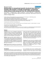
outside their derivation settings (Figure 1).
Design
This is a multi-centre prospective observational study of
consecutive children presenting with head injuries to
paediatric EDs. All data points necessary for analysis including predictor variables and outcome data for the
three clinical rules under investigation (Tables 1, 2 and
3) will be collected for all patients but treating clinicians
will manage patients as per their usual practice. The
study has been registered with the Australian New
Zealand Clinical Trials Registry (ACTRN12614000463673).
The study follows the STAndards for the Reporting of
Diagnostic accuracy studies (STARD) guidelines [22].
Setting
The study is taking place at 9 tertiary paediatric EDs, and
1 large combined adult and paediatric ED in Australia and
New Zealand. These centres are members of the Paediatric Research in Emergency Departments International
Collaborative (PREDICT) [23]: in New Zealand Kidz First
Children’s Hospital, Auckland, and Starship Children’s
Hospital, Auckland; in Australia Monash Medical Centre,
Clayton, VIC, Children’s Hospital at Westmead, Sydney,
NSW, Royal Children’s Hospital, Melbourne, VIC, Royal
Children’s Hospital, Brisbane, QLD, Mater Children’s
Hospital, Brisbane, QLD, Princess Margaret Hospital for
Children, Perth, WA, Women’s & Children’s Hospital,
Adelaide, SA, and Townsville Hospital, Townsville, QLD.
The annual paediatric census of the 10 participating EDs
is >400,000. The central site for the study is the Murdoch
Children’s Research Institute, which is affiliated with the
Royal Children’s Hospital Melbourne.
Babl et al. BMC Pediatrics 2014, 14:148
/>
Page 5 of 10
Table 3 Comparison of outcomes [11,15-17]
Primary outcome
Secondary outcomes
CATCH
Need for neurological intervention, defined as death within
7 days secondary to the head injury or need for any of the
following within 7 days: craniotomy, elevation of skull fracture,
monitoring of intracranial pressure, insertion of endotracheal
tube for the management of head injury
Brain injury on CT, defined as any acute intracranial finding
revealed on CT attributable to acute injury, including closed
depressed skull fracture (depressed past the inner table) and
pneumocephalus but excluding non-depressed skull fractures
and basilar skull fractures
CHALICE
Clinically significant intracranial injury (CSII), defined as death
as a result of head injury, requirement for neurosurgical intervention,
marked abnormality on CT (any new, acute, traumatic intracranial
pathology as reported by consultant radiologist, including intracranial
haematomas of any size, cerebral contusion, diffuse cerebral oedema
and depressed skull fractures)
Presence of skull fracture
Admission to hospital
PECARN
Clinically important traumatic brain injury (ciTBI), defined as death
from TBI, neurosurgical intervention for TBI (intracranial pressure
monitoring, elevation of depressed skull fracture, ventriculostomy,
haematoma evacuation, lobectomy, tissue debridement, dura repair,
other), intubation of more than 24 h for TBI or hospital admission of
2 nights or more for TBI* in association with TBI on CT**
None
Reproduced from Lyttle M, et al. [15] Copyright 2012, with permission from BMJ Publishing Group Ltd.
*Admission for persistent neurological symptoms or signs such as persistent alteration in mental status, recurrent emesis due to head injury, persistent severe
headache or ongoing seizure management.
**Intracranial haemorrhage or contusion, cerebral oedema, traumatic infarction, diffuse axonal injury, shearing injury, sigmoid sinus thrombosis, midline shift of
intracranial contents or signs of brain herniation, diastasis of the skull, pneumocephalus, skull fracture depressed by at least the width of the table of the skull.
CATCH Canadian Assessment of Tomography for Childhood Head Injury.
CHALICE Children’s Head Injury Algorithm for the Prediction of Important Clinical Events.
PECARN Pediatric Emergency Care Applied Research Network.
CT computed tomography.
Inclusion criteria
Patients less than 18 years of age with head injuries of
all severities irrespective of length of time from injury to
presentation will be included. The definition of head
injury does not include patients who have sustained a
trivial facial injury (ground level fall or walking or running into an object with no signs or symptoms of injury
other than facial abrasions or lacerations below the
eyebrows).
Exclusion criteria
We will exclude patients and families who refuse to
participate, are being referred directly from ED triage
to a general practitioner or other external provider
(i.e. not seen in the ED), or who do not wait to be
seen. We will exclude from analysis patients with neuroimaging prior to transfer (Figure 1). Individual exclusion criteria (relevant to each CDR (Table 2)) will
be applied during analysis.
Primary outcome measure
Primary outcome will be the performance accuracy (sensitivity, specificity, negative predictive value (NPV), and
positive predictive value (PPV)) of each CDR in identifying rule specific outcomes (Table 3) when applied to
those patients who meet the individual inclusion and exclusion criteria (Table 2).
Secondary outcome measures
1. Rate of clinically important traumatic brain injury
(ciTBI) [17] and clinically significant intracranial
injury (CSII) [16] in the study population.
2. Rate of neurosurgical intervention in the study
population.
3. Rate of cranial CT use in the study population.
4. Number of missed ciTBI and CSII in the study
population.
5. Characteristics of missed significant intracranial
injuries that would have been identified by the
application of each CDR to the study population.
6. Number of extra cranial CT scans that would be
performed by applying each CDR.
7. Sensitivity, specificity, NPV and PPV of PECARN in
identifying traumatic brain injury on cranial CT.
8. Diagnostic accuracy of each of the CDRs when
applied to those patients attending with head injury
who do not meet the specific individual inclusion
and exclusion criteria.
9. Rule performance in patients with bleeding diathesis,
ventriculoperitoneal shunt, non-accidental injuries
and pre-existing neurological conditions.
10.Economic evaluation of financial savings or burden
of implementing each CDR.
11.Rate of prolonged symptoms following a non-severe
head injury.
Babl et al. BMC Pediatrics 2014, 14:148
/>
Page 6 of 10
Patients with head injuries
of any severity* assessed
for eligibility (n)
Missed patients (n)
Patients eligible (n)
Excluded patients (n)
Refusal
Referred to external clinician
Did not wait
Cranial CT prior to transfer
Patients enrolled (n)
Patients lost to follow up (n)
Total number of
evaluable patients for
analysis (n)
Patients applicable for
CATCH rule (n)
Patients applicable for
CHALICE rule (n)
Patients applicable for
PECARN rule
<2years (n)
Patients applicable for
PECARN rule
2years (n)
Analysis of sensitivity
and specificity based on
outcomes of CATCH
rule.
Analysis of sensitivity and
specificity based on
outcomes of CHALICE
rule.
Analysis of sensitivity
and specificity based on
outcomes of PECARN
rule <2 years
Analysis of sensitivity
and specificity based on
outcomes of PECARN
rule > 2 years.
Figure 1 Algorithm for patient eligibility and analysis. *Head injuries not including trivial facial injuries defined as a ground level fall or
walking or running into an object with no signs or symptoms of injury other than facial abrasions or lacerations below the eyebrows. CT
computed tomography. LTFU lost to follow up. CHALICE Children’s Head Injury Algorithm for the Prediction of Important Clinical Events. CATCH
Canadian Assessment of Tomography for Childhood Head Injury. PECARN Pediatric Emergency Care Applied Research Network.
Patient recruitment, study procedure and data collection
Patients with head injuries will be identified at the ED
triage desk using electronic alerts and visual reminders
for patients who receive a head injury type injury code.
Triage nurses will attach a clinician study clinical report
form (CRF) to the patient record. Patients will be enrolled in the study by the treating clinician. Verbal consent for participation will be sought and documented by
the treating clinician; consent for participation will include permission to telephone families 14–90 days after
the ED visit for follow-up. Consent will be sought at the
time of the initial ED visit. Should the parent or guardian of the child not be available at that time, we will seek
consent for involvement in the study either during the
in-patient stay (where admitted) or at the time of telephone follow up (where discharged from ED). Identification of missed eligible patients will be undertaken by the
research assistant in each participating centre through a
review of the daily ED attendance record.
Data collected by the ED treating clinicians will include the predictor variables from the three CDRs
(CATCH [11], PECARN [17], CHALICE [16]). The initial ED assessment data will be documented prior to
management decisions.
A separate CRF will be completed by the site research
assistant during the hospital stay (in admitted patients)
Babl et al. BMC Pediatrics 2014, 14:148
/>
or after ED discharge (in those patients discharged direct
from ED) once outcome data are available. It will collect
the following parameters: detailed demographics, time
lines (times of triage, clinician evaluation, ED and hospital discharge), ED observation and duration of observation, admission status and duration of admission,
intensive care admission, intubation and ventilation and
duration of ventilation, imaging and results, neurosurgical interventions and mortality.
The telephone follow-up to screen for possible initially missed intracranial injuries will be completed by
the research assistant or the site physician investigators
14–90 days after the injury if no cranial CT is performed. Data on ongoing signs and symptoms, neuroimaging, admission and neurosurgery will be elicited.
Six contact attempts will be made. If more than 90 days
have elapsed from the time of injury, or if there have
been six failed contact attempts, the patient follow up
will be regarded as unsuccessful and the patient deemed
lost to follow up.
All study materials have been piloted at a single site
(Royal Children’s Hospital Melbourne) [23]; modification
of the materials to comply with local patient flow and
administrative requirements have been assessed and approved by the study steering committee.
CRFs will be de-identified after all data points have
been completed and any data queries have been addressed. Data collation and analysis will take place at the
central study site (Murdoch Childrens Research Institute, Melbourne).
All participating clinicians (physicians and nurse practitioners) at all sites receive formal training in the completion of the clinician CRF prior to the commencement
of the study. Research assistants collecting data on the
accompanying CRFs undergo formal training at the
central site prior to the commencement of the study.
Standardised teaching materials have been created and
provided to participating sites. The study coordinator
will ensure that all staff have received appropriate orientation and training and will ensure compliance with
study protocol through site visits. Investigators and
research assistants are not blinded to the results of the
collected outcome data.
Determination of outcome
Patient outcome will be determined by:
1. Consultant radiologist reports of CTs.
2. Operative reports for those who required
neurosurgical intervention.
3. Review of medical record for the duration of
admission and secondary outcomes.
4. Structured telephone follow up at 14–90 days post
injury for patients discharged without neuroimaging.
Page 7 of 10
5. Patients for whom final outcome data are not
available will be excluded from data analysis.
This process will permit the identification of the presence and extent of injury allowing classification as per
the definitions of each head injury CDR.
Definitions
CDR specific definitions of inclusion and exclusion
criteria, predictor variables and outcomes are set out in
Tables 1, 2 and 3.
Further definitions used:
ED observation: Ongoing clinical assessment and
observation of the patient in ED for less than 6 hours
post initial clinical assessment.
Admission: Transfer from ED to a hospital inpatient
unit (including short stay, observation, or intensive care
unit) for longer than 6 hours.
Neurosurgical interventions will be categorised based
on operative reports into the following categories:
Dura repair of cerebrospinal fluid leaks, skull fracture
elevation, haematoma drainage, intracranial pressure
(ICP) monitoring, lobectomy, tissue debridement,
ventriculostomy, other.
Head imaging (CT and magnetic resonance imaging)
will be categorised as follows based on reports by
consultant radiologists: Cerebellar haemorrhage,
cerebral contusion, cerebral oedema, cerebral
haemorrhage, intracerebral haematoma, diastasis of the
skull, extradural/epidural haematoma, extra-axial
haematoma, intraventricular haemorrhage, midline
shift/shift of brain structures, pneumocephalus, skull
fracture (and depth of depression), subarachnoid
haemorrhage, subdural haematoma, traumatic infarction.
Statistical methods
When applying each CDR, items will be scored as present,
absent or unknown. Sensitivity, specificity, negative
predictive value (NPV), and positive predictive value
(PPV) of each of the CDRs will be calculated using the
definitions and parameters set out in the derivation
studies as published [11,16,17]. In addition, the two CDRs
limited to minor head injuries (CATCH and PECARN)
will also be applied to patients of all head injury severities
to assess their performance in this extended patient
group. Likewise, the CHALICE CDR, though derived
for all severities of head injury, will undergo separate
analysis in minor head injury to allow comparison of
performance accuracy of the three CDRs in that population. Performance accuracy will also be calculated in
patient subgroups including but not restricted to patients with bleeding diathesis and ventriculoperitoneal
shunts. Rates of secondary outcomes such as cranial
Babl et al. BMC Pediatrics 2014, 14:148
/>
Page 8 of 10
CT, neurosurgical intervention, ciTBI and CSII and
missed ciTBI and CSII will be calculated. Key percentages
will be presented with 95% confidence intervals. Data
will be entered using Epidata (The Epidata Association,
Odense, Denmark) and analysed using Stata 12 (Statacorp,
College Station, Texas, USA).
Sample size and power calculation
In deriving a sample size for patient subgroups we extrapolated from the PECARN data as it is the only CDR
which differentiates between children aged less than two
years and children aged two years and older [17].
Based on PECARN’s ciTBI rate of 1%, and the ability
to determine the sensitivity and specificity of the CDRs
to a precision level of between 94% and 100%, we determined that we would require 10,000 patients to be enrolled in our study in order to maintain the precision for
the two subgroups in the PECARN CDR, children aged
less than two years and children aged two years and
older (i.e. 5,000 children in each age sub-group). Previous retrospective research of children diagnosed with a
head injury, conducted at Royal Children’s Hospital
Melbourne, had identified a 1:1 ratio between children
aged less than two years and those aged two years or
greater [24]. After an analysis of the first 1,000 patients
enrolled in the APHIRST study [23] this premise was
found to be incorrect and in the prospectively enrolled
patients the true ratio of children less than 2 years presenting with a head injury to children 2 years of age or
older presenting with a head injury was 1:4. Therefore,
to preserve the precision of the study in the younger age
group of children for the PECARN CDR the sample size
was recalculated to 20,000 children. Table 4 illustrates
the precision that would be achieved (using 95% confidence intervals) based on these assumptions for several
different plausible values for sensitivity for the outcomes
(i) ciTBI (ii) need for neurosurgery and (iii) brain injury
on CT (as based on PECARN data [17]).
Ethical issues and consent
In this observational non-interventional study parental
verbal consent and participant verbal assent (for patients
deemed capable to understand and appropriately answer
questions) will be obtained for all patients; it will include
permission to conduct a follow-up telephone call to determine outcome. Delayed consent at the time of the
phone call if necessary has been approved for patients
not enrolled during the initial ED visit. Ethics approval
has been granted at all 10 study sites.
Patients who refuse consent or withdraw will continue
to be managed as per the treating clinician.
As this is an observational study we are not anticipating adverse events.
Limitations
Ideally, all patients with head injuries would receive a
cranial CT to determine the presence or absence of significant intracranial injuries. However, this would expose a large number of patients to unnecessary CTs
and the associated cancer related risks; therefore, similar to the methodology used in the derivation and validation studies for CATCH [11] and PECARN [17] this
study relies on patient follow up by telephone. In doing
so we will establish whether a relevant outcome has
occurred or not.
CT rates in Australia and New Zealand may be lower
[23,25] than in North America and as reported for the
CATCH and PECARN studies [8,10,11,17] and higher
than the baseline rates reported from the United Kingdom
in the CHALICE study [16]. This highlights one of the potential key strengths of this study as it tests the CDRs in a
setting different to that in which each one was derived.
Finally, while we were provided with copies of the telephone follow up questionnaires used by the CATCH
and PECARN investigators (personal communication,
Dr Martin Osmond and Dr Nathan Kuppermann) we
reconstructed the predictor variables for the three CDRs
Table 4 Projected sensitivity for outcomes of clinically important traumatic brain injury (ciTBI), need for neurosurgery
and brain injury on computed tomography (CT) based on PECARN data [17]
Outcome
ciTBI
Need for neurosurgery
Brain injury on CT
CDR clinical decision rule.
Number of patients predicted
Projected performance of CDR
in predicting outcome (sensitivity)
Sensitivity%
95% confidence interval
50
50/50
100
93-100
50
49/50
98
89-100
50
48/50
96
86-99.5
50
47/50
94
83-99
30
30/30
100
88-100
30
29/30
96.5
83-100
300
300/300
100
98.8-100
300
290/300
97
94-98
Babl et al. BMC Pediatrics 2014, 14:148
/>
solely from the published papers [11,16,17]. This may
have introduced an element of interpretation in terms
of the most precise wording to be used in a clinical
emergency setting.
Discussion
This study will allow the simultaneous comparative application and validation of three major paediatric head
injury clinical decision rules outside their derivation setting. In addition to a high recruitment rate, the study
will depend on high follow up rates to ensure that our
results accurately represent the whole population of children presenting with head injuries.
Time plan
We have so far recruited more than 10,000 of the planned
20,000 patients. We will complete recruitment by the end
of 2014.
Abbreviations
CHALICE: Children’s head injury algorithm for the prediction of important
clinical events; CATCH: Canadian assessment of tomography for childhood
head injury; PECARN: Pediatric emergency care applied research network;
CT: Computed tomography; CSII: Clinically significant intracranial injury;
ciTBI: Clinically important traumatic brain injury; CDR: Clinical decision rule;
ED: Emergency Department; NPV: Negative predictive value; PPV: Positive
predictive value.
Competing interests
None of the authors have any competing interests arising from this research.
Authors’ contributions
FEB was responsible for identifying the research question and the design of the
study. FEB, MDL and EO were responsible for refining the design and developing
the research protocol. All authors have contributed to the development of the
protocol, the implementation of the study at participating sites and the
enrolment of patients. FEB was responsible for the drafting of this paper. All
authors provided comments on the drafts and have read and approved the final
version. FEB takes responsibility for the manuscript as a whole.
Acknowledgements
We would like to thank participating families, emergency department staff
and the site research assistants. The study is funded by grants from the
National Health and Medical Research Council (project grant GNT1046727,
Centre of Research Excellence for Paediatric Emergency Medicine
GNT1058560), Canberra, Australia; the Murdoch Childrens Research Institute,
Melbourne, Australia; the Queensland Emergency Medicine Research
Foundation (EMPJ-11162), Brisbane, Australia; Perpetual Philanthropic Services
(2012/1140), Australia; Auckland Medical Research Foundation (No. 3112011)
and the A + Trust (Auckland District Health Board), Auckland, New Zealand;
WA Health Targeted Research Funds 2013, Perth, Australia; the Townsville
Hospital and Health Service Private Practice Research and Education Trust
Fund, Townsville, Australia; and the Victorian Government’s Infrastructure
Support Program, Melbourne, Australia. FEB’s time was part funded by a
grant from the Murdoch Children’s Research Institute. SRDs time was part
funded by the Health Research Council of New Zealand (HRC13/556).
Author details
1
Department of Emergency Medicine, Royal Children’s Hospital, Flemington
Rd, Parkville, Vic 3052, Australia. 2Murdoch Childrens Research Institute,
Parkville, VIC, Australia. 3Department of Paediatrics, Faculty of Medicine,
Dentistry and Health Sciences, University of Melbourne, Melbourne, VIC 3010,
Australia. 4National Trauma Research Institute, Prahan, VIC, Australia. 5Bristol
Royal Hospital for Children, Bristol, UK. 6Academic Department of Emergency
Care, University of the West of England, Bristol, UK. 7University of Padova,
Padova, Italy. 8Princess Margaret Hospital for Children, Perth, Australia. 9Royal
Page 9 of 10
Children's Hospital and Queensland Children's Medical Research Institute,
Queensland University, Brisbane, Australia. 10Women’s & Children’s Hospital,
Adelaide, Australia. 11Starship Hospital, Auckland, New Zealand. 12Liggins
Institute, University of Auckland, Auckland, New Zealand. 13The Children’s
Hospital at Westmead, Sydney, Australia. 14Monash Medical Centre, Clayton,
VIC, Australia. 15Townsville Hospital, Townsville, Australia. 16Mater Children’s
Hospital, Brisbane, Australia. 17Kidzfirst Middlemore Hospital, Auckland, New
Zealand.
Received: 4 May 2014 Accepted: 27 May 2014
Published: 13 June 2014
References
1. Brenner DJ, Hall EJ: Computed tomography – an increasing source of
radiation exposure. N Engl J Med 2007, 357:2277–2284.
2. Pearce MS, Salotti JA, Little MP, McHugh K, Lee C, Kim KP, Howe NL,
Ronckers CM, Rajaraman P, Sir Craft AW, Parker L, Berrington de González A:
Radiation exposure from CT scans in childhood and subsequent risk of
leukaemia and brain tumours: a retrospective cohort study. Lancet 2012,
380:499–505.
3. Miglioretti DL, Johnson E, Williams A, Greenlee RT, Weinmann S, Solberg LI,
Feigelson HS, Roblin D, Flynn MJ, Vanneman N, Smith-Bindman R: The use
of computed tomography in pediatrics and the associated radiation
exposure and estimated cancer risk. JAMA Pediatr 2013, 167:700–707.
4. Mathews JD, Forsythe AV, Brady Z, Butler MW, Goergen SK, Byrnes GB, Giles
GG, Wallace AB, Anderson PR, Guiver TA, McGale P, Cain TM, Dowty JG,
Bickerstaffe AC, Darby SC: Cancer risk in 680,000 people exposed to
computed tomography scans in childhood or adolescence: data linkage
study of 11 million Australians. BMJ 2013, 346:f2360.
5. Conners GP, Sacks WK, Leahey NF: Variations in sedating uncooperative,
stable children for post-traumatic head CT. Pediatr Emerg Care 1999,
15:241–244.
6. Hoyle JD Jr, Callahan JM, Badawy M, Powell E, Jacobs E, Gerardi M, Melville K,
Miskin M, Atabaki SM, Dayan P, Holmes JF, Kuppermann N, Traumatic
Brain Injury Study Group for the Pediatric Emergency Care Applied
Research Network (PECARN): Pharmacological sedation for cranial
computed tomography in children after minor blunt head trauma.
Pediatr Emerg Care 2014, 30(1):1–7.
7. Gazelle GS, McMahon PM, Siebert U, Beinfeld MT: Cost-effectiveness
analysis in the assessment of diagnostic imaging technologies. Radiology
2005, 235:361–370.
8. National Center for Health Statistics Centers for Disease Control and
Prevention: Public use Data File, Emergency Department File. Hyattville MD:
National Hospital Ambulatory Medical Care Survey; 2005. />pub/Health_Statistics/NCHS/Datasets/NHAMCS/readme05.txt.
9. National Center for Health Statistics Centers for Disease Control and
Prevention: Public use Data File, Emergency Department File. Hyattville MD:
National Hospital Ambulatory Medical Care Survey; 1995. />pub/Health_Statistics/NCHS/Datasets/NHAMCS/readme95.txt.
10. Klassen TP, Reed MH, Stiell IG, Nijssen-Jordan C, Tenenbein M, Joubert G,
Jarvis A, Baldwin G, St-Vil D, Pitters C, Belanger F, McConnell D, Vandemheen K,
Hamilton MG, Sutcliffe T, Colbourne M: Variation in utilization of computed
tomography scanning for the investigation of minor head trauma in
children: a Canadian experience. Acad Emerg Med 2000, 7:739–744.
11. Osmond MH, Klassen TP, Wells GA, Correll R, Jarvis A, Joubert G, Bailey B,
Chauvin-Kimoff L, Pusic M, McConnell D, Nijssen-Jordan C, Silver N, Taylor B,
Stiell IG, Pediatric Emergency Research Canada (PERC) Head Injury Study
Group: The CATCH rule: a clinical decision rule for the use of computed
tomography of the head in children with minor head injury. Can Med
Assoc J 2010, 182:341–348.
12. Stiell IG, Wells GA: Methodologic standards for the development of clinical
decision rules in emergency medicine. Ann Emerg Med 1999, 33:437–447.
13. Maguire JL, Boutis K, Uleryk EM, Laupacis A, Parkin PC: Should a headinjured child receive a head CT scan? A systematic review of clinical
prediction rules. Pediatrics 2009, 124:e145–e154.
14. Pickering A, Harnan S, Fitzgerald P, Pandor A, Goodacre S: Clinical decision
rules for children with minor head injury: a systematic review. Arch Dis
Child 2011, 96:414–421.
15. Lyttle MD, Crowe L, Oakley E, Dunning J, Babl FE: Comparing CATCH,
CHALICE and PECARN clinical decision rules for paediatric head injuries.
Emerg Med J 2012, 29(10):785–794.
Babl et al. BMC Pediatrics 2014, 14:148
/>
Page 10 of 10
16. Dunning J, Daly JP, Lomas JP, Lecky F, Batchelor J, Mackway-Jones K,
Children’s head injury algorithm for the prediction of important clinical
events study group: Derivation of the children’s head injury algorithm for
the prediction of important clinical events decision rule for head injury
in children. Arch Dis Child 2006, 91:885–891.
17. Kuppermann N, Holmes JF, Dayan PS, Hoyle JD Jr, Atabaki SM, Holubkov R,
Nadel FM, Monroe D, Stanley RM, Borgialli DA, Badawy MK, Schunk JE,
Quayle KS, Mahajan P, Lichenstein R, Lillis KA, Tunik MG, Jacobs ES, Callahan
JM, Gorelick MH, Glass TF, Lee LK, Bachman MC, Cooper A, Powell EC,
Gerardi MJ, Melville KA, Muizelaar JP, Wisner DH, Zuspan SJ, et al:
Identification of children at very low risk of clinically-important brain
injuries after head trauma: a prospective cohort study. Lancet 2009,
374(9696):1160–1170.
18. Schonfeld D, Bressan S, Da Dalt L, Henien MN, Winnett JA, Nigrovic LE:
Pediatric emergency care applied research network head injury clinical
prediction rules are reliable in practice. Arch Dis Child 2014, 99(5):427–431.
19. Osmond M: Multicentre prospective validation of the Canadian
Assessment of tomography for Childhood Head Injury (CATCH) Rule.
CJEM 2012, 14(Supplement 1).
20. Easter JS, Bakes K, Dhaliwal J, Miller M, Caruso E, Haukoos JS: Comparison of
PECARN, CATCH, and CHALICE rules for children with minor head injury:
a prospective cohort study. Ann Emerg Med 2014. doi: 10.1016/j.
annemergmed.2014.01.030. [Epub ahead of print].
21. Lockie FD, Dalton S, Oakley E, Babl FE: Triggers for head computed
tomography following paediatric head injury: Comparison of physicians’
reported practice and clinical decision rules. Emerg Med Australas 2013,
25(1):75–82.
22. Bossuyt PM, Reitsma JB, Bruns DE, Gatsonis CA, Glasziou PP, Irwig LM, Lijmer
JG, Moher D, Rennie D, de Vet HC, Standards for Reporting of Diagnostic
Accuracy: Toward complete and accurate reporting of studies of
diagnostic accuracy: the STARD initiative. Standards for reporting of
diagnostic accuracy. Br Med J 2003, 326:41–44.
23. Lyttle MD, Cheek JA, Blackburn C, Oakley E, Ward B, Fry A, Jachno K, Babl FE:
Applicability of the CATCH, CHALICE and PECARN paediatric head injury
clinical decision rules: pilot data from a single Australian centre.
Emerg Med J 2013, 30(10):790–794.
24. Crowe L, Babl F, Anderson V, Catroppa C: The epidemiology of paediatric
head injuries: data from a referral centre in Victoria, Australia. J Paediatr
Child Health 2009, 45:346–350.
25. Crowe L, Anderson V, Babl FE: Application of the CHALICE clinical
prediction rule for intracranial injury in children outside the UK: impact
on head CT rate. Arch Dis Child 2010, 95(12):1017–1022.
doi:10.1186/1471-2431-14-148
Cite this article as: Babl et al.: A prospective observational study to
assess the diagnostic accuracy of clinical decision rules for children
presenting to emergency departments after head injuries (protocol): the
Australasian Paediatric Head Injury Rules Study (APHIRST). BMC Pediatrics
2014 14:148.
Submit your next manuscript to BioMed Central
and take full advantage of:
• Convenient online submission
• Thorough peer review
• No space constraints or color figure charges
• Immediate publication on acceptance
• Inclusion in PubMed, CAS, Scopus and Google Scholar
• Research which is freely available for redistribution
Submit your manuscript at
www.biomedcentral.com/submit