Haemophilus influenzae type b as an important cause of culture-positive acute otitis media in young children in Thailand: A tympanocentesis-based, multi-center, cross-sectional study
Bạn đang xem bản rút gọn của tài liệu. Xem và tải ngay bản đầy đủ của tài liệu tại đây (581.88 KB, 9 trang )
Intakorn et al. BMC Pediatrics 2014, 14:157
/>
RESEARCH ARTICLE
Open Access
Haemophilus influenzae type b as an important
cause of culture-positive acute otitis media
in young children in Thailand: a
tympanocentesis-based, multi-center,
cross-sectional study
Pavinee Intakorn1*, Nuntigar Sonsuwan2, Suwiwan Noknu3, Greetha Moungthong4, Jean-Yves Pirçon5,
Yanfang Liu6,7, Melissa K Van Dyke5,8 and William P Hausdorff5
Abstract
Background: Streptococcus pneumoniae (S. pneumoniae) and Haemophilus influenzae (H. influenzae) are considered
major causes of bacterial acute otitis media (AOM) worldwide, but data from Asia on primary causes of AOM are
limited. This tympanocentesis-based, multi-center, cross-sectional study assessed bacterial etiology and antimicrobial
susceptibility of AOM in Thailand.
Methods: Children 3 to 59 months presenting with AOM (< 72 hours of onset) who had not received prescribed
antibiotics, or subjects who received prescribed antibiotics but remained symptomatic after 48–72 hours (treatment
failures), were eligible. Study visits were conducted from April 2008 to August 2009. Bacteria were identified
from middle ear fluid collected by tympanocentesis or spontaneous otorrhea swab sampling (< 20% of cases).
S. pneumoniae and H. influenzae serotypes were determined and antimicrobial resistance was also assessed.
Results: Of the 123 enrolled children, 112 were included in analysis and 48% of the 118 samples were positive for
S. pneumoniae (23% (27/118)), H. influenzae (18% (21/118)), Moraxella catarrhalis (6% (7/118)) or Streptococcus
pyogenes (3% (4/118)). The most common pneumococcal serotypes were 19F (26%) and 14 (22%). The majority of
H. influenzae isolates were encapsulated (18/21), with 13 type b (Hib) representing 62% of all H. influenzae isolate or
11% of all samples (13/118), and there were only 3 non-typeable isolates. Despite high antibiotic resistance,
amoxicillin/clavulanate susceptibility was high. No pneumococcal vaccine use was reported.
Conclusions: S. pneumoniae and H. influenzae, both frequently antibiotic resistant, were leading causes of bacterial
AOM and there was an unexpectedly high burden of Hib in this population unvaccinated by any Hib conjugate
vaccine. Conjugate vaccines effective against pneumococcus and H. influenzae could potentially reduce the burden
of AOM in this population.
Keywords: Acute otitis media, Hib, Streptococcus pneumoniae, Haemophilus influenzae and antibiotic resistance
* Correspondence:
1
Department of Otolaryngology, Queen Sirikit National Institute of Child
Health, 420/8 Rajvithi Road, Rajthevee, Bangkok 10400, Thailand
Full list of author information is available at the end of the article
© 2014 Intakorn et al.; licensee BioMed Central Ltd. This is an Open Access article distributed under the terms of the Creative
Commons Attribution License ( which permits unrestricted use, distribution, and
reproduction in any medium, provided the original work is properly credited. The Creative Commons Public Domain
Dedication waiver ( applies to the data made available in this article,
unless otherwise stated.
Intakorn et al. BMC Pediatrics 2014, 14:157
/>
Background
Acute otitis media (AOM) is one of the most frequent
bacterial infections in children, and one of the primary
reasons for the prescription of antibiotics by pediatricians
[1,2]. Streptococcus pneumoniae (S. pneumoniae) and nontypeable Haemophilus influenzae (H. influenzae) have historically been considered the leading causes of bacterial
AOM [3]. Following introduction of the 7-valent pneumococcal conjugate vaccine (PCV7), in the United States, a
relative increase in non-PCV7 serotypes and non-typeable
H. influenzae (NTHi) was observed. There were few cases
of AOM due to Moraxella catarrhalis (M. catarrhalis) or
Streptococcus pyogenes (S. pyogenes) and no reported cases
due to H. influenzae type b (Hib) [4]. Even prior to the
Hib vaccination era, encapsulated H. influenzae was rarely
reported as a cause of AOM in the United States [3].
Most data on the topic come from North America and
Europe, however, and studies of the burden, etiology and
societal impact of AOM in Asia are sparse. While some
studies suggest a low estimated prevalence [5,6] and a
lower physician-reported frequency of AOM visits in Asia
than elsewhere [7], others have highlighted the importance of AOM in the region [6]. The significant regional
burden of chronic suppurative otitis media [8], a complication of AOM, suggests that AOM is indeed of public
health concern.
Regional treatment patterns of AOM may also raise
concerns given the extremely high rates of penicillin nonsusceptibility of S. pneumoniae isolates and of ampicillin/
amoxicillin resistance for H. influenzae non-invasive isolates documented in young children in East Asia [9-11]. A
recent survey reported that most of the physicians in
Asian countries use oral antibiotics as part of first line
treatment of AOM [7], despite ‘watchful waiting’ recommendations in many countries across the world [12,13].
There is thus a need for AOM etiology data in the region, ideally from tympanocentesis samples, as data extrapolated from pathogen distribution from nasopharyngeal
samples do not necessarily represent pathogen distribution
in the middle ear [14,15]. This study aimed to add to the
limited AOM data in Thailand, to characterize the bacterial etiology and serotypes of AOM cases in young children
in Thailand, where both Hib and pneumococcal conjugate
vaccine use are reported to be only <5% [16,17], and to determine antibiotic susceptibility of the pathogens. These
data could have important clinical implications for determining the best approach for prevention and treatment of
AOM in Thailand [18,19].
Methods
Study design
This was a tympanocentesis-based, multi-center, crosssectional study conducted within a routine clinical setting in several regions of Thailand: 2 centers in Bangkok,
Page 2 of 9
one in Hatyai in southern Thailand and one in Chiang
Mai in northern Thailand. Target enrollment was at least
100 patients over a year, based on the assumption that
in the context of high antibiotic use, 40% of samples
would be culture positive [3,4,20]. The study included
children 3 to 59 months of age visiting Ear Nose and
Throat (ENT) clinics for AOM, and from whom a middle ear fluid (MEF) sample was available either by tympanocentesis or careful sampling of spontaneous otorrhea
which occurred less than 24 hours prior to the visit.
Eligible patients were either subjects with a new episode of AOM (less than 72 hours since onset of
symptoms) who had not yet received any antibiotics
prescribed by a physician, or subjects who were diagnosed with AOM within 48–72 hours prior to study
enrollment, received antibiotic therapy from a physician, but remained symptomatic at the time of study
entry (treatment failures). Patients who received systemic antibiotic treatment for a disease other than AOM
in the 72 hours prior to enrollment, and patients receiving
antimicrobial prophylaxis for recurrent AOM, defined as
at least 3 episodes in the past 6 months or 4 episodes in
the past 12 months, were excluded. Children who were
hospitalized during the diagnosis or treatment of AOM
were also excluded. All study visits took place between 2
April 2008 and 28 August 2009.
During screening and enrollment, ENTs maintained a
logbook to collect anonymized demographic information
for subjects 3 to 59 months of age who were diagnosed
with AOM to determine the representativeness of the
AOM patients who were included in the study. ENTs
obtained informed consent from parents/guardians of
eligible children prior to performance of any studyspecific procedures. Once enrolled, demographics, medical history, care history and general symptoms were
collected and a clinical examination was performed;
AOM was diagnosed after otoscopic examination of the
tympanic membrane by the ENT and was classified according to the otoscopy score (8 grades) (OS-8), which
measures the severity of tympanic-membrane inflammation. The OS-8 scale is only appropriate for use in children with an intact tympanic membrane, and therefore
was not used for children with otorrhea. Spontaneous
otorrhea or an OS-8 score of at least 2 was necessary
for the child to meet AOM diagnosis criteria. The
levels of the OS-8 scale from level 2 are as follows: 2
indicates hyperemia, air-fluid level, no opacification,
meniscus noted; 3 indicates hyperemia, complete effusion, no opacification; 4 indicates hyperemia, opacification, air-fluid level observed, no bulging; 5 indicates
hyperemia, complete effusion, opacification, and no bulging; 6 indicates hyperemia, bulging rounded doughnut
appearance of tympanic membrane; 7 indicates hyperemia
with bulla formation.
Intakorn et al. BMC Pediatrics 2014, 14:157
/>
Middle ear fluid sample collection and sample analysis
MEF samples were collected by performing tympanocentesis. In cases of otorrhea, investigators were advised to remove and clean the ear canal material, and deep aspiration
of the MEF material, via needle insertion, was attempted to
avoid contamination and spurious results. Since pathogen
distribution from tympanocentesis and otorrhea may differ
[21], the study protocol limited otorrhea samples to represent no more than 20% of all subjects.
Samples were kept in Amies transport media and
transferred to the central laboratory within 16 hours for
plating at room temperature. Analysis of samples was
performed at a central laboratory to isolate bacterial
pathogens, assess serotypes and determine the antimicrobial susceptibility profile. MEF samples were inoculated in chocolate agar and blood agar with gentamycin
and otorrhea samples were inoculated in chocolate agar
with bacitracin and blood agar with gentamycin. S. pneumoniae serotyping was performed through polymerase
chain reaction (PCR) [22] and H. influenzae serotyping
was performed through monovalent antisera a, b, c, d, e
and f at the International Emerging Infections Program
of the United States Centers for Disease Control and
Prevention. After initial serotyping of H. influenzae isolates the results were confirmed by a second laboratory
which was blinded to the initial serotyping results. Definitions of antimicrobial susceptibility were based on the
Clinical and Laboratory Standards Institute 2009 standards [23]. Susceptibility to the following antibiotics was
assessed: penicillin, amoxicillin/clavulanate, cefuroxime,
cefotaxime, erythromycin, azithromycin, ampicillin, chloramphenicol, tetracycline, levofloxacin and trimethoprim/
sulfamethoxazole.
Statistical analysis
Children with bilateral infections were considered a single
episode but had 2 samples collected, one from each ear.
Descriptive statistics were used to compare demographics,
clinical characteristics, pathogen distribution and antibiotic susceptibility among enrolled children. All statistical
analyses were performed using SAS, version 9.1 or later
(SAS Institute Inc., Cary, NC, USA), and Microsoft Excel
(2002 SP3 or later), for graphical purposes.
Ethical approval
The study protocol was reviewed by the ethical review
committees of all participating hospitals and the Ethical
Review Committee for Research in Human Subjects at
the Thailand Ministry of Public Health.
Results
Study subjects
Study visits took place for 123 children experiencing AOM
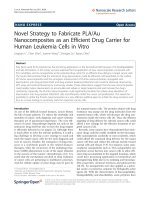
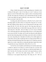
among 263 screened children (Figure 1). One hundred and
Page 3 of 9
twelve children fulfilled study criteria. Nine of the 112 (8%)
children were classified as treatment failures. Six of the
112 children were experiencing bilateral infections for
which samples from both the left and right ears were collected. Of the 118 samples collected, 91% (107/118) were
collected by tympanocentesis. The primary reason for nonenrollment was spontaneous otorrhea more than 24 hours
prior to the visit (n = 52).
Demographic characteristics and clinical history
The median age of screened children was 33.5 months
compared to a median age of 36 months among participating children (range 5–59 months) (Table 1). Nine percent (10/112) of participating children were between 3
and 11 months of age, 14% (16/112) were between 12 and
23 months, and the remainders were uniformly distributed
between the other classes of age (24–35, 36–47 and 48–59
months). Fifty-five percent (62/112) of participating children were females. Sixty-four percent (7/11) of children
with spontaneous otorrhea were less than 24 months of
age, while 19% (19/101) of children in whom tympanocentesis was used were less than 24 months. None of the
children had received any doses of a pneumococcal conjugate vaccine, while 4% (5/112) had received at least one
dose of influenza vaccine. Antibiotic use within the past
month was reported for 23% (26/112) of children. AOM
was classified as recurrent for 7% (8/112) of children.
Microbiology
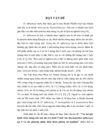
Overall, 48% (57/118) of samples yielded cultures with
one of the 4 bacterial pathogens under study (S. pneumoniae, H. influenzae, M. catarrhalis or S. pyogenes)
(Figure 2), 2 of which were positive for more than one
bacteria. The most frequently detected bacteria was S.
pneumoniae (47% (27/57)), followed by H. influenzae (37%
(21/57)), M. catarrhalis (12% (7/57)) and S. pyogenes (7%
(4/57)). Among the 11 samples collected from otorrhea
episodes, one was positive for S. pneumoniae, 2 for H.
influenzae and 3 for S. pyogenes. Two of the 9 treatment
failure samples were positive for a pathogen under study,
both of which were S. pneumoniae. The most common
pneumococcal serotypes isolated from the 27 S. pneumoniae-isolates were 19F (7/27 (26%)), 14 (6/27 (22%)) and 3
(4/27 (15%)) (Figure 2). Out of the 21 H. influenzae isolates, 13 (62%) were serotype b (Hib), 3 (14%) were nontypeable, and the remainders were serotypes a (1 isolate
(5%)), d (2 isolates (10%)) and f (1 isolate (5%)), with one
(5%) missing (Figure 2). Overall, Hib was detected in
11% of all samples (13/118). The 2 co-infected samples
were due to one co-infection of S. pneumoniae 23F and
H. influenzae serotype a, and one co-infection of Hib and
M. catarrhalis.
S. pneumoniae and Hib and non-Hib H. influenzae
were the most commonly detected pathogens in all age
Intakorn et al. BMC Pediatrics 2014, 14:157
/>
Page 4 of 9
263 children screened
140 children not enrolled
-52 spontaneous otorrhea > 24 hours
-34 no indicaƟon for tympanocentesis
-17 exceeded 20% otorrhea guideline
-11 parent refused enrollment
-26 not enrolled for other reasons
123 children enrolled
11 children excluded*
-5 received systemic anƟbioƟcs in past
72 hours for disease other than AOM
-3 onset of AOM >72 hours prior to
diagnosis
-5 did not meet criteria for AOM
-1 hospitalized during AOM
-1 provided anƟbioƟcs by ENT prior to
tympanocentesis
112 children included in cohort
-103 untreated
-9 treatment failures
118 samples**
-107 collected by tympanocentesis
-11 collected by otorrhea
61 samples – cultures yielded no
study pathogen
57 samples – cultures yielded study
pathogen
-27 S. pneumoniae
-21 H. influenzae
- 4 S. pyogenes
-7 M. catarrhalis
Figure 1 Enrollment and etiology of AOM patients included in the study.
groups (Table 1). In the youngest age range of 3–11
months, S. pneumoniae and H. influenzae were each isolated from 2/10 episodes (20%). Among children 12–35
months of age, S. pneumoniae was isolated from 7/45
(15.5%) episodes while H. influenzae was detected in
10/45 (22%). In the oldest children, 36–59 months of
age, S. pneumoniae was detected from 17/57 (30%)
episodes, and H. influenzae from 9/57 (16%). Potential
risk and protective factors, including premature birth,
HIV infection, child care attended, child breast-fed and
number of household siblings less than 5, were similar
when compared by pathogen (data not shown). Due to
small numbers, the differences in age group and potential
risk factors by pathogen were not tested for statistical
significance.
Symptoms
The most frequently reported symptom was ear pain,
reported for 95% (106/112) of episodes, followed by irritability, reported for 49% (55/112) of episodes (Table 2).
Fever was reported for 12% (3/26) of children experiencing
AOM due to S. pneumoniae but was not reported for
any children experiencing AOM due to Hib or non-Hib
H. influenzae. Trouble sleeping was reported for 2% (2/26)
of children experiencing AOM due to S. pneumoniae, 31%
(4/13) of those experiencing AOM due to Hib and 25%
(2/8) of those experiencing AOM due to non-Hib H.
influenzae (Table 2). Due to small numbers, the differences in symptoms by pathogen were not tested for statistical significance.
Hib-positive AOM
Thirty-eight percent (5/13) of Hib-positive AOM and
13% (1/8) of AOM due to other H. influenzae were in
children 12–23 months, compared to 14% (16/112) of
AOM overall. Fifteen percent (2/13) of children with
Hib-positive AOM and 13% (1/8) of children with AOM
due to other H. influenzae reported taking antibiotics in
the past month. Two of the 3 children who experienced
hearing loss had Hib-positive AOM. Irritability and ear
tugging were reported for a greater proportion of children with Hib-positive AOM compared to children with
Intakorn et al. BMC Pediatrics 2014, 14:157
/>
Page 5 of 9
Table 1 AOM pathogens analyzed by age group, gender, and sample collection method
Total
(positive and negative)
Any culture
positive
S. pneumoniae
Non-Hib
H. influenzae
Hib
M. catarrhalis
S. pyogenes
Age
3–11 months
10
5 (50%)
2
1
1
0
1
12–23 months
16
10 (63%)1
2
1
51
11
2
24–35 months
29
11 (38%)
5
2
2
2
0
36–47 months
28
14 (50%)
82
0
4
2
0
48–59 months
29
16 (55%)3
93
43
1
2
1
Total episodes
112
56 (51%)1,3
262,3 (23%)
8 (7%)3
13 (12%)1
7 (6%)1
4 (4%)
Male
51
26 (51%)
102 (20%)
5 (10%)
7 (14%)
1 (2%)
3 (6%)
Collection method
Otorrhea
11
6 (55%)
1
1
1
0
3
Tympanocentesis
107
51 (48%)
264
7
12
7
1
8 (7%)
13 (11%)
7 (6%)
4 (3%)
Total samples
4
118
57 (48%)
4
27 (23%)
Data presented per episodes in the upper part of the table and per samples in the lower part. Percentages are calculated based on the total (positive and
negative) number of episodes or samples respectively.
1
Includes one episode with a co-infection by H. influenzae and M. catarrhalis.
2
Includes one episode with a bilateral infection, from which the two collected samples were culture positive for S. pneumoniae (unknown serotype, same
susceptibility to antibiotics).
3
Includes one episode with a co-infection by S. pneumoniae and H. influenzae.
4
Two samples were collected from the 6 children presenting with a bilateral infections, leading to a total of 118 samples from the 112 episodes. No bacteria were
identified in 5 bilateral infections, the last one was positive for S. pneumoniae (27 samples from 26 episodes).
AOM due to other H. influenzae (54% (7/13) and 31%
(4/13) versus 0% and 13% (1/8), respectively). Seventyseven percent (10/13) of Hib-positive and 50% (4/8) of
other H-influenzae-positive children had an OS-8 scale
score of greater than 5.
Antibiotic susceptibility
Among the 27 S. pneumoniae isolates, all were susceptible
to amoxicillin/clavulanate and to penicillin, 11% (3/27)
were non-susceptible to cefotaxime, 63% (17/27) were
non-susceptible to cefuroxime, 67% (18/27) were nonsusceptible to erythromycin and 78% (21/27) were nonsusceptible to trimethoprim/sulfamethoxazole (Table 3).
Eighty-one percent (22/27) of S. pneumoniae isolates were
multidrug resistant. Among 19F isolates, the most prominent serotype, 2 out of 7 were non-susceptible to cefotaxime and 5 out of 7 were non-susceptible to cefuroxime. All
H. influenzae isolates were susceptible to amoxicillin/
clavulanate and to cefotaxime, 5% (1/21) was non-susceptible
to cefuroxime, and 20% (4/20) were non-susceptible to
ampicillin, with ampicillin data missing for one isolate
(Table 3). Three of the 4 isolates not susceptible to ampicillin were Hib isolates. One (Hib) of the 21 H. influenzae
isolates was beta-lactamase-negative ampicillin-resistant
but susceptible to amoxicillin/clavulanate.
Discussion
The AOM episodes seen in this study among children
who sought care from ENTs in Thailand were generally
non-recurrent episodes assessed by tympanocentesis. In
this study environment, where there was minimal use of
either Hib or pneumococcal vaccine, bacterial pathogens
were an important cause of AOM. The leading causes of
bacterial AOM were S. pneumoniae and H. influenzae,
representing 47% (27/57) and 37% (21/57) of culturepositive samples, respectively. The majority of H. influenzae was serotype b (62% (13/21)). Forty-eight percent of
samples were culture-positive for one of the pathogens
under study, slightly lower than the 53-58% reported in
other settings [24], but consistent with the assumption
that isolation of bacteria may be lower in an environment
with high antibiotic use [25]. Other studies have found
that PCR can detect bacteria in culture-negative MEF
[26], so it is possible that these pathogens play a greater
role in AOM than what was detected here.
We found slightly more S. pneumoniae than H. influenzae, consistent with what was seen elsewhere in the prePCV7 era [3]. In this population, AOM episodes were
generally comprised of relatively mild, sporadic cases, rather than severe or recurrent. H. influenzae was slightly
more prominent than S. pneumoniae in children 12–23
months of age while the reverse was true in children
24–59 months of age. Overall, the symptom profiles and
potential risk factor profiles of S. pneumoniae and
H. influenzae were generally similar.
One unexpected finding in the studywas the higher
than expected presence of Hib. This was a surprise in
part because available data suggest a low incidence of
Hib-associated invasive disease in Thailand [27], although there are concerns that existing data from Asia
Intakorn et al. BMC Pediatrics 2014, 14:157
/>
Page 6 of 9
Figure 2 Culture results and pathogens under study identified from middle ear fluid samples (N = 118). Culture results from middle ear
fluid samples including serotype distribution for S. pneumoniae (Spn, n = 27), and H. influenzae (H. inf, n = 21). There were two co-infected samples
due to one co-infection of S. pneumoniae 23F and H. influenzae serotype a, and one co-infection of Hib and M. catarrhalis.
Table 2 Symptoms reported at the visit for AOM patients in the study
S. pneumoniae positive (N = 26)
Non-Hib H. influenzae positive (N = 8)
Hib positive (N = 13)
Total (N = 112)
Ear pain
25 (96%)
8 (100%)
13 (100%)
106 (95%)
OS-8 > 5
15 (58%)
4 (50%)
10 (77%)
64 (57%)
Irritability
16 (62%)
0 (0%)
7 (54%)
55 (49%)
Tugging
6 (23%)
1 (13%)
4 (31%)
33 (29%)
37.5-39.0°C
12 (46%)
2 (25%)
3 (23%)
30 (27%)
> 39.0°C
3 (12%)
0 (0%)
0 (0%)
6 (5%)
2 (8%)
2 (25%)
4 (31%)
28 (25%)
Anorexia
4 (15%)
0 (0%)
1 (8%)
16 (14%)
Vomiting
3 (12%)
0 (0%)
1 (8%)
10 (9%)
Diarrhea
0 (0%)
0 (0%)
1 (8%)
4 (4%)
Hearing loss
1 (4%)
0 (0%)
2 (15%)
3 (3%)
Temperature – axillary
Trouble sleeping
Conjunctivitis
0 (0%)
0 (0%)
0 (0%)
2 (2%)
Lethargy
2 (8%)
0 (0%)
0 (0%)
2 (2%)
N = number of episodes, data presented as n (%).
Intakorn et al. BMC Pediatrics 2014, 14:157
/>
Page 7 of 9
Table 3 Antibacterial non-susceptibility of S. pneumoniae and H. influenzae isolates
Number of non-susceptible1 isolates
Antibiotic
Amoxicillin/Clavulanate
S. pneumoniae isolates (N = 27)2
H. influenzae isolates (N = 21)
0 (0%)
0 (0%)
-
4 (20%)
26 (96%)
2 (10%)
3
Ampicillin
Azithromycin
Cefotaxime
3 (11%)
0 (0%)
Cefuroxime
17 (63%)
1 (5%)
Chloramphenicol
7 (26%)
2 (10%)
Erythromycin4
18 (67%)
-
Levofloxacin
0 (0%)
0 (0%)
Penicillin4
0 (0%)
-
Tetracycline
18 (67%)
2 (10%)
Trimethoprim/Sulfamethoxazole
21 (78%)
7 (33%)
1
Intermediate or resistant based on the Clinical and Laboratory Standards Institute 2009 standards.
Two isolates are coming from the same child with a bilateral infection.
Ampicillin resistance data missing for one H. influenzae isolate. Ampicillin sensitivity was not performed for S. pneumoniae.
4
For H. influenzae, the median value of MIC was equal to 4.0 for Erythromycin and 0.250 for Penicillin.
2
3
underestimate the true burden [28,29]. Additionally, on
a global level, Hib is generally perceived not to be an important AOM pathogen. Before the introduction of the
Hib vaccine in the United States, for example, Hib only
represented 10% of H. influenzae AOM cases [30], while
in our study, Hib was seen in 62% of the H. influenzae
isolates. Another exception to the general observation
that encapsulated H. influenzae are not important causes
of AOM comes from a recent, tympanocentesis-based
study in Venezuela where 31% of H. influenzae AOM
were encapsulated a, c, d and f strains (Venezuela has
universal Hib immunization) [31]. Interestingly, based
on the OS-8 scale, the Thai Hib cases seemed to be
slightly more severe than S. pneumoniae or non-Hib H.
influenzae cases.
A second surprising finding was that the median age
of children in the study was 36 months, which is unusual given that AOM incidence elsewhere generally
peaks at 6–18 months of age. Since the age of the
screened cohort was only slightly younger than the enrolled cohort it does not appear that there was significant bias in the final study sample (i.e., those who
received tympanocentesis) compared to all children who
came to the ENT with suspected AOM. While it is possible that the true burden of AOM in Thailand tends to
be in older children, it also may be that younger children
with AOM are more often treated at home or by general
practitioners and do not tend to visit the ENT. We note
that a number of children could not be enrolled because
of otorrhea for greater than 24 hours, which may suggest
more severe AOM or may suggest that access to prompt
care is limited, by distance or other factors.
The distribution of S. pneumoniae serotypes was similar to what has been reported in the literature [10,32]
prior to PCV introduction. The generally mild profile
of AOM experienced by the children in our study may
explain the slightly higher than expected proportion of
M. catarrhalis isolates, as this pathogen is often associated with milder disease [33].
Due to the risk of treatment failures, up-to-date information on antibiotic resistance has important clinical
implications for determining the best approach for treatment of AOM [19]. Our results show high levels of resistance of S. pneumoniae to some antibiotics commonly
given in Thailand for respiratory infections (Azithromycin, Cefuroxime, Erythromycin, Tetracycline, Trimethoprim/Sulfamethoxazole), and a high level of multidrug
resistance. This was consistent with results from another
study in Asian countries [34], which also noted a high
level of resistance to macrolides. In our study only a low
rate of cefotaxime non-susceptibility was seen, likely due
to the fact that cephalosporins are generally only prescribed for children presenting with severe illness (moderate to severe otalgia or fever of 39°C ) at first visit or for
patients who do not respond to initial treatment. Antibiotic resistance was less common for H. influenzae, and
was similar to previously published estimates, though our
isolates had lower levels of resistance to chloramphenicol
(10% versus 25%) and ampicillin (15% versus 48%) [35]. It
is possible that more severe AOM cases than were seen in
this study would be enriched for more resistant AOM.
Currently Hib vaccine use in Thailand is extremely limited as it is not on the Expanded Program of Immunization
for Thailand [35]. Uptake of PCV7 in Thailand, which
is mainly used in private settings, has also been low
[10], and there were no reports of pneumococcal vaccine
use in the children in our study. Two other pneumococcal
vaccines, Prevenar/Prevnar 13™ (Wyeth, LLC) (PCV13)
Intakorn et al. BMC Pediatrics 2014, 14:157
/>
and Synflorix™ (GlaxoSmithKline Vaccines) (PHiD-CV),
have been licensed in recent years, and differ from PCV7
in the inclusion of 6 (1, 3, 5, 6A, 7F, 19A) and 3 (1, 5, 7F)
additional serotypes, respectively. PHiD-CV also utilizes
as the predominant carrier protein an outer membrane
protein (protein D) derived from H. influenzae, as a protein D-containing 11-valent precursor formulation of
PHiD-CV was previously shown to be efficacious against
both pneumococcal and H. influenzae AOM [36]. Efficacy
of PHiD-CV itself against AOM was also recently demonstrated in another double-blind randomized clinical study
[37]. Although PCV13 efficacy against AOM has not yet
been assessed, such data do exist for its predecessor formulation PCV7 [38]. Of the 22 pneumococcal isolates
whose serotype could be identified, at least 16 (73%) represent a serotype contained in each of the two higher
valent vaccines. Serotype 3, contained only in PCV13, was
also identified in 4/22 (18%) of those pneumococcal isolates, but it remains unclear whether serotype 3 disease is
vaccine-preventable [39]. Our results thus suggest that either vaccine would likely prevent a significant proportion
of AOM cases.
The study was successful in adding to the limited data
on AOM in Thailand, but there are important limitations, including few cases in the youngest children, small
sample size and lack of a clear population denominator.
The study did cover several, but not all, regions of
Thailand, and therefore is somewhat limited in geographical representativeness. As the use of a Hib vaccine is
known to be very limited in Thailand, we did not collect
individual Hib vaccination status, though it could have
provided further insight into the previously unrecognized
burden of Hib in AOM cases that was identified in this
study. An additional limitation is that the over-thecounter availability of antibiotics in Thailand could mean
that some children may have received antibiotics before
the study visit, This could have decreased the proportion
of culture positives, and meant that bacteria that were isolated from such patients may have been those with greater
non-susceptibility. However, as it was impossible to know
whether any antibiotics received in this manner were appropriate for AOM and/or provided in sufficient dosage,
only patients receiving antibiotics prescribed by a physician 48–72 hours prior to the study visit were considered
treatment failures, as per protocol.
Conclusions
In summary, this assessment of AOM etiology in Thai
children 3 to 59 months of age visiting ENT clinics
for AOM showed an unexpectedly high burden of Hib.
S. pneumoniae and H. influenzae were the leading causes
of AOM across all age groups, similar to what has been
seen in Europe, the United States, and Latin America, and
with pneumococcal serotypes similar to those found
Page 8 of 9
elsewhere [32,40]. These findings contribute to the scarce
tympanocentesis literature in this region, and suggest that
conjugate vaccines effective against pneumococcus and
H. influenzae, both encapsulated (Hib) and unencapsulated, may be important in attempts to reduce bacterial
AOM in the region.
Trademark
Prevnar and Prevnar 13 are trademarks of Wyeth LLC.
Synflorix is a trademark of the GlaxoSmithKline group
of companies.
Competing interest
GlaxoSmithKline Biologicals SA funded all costs associated with the study
and with the development and publishing of the present manuscript. GM
and SN declare no conflicts of interest. PI received an institutional grant and
a travel grant from the GlaxoSmithKline group of companies. NS has
received a grant, travel grant funding and payment for lectures from the
GlaxoSmithKline group of companies. JYP and WPH are employees of the
GlaxoSmithKline group of companies. WPH own stock in GlaxoSmithKline
Biologicals and is co-holder of the patent for Prevnar 13™. YFL and MVD
were previously employed by the GlaxoSmithKline group of companies and
had stock options.
Authors’ contributions
PI, YL and WPH participated in the conception and design of the study and
together with NS, MKV, SN and GM contributed to the development of the
protocol. PI, NS, SN and GM contributed to the acquisition of data. JYP
(study and project statistician), PI, GM, NS, SN, YL and MKV contributed to
data processing, to the statistical analysis and to the study report. MKV
contributed to the interpretation of the statistical analysis and together with
NS, WPH and YL to the development of the manuscript. All authors had full
access to the data, read and reviewed drafts of the manuscripts and
approved its final content.
Acknowledgements
The authors thank Drs Barbara Pelgrims, Véronique Mouton and Marie-Line
Seret (XPE Pharma & Science c/o GlaxoSmithKline Vaccines) for editorial
assistance and manuscript coordination and Dr Anna Dow (Freelance) for
scientific writing support on behalf of the GlaxoSmithKline Group of
companies.
Author details
1
Department of Otolaryngology, Queen Sirikit National Institute of Child
Health, 420/8 Rajvithi Road, Rajthevee, Bangkok 10400, Thailand.
2
Department of Otolaryngology, Faculty of Medicine, Chiang Mai University,
110 Intawaroros Road, Muang District, Chiang Mai 50200, Thailand.
3
Department of Otolaryngology, Hatyai Hospital, 182 Ratakan Haiyai,
Songkhla, 90110, Thailand. 4Department of Otolaryngology, Phramongkutklao
Hospital of the Royal Thai Army, 315 Rajvithi Road, Rajthevee, Bangkok,
Thailand. 5GlaxoSmithKline Vaccines, Avenue Fleming 20, 1300 Wavre,
Belgium. 6GlaxoSmithKline Vaccine Singapore, 150 Beach Road, Gateway
West, 22-00, 189720 Singapore, Singapore. 7Current affiliation: Janssen
Pharmaceutical companies of Johnson and Johnson, 2 International Business
Par, 07-00, The Strategy, Singapore 609930, Singapore. 8Current affiliation:
Amgen, Inc., 1 Amgen Center Dr, Thousand Oaks, CA 91320, USA.
Received: 12 October 2013 Accepted: 12 June 2014
Published: 20 June 2014
References
1. Klein JO: Otitis media. Clin Infect Dis 1994, 19:823–833.
2. McCaig LF, Hughes JM: Trends in antimicrobial drug prescribing among
office-based physicians in the United States. JAMA 1995, 273:214–219.
3. Leibovitz E, Jacobs MR, Dagan R: Haemophilus influenzae: a significant
pathogen in acute otitis media. Pediatr Infect Dis J 2004, 23:1142–1152.
4. Block SL, Hedrick J, Harrison CJ, Tyler R, Smith A, Findlay R, Keegan E:
Community-wide vaccination with the heptavalent pneumococcal
Intakorn et al. BMC Pediatrics 2014, 14:157
/>
5.
6.
7.
8.
9.
10.
11.
12.
13.
14.
15.
16.
17.
18.
19.
20.
21.
22.
23.
24.
25.
conjugate significantly alters the microbiology of acute otitis media.
Pediatr Infect Dis J 2004, 23:829–833.
Berman S: Otitis media in developing countries. Pediatrics 1995, 96:126–131.
Mahadevan M, Navarro-Locsin G, Tan HK, Yamanaka N, Sonsuwan N, Wang PC,
Dung NT, Restuti RD, Hashim SS, Vijayasekaran S: A review of the burden of
disease due to otitis media in the Asia-Pacific. Int J Pediatr Otorhinolaryngol
2012, 76:623–635.
Arguedas A, Kvaerner K, Liese J, Schilder AG, Pelton SI: Otitis media across
nine countries: Disease burden and management. Int J Pediatr
Otorhinolaryngol 2010, 74:1419–1424.
World Health Organization: Chronic suppurative otitis media; burden of
illness and management options. 2004, Available at />deafness/activities/hearing_care/otitis_media.pdf. Accessed 01 February 2011.
Levine S, Dejsirilert S, Sangsuk L, Chantra S, Feikin DR, Dowell SF, Olsen SJ:
Serotypes and antimicrobial resistance of Streptococcus pneumoniae in
Thailand 2002–2004. Pediatr Infect Dis J 2006, 25:176–178.
Srifeungfung S, Tribuddharat C, Comerungsee S, Chatsuwan T,
Treerauthanaweeraphong V, Rungnobhakhun P, Nunthapisud P,
Chokephaibulkit K: Serotype coverage of pneumococcal conjugate
vaccine and drug susceptibility of Streptococcus pneumoniae isolated
from invasive or non-invasive diseases in central Thailand, 2006–2009.
Vaccine 2010, 28:3440–3444.
Lynch JP 3rd, Zhanel GG: Streptococcus pneumoniae: epidemiology and
risk factors, evolution of antimicrobial resistance, and impact of vaccines.
Curr Opin Pulm Med 2010, 16:217–225.
Vergison A, Dagan R, Arguedas A, Bonhoeffer J, Cohen R, Dhooge I,
Hoberman A, Liese J, Marchisio P, Palmu AA, Ray GT, Sanders EA, Simoes EA,
Uhari M, van Eldere J, Pelton SI: Otitis media and its consequences:
beyond the earache. Lancet Infect Dis 2010, 10:195–203.
Cohen R, Ovetchkine P, Gehanno P: Current approaches to otitis media.
Curr Opin Infect Dis 2001, 14:337–342.
Syrjanen RK, Herva EE, Makela PH, Puhakka HJ, Auranen KJ, Takala AK, Kilpi
TM: The value of nasopharyngeal culture in predicting the etiology of
acute otitis media in children less than two years of age. Pediatr Infect Dis
J 2006, 25:1032–1036.
Eldan M, Leibovitz E, Piglansky L, Raiz S, Press J, Yagupsky P, Leiberman A,
Dagan R: Predictive value of pneumococcal nasopharyngeal cultures
for the assessment of nonresponsive acute otitis media in children.
Pediatr Infect Dis J 2000, 19:298–303.
WHO vaccine-preventable diseases: monitoring system 2012 global
summary. [ />Wongsawat J, Chokephaibulkit K: Implication of pneumococcal conjugate
vaccines to public health: Thailand perspective. J Med Assoc Thai 2010,
93(Suppl 5):S53–S60.
Muangchana C, Thamapornpilas P, Karnkawinpong O: Immunization policy
development in Thailand: the role of the Advisory Committee on
Immunization Practice. Vaccine 2010, 28(Suppl 1):A104–A109.
Dagan R: Appropriate treatment of acute otitis media in the era of
antibiotic resistance. Paediatr Drugs 2010, 12(Suppl 1):3–9.
Casey JR, Pichichero ME: Changes in frequency and pathogens causing
acute otitis media in 1995–2003. Pediatr Infect Dis J 2004, 23:824–828.
Leibovitz E, Serebro M, Givon-Lavi N, Greenberg D, Broides A, Leiberman A,
Dagan R: Epidemiologic and microbiologic characteristics of culturepositive spontaneous otorrhea in children with acute otitis media.
Pediatr Infect Dis J 2009, 28:381–384.
Pai R, Gertz RE, Beall B: Sequential multiplex PCR approach for
determining capsular serotypes of Streptococcus pneumoniae isolates.
J Clin Microbiol 2006, 44:124–131.
Clinical and Laboratory Standards Institute: Performance Standards for
Antimicrobial Disk Susceptibility Tests; Approved Standard - Tenth
Edition (M02-A10). 2009, />Li WC, Chiu NC, Hsu CH, Lee KS, Hwang HK, Huang FY: Pathogens in the
middle ear effusion of children with persistent otitis media: implications
of drug resistance and complications. J Microbiol Immunol Infect 2001,
34:190–194.
Rhodes J, Hyder JA, Peruski LF, Fisher C, Jorakate P, Kaewpan A, Dejsirilert S,
Thamthitiwat S, Olsen SJ, Dowell SF, Chantra S, Tanwisaid K, Maloney SA,
Baggett HC: Antibiotic use in Thailand: quantifying impact on blood culture
yield and estimates of pneumococcal bacteremia incidence. Am J Trop Med
Hyg 2010, 83:301–306.
Page 9 of 9
26. Couloigner V, Levy C, Francois M, Bidet P, Hausdorff WP, Pascal T, Boucherat
M, Bingen E, Mariani P, Pierrot S, Bille E, Carbonnelle E, Varon E, Cohen R:
Pathogens implicated in acute otitis media failures after 7-valent
pneumococcal conjugate vaccine implementation in France: distribution,
serotypes, and resistance levels. Pediatr Infect Dis J 2012, 31:154–158.
27. Rerks-Ngarm S, Treleaven SC, Chunsuttiwat S, Muangchana C, Jolley D,
Brooks A, Dejsirilert S, Warintrawat S, Guiver M, Kunasol P, Maynard JE, Biggs
BA, Steinhoff M: Prospective population-based incidence of Haemophilus
influenzae type b meningitis in Thailand. Vaccine 2004, 22:975–983.
28. Broker M: Burden of invasive disease caused by Haemophilus influenzae
type b in Asia. Jpn J Infect Dis 2009, 62:87–92.
29. Shetty S, Cohen AL, Edmond K, Ojo L, Loo J, O'Loughlin R, Hajjeh R: A
systematic review and critical evaluation of invasive Haemophilus
influenzae type B disease burden studies in Asia from the last decade:
lessons learned for invasive bacterial disease surveillance. Pediatr Infect
Dis J 2010, 29:653–661.
30. Bluestone C: Terminology and Classification. In Evidence-Based Otitis Media.
Edited by Bluestone C. Saint Louis: Hamilton; 1999:85–103.
31. Naranjo L, Suarez JA, DeAntonio R, Sanchez F, Calvo A, Spadola E, Rodriguez N,
Andrade O, Bertuglia F, Marquez N, Castrejon MM, Ortega-Barria E, Colindres
RE: Non-capsulated and capsulated Haemophilus influenzae in children
with acute otitis media in Venezuela: a prospective epidemiological study.
BMC Infect Dis 2012, 12:40.
32. Rodgers GL, Arguedas A, Cohen R, Dagan R: Global serotype distribution
among Streptococcus pneumoniae isolates causing otitis media in
children: potential implications for pneumococcal conjugate vaccines.
Vaccine 2009, 27:3802–3810.
33. Broides A, Dagan R, Greenberg D, Givon-Lavi N, Leibovitz E: Acute otitis
media caused by Moraxella catarrhalis: epidemiologic and clinical
characteristics. Clin Infect Dis 2009, 49:1641–1647.
34. Kim SH, Song JH, Chung DR, Thamlikitkul V, Yang Y, Wang H, Lu M, So TM,
Hsueh PR, Yasin RM, Carlos CC, Pham HV, Lalitha MK, Shimono N, Perera J,
Shibl AM, Baek JY, Kang CI, Ko KS, Peck KR: Changing trends in
antimicrobial resistance and serotypes of Streptococcus pneumoniae
isolates in Asian countries: an Asian Network for Surveillance of
Resistant Pathogens (ANSORP) study. Antimicrob Agents Chemother 2012,
56:1418–1426.
35. Srifuengfung S, Chayakulkeeree M, Chokephaibulkit K, Tribuddharat C:
Five-year study of antimicrobial susceptibility and beta-lactamase
production in Haemophilus influenzae. Southeast Asian J Trop Med
Public Health 2007, 38:732–736.
36. Prymula R, Peeters P, Chrobok V, Kriz P, Novakova E, Kaliskova E, Kohl I,
Lommel P, Poolman J, Prieels JP, Schuerman L: Pneumococcal capsular
polysaccharides conjugated to protein D for prevention of acute otitis
media caused by both Streptococcus pneumoniae and non-typable
Haemophilus influenzae: a randomised double-blind efficacy study. Lancet
2006, 367:740–748.
37. Tregnaghi MW, Sáez-Llorens X, López P, Abate H, Smith E, Pósleman A, Calvo A,
Wong D, Cortes-Barbosa C, Ceballos A, Tregnaghi M, Sierra A, Rodriguez M,
Troitiño M, Carabajal C, Falaschi A, Leandro A, Castrejón MM, Lepetic A, Lommel P,
Hausdorff WP, Borys D, Ruiz Guiñazú J, Ortega-Barría E, Yarzábal JP, Schuerman L,
on behalf of the COMPAS Group: Efficacy of PHiD-CV in Young Latin American
Children: A Double-Blind, Randomized Controlled Trial. PLoS Med, 2014,
11(6):e1001657.
38. Eskola J, Kilpi T, Palmu A, Jokinen J, Haapakoski J, Herva E, Takala A, Kayhty H,
Karma P, Kohberger R, Siber G, Makela PH: Efficacy of a pneumococcal
conjugate vaccine against acute otitis media. N Engl J Med 2001, 344:403–409.
39. Mrkvan T, Clarke C, Hausdorff WP: Should the next generation of
pneumococcal conjugate vaccines (PCVs) contain serotype 3? Lessons learned
and rediscovered. Denver, USA: Third Interscience Conference on
Antimicrobial Agents and Chemotherapy (ICAAC); 2013. Abstract B-498.
40. Hausdorff WP, Yothers G, Dagan R, Kilpi T, Pelton SI, Cohen R, Jacobs MR,
Kaplan SL, Levy C, Lopez EL, Mason EO Jr, Syriopoulou V, Wynne B, Bryant J:
Multinational study of pneumococcal serotypes causing acute otitis
media in children. Pediatr Infect Dis J 2002, 21:1008–1016.
doi:10.1186/1471-2431-14-157
Cite this article as: Intakorn et al.: Haemophilus influenzae type b as an
important cause of culture-positive acute otitis media in young children
in Thailand: a tympanocentesis-based, multi-center, cross-sectional
study. BMC Pediatrics 2014 14:157.