Perinatal mortality in pregnancies with omphalocele: Data from the Chinese national birth defects monitoring network, 1996–2006
Bạn đang xem bản rút gọn của tài liệu. Xem và tải ngay bản đầy đủ của tài liệu tại đây (244.36 KB, 7 trang )
Deng et al. BMC Pediatrics 2014, 14:160
/>
RESEARCH ARTICLE
Open Access
Perinatal mortality in pregnancies with
omphalocele: data from the Chinese national
birth defects monitoring network, 1996–2006
Kui Deng1,2†, Jie Qiu3†, Li Dai1, Ling Yi1, Changfei Deng1, Yi Mu1 and Jun Zhu1,2*
Abstract
Background: Previous studies on the mortality rate of omphalocele are limited. The risk of death of non-isolated
omphalocele and that of cases of omphalocele that are diagnosed prenatally by ultrasound are unclear. This study
aimed to estimate the perinatal mortality of pregnancies with omphalocele. This study also examined the potential
risk of death of non-isolated omphalocele and that of cases that are prenatally diagnosed by ultrasound.
Methods: Data were retrieved from the national birth defects registry in China, for 1996–2006. Multinomial logistic
regression was used to calculate the adjusted odds ratios (AORs) and 95% confidence intervals (CIs) between
perinatal mortality and selected maternal and fetal characteristics.
Results: Among 827 cases of omphalocele, 309 (37.4%) cases resulted in termination of pregnancy and stillbirth,
and 124 (15.0%) cases resulted in death in the first 7 days after delivery, yielding a perinatal mortality rate of 52.4%
(95% CI: 49.0–55.8%). The late fetal death rate (LFDR) of omphalocele that was diagnosed prenatally by ultrasound
was 15.91-fold (AOR: 15.91, 95% CI: 10.18–24.87) higher than that of postnatally diagnosed cases. The LFDR of
non-isolated omphalocele was 2.64-fold (AOR: 2.64, 95% CI: 1.62–4.29) higher than that of isolated cases. For the
early neonatal death rate, neonates with non-isolated omphalocele had a 2.96-fold (AOR: 2.96, 95% CI: 1.82–4.81)
higher risk than isolated cases, but the difference between prenatal ultrasound diagnosis and postnatal diagnosis
was not significant.
Conclusions: Selected fetal characteristics are significantly associated with the perinatal risk of death from
omphalocele. Our findings suggest that improving pregnancy and delivery care, as well as management for
omphalocele are important.
Keywords: Omphalocele, Abdominal wall defects, Mortality, Perinatal outcome, Associated anomalies, Prenatal
diagnosis, Ultrasound
Background
Omphalocele is among the more common anterior abdominal wall defects, and it is characterized as the absence
of abdominal muscles, fascia, and skin. With omphalocele,
there is herniation of the abdominal contents into the base
of the umbilical cord, and these contents are covered by a
* Correspondence:
†
Equal contributors
1
National Center for Birth Defects monitoring of China, West China Second
University Hospital, Sichuan University, 17, Section3, Ren Min South Road,
Chengdu, China
2
Key Laboratory of Birth Defects and Related Diseases of Women and
Children (Sichuan University), Ministry of Education, Chengdu, China
Full list of author information is available at the end of the article
membranous sac consisting of peritoneum and amnion
[1,2]. Previous studies have estimated that the overall
prevalence of omphalocele ranges from 2 to 3 per
10,000 births worldwide [3-10]. Omphalocele is also associated with a substantial risk of infant morbidity and
mortality, which is a severe disadvantage to the shortand long-term life of affected newborns. Early surgical
repair can improve the prognosis and increase the survival rate of omphalocele.
Previous reports regarding the mortality rate of omphalocele are limited. Few studies have reported the
mortality of omphalocele using data from a large general
population based on congenital malformation registries
© 2014 Deng et al.; licensee BioMed Central Ltd. This is an Open Access article distributed under the terms of the Creative
Commons Attribution License ( which permits unrestricted use, distribution, and
reproduction in any medium, provided the original work is properly credited.
Deng et al. BMC Pediatrics 2014, 14:160
/>
and national/regional epidemiological surveys. More recent studies have estimated the neonatal mortality of omphalocele using prenatal and neonatal databases from
certain hospitals. However, these databases were confined
to one hospital with a small number of cases, and the reported estimates with a wide range did not truly present
the risk of death from omphalocele in the general population [11-14].
Several risk factors are associated with the perinatal outcome of pregnancies with omphalocele. As reported in
most studies, omphalocele of concurrent with chromosomal anomalies or other structural malformations is
more likely to be terminated electively and the fetus dies
in utero [14-17]. Routine prenatal ultrasound screening
allows identification of the majority of omphalocele early
in gestation, but a high proportion of prenatally diagnosed
cases of omphalocele end with termination of pregnancy
or intrauterine death [3,14,18-20]. Previous studies did
not independently investigate the risk of death from
omphalocele with associated malformations using prenatal diagnosis by ultrasound because of potential confounding effects. The magnitude of effect estimates for
the risk of death from non-isolated omphalocele and
prenatally diagnosed cases by ultrasound has been imprecisely assessed.
This study aimed to estimate the perinatal mortality of
omphalocele using consecutive data for 11 years from the
national birth defects registry. We also aimed to examine
the potential risk of death of fetuses and neonates with
omphalocele by comparing the outcomes from different
groups (non-isolated vs isolated groups and prenatal ultrasound diagnosis group vs postnatal diagnosis group).
Methods
Ascertainment of cases
Data on cases with omphalocele were obtained from the
Chinese Birth Defects Monitoring Network (CBDMN)
from January 1996 to September 2006. This network is a
nationwide and hospital-based birth defects surveillance
network covering a total of 450–471 hospitals (county
level, city level, and provincial level) in China. The number
of monitored births accounted for approximately 8–10%
of the annual births in China [21]. The CBDMN used the
passive case ascertainment method to identify congenital
malformations, including live births, stillbirths, and termination of pregnancy in the member hospitals. The surveillance period for each abnormality was from 28 weeks
of gestations to the first 7 days after birth. These cases
were recognized by physical examination by trained obstetric and pediatric clinicians. The cases were confirmed
by documentation of the postnatal diagnosis and narrative
descriptions of abnormalities in the medical records. Cases
diagnosed by prenatal ultrasonography were also confirmed by the postnatal records after delivery. A trained
Page 2 of 7
midwife was then asked to complete the “Birth Defects
Register Form” registry and conduct online reporting
quarterly, after which CBDMN staff reviewed all of these
forms again. Incomplete forms and a nonspecific diagnosis
were controlled by the midwives within 7 days to correct
the final information. Pregnancies ending in stillbirth or
elective termination, including an autopsy report where
available, were reviewed to confirm or amend the final
diagnosis. Written informed consent was obtained from
the parents of neonates before they were discharged from
the hospital. The consent mainly included the aims and
importance of monitoring birth defects. This study was
approved by the Ethic Review Committee of Sichuan
University.
According to the International Clearinghouse for Birth
Defects Monitoring Systems, omphalocele was defined as
a midline abdominal wall defect, which was limited to an
open umbilical cord. The viscera herniates into the base of
the umbilical cord and is surrounded by the peritoneum
and amniotic membranes [22]. The International Classification of Diseases, Tenth Revision, was used to code the
diagnosis for omphalocele (Q79.2) in the national birth
defects registry of China.
Permission was authored by National Health and Family
Planning Commission to access data from the CBDMN.
Data for this analysis were extracted based on the diagnosis code from the national birth defects registry that was
developed by the CBDMN. The following variables were
considered in our analysis: geographical location, maternal
residence, maternal age, maternal education, gestational
age, birthweight, presence or absence of other anomalies,
prenatal or postnatal diagnosis, and the year of birth. Geographical location was divided into coastal areas, inland
areas, and remote areas. Maternal age was categorized as
20–24 years old, 25–29 years old, 30–34 years old, and
35–39 years old. Residence referred to that of the mothers,
and was divided into rural (countries or suburban areas)
and urban (towns or cities) areas. Gestational age was divided into 28–36 weeks and 37–42 weeks. Birthweight
was grouped into <2500 g and ≥2500 g. Isolated omphalocele was defined as omphalocele, which occurred without
chromosomal or structural malformations. Non-isolated
omphalocele was defined as cases with associated chromosomal or structural malformations, which were not related
to omphalocele. Prenatal diagnosis refers to cases of omphalocele that were detected prenatally by ultrasound.
Postnatal diagnosis refers to cases of omphalocele that
were detected by physical examination after birth. The response variable was perinatal mortality of birth with omphalocele, which was categorized into late fetal death
(LFD) and early neonatal death (ENND). LFD was defined
as the death of a fetus later than the gestational age of
28 weeks. ENND was defined as the death of a neonate in
the first 7 days of life. Data on the mortality of affected
Deng et al. BMC Pediatrics 2014, 14:160
/>
pregnancies were also extracted from the national birth
defects registry.
Data quality management
Data quality management (DQM) was routinely evaluated for surveillance data. The DQM teams consisted of
five upper-level CBDMN experts. These experts verified
data collection, data reporting, diagnosis of defects, and
obstetric and pediatric medical records according to the
surveillance manual. This was performed to improve the
accuracy, comparability, completeness, and timeliness of
the registered data. For DQM at the county level, all of
the member hospitals were investigated quarterly. For
DQM at the provincial and national levels, cluster sampling covered approximately one-third and 10% of the
member hospitals, respectively. Provincial- and nationallevel DQM were conducted semi-annually and annually,
respectively. The under-reporting rate of live births or
malformations needed to be no greater than 1%, and errors or missing information on the report form had to
be no greater than 1%. At each level, a panel of senior
health professionals evaluated the completeness, accuracy, and timeliness of the data.
Statistical analysis
The perinatal mortality rate of omphalocele was the sum
of the late fetal death rate (LFDR) and early neonatal
death rate (ENNDR). The LFDR was calculated by the
number of stillbirths and termination of pregnancies divided by the total number of births with omphalocele.
The ENNDR was calculated by the number of neonatal
deaths within the first 7 days after birth divided by the
total number of births with omphalocele.
The Cochran–Armitage trend test was used to assess
the changes in mortality of omphalocele over time. Because the response variables in our analysis were nominal
and for which there were two categories, multinomial logistic regression was used to generate the odds ratios and
95% confidence intervals (CIs) between the rate of LFD/
ENND and selected maternal/fetal characteristics, while
controlling for confounding factors to evaluate the effect
of each variable. The estimated risks were adjusted by
potential confounders, which were selected on the basis
of the results of the bivariate analysis and previously reported evidence. All tests of hypotheses were two-tailed
with a type 1 error rate fixed at 5%. Statistical analyses
were performed using SAS 9.1 software (SAS Institute
Inc., Cary, NC).
Since September 2006, the number of the member
hospitals has almost doubled. Therefore, to ensure the
comparability of registered data from the member hospitals, our study period was confined to January 1996 to
September 2006. During the study period, because the
number of births in some of the sampled surveillance
Page 3 of 7
hospitals had declined, we increased the number of hospitals that were selected from neighboring counties to
monitor a sufficient amount of births to ensure a representative sampled population. Additionally, some of the
member hospitals were replaced by other neighboring
hospitals because of reorganization of their medical services. Therefore, the number of hospitals over the study
period changed in our analysis.
Results
From January 1996 to September 2006, a total of 827
cases of omphalocele were identified from the CBDMN,
which included termination of pregnancy, stillbirths, and
live births. Of these, 322 (39.3%) cases were diagnosed
antenatally by ultrasound and 501 cases (60.6%) were confirmed by physical examination after birth. Four cases had
missing diagnosis information. Among the 827 cases, isolated omphalocele occurred in 596 (72.1%) cases and 231
(27.9%) cases were non-isolated omphalocele. The most
commonly associated anomaly with omphalocele was cleft
lip with or without cleft palate or cleft palate (n = 45),
followed by neural tube defects (n = 36), and then polydactyly or syndactyly (n = 33).
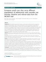
The perinatal mortality of pregnancies with omphalocele is shown in Table 1. A total of 309 fetuses died after
28 weeks’ gestation and 124 neonates were dead within
the first 7 days after birth. The perinatal mortality rate,
LFDR, and ENNDR of omphalocele was 52.4% (95% CI:
49.0–55.8%), 37.4% (95% CI: 34.1–40.7%), and 15.0%
(95% CI: 12.6–17.4%), respectively. The risk of death of a
fetus was nearly 2.5 times higher compared with that of
a neonate.
Over the study period, there were annual fluctuations
for the ENNDR. The highest rate was in 1996 (26.8%,
95% CI: 15.2–38.4%) and the lowest was in 2004 (5.0%,
95% CI: 0.7–9.2%). There was a significant difference in
the annual ENNDR (p < 0.05) during the study period,
but not for the LFDR (p > 0.05) and perinatal mortality
rate (p > 0.05). Furthermore, an upward trend was observed
for the LFDR but a downward trend was observed for the
ENNDR for 1996–2006, using the Cochran-Armitage trend
test (both p < 0.05). In contrast, no trend was shown for
the perinatal mortality rate (p > 0.05).
Table 2 shows the association between the mortality of
fetuses or neonates and selected maternal characteristics.
Fetuses or neonates with omphalocele who were located
in inland areas had a 1.56-fold or 1.72-fold higher mortality than those in coastal areas [adjusted odds ratio (AOR):
1.56, 95% CI: 1.09–2.24; AOR: 1.72, 95% CI: 1.03–2.85, respectively]. However, there was no significant difference in
mortality for the LFDR or ENNDR in remote areas compared with coastal areas (AOR: 1.07, 95% CI: 0.73–1.57;
AOR: 1.53, 95% CI: 0.91–2.59, respectively). Neonates
born to mothers with primary school or unschooled
Deng et al. BMC Pediatrics 2014, 14:160
/>
Page 4 of 7
Table 1 Perinatal mortality of pregnancies with omphalocele from 1996 to 2006, China
Years
Total
N
Late fetal death
Early neonatal death
Perinatal mortality
No.
Rate* (95% CI)
No.
Rate* (95% CI)
No.
Rate* (95% CI)
1996
56
19
33.9 (21.5, 46.3)
15
26.8 (15.2, 38.4)
34
60.7 (47.9, 73.5)
1997
46
19
41.3 (27.1, 55.5)
8
17.4 (6.4, 28.3)
27
58.7 (44.5, 72.9)
1998
71
19
26.8 (16.5, 37.1)
9
12.7 (4.9, 20.4)
28
39.4 (28.1, 50.8)
1999
83
29
34.9 (24.7, 45.2)
10
12.1 (5.0, 19.1)
39
47.0 (36.3, 57.7)
2000
83
30
36.1 (25.8, 46.5)
15
18.1 (9.8, 26.4)
45
54.2 (43.5, 64.9)
2001
67
19
28.4 (17.6, 39.2)
11
16.4 (7.6, 25.3)
30
44.8 (32.9, 56.7)
2002
75
28
37.3 (26.4, 48.3)
12
16.0 (7.7, 24.3)
40
53.3 (42.0, 64.6)
2003
66
20
30.3 (19.2, 41.4)
16
24.2 (13.9, 34.6)
36
54.6 (42.5, 66.6)
2004
101
45
44.6 (34.9, 54.3)
5
5.0 (0.7, 9.2)
50
49.5 (39.8, 59.3)
2005
115
53
46.1 (37.0, 55.2)
16
13.9 (7.6, 20.2)
69
60.0 (51.1, 69.0)
2006
64
28
43.8 (31.6, 55.9)
7
10.9 (3.3, 18.6)
35
54.7 (42.5, 66.9)
Total
827
309
37.4 (34.1, 40.7)
124
15.0 (12.6, 17.4)
433
52.4 (49.0, 55.8)
*Rate is the number of death per 100 fetus and neonates with omphalocele from 28 weeks of gestations to the first 7 days of life.
CI, confidence interval.
education had a 2.76-fold higher ENNDR compared
with those born to mothers who had gone to high
school (AOR: 2.76, 95% CI: 1.35–5.63), whereas this
phenomenon did not occur with the LFDR (AOR: 0.89,
95% CI: 0.51–1.56). The LFDR and ENNDR of neonates
born to mothers with more than high school education
were not significantly different from those born to
mothers with high school education (AOR: 0.97, 95%
CI: 0.61–1.54; AOR: 0.97, 95% CI:0.45–2.09, respectively).
Neonates of women who resided in rural areas had a
Table 2 Association with perinatal death of omphaloceles by the selected maternal characteristics, China, 1996-2006
Characteristics
Total
N
Late fetal death
Early neonatal death
No. Rate* (95% CI) COR (95% CI) AOR† (95% CI) No. Rate* (95% CI) COR (95% CI) AOR† (95% CI)
Geographical location
Coastal areas
308
108 35.1 (29.7, 40.4) Ref.
Ref.
37
12.0 (8.4, 15.6)
Ref.
Ref.
Inland areas
285
121 42.5 (36.7, 48.2) 1.55 (1.09, 2.20) 1.56 (1.09, 2.24) 46
16.1 (11.9, 20.4)
1.72 (1.05, 2.81) 1.72 (1.03, 2.85)
Remote areas
234
80
17.5 (12.7, 22.4)
1.60 (0.97, 2.65) 1.53 (0.91, 2.59)
Urban
518
190 36.7 (32.5, 40.8) Ref.
12.2 (9.3, 15.0)
Ref.
Rural
309
119 38.5 (33.1, 43.9) 1.29 (0.94, 1.76) 1.34 (0.91, 1.97) 61
19.7 (15.3, 24.2)
1.99 (1.32, 3.00) 1.32 (0.81, 2.18)
34.2 (28.1, 40.3) 1.07 (0.73, 1.56) 1.07 (0.73, 1.57) 41
Maternal residence
Ref.
63
Ref.
Maternal age$ (years)
20–24
277
104 37.5 (31.8, 43.2) 1.12 (0.79, 1.58) 1.05 (0.73, 1.52) 54
19.5 (14.8, 24.2)
1.57 (1.00, 2.46) 1.16 (0.72, 1.88)
25–29
358
135 37.7 (32.7, 42.7) Ref.
14.0 (10.4, 17.6)
Ref.
Ref.
50
Ref.
30–34
131
47
35.9 (27.7, 44.1) 0.87 (0.57, 1.35) 0.90 (0.58, 1.41) 15
11.5 (6.0, 16.9)
0.75 (0.40, 1.43) 0.70 (0.36, 1.35)
35–39
59
22
37.3 (24.9, 49.6) 0.88 (0.49, 1.59) 0.90 (0.50, 1.63) 5
8.5 (1.4, 15.6)
0.54 (0.20, 1.46) 0.50 (0.18, 1.37)
40
33.1 (24.7, 41.4) 1.07 (0.65, 1.76) 0.89 (0.51, 1.56) 32
26.4 (18.6, 34.3)
3.35 (1.78, 6.30) 2.76 (1.35, 5.63)
Maternal education#
Primary school/unschooled 121
Junior school
337
125 37.1 (31.9, 42.2) 1.06 (0.74, 1.52) 0.93 (0.62, 1.39) 57
16.9 (12.9, 20.9)
1.89 (1.10, 3.24) 1.66 (0.93, 2.99)
High school
231
90
39.0 (32.7, 45.2) Ref.
10.0 (6.1, 13.8)
Ref.
More than high school
131
50
38.2 (29.8, 46.5) 0.95 (0.60, 1.50) 0.97 (0.61, 1.54) 12
9.2 (4.2, 14.1)
0.89 (0.42, 1.91) 0.97 (0.45, 2.09)
*
Ref.
23
Ref.
Rate is the number of death per 100 fetus and neonates with omphalocele from 28 weeks of gestations to the first 7 days of life.
†
ORs were adjusted by geographical location, maternal residence, maternal age, and maternal education.
$
Two cases with unregistered maternal age were excluded in this analysis. Group of <20 years was combined into group of 20–24 years and group of ≥40 years
was also combined into group of 35–39 years because of the small number of cases in these groups.
#
Seven cases with unknown maternal education were excluded in this analysis.
COR, crude odds ratio; AOR, adjusted odds ratio; CI, confidence interval; Ref., reference group.
Deng et al. BMC Pediatrics 2014, 14:160
/>
higher risk of LFD or ENND than those of women in
urban areas, but this difference was not significant
(AOR: 1.34, 95% CI: 0.91–1.97; AOR: 1.32, 95% CI:
0.81–2.18, respectively). Similarly, the mortality of fetuses or neonates born to women in the lower maternal
age groups had a higher risk of death compared with
those born to women in the higher maternal age groups,
but this difference was not significant.
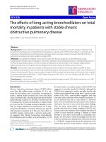
Table 3 shows the association between the mortality of
omphalocele-affected pregnancies and selected fetal
characteristics. The LFDR at the gestational ages of 28–36
weeks was 2.42-fold higher than that of 37–42 gestational
weeks (AOR: 2.42, 95% CI: 1.52–3.86). Additionally, birthweight of <2500 g was 3.17-fold higher, non-isolated omphalocele was 2.64-fold higher, and diagnosis by prenatal
ultrasound was 15.91-fold higher compared with birthweight of ≥2500 g, isolated omphalocele, and diagnosis by
postnatal ultrasound, respectively (AOR: 3.17, 95% CI:
1.97–5.09; AOR: 2.64, 95% CI: 1.62–4.29; AOR: 15.91,
95% CI: 10.18–24.87, respectively). The LFDR of omphalocele that was diagnosed by prenatal ultrasound
was the highest, followed by <2500 g birthweight, and
then non-isolated omphalocele. Neonates with <2500 g
birthweight and non-isolated omphalocele had a higher
ENNDR than that of the reference groups (AOR: 1.72,
95% CI: 1.04–2.82; AOR: 2.96, 95% CI: 1.82–4.81, respectively). The mortality of neonates who were born at
28–36 gestational weeks and the LFDR of cases of prenatally diagnosed omphalocele were slightly higher than
those of the reference groups, but these differences were
not significant (AOR: 1.18, 95% CI: 0.72–1.96; AOR:
1.42, 95% CI: 0.81–2.50, respectively). Neonates with
omphalocele and non-isolated abnormalities had the
highest rate of death in the first 7 days of life, followed
by neonates who were <2500 g birthweight at birth.
Discussion
We found that the perinatal mortality of pregnancies
with omphalocele was 52.4%, late fetal mortality was
37.4%, and early neonatal mortality was 15.0%. These estimates are in line with previous research showing that
39–41% of cases of omphalocele result in termination of
pregnancy and stillbirth, and 12% of cases result in neonatal death [4,5]. However, higher estimates than our results have been reported in other birth defects registries
[6,9,12,18,23,24]. The inconsistency in the mortality rate
for omphalocele may be owing to the registered gestational weeks of pregnancy, prenatal detection, follow-up
period, and prenatal and postnatal care, as well as management of omphalocele.
After controlling for confounding factors, we observed
that prenatally diagnosed omphalocele was more likely
to result in LFD compared with non-isolated omphalocele. This finding is supported by previous studies on
Page 5 of 7
the perinatal outcome of fetal omphalocele following
prenatal diagnosis [12,14,20]. However, most of these
previous results were mixed by the effect of non-isolated
and prenatal diagnosis, showing that a high proportion
of omphalocele was prenatally diagnosed by ultrasound,
and fetuses were electively terminated or died in utero.
If parents were properly counseled by a pediatrician
and intrauterine transfer occurred to tertiary units with
pediatric surgical facilities, the outcome of prenatally diagnosed omphalocele would be more favorable [13].
For neonates within 7 days old, those with non-isolated
omphalocele had a higher risk of death than prenatally
diagnosed cases. Up to 70% of omphalocele cases are associated with other structural malformations, chromosomal abnormalities, and genetic syndromes, and this
phenomenon is significantly associated with the ultimate prognosis for these fetuses [15,25-27]. In addition,
prenatal ultrasound diagnosis did not significantly increase the risk of death of a neonate in early life, with
similar results found in another study [20]. This finding
can be explained by the following two points. First, prenatal ultrasonography has become a routine examination
in pregnancy. The sensitivity of prenatal ultrasound screening in detecting omphalocele is 75% in the second trimester
of pregnancy, ranking second among all of the congenital
malformations that are diagnosed prenatally by ultrasound
(anencephaly is the first) [24,28]. Second, prenatally diagnosed cases include more fetuses with a giant omphalocele or liver herniation compared with those postnatally
diagnosed, and most women opt for termination of pregnancy or intrauterine death occurs [20].
There are several limitations to our study. First, we could
not distinguish between termination of pregnancy from
stillbirth in our analysis. Therefore, we could not estimate
the proportion of electively terminated pregnancies and
the proportion of stillbirths. Second, our monitoring period
covered the period from 28 gestational weeks to the first
7 days after delivery. This means that we did not investigate the death of fetuses before the age of 28 gestational
weeks. Consequently, our reported mortality may underestimate the true risk of death from omphalocele. Third,
because some women who had fetuses with omphalocele
terminated pregnancy or death occurred in utero, they refused an autopsy. Therefore, the characteristics of these
cases could not be verified postnatally, resulting in the
misclassification of other associated anomalies as isolated
omphalocele and the misclassification of gastroschisis and
other abdominal wall defects as omphalocele. Finally, because this study was hospital-based and it focused on selected hospitals rather than all deliveries in a region, the
hospital-based samples may have introduced referral bias.
However, because of the wide geographic coverage, consistent case ascertainment, and the large sample size, the
CBDMN data used in our study were reliable.
Total
N
Late fetal death
No.
Rate* (95% CI)
COR (95% CI)
Early neonatal death
AOR† (95% CI)
No.
Rate * (95% CI)
COR (95% CI)
AOR† (95% CI)
Gestational age$ (weeks)
37–42
436
83
19.0 (15.4, 22.7)
Ref.
Ref.
74
17.0 (13.4, 20.5)
Ref.
Ref.
28–36
388
224
57.7 (52.8, 62.6)
6.61 (4.73, 9.22)
2.42 (1.52, 3.86)
50
12.9 (9.6, 16.2)
1.65 (1.09, 2.52)
1.18 (0.72, 1.96)
≥2500
428
80
18.7 (15.0, 22.4)
Ref.
Ref.
65
15.2 (11.8, 18.6)
Ref.
Ref.
<2500
395
226
57.2 (52.3, 62.1)
7.20 (5.15, 10.83)
3.17 (1.97, 5.09)
58
14.7 (11.2, 18.2)
2.28 (1.50, 3.45)
1.72 (1.04, 2.82)
Deng et al. BMC Pediatrics 2014, 14:160
/>
Table 3 Association with perinatal mortality of omphaloceles by the selected fetal characteristics, China, 1996-2006
Birthweight# (g)
Non-isolated omphalocele
No
596
198
33.2 (29.4, 37.0)
Ref.
Ref.
76
12.8 (10.1, 15.4)
Ref.
Ref.
Yes
231
111
48.1 (41.6, 54.5)
2.51 (1.78, 3.54)
2.64 (1.62, 4.29)
48
20.8 (15.5, 26.0)
2.82 (1.81, 4.40)
2.96 (1.82, 4.81)
No
498
70
14.1 (11.0, 17.1)
Ref.
Ref.
99
19.9 (16.4, 23.4)
Ref.
Ref.
Yes
322
235
73.0 (68.1, 77.8)
17.81 (12.17, 26.06)
15.91 (10.18, 24.87)
25
7.8 (4.8, 10.7)
1.34 (0.80, 2.25)
1.42 (0.81, 2.50)
Prenatal ultrasound diagnosis§
*
Rate is the number of death per 100 fetus and neonates with omphalocele from 28 weeks of gestations to the first 7 days of life.
†
ORs were adjusted by geographical location, maternal residence, maternal age, maternal education, gestational age, birthweight, non-isolated omphalocele and diagnosis by prenatal ultrasonography.
$
Three cases with unknown gestation age were excluded from this analysis.
#
Four cases with unknown birthweight were excluded from this analysis.
§
Seven cases with unknown prenatal ultrasound diagnosis were excluded from this analysis.
COR, crude odds ratio; AOR, adjusted odds ratio; CI, confidence interval; Ref., reference group.
Page 6 of 7
Deng et al. BMC Pediatrics 2014, 14:160
/>
Conclusions
Our findings contribute to the growing body of estimates
regarding perinatal mortality in fetuses and neonates with
omphalocele. Cases of prenatally diagnosed omphalocele
have a higher risk of LFD, while there is no significant risk
of death for neonates with omphalocele when they are diagnosed prenatally. Those with non-isolated omphalocele
are more likely to die in the early neonatal period. Improving pregnancy and delivery care, as well as management
for omphalocele are important. Further studies are needed
to include more current data to investigate the perinatal
mortality of pregnancies with omphalocele.
Page 7 of 7
7.
8.
9.
10.
11.
12.
Abbreviations
CBDMN: Chinese birth defects monitoring network; LFD: Late fetal death;
ENND: Early neonatal death rate; DQM: Data quality management; LFDR: Late
fetal death rate; ENNDR: Early neonatal death rate; COR: Crude odds ratio;
AOR: Adjusted odds ratio; CI: Confidence interval.
13.
14.
Competing interests
The authors declare that they have no competing interests.
15.
Authors’ contributions
DK and QJ were joint first authors and participated equally in the study
design, literature review, data analysis, manuscript writing, and final revision
of the article; DL, YL, DCF, and MY participated in the acquisition of data and
the interpretation of data; ZJ participated in the study design, coordination
and critical revision of the manuscript. All authors read and approved the
final manuscript.
Acknowledgements
The authors would like to thank the staff of Chinese National Birth Defects
Monitoring Network for help with the collection of the national birth defects
registry. We are grateful to the obstetricians, pediatricians, pathologists and
other participants in member hospitals for their continued collaboration and
support of the national birth defects registry. This study was supported by
grants from Program for Changjiang Scholars and Innovative Research Team
in University (IRT0935).
Author details
1
National Center for Birth Defects monitoring of China, West China Second
University Hospital, Sichuan University, 17, Section3, Ren Min South Road,
Chengdu, China. 2Key Laboratory of Birth Defects and Related Diseases of
Women and Children (Sichuan University), Ministry of Education, Chengdu,
China. 3Department of Maternal and Children Health, National Health and
Family Planning Commission of the People's Republic of China, Beijing,
China.
Received: 3 September 2013 Accepted: 12 June 2014
Published: 23 June 2014
References
1. Ledbetter DJ: Gastroschisis and omphalocele. Surg Clin North Am 2006,
86(2):249–260. vii.
2. Wilson RD, Johnson MP: Congenital abdominal wall defects: an update.
Fetal Diagn Ther 2004, 19(5):385–398.
3. Calzolari E, Bianchi F, Dolk H, Milan M, EUROCAT Working Group:
Omphalocele and gastroschisis in Europe: a survey of 3 million births
1980-1990. Am J Med Genet 1995, 58(2):187–194.
4. Byron-Scott R, Haan E, Chan A, Bower C, Scott H, Clark K: A population-based
study of abdominal wall defects in South Australia and Western Australia.
Paediatr Perinat Epidemiol 1998, 12(2):136–151.
5. Forrester MB, Merz RD: Epidemiology of abdominal wall defects, Hawaii,
1986-1997. Teratology 1999, 60(3):117–123.
6. Rankin J, Dillon E, Wright C: Congenital anterior abdominal wall defects in
the north of England, 1986-1996: occurrence and outcome. Prenat Diagn
1999, 19(7):662–668.
16.
17.
18.
19.
20.
21.
22.
23.
24.
25.
26.
27.
28.
Suita S, Okamatsu T, Yamamoto T, Handa N, Nirasawa Y, Watanabe Y,
Yanagihara J, Nishijima E, Hirobe S, Nio M, Gomi A, Horisawa M: Changing
profile of abdominal wall defects in Japan: results of a national survey.
J Pediatr Surg 2000, 35(1):66–71. discussion 72.
Salihu HM, Pierre-Louis BJ, Druschel CM, Kirby RS: Omphalocele and
gastroschisis in the State of New York, 1992-1999. Birth Defects Res A
Clin Mol Teratol 2003, 67(9):630–636.
Tan KB, Tan KH, Chew SK, Yeo GS: Gastroschisis and omphalocele in
Singapore: a ten-year series from 1993 to 2002. Singapore Med J 2008,
49(1):31–36.
Zhou GX, Liang J, Zhu J, Dai L, Wang YP, Miao L: An epidemiological study
on omphalocele in China during 1996 to 2000. Zhonghua Yu Fang Yi Xue
Za Zhi 2004, 38(5):328–330.
Kominiarek MA, Zork N, Pierce SM, Zollinger T: Perinatal outcome in the
live-born infant with prenatally diagnosed omphalocele. Am J Perinatol
2011, 28(8):627–634.
Brantberg A, Blaas HG, Haugen SE, Eik-Nes SH: Characteristics and outcome
of 90 cases of fetal omphalocele. Ultrasound Obstet Gynecol 2005,
26(5):527–537.
Boyd PA, Bhattacharjee A, Gould S, Manning N, Chamberlain P: Outcome of
prenatally diagnosed anterior abdominal wall defects. Arch Dis Child Fetal
Neonatal Ed 1998, 78(3):F209–F213.
Lakasing L, Cicero S, Davenport M, Patel S, Nicolaides KH: Current outcome
of antenatally diagnosed exomphalos: an 11 year review. J Pediatr Surg
2006, 41(8):1403–1406.
Sermer M, Benzie RJ, Pitson L, Carr M, Skidmore M: Prenatal diagnosis and
management of congenital defects of the anterior abdominal wall. Am J
Obstet Gynecol 1987, 156(2):308–312.
Christison-Lagay ER, Kelleher CM, Langer JC: Neonatal abdominal wall
defects. Semin Fetal Neonatal Med 2011, 16(3):164–172.
Mitanchez D, Walter-Nicolet E, Humblot A, Rousseau V, Revillon Y, Hubert P:
Neonatal care in patients with giant ompholocele: arduous management
but favorable outcomes. J Pediatr Surg 2010, 45(8):1727–1733.
Islam S: Clinical care outcomes in abdominal wall defects. Curr Opin
Pediatr 2008, 20(3):305–310.
Garne E, Loane M, Dolk H, Group EW: Gastrointestinal malformations:
impact of prenatal diagnosis on gestational age at birth. Paediatr Perinat
Epidemiol 2007, 21(4):370–375.
Cohen-Overbeek TE, Tong WH, Hatzmann TR, Wilms JF, Govaerts LC,
Galjaard RJ, Steegers EA, Hop WC, Wladimiroff JW, Tibboel D: Omphalocele:
comparison of outcome following prenatal or postnatal diagnosis.
Ultrasound Obstet Gynecol 2010, 36(6):687–692.
Dai L, Zhu J, Liang J, Wang YP, Wang H, Mao M: Birth defects surveillance
in China. World J Pediatr 2011, 7(4):302–310.
International Clearinghouse for Birth Defects Surveillance and Research:
International Clearinghouse for Birth Defects Surveillance and Research
Annual report 2012. In Roma, Italy: The Internaltional Center on Birth
Defects - ICBDSR Center; 2012. />ar2005/Report2012.pdf (accessed June 21, 2013).
Stoll C, Alembik Y, Dott B, Roth MP: Risk factors in congenital abdominal
wall defects (omphalocele and gastroschisi): a study in a series of
265,858 consecutive births. Ann Genet 2001, 44(4):201–208.
Barisic I, Clementi M, Hausler M, Gjergja R, Kern J, Stoll C: Evaluation of
prenatal ultrasound diagnosis of fetal abdominal wall defects by 19
European registries. Ultrasound Obstet Gynecol 2001, 18(4):309–316.
Heider AL, Strauss RA, Kuller JA: Omphalocele: clinical outcomes in cases
with normal karyotypes. Am J Obstet Gynecol 2004, 190(1):135–141.
Mann S, Blinman TA, Douglas Wilson R: Prenatal and postnatal
management of omphalocele. Prenat Diagn 2008, 28(7):626–632.
Paidas MJ, Crombleholme TM, Robertson FM: Prenatal diagnosis and
management of the fetus with an abdominal wall defect. Semin Perinatol
1994, 18(3):196–214.
Stoll C, Tenconi R, Clementi M: Detection of congenital anomalies by fetal
ultrasonographic examination across Europe. Community Genet 2001,
4(4):225–232.
doi:10.1186/1471-2431-14-160
Cite this article as: Deng et al.: Perinatal mortality in pregnancies with
omphalocele: data from the Chinese national birth defects monitoring
network, 1996–2006. BMC Pediatrics 2014 14:160.