Performance of capnometry in non-intubated infants in the pediatric intensive care unit
Bạn đang xem bản rút gọn của tài liệu. Xem và tải ngay bản đầy đủ của tài liệu tại đây (648.82 KB, 6 trang )
Coates et al. BMC Pediatrics 2014, 14:163
/>
RESEARCH ARTICLE
Open Access
Performance of capnometry in non-intubated
infants in the pediatric intensive care unit
Bria M Coates1*, Robin Chaize2, Denise M Goodman1 and Ranna A Rozenfeld1
Abstract
Background: Assessing the ventilatory status of non-intubated infants in the Pediatric Intensive Care Unit (PICU) is
a constant challenge. Methods to evaluate ventilation include arterial blood gas analysis (ABG), which is invasive
and intermittent, and transcutaneous carbon dioxide monitoring (PtcCO2), which, while non-invasive, is also intermittent.
A method that is non-invasive and continuous would be of great benefit in this population. We hypothesized that
non-invasive capnometry via sidestream monitoring of exhaled carbon dioxide (CO2) would provide an acceptable
measurement of ventilatory status when compared to ABG or PtcCO2.
Methods: Preliminary prospective study of infants less than one year of age admitted to the PICU in a large urban
teaching hospital. Infants not intubated and not requiring non-invasive ventilation were eligible. A sidestream CO2
reading was obtained in a convenience sample of 39 patients. A simultaneous ABG was collected in those with an
arterial catheter, and a PtcCO2 was obtained in those without.
Results: Correlation of sidestream CO2 with ABG was excellent (r2 = 0.907). Sidestream correlated less well with PtcCO2
(r2 = 0.649). Results were not significantly altered when weight and respiratory rate were added as independent
variables. Bland-Altman analysis revealed a bias of -2.7 with a precision of ±6.5 when comparing sidestream CO2 to
ABG, and a bias of -1.7 with a precision of ±9.9 when comparing sidestream CO2 to PtcCO2.
Conclusions: Performance of sidestream monitoring of exhaled CO2 is acceptable clinical trending to assess the
effectiveness of ventilation in non-intubated infants in the PICU.
Keywords: Capnometry, Ventilation, Monitoring, Infants, Microstream, Carbon dioxide
Background
Respiratory monitoring of non-intubated children in the
pediatric intensive care unit (PICU) is a constant challenge. Effectiveness of the patient’s respiratory effort can
be monitored, to some extent, by visual observation of
chest expansion, rate and depth of respirations, use of
accessory muscles, and the quality and quantity of breath
sounds. These subjective findings can be misleading, however, and objective means of following oxygenation and
ventilation are needed in the majority of PICU patients.
Pulse oximetry has become the standard of care for oxygenation monitoring in both the PICU and the general
ward [1]. Monitoring of ventilation poses a more difficult
challenge. Except in extreme cases, pulse oximetry will not
* Correspondence:
1
Division of Critical Care, Northwestern University Feinberg School of
Medicine and Ann & Robert H. Lurie Children’s Hospital of Chicago, 225 E.
Chicago Ave, Box 73, Chicago, Illinois 60611, USA
Full list of author information is available at the end of the article
reliably detect hypoventilation. Measurement of the partial
pressure of carbon dioxide (PaCO2) through arterial blood
gas analysis (ABG) is the gold standard for assessing ventilation. While a true reflection of ventilation, measurement
of the PaCO2 is both invasive and intermittent, which
greatly limits its use. Consequently, other measures of
ventilatory monitoring have been established.
Transcutaneous carbon dioxide monitoring (PtcCO2)
estimates the PaCO2 by warming the skin to induce hyperperfusion, enabling the electrochemical measurement
of the partial pressure of oxygen and carbon dioxide [2].
Transcutaneous monitoring is considered a safe procedure,
however tissue injury may occur at the measuring site,
including blisters, burns, and skin tears. These complications are rare with current technology, and primarily occur
when the PtcCO2 is left in place for long periods of time, so
continuous monitoring is generally avoided. In patients
with poor skin integrity or adhesive allergy, transcutaneous
© 2014 Coates et al.; licensee BioMed Central Ltd. This is an Open Access article distributed under the terms of the Creative
Commons Attribution License ( which permits unrestricted use, distribution, and
reproduction in any medium, provided the original work is properly credited. The Creative Commons Public Domain
Dedication waiver ( applies to the data made available in this article,
unless otherwise stated.
Coates et al. BMC Pediatrics 2014, 14:163
/>
Page 2 of 6
monitoring may be relatively contraindicated [3,4]. Various
clinical factors may increase the discrepancy between arterial and transcutaneous values of carbon dioxide, including
hyperoxia (PaO2 > 100 torr), hypoperfusion, improper electrode placement or application, body wall edema, and the
thickness of the patient’s skin or subcutaneous tissue [3-5].
Capnography is regularly used in operating rooms and
intensive care units to monitor carbon dioxide clearance
in tracheally intubated patients [6-8]. Exhaled carbon
dioxide generally reflects PaCO2, but the correlation
decreases predictably with increasing dead space ventilation [9]. In recent years, there has been widening use of
oral and nasal capnometry for monitoring ventilation in
non-intubated adults and children [10,11]. Noninvasive
capnography has been used in emergency departments,
pediatric intensive care units, during polysomnography,
during sedation, and during interfacility transport [10-18].
However, the ability of these devices to reliably capture exhaled CO2 from non-intubated infants with high respiratory rates and low tidal volumes is unknown [19]. We
prospectively compared sidestream carbon dioxide (CO2),
with PtcCO2 and/or PaCO2 in infants less than one year of
age admitted to the PICU to determine if sidestream monitoring provides an acceptable measurement of the effectiveness of ventilation. Should the performance of this
technology prove acceptable for clinical trending, it would
provide the benefits of both non-invasive and continuous
monitoring of ventilatory status.
machine was calibrated prior to placing the sidestream
cannula on the subject. The PtcCO2 electrode was placed at
the same time as the sidestream nasal cannula and the
reading was recorded when no further increase was
observed for 30-60 seconds. In those subjects who had an
indwelling arterial catheter in place as part of their routine
care, an arterial blood gas was drawn immediately after
the sidestream reading was recorded. The majority of
subjects had a single sidestream CO2 measurement and
either a PtcCO2 or a PaCO2, but 4 subjects had both a PtcCO2
and a PaCO2.
The correlation between a single sidestream CO2 reading and the simultaneous PtcCO2 or PaCO2 was examined
using Spearman’s rho. Since correlation is expected when
two methods attempt to measure the same physiologic
parameter, a Bland Altman analysis was also conducted
[20] to analyze the differences between the sidestream
reading and either the PtcCO2 or the PaCO2. Bias, the mean
difference between values, and precision, the standard
deviation (SD) of the bias, were calculated for PtcCO2 to
sidestream and PaCO2 to sidestream differences.
The effect of respiratory rate and weight on the relationship between PtcCO2 or PaCO2 and sidestream CO2 was
assessed by simple linear regression models for the
unadjusted effect and multiple linear regression models
for the adjusted effect. We examined both absolute values
of sidestream CO2 and log-transformed sidestream due to
the non-normal distribution of the sidestream values.
Methods
This study was approved by the Institutional Review Board
at Children’s Memorial Hospital in Chicago, Illinois, now
the Ann & Robert H. Lurie Children’s Hospital of Chicago.
Infants admitted to the PICU who were age 1 year or less,
without a tracheostomy or immediate need for invasive
or non-invasive ventilation were eligible for enrollment.
Additional information was recorded on each subject
including age in months, weight in kilograms (kg), respiratory rate, and diagnosis. A convenience sample of subjects
was enrolled from March 2007 – September 2008.
Sidestream sampling was performed on all subjects in
the study. The sidestream cannula (Philips Microstream
EtCo2 circuit, Smart Capnoline O2), a two prong nasal
cannula with a CO2 detection port that hangs in front of
the mouth, was placed on the subject and left in place
until a steady state reading was obtained. The sampling
rate is 50 ml/min and the sampling line is 100 cm long.
Oxygen can be delivered through the nasal prongs, if
desired, and was only used as indicated for patient care.
The value of the exhaled CO2 was recorded (Microcap
Microstream Oridion Machine) after a consistent reading
had been present for 2 minutes. For those subjects without an arterial catheter in place, a PtcCO2 (Radiometer
Copenhagen, Tina TCM 4) was obtained. The PtcCO2
Results
Forty-three sample sets were obtained from 39 subjects
for analysis. Please see Table 1 for full demographic data.
In the PtcCO2 comparison group there were 29 subjects.
Admission diagnoses included respiratory illness (n = 16),
cardiothoracic surgery (n = 9), and other (n = 4). In the
ABG comparison group there were 14 subjects. Admission
diagnoses included respiratory illness (n = 1), cardiothoracic surgery (n = 12), and other (n = 1). The predominance
of cardiothoracic surgery subjects in the ABG group
reflects the practice patterns at our institution for placing
and keeping arterial lines in non-intubated subjects
(Table 1).
Table 1 Demographics
PtcCO2
ABG
Number of patients
29
14
Age (months)
3.4 (2.8, 0.5-11)
4.1 (3.3, 0.3-11)
Weight (kg)
5.3 (1.8, 3-9.4)
5.3 (1.3, 3-7.1)
Diagnosis: respiratory
16
1
Cardiac
9
12
Other
4
1
Respiratory rate (breaths/min)
44 (15, 25-82)
45 (12, 32-69)
Results expressed as mean (SD, range).
Coates et al. BMC Pediatrics 2014, 14:163
/>
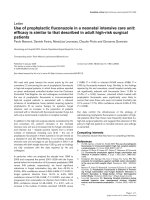
The correlation between sidestream CO2 and PaCO2
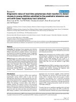
was excellent (r2 0.907, Figure 1A). Sidestream CO2 correlated less well with PtcCO2 values (r2 0.649, Figure 2A).
The Bland-Altman analysis revealed good agreement between PaCO2 and sidestream CO2 (bias –2.7, precision ±6.5,
Figure 1B). Agreement was less robust when comparing
Page 3 of 6
sidestream CO2 and PtcCO2 (bias –1.7, precision ±9.9,
Figure 2B).
The influence of respiratory rate and weight on performance of sidestream CO2 monitoring is presented in
Table 2. Sidestream performance was unaffected by either
factor. Therefore, sidestream measurements were accurate
Figure 1 Comparison of sidestream CO2 to PaCO2. A: Correlation between sidestream CO2 and PaCO2 values (r2 = 0.907). B: Bland-Altman analysis
of difference in CO2 between sidestream and PaCO2 (y-axis) versus average of measured sidestream and PaCO2 (x-axis), bias -2.7, precision ± 6.5.
Coates et al. BMC Pediatrics 2014, 14:163
/>
Page 4 of 6
Figure 2 Comparison of sidestream CO2 to PtcCO2. A: Correlation between sidestream CO2 and PtcCO2 values (r2 = 0.649). B: Bland-Altman analysis
of difference in CO2 between sidestream and PtcCO2 (y-axis) versus average of measured sidestream and PtcCO2 (x-axis), bias -1.7, precision ±9.9.
across the range of size and respiratory rates found in the
infants in this study.
Discussion
This study demonstrates that sidestream CO2 monitoring
can provide an acceptable estimation of PaCO2 in infants
with varying degrees of tachypnea. It is comparable to
another commonly used non-invasive monitor, the transcutaneous CO2. To our knowledge, this is the only study
comparing these different techniques in this age range.
One retrospective study compared end tidal CO2 with
venous CO2 and found them to be highly correlated [21].
This study included children age 5.5 months-20 years with
a median age of 5.7 years. Additional studies in infants
Coates et al. BMC Pediatrics 2014, 14:163
/>
Page 5 of 6
Table 2 Linear regression (dependent variable: log
transformed sidestream CO2)
Independent
variables
β (Unstandardized
coefficient)
R2
p value
Unadjusted effect
Respiratory rate
-.003
.043 .195
Weight
-.019
.033 .248
PtcCO2
.016
.650 <.001
PaCO2
.019
.855 <.001
2
Adjusted model, R = .668
Respiratory rate
-.002
.266
Weight
.001
.965
PtcCO2
.016
<.001
Adjusted model, R2 = .866
Respiratory rate
.002
.395
Weight
.001
.936
PaCO2
.019
<.001
have examined the difference between sidestream CO2
and capillary CO2, or used exhaled mainstream CO2 to
analyze the capnographic indices associated with bronchopulmonary dysplasia (BPD) [22,23]. One of the studies was
consistent with our findings, showing good correlation
between sidestream CO2 and capillary CO2 in preterm
infants without lung disease [22]. However, the correlation
between sidestream CO2 and capillary CO2 was lost in
infants with BPD. The gradient between the sidestream
and capillary CO2 values in the BPD group was attributed
to dead space ventilation and the ventilation-perfusion
mismatch characteristic of this lung disease. None of these
studies specifically evaluated the performance of sidestream CO2 in comparison to PtcCO2 or PaCO2 in infants.
In most cases, an estimated CO2 that is within 5 mm
Hg of the PaCO2 would be considered acceptable for making clinical decisions when an arterial value is unavailable.
In this study, the majority of measurements were within
this range. Our results contrast with an adult study that
did not show good correlation between sidestream CO2
and PaCO2 in non-intubated patients [24]. These adults
were very tachypneic, and the authors speculated that this
was a reflection of abnormal lung function, leading them
to conclude that the usefulness of sidestream monitoring
was limited in the presence of lung disease. As with all
end-tidal CO2 monitoring, it is important to note that
dead space ventilation will increase the discrepancy
between exhaled CO2 and PaCO2 or PtcCO2 [9]. This may
explain the gradients seen between sidestream CO2 and
PaCO2 in this adult study, and sidestream CO2 and capillary CO2 in the study of premature infants with BPD mentioned above. However, in our population, sidestream CO2
closely approximated PaCO2, and to a slightly lesser degree,
PtcCO2, over a wide range of respiratory rates and degrees
of distress in these infants, suggesting that sidestream
monitoring of exhaled CO2 could be quite useful in this
population.
Accurate sidestream monitoring is advantageous over
ABG or PtcCO2 as it provides both a continuous and
non-invasive means of monitoring ventilation. It is also
much less labor intensive when compared to the alternate methods. Effective transcutaneous monitoring is
dependent on both technical and patient factors. Repetitive ABG analysis requires the presence of an indwelling
arterial catheter which can be difficult, painful, and time
consuming to place.
The primary limitation to this preliminary study was
its small sample size. Larger numbers and repeated measurements would enable further elucidation of patient or
operator characteristics that impact the precision and
reliability of these measurements. As the gold standard,
PaCO2 should still be obtained when either PtcCO2 or
sidestream readings seem to contradict clinical assess
ment.
Conclusion
Performance of sidestream monitoring of exhaled CO2 is
acceptable for assessing the effectiveness of ventilation
in non-intubated infants in the PICU. It should be
considered when continuous monitoring of ventilation is
desired to aid in the early detection of changes in clinical
status.
Abbreviations
ABG: Arterial blood gas; BPD: Bronchopulmonary dysplasia; CO2: Carbon
dioxide; kg: Kilograms; PaCO2: Arterial partial pressure of carbon dioxide;
PICU: Pediatric intensive care unit; PtcCO2: Transcutaneous carbon dioxide
monitoring; SD: Standard deviation.
Competing interests
The authors declare that they have no competing interests.
Authors’ contributions
BMC recruited subjects and collected data for the study, drafted the initial
manuscript, and approved the final manuscript as submitted. RC developed
initial study design, recruited subjects and collected data for the study, and
approved the final manuscript as submitted. DMG helped with initial
conceptualization of the study, performed the statistical analysis, and
approved the final manuscript as submitted. RAR developed final study
design, recruited subjects and collected data for the study, revised the
manuscript, and approved the final manuscript as submitted.
Acknowledgements
No funding source was used for the study design, data collection, analysis,
interpretation, in the writing of the manuscript or in the decision to submit
the manuscript for publication.
Author details
1
Division of Critical Care, Northwestern University Feinberg School of
Medicine and Ann & Robert H. Lurie Children’s Hospital of Chicago, 225 E.
Chicago Ave, Box 73, Chicago, Illinois 60611, USA. 2Joe DiMaggio Children’s
Hospital, 1005 Joe DiMaggio Drive Hollywood, Florida 33021, USA.
Received: 11 November 2013 Accepted: 19 June 2014
Published: 25 June 2014
Coates et al. BMC Pediatrics 2014, 14:163
/>
References
1. Fouzas S, Priftis KN, Anthracopoulos MB: Pulse oximetry in pediatric
practice. Pediatrics 2011, 128:740–752.
2. Monaco F, Nickerson BG, McQuitty JC: Continuous transcutaneous oxygen
and carbon dioxide monitoring in the pediatric ICU. Crit Care Med 1982,
10:765–766.
3. Restrepo RD, Hirst KR, Wittnebel L, Wettstein R: AARC clinical practice
guideline: transcutaneous monitoring of carbon dioxide and oxygen.
Respir Care 2012, 2012:1955–1962.
4. Tobias JD: Transcutaneous carbon dioxide monitoring in infants and
children. Pediatr Anesth 2009, 19:434–444.
5. Tobias JD, Wilson WR, Meyer DJ: Transcutaneous monitoring of carbon
dioxide tension after cardiothoracic surgery in infants and children.
Anesth Analg 1999, 88:531–534.
6. Eipe N, Doherty DR: A review of pediatric capnography. J Clin Monit
Comput 2010, 24:261–268.
7. Tobias JD, Meyer DJ: Noninvasive monitoring of carbon dioxide during
respiratory failure in toddlers and infants: end-tidal versus transcutaneous
carbon dioxide. Anesth Analg 1997, 85:55–58.
8. Sivarajan VB, Bohn D: Monitoring of standard hemodynamic parameters:
heart rate, systemic blood pressure, atrial pressure, pulse oximetry, and
end-tidal CO2. Pediatr Crit Care Med 2011, 12:S2–S11.
9. McSwain SD, Hamel DS, Smith PB, Gentile MA, Srinivasan S, Meliones JN,
Cheifetz IM: End-tidal and arterial carbon dioxide measurements
correlate across all levels of physiologic dead space.
Respir Care 2010, 55:288–293.
10. Bhende M: Capnography in the pediatric emergency department.
Pediatr Emerg Care 1999, 15:64–69.
11. Yosefy C, Hay E, Nasri Y, Magen E, Reisin L: End tidal carbon dioxide as a
predictor of the arterial PCO2 in the emergency department setting.
Emerg Med J 2004, 21:557–559.
12. Kirk VG, Batuyong ED, Bohn SG: Transcutaneous carbon dioxide
monitoring and capnography during pediatric polysomnography.
Sleep 2006, 29:1601–1608.
13. Sullivan KJ, Kissoon N, Goodwin SR: End-tidal carbon dioxide monitoring
in pediatric emergencies. Pediatr Emerg Care 2005, 21:327–332.
14. Langhan ML, Chen L: Current utilization of continuous end-tidal carbon
dioxide monitoring in pediatric emergency departments.
Pediatr Emerg Care 2008, 24:211–213.
15. Langhan M: Continuous end-tidal carbon dioxide monitoring in pediatric
intensive care units. J Crit Care 2009, 24:227–230.
16. Price DD, Wilson SR, Fee ME: Sidestream end-tidal carbon dioxide monitoring
during helicopter transport. Air Med J 2007, 26:55–59.
17. Krauss B, Hess DR: Capnography for procedural sedation and analgesia in
the emergency department. Ann Emerg Med 2007, 50:172–181.
18. Lightdale JR, Goldmann DA, Feldman HA, Newburg AR, DiNardo JA, Fox VL:
Microstream capnography improves patient monitoring during moderate
sedation: a randomized, controlled trial. Pediatrics 2006, 117:e1170–e1178.
19. Nobel JJ: Carbon dioxide monitors, exhaled gas. Pediatr Emerg Care 1996,
12:239–240.
20. Bland JM, Altman DG: Statistical methods for assessing agreement between
two methods of clinical measurement. Lancet 1986, 1(8476):307–310.
21. Moses JM, Alexander JL, Agus MS: The correlation and level of agreement
between end-tidal and blood gas pCO2 in children with respiratory
distress: a retrospective analysis. BMC Pediatr 2009, 9:20.
22. Lopez E, Mathlouthi J, Lescure S, Krauss B, Jarreau PH, Moriette G:
Capnography in spontaneously breathing preterm infants with
bronchopulmonary dysplasia. Pediatr Pulmonol 2011, 46:896–902.
23. Fouzas S, Häcki C, Latzin P, Proietti E, Schulzke S, Frey U, Delgado-Eckert E:
Volumetric capnography in infants with bronchopulmonary dysplasia.
J Pediatr 2014, 164:283–288.
24. Jabre P, Jacob L, Auger H, Jaulin C, Monribot M, Aurore A, Margenet A,
Marty J, Combes X: Capnography monitoring in nonintubated patients
with respiratory distress. Am J Emerg Med 2009, 27:1056–1059.
Page 6 of 6
Submit your next manuscript to BioMed Central
and take full advantage of:
• Convenient online submission
• Thorough peer review
• No space constraints or color figure charges
doi:10.1186/1471-2431-14-163
Cite this article as: Coates et al.: Performance of capnometry in
non-intubated infants in the pediatric intensive care unit.
BMC Pediatrics 2014 14:163.
• Immediate publication on acceptance
• Inclusion in PubMed, CAS, Scopus and Google Scholar
• Research which is freely available for redistribution
Submit your manuscript at
www.biomedcentral.com/submit