Femur shaft fracture at a young age and the risk of subsequent severe injuries during childhood: A cohort study
Bạn đang xem bản rút gọn của tài liệu. Xem và tải ngay bản đầy đủ của tài liệu tại đây (281.74 KB, 8 trang )
von Heideken et al. BMC Pediatrics 2014, 14:62
/>
RESEARCH ARTICLE
Open Access
Femur shaft fracture at a young age and the risk
of subsequent severe injuries during childhood: a
cohort study
Johan von Heideken1*, Tobias Svensson2, Maura Iversen1,3,4, Anders Ekbom2 and Per-Mats Janarv1,5
Abstract
Background: A child who suffers a fracture or a soft-tissue injury at a young age faces an increased risk of
subsequent injuries during childhood. This risk could be related to personal and family characteristics or to lowerthan-average bone-mineral density. The purpose of this nationwide cohort study was to estimate the association
between a femur shaft fracture at a young age and the subsequent risk of hospitalization for injuries during
childhood.
Methods: We compared the subsequent risk of hospitalization for injuries during childhood among 1,404 children
(exposed) who were one to three years of age when they suffered a femur shaft fracture with the risk among
13,814 randomly selected, gender- and age-matched femur fracture–free children (unexposed). Hazard ratios (HRs)
and 95% confidence intervals (CIs) for severe injuries defined as fractures or soft-tissue injuries requiring hospital
admission were estimated in a Cox proportional hazards model.
Results: Exposed children exhibited no significantly increased risk of upper-extremity fractures or soft-tissue injuries
during childhood, regardless of sex and follow-up time. Boys exhibited a 162% increased risk of suffering a lower
leg fracture requiring hospital admission (HR = 2.62, 95% CI: 1.45–4.71), but the refracture risk was not significant for
girls 2.02 (0.58–6.97).
Conclusions: We found an increased risk for subsequent fractures in the lower leg that requires inpatient care
during childhood for boys, but not for girls, who were one to three years of age when they first suffered a femur
shaft fracture. This increased fracture risk is probably not simply the result of greater risk-taking among boys. The
explanation might relate to factors affecting the bone quality of the lower leg.
Keywords: Gender differences, Femoral, Trauma, Sweden
Background
The incidence of femur shaft fractures for boys and girls
peaks among children aged one to three years, and the
incidence is three times higher among boys than among
girls [1,2]. Although all fracture types are more frequent
in boys, such a great gender difference in this age group
does not occur for other types of fractures [3,4]. The
reasons for the difference are unknown, but studies
examining both the behavior of children in this age
group and parent-child interactions describe greater risk
* Correspondence:
1
Department of Women’s and Children’s Health, Karolinska Institutet,
Karolinska University Hospital, Solna, SE 171 77, Stockholm, Sweden
Full list of author information is available at the end of the article
taking among boys and higher parental protectiveness
toward girls [5].
Studies have also shown that a child with one hospital
admission for an accident in the first five years of life
runs an increased risk of experiencing another accidentrelated admission, compared with children of the same
age and sex with no previous admissions for accidents
[6-8]. An increased risk of repeated fractures could relate to the personal and family characteristics of these
accident-prone children [9-11].
Data indicate that children without obvious metabolic
bone diseases who experience their first fractures early
© 2014 von Heideken et al.; licensee BioMed Central Ltd. This is an Open Access article distributed under the terms of the
Creative Commons Attribution License ( which permits unrestricted use,
distribution, and reproduction in any medium, provided the original work is properly credited. The Creative Commons Public
Domain Dedication waiver ( applies to the data made available in this
article, unless otherwise stated.
von Heideken et al. BMC Pediatrics 2014, 14:62
/>
in life are especially vulnerable to further fractures [3,12],
perhaps owing to lower-than-average bone-mineral density [13-17]. Children aged one to three years who suffer a
femur shaft fracture are usually treated with spica casting
or traction for four weeks. This period of immobilization
and no weight bearing, along with the associated inactivity, results in a loss of bone-mineral tissue and in
muscle atrophy, which may influence the risk of further
fractures [18-20].
The risk of further injuries demanding inpatient care
among children who experience a severe fracture at
young age has not, to our knowledge, previously been
analyzed. The pediatric femur shaft fracture is a significant injury with a unique distribution of incidence regarding age and gender in one- to three-year-olds. In
this study using Swedish databases, we hypothesized that
a femur shaft fracture at this age is a predictor for further severe injuries during childhood and that fracture
risk would differ between boys and girls.
Methods
For the purpose of this population-based cohort study,
we used data from the following three Swedish national
registers: the Swedish Inpatient Register, the Swedish
Medical Birth Registry, and the Cause of Death Registry.
Record linkage of these registers was possible because a
personal identification number is issued to every resident of Sweden [21].
We created the database, including a study cohort and a
comparison cohort, using four steps. First, we identified
children who suffered a femur shaft fracture between
January 1, 1990, and December 31, 2005, in the Swedish
Inpatient Register. The Swedish Inpatient Register contains information on all hospitalizations in Sweden, including each patient’s individual personal identification
number, dates of hospitalizations, discharge code, and
E-codes (external causes) according to the International
Classification of Diseases (ICD-9: 1990 to 1996, ICD10: 1997 to 2005) [22]. This study cohort examined the
5,124 children with femur shaft fractures considered in
our recent paper on incidence and trends in femur
shaft fractures [2].
Exposed individuals were defined as children who were
one to three years of age when they suffered a fracture of
shaft of femur, primary or secondary diagnostic code
ICD: ICD-9/ICD-10: 821*/S723*. In ICD-9, fracture of
femur, part unspecified, is included under diagnostic
code 821*, and we chose therefore to include 15 children
with ICD-10, diagnostic code fracture of femur, part
unspecified (S72.9).
We excluded 56 children from the analysis with congenital medical conditions affecting the bone quality or
the risk of trauma, regardless time of diagnosis. Exposed
children (n = 12) were censored if they had received a
Page 2 of 8
diagnosis that might affect their bone quality or risk of
trauma—namely, bone tumor/cyst (malign or benign),
epilepsy, and attention deficit/hyperactivity disorder
(ADHD) (Table 1). If a child received a censoring diagnosis before the femur shaft fracture, the child was excluded from the analysis (n = 6).
Second, up to 10 unexposed children (median 10;
range of unexposed per case 7–10) were randomly selected for each exposed child from the Swedish Medical
Birth Registry, which includes data on practically all deliveries in Sweden [23]. Unexposed children were individually matched by sex, year of birth, and county of
residence. The unexposed had no diagnosis of fracture of
femur in the Swedish Inpatient Register, nor were they
siblings of a child with a femur shaft fracture. Using the
same definition as for the exposed group, we excluded
from the unexposed group 111 children with histories of
medical conditions that affect bone quality or risk of
trauma. Unexposed children (n = 69) were censored
according to the same criteria applied to the exposed
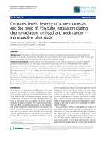
children. A total of 1,404 children and their 13,814 unexposed controls met the inclusion criteria (Figure 1).
Third, additional information regarding all hospital admissions since birth, until the age of 14 years or until
December 31, 2005, whichever came first, was identified
for exposed and unexposed children from the Swedish
Inpatient Register. The rationale for the choice of age
14 is that patients in Sweden under the age of 15 are
considered children regarding diagnoses of conditions
caused by trauma.
Fourth, data were linked with the Cause of Death
Registry in order to retrieve dates of death [24].
These two groups were compared to assess the risk
of injury ICD-9/ICD-10: 800–848/S00–S99, T00–T14
(except for femur fractures, regardless of localization,
ICD-9/ICD-10: 820–821/S72*). Diagnostic codes and
E-codes (external causes) according to ICD-9 (E967,
995F, V61C) and ICD-10 (T74*, Y07*) were used to
identify injuries caused by non- accidental trauma. To
identify injuries classified as undetermined whether accidentally or purposely inflicted, diagnostic codes and
E-codes according to ICD-9 (E988) and ICD-10 (Y33*
and Y34*) were used. Only severe injuries defined as
fractures or soft-tissue injuries (defined as all injury types
except fractures) requiring hospital admission were identified because minor injuries are treated in outpatient settings. For the exposed, all injuries that occurred at the
index date were not considered within the first 18 months
of follow-up; these admissions were likely related to the
injuries at index date and not to new injuries. We used
the same washout period for the same injuries regarding
the matched unexposed children.
The Stockholm Regional Ethical Review Board approved the study (Dnr2006/399–31).
von Heideken et al. BMC Pediatrics 2014, 14:62
/>
Page 3 of 8
Table 1 Congenital medical conditions affecting the bone quality or the risk for injuries, diagnostic codes ICD-9/ICD-10
Serious congenital medical conditions affecting bone quality
ICD-9
ICD-10
Number of
exposed children
Number of
unexposed children
Osteogenesis imperfecta and Osteopetrosis
756.51, 756.52
Q78.0, Q78.2
23
0
Congenital malformations of the nervous system
740*–742*
Q00*–Q07*
17
24
Cerebral palsy and other paralytic syndromes
342*–344*
G80*–G83*
5
30
Pervasive developmental disorders
299*
F84*
3
16
Osteoporosis
733.0
M80*–M81*
3
0
Cystic fibrosis
277.0
E84*
2
2
Spinal muscular atrophy and related syndromes
335*
G12*
2
1
Hypopituitarism
253.3
E23.0
1
9
Down syndrome
758.0
Q90*
0
16
Marfan syndrome or Congenital malformation syndromes
predominantly associated with short stature
759.81, 759.82
Q87.4*, Q87.1
0
5
Reduction defects of lower limb
755.3
Q72*
0
3
Muscular dystrophy
359.1
G71.0
0
3
Arthrogryposis multiplex congenita
754.89
Q74.3
0
1
Ehlers-Danlos syndrome
756.83
Q79.6
0
1
56
111
4
23
Total number of children with congenital medical conditions
affecting bone quality
Non-congenital medical conditions affecting bone quality or
risk of trauma
Epilepsy
345*
G40*–G41*
Attention deficit/hyperactivity disorder
314*
F90*
2
8
Solitary bone cyst or Aneurysmal bone cyst
733.21, 733.22
M85.4, M85.5
2
1
Malignant neoplasm of lymphatic and hematopoietic tissue
200*–208*
C81*–C96*
1
13
Visual disturbances and blindness
368*-369*
H53*–H54*
1
9
Juvenile idiopathic arthritis
714.3*
M08*
1
3
Organ or tissue replaced by transplant
V42*
Z94*
1
0
Ulcerative colitis
556*
K51*
0
9
Benign neoplasm of long bones of lower limb
213.7
D16.2–D16.3
0
3
Malignant neoplasm of long bones of lower limb
170.7
C40.2–C40.9
0
0
Vitamin D deficiency
268*
E55.0, E55.9
0
0
12
69
Total number of children with non-congenital medical
conditions affecting bone quality or risk of trauma
*Includes all forth and fifth positions.
Statistical analysis
Descriptive statistics employed frequency and percentages. The two primary outcomes for this study were
fracture and soft-tissue injury. The risk of having an outcome was calculated using Cox proportional hazards
models and expressed as hazard ratios (HRs) with 95%
confidence intervals (CIs). The hazard ratio was adjusted
for year of fracture and the corresponding date for the
unexposed, year of birth, and sex. The start of follow-up
was defined as the date of the femur shaft fracture for
exposed cases and as the corresponding date for the
matched controls. Follow-up continued until the patient
received an injury diagnosis or a diagnosis that excluded
him or her from study, died, or reached age 15, or until
December 31, 2005, whichever came first. A person
could have more than one study endpoint (different
injuries). Data were stratified by gender, and separate
analyses examined fractures and soft-tissue injuries. Furthermore, fractures in upper extremities and in lower extremities (except for femur fractures, regardless of
localization), were analyzed. The rationale for excluding
femur fractures as an endpoint for the exposed children
is that the unexposed had no diagnosis of fracture of
femur in the Swedish Inpatient Register. In addition, the
number of children with the most common type of
upper limb fracture, lower limb fracture and soft tissue
von Heideken et al. BMC Pediatrics 2014, 14:62
/>
Page 4 of 8
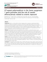
Exposed
Children one to three years of age
who suffered a fracture of the femur
shaft during the study period
n =1,466
Excluded
owing to a congenital
medical condition
affecting the bone
quality
or a diagnosis
indicating censoring
prior to femur shaft
fracture
n = 62
Unexposed
Up to 10 randomly selected
controls for each case
n = 13,925
Excluded
owing to a congenital
medical condition
affecting the bone
quality
or a diagnosis
indicating censoring
prior to femur shaft
fracture
n = 111
Study base
n = 1,404 exposed
n = 13,814 unexposed
Figure 1 Flow chart of cases and matched controls.
injury were reported. Multiple fractures and multiple
soft-tissue injuries as well as injuries related to determined and undetermined non- accidental trauma were
also stated. The assumption of proportional hazard was
verified by comparing the difference in HR for follow-up
time to injury between children with a follow-up period
shorter than three years and children with a follow-up
period of more than three years. No signs of insufficient
proportionality were detected. An HR was considered
significant if the 95% CI did not include 1.00. All statistical
analyses were performed using SAS 9.3 for Windows
(SAS Institute Inc., Cary, NC, USA) and IBM SPSS Statistics software, version 20 for Windows (SPSS Inc., Chicago,
IL, USA).
Results
Our cohort comprised 1,404 children, each exposed to a
femur shaft fracture between the ages of one and three
years (hereafter exposed children) and 13,814 matched
controls (hereafter unexposed children). Five (three
within 30 days after the femur shaft fracture) of the exposed children and 16 of the unexposed children died
before the age of 15 years. We observed 97 children with
injuries that required hospital admission among the exposed children during 12,234 person-years of follow-up
(mean per child 8,7 years), compared to 885 injuries that
required hospital admission among the unexposed children during 120,849 person-years of follow-up (mean
per child 8,7 years).
The cohort characteristics, as well as rates of the different types of injuries, stratified by gender, are summarized in Table 2. The risk of injury for an exposed child
was not higher than that among matched children with
no history of femur shaft fracture (HR = 1.08, 95% CI:
0.88–1.33) (n = 97). When the type of injury was
assessed in separate analyses, the risk of an injury resulting in a fracture was 38% higher among the exposed
children (HR = 1.38, 95% CI: 1.04–1.84) (n = 54). However, the increased risk was seen only among boys, and
it rose to 50% (HR = 1.50, 95% CI: 1.10–2.03) (n = 47).
The association between a femur shaft fracture and future fractures was seen only in lower leg fractures
(HR = 2.49, 95% CI: 1.46–4.23) (n = 17). This risk could
only be linked to boys, who demonstrated a 162% increased risk (HR = 2.62, 95% CI: 1.45–4.71) (n = 14) of suffering a fracture in a lower limb that required hospital
admission. The increased risk for boys was significant regardless of whether the lower leg fracture occurred within
three years or more than three years after the exposure to
a femur shaft fracture. The association between a femur
von Heideken et al. BMC Pediatrics 2014, 14:62
/>
Page 5 of 8
Table 2 Characteristics of the study subjects
Variable
Exposed (N = 1,404)
Unexposed (N = 13,814)
Boys
Girls
Boys
Girls
1,070 (76.2)
334 (23.8)
10,516 (76.1)
3,298 (23.9)
Number of children with injuries
83 (7.8)
14 (4.2)
717 (6.8)
168 (5.1)
Number of children with fracturesb
47 (4.4)
7 (2.1)
312 (3.0)
76 (2.3)
Number of children with fractures, upper limb
29 (2.7)
4 (1.2)
239 (2.3)
58 (1.8)
Number of children with fracture of the lower end of radius or ulnad
13 (1.2)
0 (0.0)
90 (0.9)
21 (0.6)
Number of children with fractures, lower limbb, c
14 (1.3)
3 (0.9)
53 (0.5)
15 (0.5)
Number of children with fracture of shaft of tibia and fibulae
10 (0.9)
2 (0.6)
16 (0.2)
4 (0.1)
Total, N (%)
a
c
Number of children with soft-tissue injury
38 (3.6)
8 (2.4)
457 (4.3)
105 (3.2)
Number of children with intracranial injuries, the most common type of soft-tissue injuryf
23 (2.1)
5 (1.5)
278 (2.6)
62 (1.9)
Number of children with multiple fracturesg
9 (0.8)
1 (0.3)
42 (0.4)
14 (0.4)
Number of children with multiple soft-tissue injuriesg
4 (0.4)
1 (0.3)
47 (0.4)
12 (0.4)
Number of children with injuries caused by non-accidental trauma
0 (0.0)
1 (0.3)
5 (0.0)
0 (0.0)
Number of children with injuries undetermined whether accidentally or purposely inflicted
0 (0.0)
0 (0.0)
2 (0.0)
0 (0.0)
a
b
c
Injuries resulting in a fracture or soft-tissue injury requiring hospital admission. Except for femur fractures (regardless of localization). Some children had
experienced both upper- and lower-limb fractures. dThe most common type of upper limb fracture. Diagnostic codes according to ICD-9 (813.4 – 813.5) and
ICD-10 (S525*-526*). eThe most common type of lower limb fracture. Diagnostic codes according to ICD-9 (823.2 – 823.3) and ICD-10 (S822*). fDiagnostic codes
according to ICD-9 (850 - 854) and ICD-10 (S06*). gMultiple is defined as involving more than one fracture or soft-tissue injury requiring hospital admission during
the study period.
shaft fracture and soft-tissue injuries was not significant
regardless of gender and follow-up time (Table 3).
Discussion
In this nationwide registry-based cohort study, we confirmed the previously known fact that there is an increased risk of a repeated fracture during childhood for
a child who has been admitted to hospital for a fracture
at a young age [3,12]. The novel aspect of this study is
the retrospective analysis that allows us to test the hypothesis that the risk of hospitalization for injuries, both
fractures and soft-tissue injuries during childhood, is influenced by a femur shaft fracture at young age and to
determine whether fracture risk differs between boys
and girls. Previous studies on repeated injuries have not
evaluated soft-tissue injuries and fractures separately
[6-8], and studies showing that an earlier fracture is associated with increased risk of new fractures during
childhood have not included the risk of soft-tissue injuries in their analyses [3,12].
The incidence of femur shaft fractures among boys
and girls peaks in the age group of one to three years,
and the incidence rate ratio of boys to girls is 3:1 [2].
The reasons for the difference are unknown, but they
probably relate to higher levels of risk taking among
boys, which may correlate to a possible imbalance between demands placed on the femur and bone-mineral
density [5,25].
Fracture risk, regardless of location and the child’s age,
is higher among boys than among girls, and it has been
suggested that a greater skeletal fragility relative to body
size contributes to this gender difference [26]. A recent
meta-analysis by Clark et al., concluded that children
who experience fractures have lower bone-mineral density than children who do not experience fractures [27].
This concurs with the first study on the subject by
Landin and Nilsson [14], who analyzed bone-mineral
content in children with fractures. The patients were reexamined almost 30 years later, and the results showed
that males with a fracture in childhood had a lower bone
mass and smaller bone size at follow-up [28].
The present study found that boys who suffered a
femur shaft fracture between the ages of one and three
had an increased risk of lower leg fracture during childhood. Interestingly, there were no significantly increased
risks for upper-limb fractures or soft-tissue injures for
boys or girls. We can only speculate on the underlying
explanations for the study findings, but the increased
risk cannot be explained by simply pointing to higher
levels of risk taking among boys or by positing that children with fractures generally have lower bone mass or
slenderer bones than children without fractures do.
The literature has identified several risk factors for injuries in children, including inherited factors and lifestyle
factors (e.g., nutritional factors and vigorous physical activity), as well as behavioral characteristics of the child,
the family, and the social and physical environment
[10,29]. The results of this study may be associated with
factors affecting the bone strength of the lower legs.
Immobilization and associated periods of inactivity are
known to induce bone-mineral loss and muscle atrophy,
and they affect the lower limb distal to the fracture site,
Regardless of follow-up time
Follow-up, up to three years
Follow-up, more than three years
P-valuea
Number
of events
exposed
Number
of events
unexposed
HRb (CI)
Number
of events
exposed
Number
of events
unexposed
HRb (CI)
Number
of events
exposed
Number
of events
unexposed
HRb (CI)
All injuriesc
97
885
1.08 (0.88–1.33)
23
241
0.94 (0.61–1.44)
74
644
1.13 (0.89–1.44)
Boys
83
717
1.14 (0.91–1.43)
22
199
1.09 (0.70–1.69)
61
518
1.16 (0.89–1.51)
0.80
Girls
14
168
0.91 (0.42–1.98)
1
42
0.23 (0.03-1.69)
13
126
1.02 (0.57–1.80)
0.16
0.45
All fractures
54
388
1.38 (1.04–1.84)
10
73
1.35 (0.70–2.62)
44
315
1.39 (1.01–1.90)
0.94
Boys
47
312
1.50 (1.10–2.03)
10
57
1.73 (0.88–3.38)
37
255
1.44 (1.02–2.04)
0.64
Girls
7
76
0.91 (0.42–1.98)
0
16
N.A.
7
60
1.15 (0.53–2.52)
0.98
Upper-limb fracturesd
33
297
1.09 (0.76–1.57)
5
53
0.93 (0.37–2.32)
28
244
1.13 (0.76–1.67)
0.70
Boys
29
239
1.19 (0.81–1.75)
5
42
1.17 (0.46–2.95)
24
197
1.20 (0.79–1.83)
0.96
Girls
4
58
0.68 (0.25–1.86)
0
11
N.A.
4
47
0.83 (0.30–2.31)
0.98
17
68
2.49 (1.46–4.23)
5
15
3.27 (1.19–9.00)
12
53
2.26 (1.21–4.23)
0.54
14
53
2.62 (1.45–4.71)
5
10
4.90 (1.67–14.33)
9
43
2.08 (1.01–4.26)
0.19
Lower-limb fracturesd,
Boys
e
Girls
3
15
2.02 (0.58–6.97)
0
5
N.A.
3
10
3.05 (0.84–11.10)
0.99
Soft-tissue injury
46
562
0.80 (0.59–1.08)
13
171
0.74 (0.43–1.31)
33
391
0.83 (0.58–1.18)
0.77
Boys
38
457
0.81 (0.58–1.13)
12
145
0.81 (0.45–1.47)
26
312
0.81 (0.54–1.21)
0.99
Girls
8
105
0.76 (0.37–1.55)
1
26
0.38 (0.05–2.77)
7
79
0.89 (0.41–1.92)
0.43
a
b
von Heideken et al. BMC Pediatrics 2014, 14:62
/>
Table 3 Association between femur shaft fractures and injury requiring hospital admission grouped according to follow-up time after injury among 1,404
children (exposed) who were one to three years of age when they suffered a femur shaft fracture with the risk among 13,814 (unexposed) randomly selected,
gender- and age-matched femur fracture–free children
c
P-value for difference between HR up to three years and HR more than three years. Adjusted by matching for year of fracture and corresponding date for unexposed, age, and sex. Injuries resulting in a fracture or
soft-tissue injury requiring hospital admission. dSome children had experienced both upper- and lower-limb fractures. eExcept for femur fractures (regardless of localization). Significant values are written in bold type.
Page 6 of 8
von Heideken et al. BMC Pediatrics 2014, 14:62
/>
thus influencing the risk of further fractures [18,19]. On
the other hand, in a prospective study by Ceroni et al.,
examining bone mass in adolescents after a lower-limb
fracture, a full bone recovery was seen after 18 months
[20]. This contradicts our finding of an increased risk for
boys regardless of whether the lower leg fracture occurred within three years or more than three years after
the femur shaft fracture. There is, of course, a possibility
that factors act together. For example, impaired bone
strength of the lower limb after a femur shaft fracture
may affect both girls and boys but increase the risk for
subsequent lower limb fractures only among boys because of their greater tendency, to engage in risk-taking
behavior.
There are limitations to our study. Yeh et al., found
that only 13.4% of children with confirmed fractures
were admitted to hospital, whereas 86.6% received outpatient care [12]. This is consistent with the results from
the Swedish study by Hedström et al., who found that
the overall fracture incidence for children ages 0–16
years was 208 per 100,000, compared to an incidence of
admittances owing to fractures of 40 per 100,000 children [30]. Most childhood fractures affect the upper
limbs, but lower limb fractures are to a greater degree
associated with severe trauma that requires hospital admission. Because we did not have information on fractures treated in outpatient settings, we cannot directly
compare our results with those of previous studies that
found increased risk of repeated fractures among children [3,12]. On the other hand, previous studies on repeated childhood trauma deal with injuries regardless of
whether they were benign or severe. In contrast, we examined injuries that required hospital admission—by
definition, significant injuries. This is a registry-based
study, and we did not have access to the charts or radiographs to confirm the diagnosis or side of each extremity
injury. Therefore, some selection bias may have occurred. However, the quality of the Swedish Inpatient
Register data has been systematically reviewed, and the
accuracy of the coding is reported to be high [22]. We
therefore believe that the present study likely includes
all patients ages one to three who were hospitalized with
femur shaft fractures in Sweden during the observation
period. Non-accidential trauma (NAT) may be a confounder to the risk of subsequent fracture. Though, our
method of using ICD-codes to identify undetermined intent or NAT probably resulted in an underestimation of
the rate of physical abuse [31]. Another limitation is that
we did not have information regarding body mass index.
Obesity in children have been reported to be associated
to an increased risk of lower leg fractures [32]. Although
our group of unexposed children did not differ from the
exposed children in age, gender, or county of residence,
the unexposed children had no diagnosis of fracture of
Page 7 of 8
femur in the Swedish Inpatient Register. However, the
intent of this analysis was to focus on new traumas and
not on fractures at the same site (femur). Children were
excluded from our study if they were diagnosed with
ADHD since such children are more likely to suffer injury [33]. The rationale for not excluding them from the
start of the study period is that the onset for ADHD varies; however, since we did not use information from prescriptions, there may be children in our study (in both
the exposed and the unexposed group) with diagnosed
or undiagnosed ADHD. It is possible that the experience
of a femur shaft fracture at a young age changes children’s behavioral habits and potentially their parents’ attitudes toward childhood injury risk. This change in
attitude could affect the likelihood that they would seek
medical care for their child. However, the decision to
admit a patient overnight is made by the treating doctor
and not by parents, and this should minimize the risk of
ascertainment bias.
This study includes hospital admissions for trauma
over two decades. A fracture is a precise injury, and even
if the treatment options for some fractures have changed
during the study period, based on clinical experience we
believe that the indication for fractures treated in inpatient care at the beginning and at the end of the study
period are similar. In the previously mentioned study by
Hedstrom et al., the incidence of fractures requiring admission increased by 38% between 1997 and 2007 in the
northern part of Sweden [30]. Moreover, the threshold
for admission for soft-tissue injuries has changed over
time owing to new injury algorithms (e.g., computed
tomography and head trauma) [34]. Hence, these factors
will affect injury reporting in both the exposed and the
unexposed children. In a previous study, we reported
that sociodemographic variables influence the rate of
femur shaft fractures, but we have not adjusted for this
potential confounder [35].
This study provides valuable information regarding the
risk of subsequent severe injuries during childhood.
However, even though repeat accidents contribute little
to the overall accident burden, additional studies are
needed to better understand the bone health of children,
especially boys, who suffer a femur shaft fracture at a
young age.
Conclusions
We found an increased risk for subsequent fractures in
the lower leg that requires inpatient care during childhood for boys, but not for girls, who were one to three
years of age when they first suffered a femur shaft fracture. This increased fracture risk is probably not simply
the result of greater risk-taking among boys. The explanation might relate to factors affecting the bone quality of
the lower leg.
von Heideken et al. BMC Pediatrics 2014, 14:62
/>
Abbreviations
HR: Hazard ratio; CI: Confidence intervals; ADHD: Attention deficit/
hyperactivity disorder; ICD: International Classification of Diseases;
NAT: Non-accidential trauma.
Competing interests
The authors declare that they have no competing interests.
Authors’ contributions
JvH had primary responsibility for study design, data analysis, statistics,
writing, and manuscript editing. TS participated in study design, data
analysis, statistics, writing, and manuscript editing. MI participated in study
design, writing, and manuscript editing. AE participated in study design,
writing, and manuscript editing. PMJ participated in study design, writing,
and manuscript editing. All authors read and approved the final manuscript.
Acknowledgment
Funding for this study was provided by the research foundations: Capio
Forskningsstiftelse, Stiftelsen Samariten, Stiftelsen för Barnavård and Stiftelsen
Konung Oscar II:s och Drottning Sophias Guldbröllopsminne.
Author details
1
Department of Women’s and Children’s Health, Karolinska Institutet,
Karolinska University Hospital, Solna, SE 171 77, Stockholm, Sweden.
2
Department of Medicine, Solna, Clinical Epidemiology Unit, Karolinska
Institutet, Stockholm, Sweden. 3Department of Physical Therapy, Movement
and Rehabilitation Sciences, Northeastern University, Boston, Massachusetts.
4
Division of Rheumatology, Immunology, and Allergy, Brigham and Women’s
Hospital, Harvard Medical School, Boston, MA, USA. 5Capio Artro Clinic,
Stockholm, Sweden.
Received: 19 December 2013 Accepted: 24 February 2014
Published: 3 March 2014
References
1. Hedlund R, Lindgren U: The incidence of femoral shaft fractures in
children and adolescents. J Pediatr Orthop 1986, 6(1):47–50.
2. Heideken J, Svensson T, Blomqvist P, Haglund-Akerlind Y, Janarv PM:
Incidence and trends in femur shaft fractures in Swedish children
between 1987 and 2005. J Pediatr Orthop 2011, 31(5):512–519.
3. Landin LA: Fracture patterns in children. Analysis of 8,682 fractures with
special reference to incidence, etiology and secular changes in a
Swedish urban population 1950-1979. Acta Orthop Scand Suppl 1983,
202:1–109.
4. Rennie L, Court-Brown CM, Mok JY, Beattie TF: The epidemiology of
fractures in children. Injury 2007, 38(8):913–922.
5. Morrongiello BA, Ondejko L, Littlejohn A: Understanding toddlers’ in-home
injuries: I. Context, correlates, and determinants. J Pediatr Psychol 2004,
29(6):415–431.
6. Manheimer DI, Dewey J, Mellinger GD, Corsa L Jr: 50,000 child-years of
accidental injuries. Public Health Rep 1966, 81(6):519–533.
7. Eminson CJ, Jones H, Goldacre M: Repetition of accidents in young
children. J Epidemiol Community Health 1986, 40(2):170–173.
8. Bijur PE, Golding J, Haslum M: Persistence of occurrence of injury: can
injuries of preschool children predict injuries of school-aged children?
Pediatrics 1988, 82(5):707–712.
9. Visser E, Pijl YJ, Stolk RP, Neeleman J, Rosmalen JG: Accident proneness,
does it exist? A review and meta-analysis. Accid Anal Prev 2007,
39(3):556–564.
10. Goulding A: Risk factors for fractures in normally active children and
adolescents. Med Sport Sci 2007, 51:102–120.
11. Nathorst Westfelt JA: Environmental factors in childhood accidents. A
prospective study in Goteborg, Sweden. Acta Paediatr Scand Suppl 1982,
291:1–75.
12. Yeh FJ, Grant AM, Williams SM, Goulding A: Children who experience their
first fracture at a young age have high rates of fracture. Osteoporos Int
2006, 17(2):267–272.
13. Ryan LM, Teach SJ, Singer SA, Wood R, Freishtat R, Wright JL, McCarter R,
Tosi L, Chamberlain JM: Bone mineral density and vitamin D status
among African American children with forearm fractures. Pediatrics 2012,
130(3):e553–e560.
Page 8 of 8
14. Landin L, Nilsson BE: Bone mineral content in children with fractures.
Clin Orthop Relat Res 1983, 178:292–296.
15. Manias K, McCabe D, Bishop N: Fractures and recurrent fractures in
children; varying effects of environmental factors as well as bone size
and mass. Bone 2006, 39(3):652–657.
16. Ferrari SL, Chevalley T, Bonjour JP, Rizzoli R: Childhood fractures are
associated with decreased bone mass gain during puberty: an early
marker of persistent bone fragility? J Bone Miner Res 2006, 21(4):501–507.
17. Goulding A, Jones IE, Taylor RW, Williams SM, Manning PJ: Bone mineral
density and body composition in boys with distal forearm fractures: a
dual-energy x-ray absorptiometry study. J Pediatr 2001, 139(4):509–515.
18. Szalay EA, Harriman D, Eastlund B, Mercer D: Quantifying postoperative
bone loss in children. J Pediatr Orthop 2008, 28(3):320–323.
19. Henderson RC, Kemp GJ, Campion ER: Residual bone-mineral density and
muscle strength after fractures of the tibia or femur in children. J Bone
Joint Surg Am 1992, 74(2):211–218.
20. Ceroni D, Martin XE, Delhumeau C, Farpour-Lambert NJ, De Coulon G,
Dubois-Ferrière V, Rizzoli R: Recovery of decreased bone mineral mass
after lower-limb fractures in adolescents. J Bone Joint Surg Am 2013,
95(11):1037–1043.
21. Ludvigsson JF, Otterblad-Olausson P, Pettersson BU, Ekbom A: The Swedish
personal identity number: possibilities and pitfalls in healthcare and
medical research. Eur J Epidemiol 2009, 24(11):659–667.
22. Ludvigsson JF, Andersson E, Ekbom A, Feychting M, Kim JL, Reuterwall C,
Heurgren M, Olausson PO: External review and validation of the Swedish
national inpatient register. BMC Public Health 2011, 11:450.
23. Socialstyrelsen: the Swedish Medical Birth Register. Available from: www.
socialstyrelsen.se/register/halsodataregister/medicinskafodelseregistret/inenglish.
24. Socialstyrelsen: The Cause of Death Register. Available from: www.socialstyrelsen.
se/register/dodsorsaksregistret.
25. Jones G, Ma D: Skeletal age deviation assessed by the TannerWhitehouse 2 method is associated with bone mass and fracture risk in
children. Bone 2005, 36(2):352–357.
26. Clark EM, Ness AR, Bishop NJ, Tobias JH: Association between bone mass
and fractures in children: a prospective cohort study. J Bone Miner Res
2006, 21(9):1489–1495.
27. Clark EM, Tobias JH, Ness AR: Association between bone density and
fractures in children: a systematic review and meta-analysis. Pediatrics
2006, 117(2):e291–e297.
28. Buttazzoni C, Rosengren BE, Tveit M, Landin L, Nilsson JA, Karlsson MK: Does
a childhood fracture predict low bone mass in young adulthood? A
27-year prospective controlled study. J Bone Miner Res 2013,
28(2):351–359.
29. Clark EM, Ness AR, Tobias JH: Vigorous physical activity increases fracture
risk in children irrespective of bone mass: a prospective study of the
independent risk factors for fractures in healthy children. J Bone Miner
Res 2008, 23(7):1012–1022.
30. Hedstrom EM, Svensson O, Bergstrom U, Michno P: Epidemiology of
fractures in children and adolescents. Acta Orthop 2010, 81(1):148–153.
31. Hooft A, Ronda J, Schaeffer P, Asnes AG, Leventhal JM: Identification of
physical abuse cases in hospitalized children: accuracy of international
classification of diseases codes. J Pediatr 2013, 162(1):80–85.
32. Kessler J, Koebnick C, Smith N, Adams A: Childhood obesity is associated
with increased risk of most lower extremity fractures. Clin Orthop Relat
Res 2013, 471(4):1199–1207.
33. Kang JH, Lin HC, Chung SD: Attention-deficit/hyperactivity disorder
increased the risk of injury: a population-based follow-up study.
Acta Paediatrica 2013, 102(6):640–643.
34. Pickering A, Harnan S, Fitzgerald P, Pandor A, Goodacre S: Clinical decision
rules for children with minor head injury: a systematic review. Arch Dis
Child 2011, 96(5):414–421.
35. von Heideken J, Svensson T, Iversen M, Blomqvist P, Haglund-Akerlind Y,
Janarv PM: Sociodemographic factors influence the risk for femur shaft
fractures in children: a Swedish case-control study, 1997-2005.
Acta paediatrica 2013, 102(4):431–437.
doi:10.1186/1471-2431-14-62
Cite this article as: von Heideken et al.: Femur shaft fracture at a young
age and the risk of subsequent severe injuries during childhood: a
cohort study. BMC Pediatrics 2014 14:62.