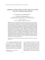
Identification and antimicrobial activity of actinomycetes strains isolated from marine samples in the coastal area of Thanh Hoa – Quang Binh – Quang Tri
Bạn đang xem bản rút gọn của tài liệu. Xem và tải ngay bản đầy đủ của tài liệu tại đây (965.9 KB, 10 trang )
Vietnam Journal of Science and Technology 56 (4) (2018) 424-433
DOI: 10.15625/2525-2518/56/4/10663
IDENTIFICATION AND ANTIMICROBIAL ACTIVITY
OF ACTINOMYCETES STRAINS ISOLATED FROM
MARINE SAMPLES IN THE COASTAL AREA OF
THANH HOA – QUANG BINH – QUANG TRI
Cao Duc Danh1, 3, Cao Duc Tuan1, 2, 3, Vu Thi Quyen1, Nguyen Mai Anh1,
Doan Thi Mai Huong1, Pham Van Cuong1, Chau Van Minh, Le Thi Hong Minh1, *
1
2
Institute of Marine Biochemitry, VAST, 18 Hoang Quoc Viet, Cau Giay, Ha Noi
Hai Phong University of Medicine and Pharmacy, 72A, Nguyen Binh Khiem, Hai Phong
3
Graduate University of Science and Technology, VAST, 18 Hoang Quoc Viet, Cau Giay, Ha Noi
*
Email:
Received: 12 September 2017; Accepted for publication: 29 June 2018
Abstract. In this study, 46 strains of actinomycetes were isolated from 40 samples including:
sediments, sponges, soft corals, echinoderms and starfish collected from three coastal areas of
Vietnam: Thanh Hoa – Quang Binh – Quang Tri. The strains were fermented in A+ medium and
fermentation broths were extracted 5 times with ethyl acetate then the extracts were evaporated
under reduced pressure to yield crude extracts. Quantitative assay was used to determine MIC
(Minimum inhibitory concentration) of extract against 7 reference strains. From the results of
screening, five strains of actinomycetes exhibited the highest biological activity were chosen
(Code: G212, G222, G233, G227 and G241). In particular, strains G222, G233, G227 and G241
were resistant 6/7 strains of microorganisms test, with MIC values from 64 µg/ml to 256 µg/ml;
Moreover. All of the five strains were highly resistant to yeast Candida albicans
ATCC10231. These strains were then subjected to morphological and phylogenetic
investigations based on 16S rDNA gene sequences. The results showed that strains G212, G222
andG227 belonged to the genus Streptomyces; strains G233 and G241were identified as a
member of the genus Micromonospora.
Keywords: Micromonospora, Streptomyces, antimicrobial activity, MIC, 16S rDNA gene
sequences.
Classification numbers: 1.5.3; 3.4.4
1. INTRODUCTION
Actinomycetes are Gram-positive bacteria that grows in various environments, with a
filamentous form similar to fungi. They are ubiquitous in freshwater and marine water habitats
[1]. Actinomycetes are of biological importance because of their efficiency in antibiotic
production. They are considered highly valuable as they can produce various antibiotics and
Identification and antimicrobial activity of actinomycetes strains isolated from marine …
other therapeutically useful compounds with diverse biological activities. Many of the presently
used antibiotics such as streptomycin, gentamicin, rifamycin and erythromycin are the products
of actinomycetes. Need of new antimicrobial agents is greater than ever because of emergence of
multidrug resistance in common pathogens, the rapid emergence of new infections and the use of
multidrug resistant pathogens in bioterrorism [2]. Resistance of bacteria to the effects of
antibiotics has been a major problem in the treatment of diseases [3].
The genus Streptomyces is represented in nature by the largest number of species and
varieties, producing the majority of known antibiotics among the family Actinomycetaceae.
Streptomyces are well known sources of antibiotics and other important novel metabolites,
including antifungal agents [4], antitumor agents [5], antihelminthic agents [6] and herbicides.
Marine environment contains a wide range of distinct Streptomyces that are not present in
the terrestrial environment. Though some reports are available on antibiotic and enzyme
production by marine actinomycetes, the marine environment is still a potential source for new
actinomycetes, which can yield novel bioactive compounds and industrially important enzymes
[7].
In addition, Micromonospora species – the dominant actinomycetes are possible to be
isolated from aquatic habitats such as streams, lake mud, river sediments, beach sands, sponge
and marine sediments [8, 9]. Micromonospora species, together with Streptomyces species are
best known for synthesizing antibiotics, especially aminoglycoside, enediyne, and
oligosaccharide antibiotics. Thus, their impact on medicine is considerable. Of common
antibiotics in the medical field, gentamicin and netamicin belong to the aminoglycoside
antibiotics yielded by Micromonospora [10].
It is obviously that actinomycetes serve as an abundant source of bioactive compounds. In
the future, manifold novel compounds would be potentially discovered from them. Herein, we
reported on the isolation, taxonomic characterization, extraction fermentation broths with ethyl
acetate of these actinomycete strains isolated from samples collected in Thanh Hoa – Quang
Binh – Quang Tri of Viet Nam and also reported on their antimicrobial activity.
2. MATERIALS AND METHODS
2.1. Material
Chemicals
The genomic DNA isolation kits were purchased from Promega (Madison, WI, USA). The
mini-prep and DNA gel extraction kits were purchased from Qiagen (Mannheim, Germany).
PCR master mix was purchased from Bioneer. Glucose and all other chemicals (for media) were
obtained from Himedia (India), Duc Giang (Viet Nam) and Sigma-Aldrich (St. Louis, MO, USA).
Microorganisms test
Seven standard reference microorganisms were come from ATCC Bacteriology Collection
including: Three Gram negative bacteria (Escherichia coli ATCC25922, Pseudomonas
aeraginosa ATCC27853, Salmonella enterica ATCC13076), and three Gram positive bacteria
(Enterococcus faecalis ATCC29212, Stapphylococus aureus ATCC25923, Bacillus cereus
ATCC 13245), one yeast strain Candida albicans ATCC10231.
425
Le Thi Hong Minh, et al.
2.2. Samples collection
The marine samples were collected using Ponar from three locations at 4 m-24 m in depth
with different geographic coordinates; temperature of water was 26-29 oC in Thanh Hoa –
Quang Binh – Quang Tri. The samples were collected into 15 mL or 50 mL sterile Falcon tubes
and preserved in ice-box and processed within 24 h.
2.3. Isolation of actinomycetes
First, 0.5 g of sample was suspended in 4.5 mL of sterile distilled water, homogenized by
vortexing for 1 min, and the suspension was treated using a wet-heat technique (60 oC for 6
min). Next, 0.5 mL of this suspension was transferred to another 4.5 mL sterile distilled water
and this step was repeated to set up a tenfold dilution series to 10-3. At the final dilution step,
aliquots of 50 µL were spread on six different media like: A1 (soluble starch: 10 g/L; yeast
extract: 4 g/L; peptone: 2 g/L; instant ocean: 30 g/L; agar: 15 g/L), M1 (soluble starch: 5 g/L;
yeast extract: 2 g/L; peptone: 1 g/L; instant ocean: 30 g/L; agar: 15 g/L), SWA (instant ocean: 30
g/L; agar: 15 g/L), A+( soluble starch: 10 g/L; yeast extract: 4 g/L; peptone: 2 g/L; instant ocean:
30 g/L; CaCO3: 1 g/L; agar: 15 g/L), SCA (soluble starch: 10 g/L; K2HPO4: 2 g/L; KNO3: 2 g/L
; casitone: 300 mg/L; MgSO4·7H2O: 50 mg/L; FeSO4·7H2O: 10 mg/L; instant ocean: 30 g/L;
CaCO3: 2 mg/L; agar: 15 g/L), NZSG (soluble starch:20 g/L; yeast extract: 5 g/L; glucose: 10
g/L; NZ amine A: 5 g/L; Instant ocean: 30 g/L; agar: 15 g/L); ISP1 (soluble starch: 5 g/L; yeast
extract: 2 g/L; casitone: 5 g/L; instant ocean: 30 g/L; Agar: 15 g/L), ISP2 (soluble starch: 5 g/L;
yeast extract: 2 g/L; malt extract: 10 g/L; glucose: 10 g/L; instant ocean: 30 g/L; agar: 15 g/L).
These media were supplemented with 50 µg/mL polymycin B and cycloheximide to inhibit
Gram - negative bacterial and fungal contamination. After 21 days of aerobic incubation at 30
o
C, the colonies of actinomycete strains were transferred onto yeast extract-malt extract agar
(ISP2 medium) [11, 12].
2.2. Production of crude extracts
The actinomycetes strains were cultivated at 28 °C in sterile 1000 mL flasks containing 500
mL media A+ with glucose 1 %, pH 7.0, at 200 rpm and 30 oC. After 7 days cultivation, the
fermentation broths were filtered and then extracted with ethyl acetate (5 times). The extracts
were evaporated under reduced pressure to yield crude extracts [13].
2.3. Screening of actinomycetes for antimicrobial activity assessment of antimicrobial
activity of the extracts 2.2
Crude extracts were tested against the Gram-positive bacteria (Bacillus cereus
ATCC13245, Enterococcus faecalis ATCC29212, Staphylococcus aureus ATCC25923), the
Gram-negative bacteria (Pseudomonas aeruginosa ATCC27853, Escherichia coli ATCC25922,
Salmonella enterica ATCC13076) and the fungi Candida albicans ATCC10231. The positive
control was streptomycin for bacteria, cycloheximide for fungi Candida albicans ATCC10231.
Quantitative assay was performed based on dilution method for determination of MIC
(Minimum Inhibition Concentration) values of extracts against test bacteria. MIC means the
lowest concentration of extract at which the test microorganism did not show any visible. The
density of cells was read at 610 nm and adjusted to an optical density (OD) of 0.04 for Grampositive bacteria, and 0.05 for Gram-negative bacteria and C. albicans. Aliquots of 50 µL of
426
Identification and antimicrobial activity of actinomycetes strains isolated from marine …
bacterial or fungal suspension were incubated with each crude extract for 24 h at 30 ºC. The UV
absorption of each sample was read at 610 nm and compared against the UV absorption of the
media as control. MIC value was determined in plate 96 wells with the lowest concentration of
reagents that completely inhibits the growth of microorganisms after 24 hours of incubation and
were correctly identified based on data of cell turbidity measured by spectrophotometer
Biotek and GraphPad Prism DaTa software [14].
2.4. Identification of actinomycetes
The actinomycete strains were incubated for 14 days at 30 ºC on starch casein agar (SCA)
and its morphology was examined using scanning electron microscopy (model JSM-5410 LV;
JEOL). Samples for scanning electron microscopy (SEM) were prepared as described by Itoh et
al [15].
Sequencing 16S rDNA method was used for identification of chosen strains.
Amplifications were performed in a 25.0 µL mixture containing 16.3 µL of H2O, 2.5 µL of 10X
PCR buffer, 1.5 µL of 25 mM MgCl2, 0.5 µL of 10 mM dNTP’s, 0.2 µL of Taq polymerase,
1.0 µL for both 0.05 mM of 9 F (5'-GAGTTTGATCCTGGCTCAG3') and 0.05 mM of
1541R (5'-AAGGAGGTGATCCAACC3') primer [16] and 2.0 µL of genomic DNA. The
reaction tube was then put into MJ Thermalcycler, which had been programmed to preheat at 94
o
C for 3 min, followed by 30 cycles of denaturation at 94 oC for 1 min, annealing at 60oC for 30s
and elongation at 72 oC for 45s before a final extension of 72 oC for 10 min. Estimated product
size was about 1500 bp. PCR products were purified by DNA purification kit (Invitrogen). The
16S rDNA gene sequencing was carried by DNA Analyzer (ABI PRISM 3100, Applied
Bioscience). Gene sequences were handled by BioEdit v.2.7.5. and compared with bacterial 16S
rRNA sequences in GenBank database using NBCI Blast programme. The alignment was
manually verified and adjusted prior to the construction of a phylogenetic tree. The phylogenetic
tree was constructed by using the neighbor-joining the MEGA programme version 4.1 [17].
3. RESULTS AND DISCUSSION
From 40 marine samples were collected of Thanh Hoa – Quang Binh – Quang Tri areas
Vietnam. 46 actinomycete strains were isolated. These actinomycete strains were cultivated in
SCA medium broth. The fermentation broths were extracted 5 times with ethyl acetate then the
extracts were evaporated under reduced pressure to yield crude extracts. Crude extracts were
tested against 7 reference strains. From the results of screening, we chose 5 strains of
actinomycetes that have the highest biological activity (Code: G212, G222, G233, G227 and
G241) (Table 1).
Table 1 shows the results of the screening of the crude extract of 5 selected strains on
antibacterial activity. The results of the current study revealed that 6/7 strains of reference
microorganism were against following G222, G233, G227 and G241 with values MICs from 64
µg/mL to 256 µg/mL, whereas G212 showed moderate antibacterial against only with 4/7
reference strains. Specially, all of the five strains had not resistant to E.coli ATCC25922. In
addition, all crude extract showed a high MIC against Candida albicans (ATCC10231), from 64
µg/mL to 256 µg/mL. According to the previuos report, out of 15 strains screened in the North
East Coast of Viet Nam, there were only three strains of G115, G119, G120 being resistant to P.
aeruginosa ATCC27853, and G057 being resistant to S. enterica ATCC 13076 [18]. This result
shows that the biological activity of the strains depends very much on geographic location
427
Le Thi Hong Minh, et al.
during sample collection. The different sensitivity between Gram positive and Gram negative
bacteria could be explained by different in membrane structure of these microorganisms. Gram
negative bacteria have an outer polysaccharide membrane carrying the structural
lipopolysaccharide components. This makes the cell wall impermeable to lipophilic solutes. The
gram positive bacteria would more susceptible due to having only an outer peptidoglycan layer
which is not an effective permeability barrier [19, 20].
Table 1.Antimicrobial activity of crude extracts of ethyl acetate extracts from 7strains.
No
Isolates
Gram-positive
E.faecalis
S.aureus
Gram-negative
B.cereus
E.coli
P.aeruginosa
Yeast
S.enterica
C.albicans
ATCC29212 ATCC25923 ATCC13245 ATCC25922 ATCC27853 ATCC13076 ATCC10231
1
G212
128
-
2
G222
256
64
3
G233
256
4
G227
5
128
-
-
64
256
64
-
128
128
128
128
64
-
128
64
64
256
128
128
-
128
128
128
G241
256
256
64
-
256
64
64
Streptomycin
256
256
128
32
256
128
-
Cycloheximide
-
-
-
-
-
-
32
Units for concentration of crude extracts is MIC (µg/mL)
A
B
D
C
E
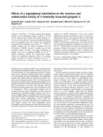
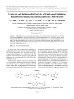
Figure 1. Scanning electron micrographs of the representative strains G212(A ); G233 (B); G222 (C);
G227 (D) and G241(E) grown on SCA agar for 2 weeks at 30 °C.
The spore morphology is considered as one of the important characteristics in the
428
Identification and antimicrobial activity of actinomycetes strains isolated from marine …
identification of Streptomyces and it greatly varies among the species. It has been found that the
majority of the marine isolates produced aerial coiled mycelia and the spores arranged in chains
as similar with the reported by Mukherjee and Sen [21] (Figs.1A, 1C, 1D). Micromonospora
species produced well-developed and branched substrate hyphae on yeast extract-malt extract
medium, but no aerial hyphae. Spores were borne singly on the substrate hyphae motile
(Figs.1B, 1E).
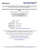
The colors of the substrate mycelium were white to vivid orange and turned to brownish
black after sporulation (Fig. 2). The morphological characteristics of these isolates were
consistent with their classification in the genus [22].
Figure 2.Morphological appearance of isolates. The colors of the substrate mycelium were vivid
orange (G241) and from white (G222, G227) turned to brownish (G212, G233).
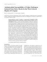
Five potential isolates were selected for identification by 16S rDNAgene sequencing. The
16S rDNA genes were amplified by PCR with compositions as above. PCR products were
verified by agarose gel electrophoresis 1 %. The result of electrophoresis image showed a
specific band with a size about 1500 bp, corresponding theoretical size 16S rDNA gene of
specific primers 9F and 1541R (Fig 3). After analysis of obtained sequences by Bioedit
programme, sequences of 5 isolates G212, G222, G227, G233 and G241 were revealed with the
size 1443 bp, 1443 bp, 1434 bp, 1436 bp and 1433 bp, respectively. Compare these sequences on
GenBank database, the results showed that 16S rDNA sequences of G212, G222 and G227
strains exhibited high similarity (99 %) with Genus Streptomyces spp; Strains G233 and G241
were identified (99 % similarity) of 16S rDNA sequences with Genus Micromonospora spp in
GenBank (Fig. 4).
429
Le Thi Hong Minh, et al.
Figure 3. Electrophores is image of PCR products16S rDNA gene of isolates,
M: Marker 1Kb,
Lanes 1-5: PCR products of G212, G222, G227, G233 and G241 isolates,
Lane 6: PCR product control without DNA template.
Figure 4. Neighbor-joining tree based on almost-complete 16S rDNA gene sequences showing
relationships between the strains in groups and representative members of the genera Streptomyces and
Micromonospora were used as an outgroup. The numbers on the branches indicate the percentage
bootstrap values of 1,000 replicates; Bar, 0.01 substitutions per nucleotide position.
The morphological differentiation of Streptomyces involves the formation of a layer of
hyphae that can differentiate into a chain of spores. The most interesting property
of Streptomyces is the ability to produce bioactive secondary metabolites such as antifungals,
antivirals, antitumoral, anti-hypertensives, and mainly antibiotics and immune suppressives [23,
24, 25]. Another characteristic is of the genus is complex multicellular development, in which
their germinating spores form hyphae, with multinuclear aerial mycelium, which forms septa at
regular intervals, creating a chain of spores [26].
Micromonospora species are best known for synthesizing antibiotics, especially
aminoglycoside gentamicin and netamicin), enediyne, and oligocharide antibiotics which
contribute their impact medicinal usage [27]. Furthermore, Micromonospora species has been
intensively investigated and isolated anticancer antibiotics such as anthraquinones, hracyclines,
alkaloids, and macrolides [28, 29].
430
Identification and antimicrobial activity of actinomycetes strains isolated from marine …
Several Micromonospora isolates were found to produce the bioactive compounds for
example: The extract of Micromonospora aurantiaca with doxorubicin (DOX) were treated on
human carcinoma of nasopharynx (KB cells). The result showed this combination caused 10fold enhanced cell death at concentrations that each agent alone is poorly effective. The
enhanced cytotoxicity of the combined treatment may result from augmentation of DOXinduced apoptosis by Micromonospora aurantiaca extract [30]. Diazepinomicin was isolated
from the marine sponge-associated strain Micromonospora sp. RV115. Results showed that
diazepinomicin exhibited antioxidant capacity using two different strategies including cell-free
and cell-based assays. Diazepinomicin was able to protect cells from toxicity and genomic
damage induced by the strong oxidant H2O2. This antioxidant activity will add a new perspective
on the use of diazepinomicin in chemoprevention therapy for different types of cancer [31]. The
results presented above confirmed that Micromonospora, especially marine Micromonospora
have the potential to make an important contribution to fight against the pathogen.
4. CONCLUSION
From 40 samples including: sediments, sponges, soft corals, echinoderms and starfish
collected from three sea areas of Vietnam: Thanh Hoa – Quang Binh – Quang Tri, we isolated
46 strains of actinomycetes. All most of the isolates exhibited antimicrobial activity.We chose 5
strains of actinomycetes that have the highest biological activity (Code: G212, G222, G233,
G227 and G241). Specificly, strains G222, G233, G227 and G241 were resistant 6/7 strains of
microorganisms test, with MICs values from 64 µg/ml to 256 µg/ml. In addition, all of the five
strains were highly resistant to yeast Candida albicans ATCC10231. The morphological and
phylogenetic investigations based on 16S rDNA gene sequences showed that: strains G212,
G222 and G227 belonged to Genus Streptomyces; strains G233 and G241 were identified as
Genus Micromonospora.
Acknowlegement: This work was financially supported by the Vietnam Academy of Science and
Technology (VAST). Code of project: VAST.TĐ.DLB.04/16-18.
REFERENCES
1.
Fenical W., Jensen P. R. - Developing a new resource for drug discovery: marine
actinomycete bacteria, Nat. Chem. Biology 2 (2006) 666-673.
2.
Spellberg B., Powers J. H., Brass E. P., Miller L. G. and Edwards J. E. - Trends in
antimicrobial drug development: implications for the future, Clinical Infectious Diseases
38 (9) (2004) 1279-1286.
Luzhetskyy, A., Pelzer S. and Bechthold A. - The future of natural products as a source of
new antibiotics, Current Opinion in Investigational Drugs 8 (8) (2007) 608-613.
3.
4.
Thakur D., Yadav A., Gogoi B. K., Bora T. C. - Isolation and screening of Streptomyces
in soil of protected forest areas from the states of Assam and Tripura, India, for
antimicrobial metabolites, J. Mycol. Med. 17 (2007) 242–249.
5.
Lee H. B., Kim C. J., Kim J. S., Hong K. S., Cho K. Y. - A bleaching herbicidal activity
of methoxyhygromycin (MHM) produced by an actinomycete strain Streptomyces sp. 8E12, Lett. Appl. Microbiol. 36 (2003) 387–391.
6.
Sanglier J. J., Haag H., Huck T. A., Fehr T. - Novel bioactive compounds from
431
Le Thi Hong Minh, et al.
actinomycetes, Res.Microbiol. 144 (1993) 633–642.
7.
Jacques P., Eric M., Nadia P., Stephane B., William F., Paul J. - Marine actinomycetes: a
new source of compounds against the human malaria parasite, PLos One 3 (2008) 2335 –
2340.
8.
Rifaat H. M. - The biodiversity of actinomycetes in the River Nile exhibiting antifungal
activity, J.Mediter Ecol. 4 (2003) 5-7.
9.
Eccleston G. P., Brooks P. R., Kurtboke D. I. - The occurrence of bioactive
micromonosporae in aquatic habitats of the sunshine coast in Australia, Mar. Drugs 6
(2008) 243-261.
10. Bérdy J. - Bioactive microbial metabolites: a personal view, Journal of Anti-biotics
(Tokyo) 58 (2005) 1-26.
11. Williams S. T. and Davies F. L. - Use of antibiotics for selective isolation and
enumeration of Actinomycetes in soil, J. of General Microbiology 38 (1965) 251-261.
12. Williams S. T., Cross T. - Actinomycetes, In: Methods in Microbiology, Academic Press
(London) 4 (1971) 295–334.
13. Cédric O., Skylar C., Bindiya K., Mashal M. A., Haipeng L., Anna O., Quan S., Van
Cuong Pham, Catherine L. S., Brian T. Murphy and Alexander S. M. - Tool for
characterizing bacterial protein synthesis inhibitors, Antimicrob, Agents Chemother 57
(2013) 5994- 6002.
14. Hadacek F., Greger H. - Test of antifungal natural products methodolagies, comparability
of result and assay choise, Phytochem, Anal. 90 (2000) 137-147.
15. Itoh T., Kudo T., Parenti F., Seino A. - Amended description of the genus
Kineosporia,based on chemotaxonomic and morphological studies, Int. J. Syst.
Bacteriology 39 (1989) 168–173.
16. Rajesh M. M., Subbaiya R., Balasubramanian M. - Ponmurugan and Masilamani Selvam.
Isolation and Identification of Actinomycetes 6 Isoptericola variabilis From Cauvery
River Soil Sample, Int. J. Curr. Microbiology 2 (2013) 236-245.
17. Saitou N., Nei M. - The neighbor-joining method: a new method for reconstructing
phylogenetic trees, Mol.Biology 4 (1987) 406-425.
18. Minh L. T. H., Quyen V. T., Anh N. M., Huong D. T. M., Murphy B. T., Minh C. V., and
Cuong P. V. - Isolation, screening and identification of microorganisms having
antimicrobial activity isolated from samples collected on seabed of Northeast Vietnam,
Vietnam Journal of Biotechnology 14 (3) (2016) 539-547.
19. BasilioA., González I., Vicente M. F., Gorrochategui J., Cabello A., González A.,
Genilloud O - Patterns of antimicrobial activities from soil actinomycetes isolated under
different conditions of pH and salinity, J. Appl. Microbiol. 95 (2003) 814-823.
20. Oskay M., Same A., Azeri C. - Antibacterial activity of some actinomycetes isolated from
farming soils of Turkey, Afr. J. Biotech. 3 (2004) 441-446.
21. Mukherjee G., Sen S. K. - Characterization and identification of chitinase producing
Streptomyces venezulae P10, Indian J. Exp. Biol. 42 (2004) 541–544.
22. Kawamoto I., Williams S. T., Sharpe M. E., Holt J. G. - Bergey’s Manual of Systematic
Bacteriology 4 (1989) 2442-2450.
23. Ōmura S., Ikeda H., Ishikawa J. - Genome sequence of an industrial microorganism
432
Identification and antimicrobial activity of actinomycetes strains isolated from marine …
Streptomyces avermitilis: deducing the ability of producing secondary metabolites, Proc.
Natl. Acad. Sci. 98 (2001) 12215-12220.
24. Khan S. T. - Streptomyces associated with a marine sponge Haliclona sp. biosynthetic
genes for secondary metabolites and products, Environ. Microbiol Black Sci.
Pub. 13 (2011) 391-403.
25. Patzer S. I., Volkmar B. - Gene cluster involved in the biosynthesis of griseobactin, a
catechol-peptide siderophore of Streptomyces sp. ATCC 700974, J. Bacteriol. 192 (2010)
426-435.
26. Ohnishi Y., Ishikawa J., Hara H. - Genome sequence of the streptomycin-producing
microorganism Streptomyces griseus IFO 13350, J. Bacteriol. 190 (2008) 4050-4060.
27. Hirsch A. M, Valdés M. - Micromonospora: An important microbe for biomedicine and
potentially for biocontrol and biofuels, Soil Biol. Biochem. 42 (4) (2010) 536-542.
28. Igarashi Y., Yanase S., Sugimoto K., Enomoto M., Miyanaga S., Trujillo M. E., Saiki I.,
Kuwahara S., Lupinacidin C. - An inhibitor of tumor cell invasion from Micromonospora
lupine, J. Nat. Prod. 74 (2011) 862-865.
29. Gaertner A., Ohlendorf B., Schulz D., Zinecker H., Wiese J., Imhoff J. F. - Levantilides
A and B, 20-membered macrolides from a Micromonospora strain isolated from the
mediterranean deep sea sediment, Mar. Drugs 9 (2011) 98-108.
30. Chantarawan S., Jantharat P., Rattanaporn S. - Extraction of Micromonospora
aurantiaca from Coastal Marine Sediments Enhances Doxorubicin Induced Apoptosis in
KB Cells, J. Physiol. Biomed. Sci. 26 (2) (2013) 76-82.
31. Usama R.A., Matthias S., Eman M.O., Tanja S., Stephanie G., Helga S and Ute H. Antioxidant and Anti-Protease Activities of Diazepinomicin from the Sponge-Associated
Micromonospora Strain RV115, Mar. Drugs 10 (2012) 2208-2221.
433