In vitro propagation of citrus species through callus induction and regeneration: A review
Bạn đang xem bản rút gọn của tài liệu. Xem và tải ngay bản đầy đủ của tài liệu tại đây (224.34 KB, 14 trang )
Int.J.Curr.Microbiol.App.Sci (2019) 8(10): 2282-2295
International Journal of Current Microbiology and Applied Sciences
ISSN: 2319-7706 Volume 8 Number 10 (2019)
Journal homepage:
Review Article
/>
In vitro Propagation of Citrus Species through Callus Induction and
Regeneration: A Review
Mudasir Iqbal1*, V. K. Wali1, Parshant Bakshi1, Kiran Kour1,
Vijay K. Razdan2, B. K. Sinha2 and K. K. Sood3
1
Division of Fruit Science, 2Division of Plant Pathology, 3Division of Agroforestry, Faculty of
Agriculture, Main Campus, Chatha, Sher-e-Kashmir University of Agricultural Sciences &
Technology of Jammu, J&K-180009, India
*Corresponding author
ABSTRACT
Keywords
Tissue culture,
Micropropagation,
Callus,
Regeneration,
Rooting,
Acclimatization
Article Info
Accepted:
17 September 2019
Available Online:
10 October 2019
Citrus, one of the most important group of fruit crops around the world, are propagated at
large scale with many difficulties. Propagation through seeds is challenging because of
Phytophthora foot rot together with recalcitrance of citrus seeds. Vegetative propagation of
Citrus species is mainly performed now-a-days by budding on seedling rootstocks. As
heavy losses are experienced among the susceptible seedlings due to Phytophthora and
Citrus tristeza virus (CTV), the interest in resistant rootstocks has greatly increased. The
potential of conventional methods of citrus plant breeding of rootstocks are limited by
physiological factors such as heterozygosity, inbreeding depression, nucellar
polyembryony and juvenility. Under such conditions advanced tissue culture techniques
provide best possible alternative for producing large number of resistant progenies from
elite citrus genotypes. Plant tissue culture provides reliable and economical method of
maintaining pathogen free plants that allows rapid multiplication and international
exchange of germplasm. Generally, when in vitro propagation protocols are developed for
any specific plant species, specialized conditions for individual genotypes, elite species
and even various developmental stages of the explants plants are selected via error-andtrial experiments. Because large diversity is observed in Citrus plant family, it takes many
months to develop protocols for most suitable culture medium, best concentrations and
combinations of plant growth regulators and other supplements for better development of
explant cultures. Therefore, in this review, we tried to put together results from difficultto-find literatures and listed all the identified findings, in which callus induction or somatic
organogenesis was used to develop citrus plants. Successful protocols of surface
sterilization method, culture establishment, shoot regeneration, in vitro rooting and
acclimatization are presented systematically.
2282
Int.J.Curr.Microbiol.App.Sci (2019) 8(10): 2282-2295
Introduction
Citrus (Citrus sp.) is collective generic term
comprising a number of species and varieties
of fruits known to the world for their
characteristic flavour, attractive range of
colours and uses (Raja, 2012). Citrus is
believed to have originated in the part of
Southeast Asia bordered by Northeastern
India, Myanmar (Burma) and the Yunnan
province of China (Scora, 1975; Gmitter and
Hu, 1990; Liu et al., 2012). They are longlived perennial crops grown in more than 100
countries across the world (Saunt, 1990). The
citrus growing belts of the world are
concentrated in tropical and subtropical
regions where suitable soil and climatic
conditions prevail (Kaur, 2016).
Citrus is considered as the number one fruit of
the world due to its high nutritional value,
great production potential and preparation of
large number of fruit products from them
(Kour and Singh, 2012). Citrus fruits are
known for their distinctly pleasant aroma,
arising due to terpenes present in the rind (Li
et al., 2014). The genus derives its commercial
importance from its fruits, which are of great
economic and health value and are consumed
fresh or pressed to obtain juice (Talon and
Gmitter Jr., 2008). Citrus peels too have no
less importance and can be candied, used as
livestock feed, in perfumeries, bakeries and in
soap industry (Dhanavade et al., 2011).
Lemon oil obtained by cold pressing of lemon
peels is extensively used in furniture polish
(Bansode et al., 2012). Citrus has been utilized
in a number of medicinal preparations for the
remedy of scores of ailments ranging from
toothache,
diarrhea,
constipation,
and
insomnia to vomiting (Singh and Rajam,
2010). It carries bioactive secondary
components which are working against cancer
and degenerative diseases (Karimi et al.,
2012). The medicinal practitioners commonly
suggest consuming citrus fruits for obtaining
minerals, vitamins and other necessary
components so as to recover weak health by
improving appetite quickly (Rakesh et al.,
2013). The flavonoids of citrus play an
important role in preventing progression of
hyperglycemia by increasing the glycogen,
hepatic glycolysis and reducing the hepatic
gluconeogenesis (Shen et al., 2012).
The primary reason for shifting citriculture
from seedling to budded plants was the
appearance of Phytophthora “foot rot” in
Azores Islands in 1842 (Singh and Naqvi,
2001). Since early 1950s extensive rootstock
trials on citrus have been conducted under
different
environmental
conditions
(Bhattacharya and Dutta, 1952; Rangacharlu
et al., 1958 and Singh, 1962). Further, the
citrus root stock scenario in India has been
reviewed by (Agarwal, 1982); (Randhawa and
Srivastava, 1986); (Patil, 1987) and (Chadha
and Singh, 1990). The dominant sour orange
rootstock has been replaced by rough lemon
rootstock which was tolerant to CTV
(Chamandoosti, 2017).
Rough lemon is highly vulnerable to
Phytophthora, which leads to main losses in
an orchard if appropriate phyto-sanitary
conditions are not followed (Mukhtar et al.,
2005a, Savita et. al. 2010, Sarma et al., 2011
and Kasprzyk-Pawelec et al., 2015). The
potential of conventional methods of
upgrading of citrus rootstocks is limited by
biological factors that hinder breeding and
selection, such as heterozygosity and
inbreeding pollen and ovule sterility, sexual
incompatibility,
apomixes,
depression,
nucellar polyembryony and juvenility (Guo
and Deng, 2001; Guo and Grosser, 2005)
(Tusa et al., 1990, Carimi et al., 1994, Savita
et al., 2010, Benabdesselam et al., 2011,
Lombardo et al., 2011). In vitro culture is a
method that can resolve this problem and can
also produce crops on a comparatively large
scale in comparison with conventional plant
2283
Int.J.Curr.Microbiol.App.Sci (2019) 8(10): 2282-2295
breeding (Kasprzyk-Pawelec et al., 2015).
Under such circumstances, in vitro culture
techniques hold potential and could present
solution to these problems
Tissue culture and micropropagation practice
have been developed from different explants
sources for number of Citrus spp., Therefore,
the aim of this review is to focus on the use of
the former pathway, most probably the
technique
previously
employed
for
micropropagation of citrus, and an attempt has
been made to present a comprehensive
available literature related to tissue culture in
Citrus species under the following headings
and sub-headings.
Tissue culture studies in citrus species
Studies on citrus tissue culture in vitro were
set off in early Nineteen Fifties with the aim
of genetic improvement of the species as well
as to get virus free plants. It has been
suggested that plant tissue culture would play
a very significant role in conservation and
genetic improvement for large scale
propagation of plants in India (Raja, 2012).
Plant tissue culture has come into view as a
powerful
tool
for
propagation
and
improvement of many woody plant species
including Citrus. The genetic and epigenetic
mechanism of callus formation, the
widespread use and knowledge of molecular
mechanisms and the underlying induction of
callus, deserve to be studied systematically
(Momoko et al., 2013). In vitro culture has the
potential to eradicate diseases and provides
scope for development of new cultivars
through somaclonal variations (Hammschlag
et al., 1995). Despite its rich genetic
resources, scientists come across difficulties in
citrus hybridization breeding due to high
sterility, heterozygosity, incompatibility and
nucellar embryos (Shen et al., 1998). With the
development of biotechnology, genetic
transformation and protoplast hybridization
have been recognized to avoid those breeding
obstacles in many fruit trees (Deng and Liu,
1996). For citrus, embryogenic callus is
extensively used in genetic transformation and
protoplast hybridization since it can simply
regenerate plants (Deng and Liu, 1996; Hao,
2000; Hao and Deng, 2002). Citrus
embryogenic calluses can be maintained in
culture at one month intervals for a long
period (Hao and Deng, 2002; Yi and Deng,
1998). However, recurrent subculture of
numerous cultures is labour intensive and
costly (Engelmann, 1997; Ashmore, 1997). To
resolve this problem, short and medium term
storage methods have been developed to
lessen growth and increase subculture
intervals. Tissue culture protocols have been
described for a number of Citrus spp. through
callus (Singh and Rajam, 2009; Savita et al.,
2010, 2011a, 2011b; Ali and Mirza, 2006;
Altaf et al., 2008; Altaf et al., 2009a,b; Khan
et al., 2009; Laskar et al., 2009; Kaur, 2018
and Taye et al., 2018).
Sterilization procedures of explants
Sterilization is a very important and basic
aspect of tissue culture, as it actually aims at
in vitro propagation of progenies of desired
genotypes free from surface and systemic
contamination. The explants collected from
field grown seedlings harbour many microbial
pathogens like fungi and bacteria, in addition
to adhered soil particles thus, it necessities a
thorough and effective surface sterilization of
explants before culturing. Mercuric chloride
seems as the best sterilizing gent as preferred
by Ali and Mirza 2006, Savita et al., 2011b,
Saini et al., 2010 and Kour, 2016 in Citrus
jambhiri at the concentration of 0.1 % treated
for 4-5 minutes, Kanwar et al., 2016 in Sour
orange at the concentration of 0.1 % treated
for 1 minute, in addition sodium hypochlorite
(NaOCl) is also used by some others
(Upadhyay et al., 2010 in Sweet orange cv.
2284
Int.J.Curr.Microbiol.App.Sci (2019) 8(10): 2282-2295
Mosambi). In addition Taye et al., 2018 used
fungicides like Kocide, Bayleton and Redimol
each with the concentration of 0.25 g/100 ml
of water for 15 and 20 minutes. The surface
sterilization of explants with 70 % aqueous
solution of ethanol for 30 seconds followed by
0.1 % mercuric chloride for 8-10 minutes and
then thoroughly washing with sterile distilled
water in citrus was reported by (Sharma et al.,
2009). Pre-sterilization of excised explants
with Benomyl (0.2%) can improve a cleanness
and aliveness of all types of explants,
especially when followed through surface
sterilization done by mercury chloride (HgCl2)
(Nurul, et al., 2012). Kour and Singh 2012
removed expanded leaves of Rough lemon as
explants and then treated them with 10 %
solution of teepol detergent for 10 minutes
followed by thorough washing with distilled
water. They further preferred treatment of
explants with 70 % ethanol for 30 seconds
followed by 0.1 % mercuric chloride treatment
for 8 minutes and then rinsing with autoclaved
distilled water three times.
Culture establishment
Callus formation is controlled by the level of
plant growth regulators (auxin and cytokinins)
in the culture media. Concentrations of plant
growth regulators can vary for each plant
species and can even depend on the sources of
explants or individual plant. Culture
conditions (temperature, light) are also
important. Protocols developed in previous
studies have shown that plant growth regulator
concentration and selection are vital for citrus
callus induction.
Explant type, media composition and callus
induction
The major advantages of using seedlings
explants over explants taken from field-grown
mature plants are their high multiplication
rates and high regeneration potentials
However, the disadvantages are very known,
including insufficient knowledge regarding
their genetic background. Das et al., (2000) in
their study developed a protocol for
micropropagation of elite plants of sweet
orange (Citrus sinensis) through nucellar
embryo culture and found that MS medium
supplemented with NAA (1.0 mg/l) or 2, 4-D
(1.0 mg/l) encouraged callus development in
both nucellar and zygotic embryos. Al-Khayri
and Al-Bhrany (2001) in their study on
micropropagation in lime Citrus aurantifolia
using nodal explants of mature tree nodes
found best multiple shoot formation, i.e. 8.0
shoots per node on MS medium supplemented
with 1.0 mg/l BAP and 0.5 mg/l kinetin.
Srivastava et al., (2001) in their study on vitro
plant regeneration of Citrus aurantifolia
through callus culture, shoot tip, epicotyls and
hypocotyl segments reported callusing on MS
medium enriched with BAP (5.0 mg/l) and
observed highest per cent of callus and shoot
regeneration with 5.0 mg/l BAP. Kamble et
al., (2002) in their study on in vitro
micropropagation and callus induction in acid
lime (Citrus aurantifolia) cv. Sai Sarbati
observed the highest callus induction with
epicotyl cultured on half-strength of MS
medium supplemented with NAA (10.0 mg/l)
and BAP (0.5 mg/l). Karwa Alka (2003)
carried research on in vitro propagation of
Citrus reticulata (Nagpur mandarin) through
mature seeds and found the highest (80%) of
shoot induction and multiple shoots per
explants when cultured on MS medium
supplemented with BA (8.80 μM), NAA (2.69
μM) and kinetin (2.32 μM). Singh et al.,
(2004) obtained multiple shoots on shoot tips
(2.0 to 3.0 mm) derived from mature plants (5
to 6-year-old) of Citrus reticulata Blanco cv.
Khasi mandarin and C. limon Burm.f. cv.
Assam lemon, when cultured on Murashige
and Skoog (MS) medium, supplemented with
1.0 mg/l BAP, 0.5 mg/l kinetin, and 0.5 mg/l
NAA. Mukhtar et al., (2005) reported that
callus induction was greatest when shoot
2285
Int.J.Curr.Microbiol.App.Sci (2019) 8(10): 2282-2295
segments of lime were cultured on MS
medium containing 2, 4-D and coconut milk.
Further, embryo proliferation was greatest on
MS medium supplemented with kinetin (1.5
mg/l). In addition, shoot induction was highest
on MS medium along with BAP (2.0 mg/l).
Ali and Mirza (2006) observed optimal callus
induction response on MS medium,
supplemented with 2,4-D 1.5 mg/l from all
types of explants, with highest response (92%)
and maximum shoot regeneration response (70
%) from callus on MS medium supplemented
with BA 3 mg/l. Saini et al., (2010 observed
maximum bud induction frequency of 83.97 %
on MS medium supplemented with BA (0.5
mg/l) with an average of 8.6 buds per explant.
Kumar et al., (2011) obtained maximum
callusing in epicotyl segments on MS medium
supplemented with NAA (10.0 mg/l) in
combination with BA (1.0 mg/l), KN (0.5
mg/l), sucrose (6%) and galactose (3%).
Savita et al., (2010) reported that the
maximum callus induction (98.66 %) were
found from leaf segments on MS medium
supplemented with 2, 4-D (4.0 mg/l). Further
in nodal segments, maximum callus induction
(96.00 %) was observed with 2, 4-D (1.0 mg/l)
and in root segments; it was 48.66 % on MS
medium supplemented with 2, 4-D (2.0 mg/l).
Savita et al., (2011b) found maximum callus
induction of 91.66 % on MS medium
supplemented with 2, 4-D (2.0 mg/l) in
combination with ME (500 mg/l). Further,
maximum shoot regeneration of 87.50 % was
observed with BA (3.0 mg/l). In vitro
multiplication of C. jambhiri through the
nodal explant on MS medium supplemented
with BAP (1.5 mg/l) and malt extract (500
mg/l) established highest number of shoots per
explant in minimum time (Kour and Singh
2012). Kasprzyk-Pawelec et al., (2015)
observed best shoot induction when the leaf
explants were cultured on Murashige and
Tucker media (MT) supplemented with BAP
(3.5 mg/l). MS medium supplemented with 2,
4-D (1.0 mg/l) in combination with BAP (1.0
mg/l) produced early and highest percentage
of callus with formation of somatic embryos
(Kaur, 2018). Taye et al., (2018), in their
study on optimization of an in vitro
regeneration protocol for Rough lemon
rootstock (Citrus jambhiri Lush.) via direct
organogenesis reported that almost all IBA
and BA treatments resulted in almost cent
percent shoot induction except IBA (0.1 mg/l),
BA (1.5 and 2.0 mg/l). Further, it was reported
that among the explant sources, nodal
segments induced a higher percentage of
longer shoots in a shorter period of time than
shoot tips.
Shoot regeneration and multiplication
The inherent capacity of plant cells to give rise
to complete plant is described as „Cellular
totipotency‟. For a differentiated cell to
express its totiotency it first reverts to
meristimatic stage and forms undifferentiated
callus tissue (dedifferentiation) followed by
forming whole plant or plant organ
(redifferentiation). Al-Khayri and Al-Bahrany
(2001) reported that multiple shoots from
nodal segment of lime (Citrus aurantifolia
(Christm.) on MS medium supplemented with
BAP, kinetin and NAA. Ali and Mirza (2006)
reported maximum shoot regeneration
response (70 %) from callus on MS medium
supplemented with BA (3.0 mg/l). PerezTornero and Tallo´nI.Porra (2008) tried
several combinations of BAP and Gibberellic
acid (GA3) to optimize the proliferation phase
and found that the numbers of shoots were
dependent on the BA and GA concentrations
and the best results were observed with 2.0
mg/l BAP and 1.0 or 2.0 mg/l GA. Sharma et
al., (2009) obtained maximum number of
shoots per plant through the callus in
Pectinifera, rough lemon and Cleopatra
mandarin on MS basal medium with 1.0 mg/l
BAP. Saini et al., (2010) reported higher
number of elongated shoots on MS medium
2286
Int.J.Curr.Microbiol.App.Sci (2019) 8(10): 2282-2295
having BA 0.5 mg/l and GA3 1.0 mg/l, while
studying direct shoot organogenesis and plant
regeneration in rough lemon. Upadhyay et al.,
(2010) found MS medium supplemented with
BAP (2.0 mg/l) in combination with KN (1.0
mg/l) and NAA (0.1 mg/l) as the best
treatment multiplication medium with
maximum shoot length and highest number of
leaves. Kumar et al., (2011) concluded that the
maximum shoot regeneration of 76.09 % was
achieved on MS medium supplemented with
NAA (0.5 mg/l) in combination with BA (3.0
mg/l) and KN (0.5 mg/l) and highest
regeneration
potential
on
medium
supplemented with sucrose (6.0 %) and
maltose (2.0 %) and it decreased ever more
with increase in the age of callus from 40 to
120 days. Savita et al., (2010) established a
protocol for micropropagation of C. jambhiri
via callus induction and regeneration and
reported that callus raised from leaf segments
showed maximum regeneration of 57 % on
MS medium supplemented with NAA (0.5
mg/l) and BA (1.0 mg/l), where as nodal
segments showed better regeneration of 71.89
% on MS medium augmented with NAA (0.5
mg/l) and BA (3.0 mg/l. Savita et al., (2011b)
further
developed
an
efficient
micropropagation protocol for Citrus jambhiri
Lush. using cotyledons as explants and
reported maximum shoot regeneration (87.50
%) on MS medium supplemented with BA
(3.0 mg/l). It was also reported that the callus
retained regeneration capacity (58.33 %) even
after 420 days of culture. Kasprzyk-Pawelec et
al., (2015) in in vitro organogenesis using
Citrus limon L. Burm cv. „Primofiore‟ leaf
explants reported the best shoot induction
when the leaf explants were cultured on
Murashige and Tucker media supplemented
with 3.5 mg/l BAP. Sarker et al., (2015) found
that semi solid MS medium having BAP (1.5
mg/l) in combination with GA3 (0.5 mg/l)
established as best medium formulation for
proper shoot regeneration and elongation.
Kanwar et al., (2016) conducted a study on
micro propagation technique for Sour Orange
(Citrus aurantium L.) using nodal explants of
mature trees, and reported that best shoot
formation of 7.4 shoots per node on MS
medium containing BAP (1.0 mg/l) combined
with Kinetin (0.5 mg/l). Kaur (2016) during in
vitro plant regeneration in Rough lemon
(Citrus jambhiri Lush.) through epicotyl
segments by direct shoot organogenesis
obtained maximum number of elongated
shoots (8.50) on MS medium having BAP (0.5
mg/l) combined with Gibberellic Acid (GA3)
(1.0 mg/l). Taye et al., (2018) observed longer
shoots with 0.1 mg/l GA3 than culture medium
without this plant growth regulator. Kaur
(2018) developed an efficient protocol for in
vitro embryogenic callus induction and
regeneration of Rough lemon (Citrus jambhiri
Lush.). It was reported that MS medium
fortified with NAA (0.5mg/l) combined with
BAP (3.0 mg/l) and kinetin (1.0 mg/l) had
good regeneration potential, highest number of
shoots and shoot length and took minimum
number of days for regeneration.
In vitro rooting
In vitro good quality of root induction is a
known phenomenon due to plant growth
regulators (auxins). The plant growth
regulators (IAA, IBA and NAA) have been
popularly considered as rooting hormones in
plant tissue culture. Paudyal and Haq (2000)
found that NAA was superior to IBA for in
vitro root induction (75%) in Pummelo when
shoots were transferred into half strength MS
medium supplemented with 1.3, 2.7 and 5.4
μM of NAA. Krishan et al., (2001) has found
good response for in vitro rooting in Mosambi
(Jaffa). Further recorded longest regenerated
roots of 5.33 cm on half strength MS medium
supplemented with NAA (0.5 mg/l) combined
with IBA (0.5 mg/l). Singh et al., (2001)
observed paclobutrazol showing significant
effect on rooting in citrus. Further, they
recorded that root length reduction was more
2287
Int.J.Curr.Microbiol.App.Sci (2019) 8(10): 2282-2295
pronounced in Assam lemon than Sweet lime,
may be due to reduced the biosynthesis of
gibberellins as a result of paclobutrazol
addition. Al-Khayri and Al-Bhrany (2001)
observed the highest rooting on medium
containing either NAA (1.5 mg/l) alone or
NAA (0.5 mgl/1) combined with (IBA 2.0
mg/l). Further they observed that the highest
number of roots were produced on a treatment
containing both NAA (2.0 mg/l) and IBA (2.0
mg/l) whereas, most of the elongated roots
were found in the treatment containing 0.5
mg/l of either NAA or IBA. Kaya and Gubbuk
(2001) conducted a study on in vitro
propagation and rooting in some citrus
rootstock through tissue culture in Troyer
citrange and Carrizo on MS medium
supplemented with BAP (1.0 mg/l), NAA (1.0
mg/l) and GA3 (1.0 mg/l). They observed that
they had optimum growth and development
other than MS supplemented with BA (1.0
mg/l) and NAA (1.0 mg/l) in Sour orange cv.
Trunk. Wang et al., (2002) achieved 87 %
rooting frequency, when in vitro raised shoots
were cultured into MT medium supplemented
with NAA at 0.5 mg/l in Citrus reticulata var.
tankan hayata. Singh et al., (2003) has studied
the effect of bio-regulators on rooting of in
vitro raised micro shoots in two Citrus
species, namely, Khasi mandarin and Sweet
lime and recorded that medium having NAA
at 0.1 mg/l resulted in the maximum rooting
(87.71 %) and longer root length of 46.79 mm.
Paclobutrazol increased root diameter but
reduced root length. The growth regulators in
Sweet lime registered a lower rooting
percentage (6.83 %) than mandarin (51.75 %).
Karwa and Chikhale (2004) studied that the
effect of various growth hormones on in vitro
clonal propagation of Citrus sinensis and
found that IBA (2.64 μM/l) as best treatment
with 100 % of the explants producing roots
among different concentration of IBA (0.98 to
4.9 μM/l). Silva et al., (2005) found that
rooting in Citrus reshni mandarin was best
achieved, when in vitro raised shoot on MS
medium half-strength was supplemented with
NAA (1.0 mg/l). Also concluded that half
strength of MT medium without auxin resulted
in the maximum rooting of regenerated shoots.
Ali and Mirza (2006) reported that MS
medium supplemented with NAA (0.5 mg/l)
provided 70 % of rooting response in Citrus
jambhiri. EI-Sawy et al., (2006) found that
rooting in citrus was best using micro shoots
regenerated from nodal explants. Treatments
including MS medium with IBA at 0.0, 0.5
and 1.0 mg/l and NAA at 0.0, 0.5 and 1.0 mg/l
were evaluated for rooting and NAA at 0.5
mg/l resulted in best rooting response among
all the treatments. Pe´rez-Tornero et al.,
(2008) obtained highest rooting percentages
on media containing IBA (3.0 mg/l) alone or
in combination with) IAA (1.0 mg/l. The
average root length was affected significantly
by the IBA and IAA concentrations. Root
length was greater when only 3.0 mg/l IBA
was used, also explants had a better
appearance, with greener and larger leaves.
While studying in vitro propagation of citrus
rootstocks viz. Rough lemon, Cleopatra
mandarin Pectinifera and Troyer citrange
Sharma et al., 2009 reported maximum
rooting of shoots (1.11 %) in rootstock Rough
lemon followed by Cleopatra mandarin for the
MS media (half strength) supplemented with
IBA (10 mg/l). Saini et al., (2010) reported
highest rooting percentage of 77 % on MS
medium containing NAA (1.0 mg/l) combined
with IBA (1.0 mg/l) in Citrus jambhiri. Savita
et al., (2010) found best rooting response (71
%) with NAA (0.5 mg/l) and reported that
callus from root segments did not regenerate
in Citrus jambhiri. While studying, an
efficient plant regeneration protocol from
callus cultures of Citrus jambhiri Lush.
(Savita et al., 2011) reported maximum
rooting response (91.67 %) on half strength
MS medium supplemented with NAA (0.5
mg/l). Kasprzyk-Pawelec et al., (2015)
reported best rooting response of 82 % using
the MS medium with NAA (1.0 mg/l). Kaur
2288
Int.J.Curr.Microbiol.App.Sci (2019) 8(10): 2282-2295
(2016) obtained highest rooting percentage of
96 % and root number on MS medium
containing IBA (0.1 mg/l) combined with IAA
(0.5 mg/l). Sarker et al., (2015) found best
root induction (100 %) on MS medium having
NAA (0.5 mg/l) in Citrus aurantifolia. Kaur
(2018) observed that rooting of regenerated
shoots was highest in MS supplemented with
NAA (1.0 mg/l) and IBA (1.0 mg/l) and took
minimum number to rooting in Citrus
jambhiri. Taye et al., (2018) reported longest
roots with MS medium (half strength)
supplemented with GA3 (0.1 mg/l).
Hardening and planting out
In vitro propagation technique has been
widely used for development of disease free
plants, their improvement and rapid
multiplication in many crop plants. However,
its wider use often gets restricted by high
percentage of plant loss or death whenever
transferred
to
natural
environmental
conditions. The acclimatization and survival
of in vitro hardened plantlets in natural field
conditions is the ultimate and important step.
Eden and Cerruti (2008) successfully
acclimatized 7-8 cm heighted and well rooted
shoots in partial shading that can initially
reduce light by 50 percent. Anita et al., (2000)
found that bacterial inoculum enhanced the
survival rate of in vitro hardened plantlets and
there was increase (30-50%) in survival rate.
Pospisilova et al., (1999) indicated different
abnormalities from in vitro acclimatized plants
due to the suddenly changed environmental
conditions. Hazarika and Parthasarathy (2002)
have also reported the beneficial effects of
reduced humidity and antitranspirants use for
successful in vitro hardening and ex vitro
survival of citrus plantlets. Darwesh and
Rasmia (2015) studied the in vitro isolated
plantlets transferred to acclimatize in
greenhouse in peat of moss and perlite (2:1)
kept in plastic cover with inside 100%
humidity and noted their better normal
growth. During the present work, the fine sand
and coco peat mixture placed in the shade with
low light intensity, succeeded in showing
normal growth and functioning of the plants.
Kumar and Rao (2012), by using lower
relative humidity, higher light intensity and
septic environmental condition, reported good
amount of success as regards hardening.
Normah et al., (1997) reported 83.33 %
survival of regenerated plantlets of Citrus
halimii under ex-vitro conditions. Al-Khayari
and Al-Baharany (2001) reported 90 %
survival of regenerated plantlets of Citrus
aurantifolia. Rani et al., (2004) reported 67%
survival rate of rooted plantlets of Kinnow.
Altaf et al., (2008) reported 76 % survival of
regenerated plantlets of Citrus jambhiri.
Citrus is vast genera comprising of many
economically important species and varieties
across the world. Citrus species are infected
by several microorganisms like bacteria,
fungi, viruses and mycoplasma causing severe
economic losses. Microorganism infestation is
easily transferred through seed as well as
vegetative means of propagation. The demand
and need of citrus industry is to develop high
yielding progenies as well as to get biotic and
abiotic stress resistant root stock as a planting
material. Therefore, like majority of vast
genera and plant species, Citrus also needs
improvement to develop resistant genotypes.
The conventional citrus breeding methods are
limited due to difficulties such as
heterozygosity,
inbreeding
depression,
nucellar polyembryony and juvenility. Under
such conditions in vitro standardized protocol
of citrus micropropagation would prove useful
for rapid multiplication of plants. It can be
concluded that the citrus species can be
successfully be micropropagated employing
seedling explants like leaf, epicotyl and nodal
segments though callus induction with good
multiplication rates and regeneration potential
on different media composition with different
combinations and concentrations of plant
growth regulators cited within the manuscript
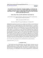
(Fig. 1).
2289
Int.J.Curr.Microbiol.App.Sci (2019) 8(10): 2282-2295
Fig.1 Typical events during propagation of Citrus spp. through callus induction as exemplified
by Citrus jambhiri. A. Inoculation of leaf explants B. Callus induction from leaf segments C.
Callus regeneration D. Shoot regeneration after subculturing E. Rooting of regenerated shoots F.
Planting out after acclimatization
C
B
A
D
E
References
Agarwal, P. K. 1982. Performance of different
citrus root stocks in India: A Review.
Agricultural Review, 3: 17-34.
Ali, S. and Mirza, B. 2006. Micropropagation
of Rough lemon (Citrus jambhiri
Lush.). Effect of explant type and
hormone concentration. Acta Botanica
Croatica, 65: 137-146.
Al-Khayri, J. M. and Al-Bahrany, A. M. 2001.
In vitro micropropagation of Citrus
aurantifolia (lime). Current Science,
81: 1242-1246.
Altaf, N., Khan, A. R., Ali, L. and Bhatti, I. A.
(2009b). In vitro culture of Kinnow
explants. Pakistan Journal of Botany,
41: 597–602.
F
Altaf, N., Khan, A. R., Ali, L. and Bhatti, I. A.
2008. Propagation of Rough lemon
(Citrus jambhiri Lush.) through in
vitro culture and adventitious rooting
in cuttings. Electronic Journal of
Environmental, Agricultural and Food
Chemistry, 7: 3326-3333.
Altaf, N., Khan, A. R., Bhatti, I. A. and Ali, L.
(2009a). Tissue culture of citrus
cultivars. Electronic Journal of
Environmental, Agricultural and Food
Chemistry, 8: 43–51.
Anita, P., Lokman, P. and Niladri, B. 2000.
Biological hardening of tissue culture
raised tea plants through rhizosphere
bacteria. Kluwer Academic Publishers
Biotech Let, 22: 1087-1091.
Ashmore, S. E. 1997. Status report on the
2290
Int.J.Curr.Microbiol.App.Sci (2019) 8(10): 2282-2295
development and application of in
vitro techniques for the conservation
and use of plant genetic resources, pp.
23-24. International Plant Genetic
Resources Institute, Rome.
Bansode, D. S., Chavan, M. D. 2012. Studies
on
antimicrobial
activity
and
phytochemical analysis of Citrus fruit
juices
against
selected
enteric
pathogens. Internat. Res. J. Pharm.,
3(11): 122–126.
Benabdesselam, F., Khettal, B. and Bedjou, F.
2011. Micropropagation of Algerian
juvenil rootstocks citrus species. Life
sciences Leaflets, 18:707 –717.
Bhattacharya, S. C. and Dutta, S. 1952. A
preliminary observation on rootstocks
for Citrus in Assam. Indian Journal of
Horticulture, 9: 1-11.
Carimi, F., De Pasquale, F. and Crescimanno,
F. G. 1994. Somatic embryogenesis
from styles of lemon (C. limon). Plant
Cell, Tissue and Organ Culture,
37:209-211.
Chadha, K. L. and Singh, H. P. 1990.
Citriculture scenario of India. In:Gill,
K. S., Kanwar, J. S. and Singh, R.
(eds.) Proceedings Citrus Show-cumSeminar, Prospects and Problems of
Kinnow Cultivation. pp 21-64, Punjab
Agricultural University, Ludhiana,
India.
Chamandoosti, F. 2017. The utilities of Citrus
tissue culture. International Journal of
Environmental
and
Agriculture
Research, 3(9): 36-46.
Darwesh and Rasmia, S. 2015. Morphology,
physiology and anatomy in vitro
affected acclimatization, ex vitro date
palm plantlets: A review. International
Journal
of
Chemistry
and
Environmental Biological Science. 3
(2): 183-190.
Das, A., Paul, A. K. and Chaudhary, S. 2000.
Micropropagation of sweet orange
(Citrus sinensis Osbeck) for the
development of nucellar seedlings.
Indian Journal of Experimental
Biology, 38: 269–272.
Deng, X. and Liu, J. H. 1996. Advances in
genetic transformation and genome
transfer of fruit crops. Journal of
Wuhan Botanical Research, 14: 357369.
Dhanavade, M. J., Jalkute, C. B., Ghosh, J. S.,
Sonawane, K. D. 2011. Study
antimicrobial activity of lemon (Citrus
lemon L.) peel extract. Brit. J. Pharm.
Toxic., 2 (3): 119–122.
Eden, A. P. and Cerruti, R. H. 2008. Preparing
tissue cultured banana plantlets for
field planting. Co. ext. series
Agriculture Research Biotechnology,
8: 1-3.
El-Sawy, A., Gomaa, A., Reda, A. and Danial,
N. 2006. Somatic embryogenesis and
plant regeneration from undeveloped
ovules of citrus. Arabian Journal of
Biotechnology, 9: 189-202.
Engelmann, F. (1997). In vitro conservation
methods. In: Callow, J. A., FordLloyd, B. V. and Newbury, H. J. (eds).
Biotechnology and plant Genetic
Resources. CABI, Oxon, pp. 119-161.
Gmitter, F. G. Jr. and Hu, X. 1990. The
possible role of Yunnan, China, in the
origin of contemporary Citrus species
(Rutaceae). Economic Botany, 44:
267-277.
Guo, W. W. and Deng, X. X. 2001. Wide
somatic hybrids of Citrus with its
related genera and their potential in
genetic improvement. Euphytica, 118:
175-183.
Guo, W. W. and Grosser, J. W. 2005. Somatic
hybrid vigor in Citrus: Direct evidence
from protoplast fusion of an
embryogenic callus line with a
transgenic
mesophyll
parent
expressing the GFP gene. Plant
Science, 168: 1541-1545.
Hammschlag, F., Ritchie, D., Werner, D.,
2291
Int.J.Curr.Microbiol.App.Sci (2019) 8(10): 2282-2295
Hashmil, G., Krusberg, L., Meyer, R.
and Huettel, R. 1995. In vitro selection
of disease resistance in fruit trees. Acta
Horticulturae, 392: 19-26.
Hao, Y- J. 2000. In vitro conservation and
genetic variation of important fruit
trees.
Ph.D.
thesis,
Huazhong
Agricultural University, Wuhan, P. R.
China.
Hao, Y-J. and Deng, X. X. 2002. Occurrence
of chromosomal variations and plant
regeneration from long-term-cultured
citrus callus. In Vitro Cellular
Developmental Biology-Plant, 38: 472476.
Hazarika B. N. and Parthasarathy V. A. 2002.
Effect of reduced humidity and
antitranspirants
in
acclimatizing
micropropagated
citrus
plantlets.
Journal of Applied Horticulture, 4 (1):
30-32.
Kamble, M., Kalim, S., Bajpai, A., Chandra,
R. and Kumar, R. 2012. In vitro
selection for wilt resistance in guava
(Psidium guajava L.) cv. Allahabad
Safeda. Biotechnology, 11(3): 163-171.
Kanwar, J., Kaul, M. K., Shaktawat, R. P. S.
and Naruka, I. S. 2016. In vitro
multiple shoots induction from nodal
explants of Sour orange (citrus
aurantium l.). The Bioscan, 11(4):
2127-2131.
Karimi, E., Oskoueian, E., Hendra, R.,
Oskoueian, A. and Jaafar, H. Z. 2012.
Phenolic compounds characterization
and biological activities of Citrus
aurantium
Bloom.
Journal
of
molecular biology, 17 (2): 1203-1218.
Karwa, A. 2003. In vitro propagation of C.
reticulata Blanco. (Nagpur mandarin).
Indian J. Gen. Plant Breed. 63: 18788.
Karwa, A. S. and Chikhale, N. J. 2004. Effect
of various growth hormones on Invitro clonal propagation of Citrus
sinensis Osbeck. Recent Trends in
Biotechnology. Scientific Publishers
(India), Jodhpur, India, pp. 192-195.
Kasprzyk-Pawelec, A., Pietrusiewicz, J.,
Szczuka, E. 2015. In vitro regeneration
induced in leaf explants of Citrus
limon L. Burm cv. „Primofiore‟. Acta
Sci. Pol. Hortorum Cultus, 14(4): 143–
153.
Kaur, S. 2016. In vitro plant regeneration in
rough lemon (Citrus jambhiri Lush.)
through epicotyl segments by direct
shoot organogenesis. Journal of
Applied and Natural Science, 8 (2):
724 – 729.
Kaur, S. 2018. In vitro somatic embryogenesis
and regeneration from epicotyl
segments of rough lemon (Citrus
jambhiri Lush.). International Journal
of Chemical Studies, 6(1): 2082-2091.
Kaya, B. and Gubbuk, H. 2001. Investigation
on the propagation of some citrus
rootstocks by tissue culture. Ziraat
Facultesi Dergisi, Akedeniz University,
14(2): 69- 76.
Khan, E. U., Xing-Zheng, F., Wang, J., QiJun, F., Xiao-San, H., Ge-Ning, Z.,
Shi, J. and Ji-Hong, L. 2009.
Regeneration and characterization of
plants derived from leaf in vitro culture
of two sweet orange (Citrus sinensis
(L.) Osbeck) cultivars. Scientia
Horticulturae, 120: 70-76.
Kour, K. and Singh, B. 2012. In vitro
multiplication of rough lemon (Citrus
jambhiri Lush.). Journal of Agriculture
and Veterinary Science, 1(4): 05-09.
Krishan, K., Dhatt, A. S. and Gill, M. I. S.
2001. In-vitro plant propagation in
sweet orange (Citrus sinensis L.
Osbeck.) cv. Mosambi and Jaffa.
Indian J. Hort., 58: 208-211.
Kumar,
K.
and
Rao,
I.
2012.
Morphophysiologicals problem in
acclimatization of micropropagated
plants in-ex vitro conditions-A review.
Journal
of
ornamental
and
2292
Int.J.Curr.Microbiol.App.Sci (2019) 8(10): 2282-2295
Horticulture Plants, 2 (4): 271-283.
Kumar, K., Kaur, H., Gill, M. I. S., Rattanpal,
H. S., Kanika and Gosal, S. S. 2011.
An efficient regeneration protocol
from callus culture in rough lemon
(Citrus jambhiri). Indian Journal of
Agricultural Sciences, 81(4): 324-9.
Laskar, M. A., Hynniewta, M. and Rao, C. S.
2009. In vitro propagation of Citrus
indica Tanaka- An endangered
progenitor species. Indian Journal of
Biotechnology, 8: 311-316.
Li, S., Lin, Y. C., Ho, C. T. 2014. Formulated
extract from multiple Citrus peels
impairs dendritic cell functions and
attenuates
allergic
contact
hypersensitivity.
International
Immunopharmacol, 20: 12 – 23.
Liu, Y., Heying, E. and Tanumihardjo, S.
2012. History, global distribution, and
nutritional importance of citrus fruits.
Comprehensive reviews in food
Science and Food Safety, 11 (6), 530545.
Lombardo, G., Alessandro, R., Scialabba, A.,
Sciandra, M. and De Pasquale, F.
2011. Direct organogenesis from
cotyledons in cultivars of Citrus
clementina Hort. Ex Tan. American
Journal of Plant Science, 2: 237 – 244.
Momoko, I., Keiko, S. and Akira, I. 2013.
Plant callus: Mechanisms of induction
and repression. American Society of
Plant Biology and the Plant Cell, 25
(9): 3159-3173.
Mukhtar, R., Khan, M. M., Rafiq. R., Shahid,
A. and Khan, F. A. 2005. In-vitro
regeneration
and
somatic
embryogenesis in Citrus aurantifolia
and Citrus sinensis. International
Journal of agricultural Biology,7(3):
518 -520.
Normah, M. N., Hamidoh, S. and Ghani, F. D.
1997. Micropropagation of Citrus
halimii- an endangered species of
South-east Asia. Plant Cell, Tissue and
Organ Culture, 50: 225-227.
Nurul, H. Daud., Shashita, J. and Rozi, M.
2012.
An
improved
surface
sterilization technique for introducing
leaf, nodal and seed explants of
Aquilaria malaccensis from field
sources into tissue culture. AsiaPacific Journal of Molecular Biology
Biotechnology, 20 (2): 55-58.
Patil, V. K. 1987. High density planting and
dwarfing rootstocks in citrus. A
review. Journal of Maharashtra
Agricultural University, 12:189-94.
Paudyal, K. P. and Haq, N. 2000. In vitro
propagation of pummelo (Citrus
grandis L. Osbeck). Journal of In Vitro
Cell, Developmental Biology of Plants,
36: 511-516.
Pe´rez-Tornero, O. and Porras, I. 2008.
Assessment of polyembryony in
lemon: rescue and in vitro culture of
immature embryos. Plant Cell Tissue
Organ Culture, 93(2): 173–180.
Pospisilova J., Ticha I., Kadlecek P., Haised.
L
and
Plzakova
S.
1999.
Acclimatization of micropropagated
plants to ex vitro conditions. Journal of
Biologia Plantarum, 42 (4): 481-497.
Raja,
W.
H.
2012.
Studies
on
micropropagation in citrus rootstocks
and effects of arbuscular mycorrhizal
inoculation on in vitro raised plants.
Ph.D. thesis, Indian Agricultural
Research Institute, New Delhi, India.
Rakesh, K., Saurabh, V. and Nawaz, K. 2013.
Comparative nutritional analysis and
antioxidant activity of fruit juices of
some Citrus spp. Octa Journal of
Bioscience, 1 (1): 44-53.
Randhawa, G. S. and Srivastava, K. C. 1986.
Citriculture in India. pp. 8-13.
Hindustan Publishing Corporation,
India.
Rangacharlu, V. S., Naidu, N., Anjaneyulu
and Narsimharao, T. L. 1958.
Performance of Sathgudi orange
2293
Int.J.Curr.Microbiol.App.Sci (2019) 8(10): 2282-2295
(C.sinensis Osbeck) on different
rootstocks at Kodur (South India).
Indian Journal of Horticulture, 15: 3558.
Rani, G., Singh, B., Sharma, S., Rehan, L.,
Zaidi, A. A., Nagpal, A. and Virk, G.
S. 2004. Micropropagation of Kinnow
(Citrus nobilis x Citrus deliciosa)
through nodal segments. Journal of
Indian Botanical Society, 83: 26-29.
Saini, H. K., Gill, M. S. and Gill, M. I. S.
2010. Direct shoot organogenesis and
plant regeneration in rough lemon
(Citrus jambhiri Lush.). Indian
Journal of Biotechnology, 9(4): 419423.
Sarker, I., Islam, J., Shaekh, M. P. E.,
Rahman,
M.
M.,
Khan,
H.,
Chowdhury, A. S. M. H. K., Mukim,
M. S. I., Islam, R. and Haque, R. 2015.
Establishment of a Standard Protocol
for In Vitro Meristem Culture and
Plant
Regeneration
of
Citrus
aurantifolia. Journal of Pharmacy and
Biological Sciences, 10(2): 61-69.
Sarma, C., Borthakur, A., Singh, S., Mod, M.
K. and Sen, P. 2011. Efficient in vitro
plant regeneration from cotyledonary
explants of Citrus reticulata L. Blanco.
Annals of Biological Research, 2(6):
341 – 348.
Saunt, J. 1990. Citrus varieties of the world,
pp. 126. Sinclair International Ltd.
Savita, Singh, B., Virk, G. S. and Nagpal, A.
(2011b). Efficient micropropagation
protocol for regeneration of Citrus
jambhiri Lush. using cotyledon as
explant. Physiology and Molecular
Biology of Plants, 17: 161-169.
Savita, Vijay, Virk, G. S. and Nagpal, A.
2010. Effect of explant types and plant
growth regulators on callus induction
and plantlet regeneration in Citrus
jambhiri Lush. Environment and WeAn International Journal of Science
and Technology, 5: 97-106.
Savita, Virk, G. S. and Nagpal, A. (2011a). In
vitro selection of calli of Citrus
jambhiri Lush. for tolerance to culture
filtrate of Phytophthora parasitica and
their regeneration. Physiology and
Molecular Biology of Plants, 17: 4147.
Scora, R. W. 1975. On the history and origin
of citrus. Bulletin of Torrey Botanical
Club, 102: 369–375.
Sharma, S., Prakash, A. and Tele, A. 2009. In
vitro propagation of Citrus rootstocks.
Notulae Botanicae Horti Agrobotanici
Cluj Napoca, 37: 84–88.
Shen, D-X., Wang, Y-Y. and Chen, L. G.
1998. Citrus genetics and breeding. pp
173. China Scientific Press, Beijing.
Shen, W., Xu, Y. and Lu, Y. H. 2012.
Inhibitory effects of citrus flavonoids
on starch digestion and antihyperglycemic effects in Hep G2 cells.
Journal
of
Agricultural
Food
Chemistry, 60 (38): 9609-9619.
Silva, R. P., Da Souza, E., Dos, S., Reboucos.
F. S. and Almeida, W. A. B. 2005.
Optimization of protocols for the invitro regeneration of plants of
mandarin (Citrus reshmi Hort. Ex
Tan.) cv. Cleoptara. Revista Brasileria
de fruticultura., 27: 484-487.
Singh, I. P., Parthasarthy, V. A. and Handique,
P. J. 2003. Effect of bioregulators on
In-vitro
raised
microshoots
of
economically important Citrus species
of the NEH region. Indian J
Horticulture., 60 :16-21.
Singh, L. B. 1962. Studies on the rootstocks
for sweet orange in the web tropics I.
Variety Rangtra. Hort. Adv, 6: 19-27
Singh, R., Sindhu, A., Singal, H. R. and Singh,
R. 2004. Biochemical basis of
resistance in chickpea (Cicer arietinum
L.) against Fusarium wilt. Acta
Phytopathologica et Entomologica
Hungarica, 38: 13-19.
Singh, S. and Naqvi, S. A. M. H. 2001. Citrus,
2294
Int.J.Curr.Microbiol.App.Sci (2019) 8(10): 2282-2295
pp.
588.
International
Book
Distributing Company, Lucknow,
India.
Singh, S. and Rajam. M. V. 2010. Highly
efficient and rapid plant regeneration
in Citrus sinensis. Journal of Plant
Biochemistry and Biotechnology, 19:
195-202.
Srivastava, R.K., Sandhu, A.S. and Sood, N
(2001). In-vitro plant regeneration of
Citrus aurantifolia through callus
culture. J. Appl. Hort., 2(1): 28 - 30.
Talon, M. and Gmitter Jr, G. F. 2008. Citrus
Genomics. International Journal of
Plant Genomics, 2008: 1-17.
Taye, M. G., Debesay, B., Tesfahun, Y. and
Brhanu, A. 2018. Optimization of an in
vitro regeneration protocol for rough
lemon rootstock (Citrus jambhiri L.)
via direct organogenesis. Advances in
Crop Science and Technology, 6 (1):
329.
Tusa, N. Grosser, J. W. and Gmitter, F. G.
1990. Plant regeneration of Valencia
Sweet orange, Femminello lemon and
the interspecific somatic hybrid
following protoplasm fusion. Journal
of the American Society for
Horticultural Science, 115 (6): 1043 –
1046.
Upadhyay, S., Syamal, M. M. and Itoo, H.
2010. Micropropagation of Sweet
orange cv. Mosambi through shoot tips
and nodal segments. Indian Journal of
Horticulture, 67: 21-25.
Wang, S., Huang, Z., Yu, R., Zou, W. and
Song, F. S. 2002. Studies on the
induction of embryogenic callus,
establishment of cell suspensions and
plant regeneration in Citrus retuculata
var. tanaka Hayata. J. South China
Agricultural University, 23 : 52 – 55.
Yi, H-L. and Deng, X-X. 1998. Preliminary
study on preservation of citrus calli.
Journal of Huazhong Agricultural
University, 17: 89-92.
How to cite this article:
Mudasir Iqbal, V. K. Wali, Parshant Bakshi, Kiran Kour, Vijay K. Razdan, B. K. Sinha and
Sood, K. K. 2019. In vitro Propagation of Citrus Species through Callus Induction and
Regeneration: A Review. Int.J.Curr.Microbiol.App.Sci. 8(10): 2282-2295.
doi: />
2295